A New Microporous Membrane for Skin Regeneration

Skin regeneration
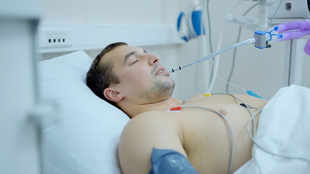
Skin regeneration has been a major goal for tissue engineering and regenerative medicine. However, natural skin is difficult to recapitulate because of its many different layers, components (such as hair follicles and sweat glands), and cell types. Each of these factors is critical to functional skin and poses a different roadblock to regenerating it. Furthermore, regenerative wound healing must compete with the faster, less functional, fibrotic scar tissue healing response. This is complicated even more by the fact the patients who need these treatments most, such as older adults and people with diabetes, are less able to tap into their body’s natural wound healing response.
One strategy currently used in the clinic is to apply an amniotic membrane to the wound. Surprisingly, this material, which typically is discarded after live births, has many of the same extracellular matrix components as skin. It also contains many growth factors that suppress inflammation (a major limitation in chronic wounds) and upregulate tissue formation. While this material improves the wound healing process, it still has room for improvement in closure speed, the size of wounds it can heal, and in limiting the formation of scar tissue.
A small change can make a big difference
Recently, researchers have reported on a manufacturing strategy that improves upon this material. [1] They hypothesized that the high density of amniotic membrane hinders the healing process by restricting cell penetration and mobility at the wound site. By applying a chemical treatment, a team at the Royan Institute for Stem Cell Biology and Technology created a membrane with a more microporous structure and tested it in diabetic rats.
They found a slight increase in tissue formation and a large increase in vascularization in their microporous amniotic membrane compared to the membranes typically used in the clinic. Additionally, their implant considerably decreased inflammation, a key issue for chronic wounds in diabetic patients.
To the best of our knowledge, the present study was the first report on generation and application of bioengineered HAM-S to enhance healing of an ischemic and delayed wound in diabetic type 1 animal model. The results demonstrated that the natural 3D micro-porous scaffold has more impact in the wound healing process compared to traditional use of HAM. It is suggested that the suitable pore size for cells adhesion, penetration, and migration are markedly positive properties of the bioengineered scaffold relative to the HAM-M. Yet, to develop a more advanced scaffold in the platform of tissue engineering to overcome the diabetic ulcer, further investigations using other synergic key players are needed.
Conclusion
This study demonstrated a clear improvement over a wound healing strategy currently used in the clinic by making only a moderate change to the existing treatment. This is promising news for people with chronic wounds; of course, the results in humans remain to be seen, and there will still be considerable room for improvement in skin regeneration beyond this new treatment.
[1] Nasiry, D., Khalatbary, A. R., Abdollahlfar, M.-A., Amini, A., Bayat, M., Noori, A., Piryael, A. (2020). Engraftment of bioengineered three-dimensional scaffold from human amniotic membrane-derived extracellular matrix accelerates ischemic diabetic wound healing. Archives of Dermatological Research, online ahead of print.