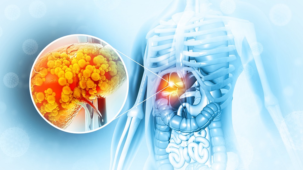
A Reason Why Livers Accumulate Fat with Age
- The problems are much more than just fat on the liver.

Researchers have discovered one of the reasons why fatty liver disease, even without alcohol consumption, increases with aging.
The earliest stage of fatty liver disease
Nonalcoholic fatty liver disease (NAFLD) begins with steatosis, the accumulation of fatty tissues in an organ [1], which can then progress to hepatitis, fibrosis, and cancer [2]. Therefore, fat accumulation in the liver isn’t just a matter of obesity or fat accumulation in general: it is a direct precursor to liver failure. NAFLD increases with aging, and this fact has been re-confirmed in multiple studies encompassing multiple populations and time periods [3, 4, 5].
The acyl-coenzyme A (acyl-CoA) dehydrogenases are key elements of lipid metabolism and synthesis in the liver, as they form acetyl-CoA, a precursor of lipid formation [6]. Blocking the formation of acetyl-CoA promotes autophagy and lengthens lifespan in simple organisms [7]. This research focuses on the short-chain form of acyl-CoA dehydrogenase (SCAD), which these researchers believe to be key to the gradual formation of NAFLD.
SCAD is linked to both liver disease and aging
This paper began with a gene set analysis taken from younger (21-45) and older (70+) people. As expected, genes related to fatty acid metabolism were strongly upregulated in the older group. ACADS, which encodes for SCAD, was significantly upregulated among this gene set. In groups of young. middle-aged, and older mice, the genes for very long, long, and medium-chain fatty acids did not significantly change; only the gene for SCAD did.
Liver tissue analysis showed similar results, with people older than 74 producing much more of it than 18- to 25-year-olds. Peripheral blood mononuclear cells (PBMCs), which are easy to access and naturally produce SCAD, also produce much more of it in this older cohort. Inducing senescence into a human liver cell line through hydrogen peroxide also caused these cells to express more SCAD along with the senescence marker p21.
Gene expression of SCAD was positively correlated with NAFLD. People with this disease were age-matched with healthy controls, and the healthy controls had far less SCAD than the people with NAFLD. Unsurprisingly, even in the NAFLD patients, the amount of SCAD was increased with age.
Life without SCAD?
The researchers utilized a mouse strain that had the ACADS gene completely knocked out, and analysis of their liver tissues confirmed that these mice did not produce any SCAD. While their total bodyweights were the same, older mice of this strain had far less evidence of liver steatosis compared to similar mice that produced SCAD. Fibrosis was also greatly diminished in the SCAD-free mice, as were standard biomarkers of cellular senescence, and embryonic fibroblasts derived from these SCAD-free mice were more able to consume lipids after multiple replications and had less DNA damage.
Additionally, the cellular housecleaning mechanism known as autophagy was strongly downregulated with SCAD. With aging, the cells of SCAD-free mice still engaged in less autophagy than younger mice, but multiple biomarkers confirmed that their autophagy was still greater than older mice that produce SCAD. Further experiments found this to be directly related to the acetyl-CoA pathway.
While most of these results were positive, one crucially negative result was found: the mice without SCAD production produced significantly less ATP, particularly with aging. This is the crucial molecule used for mitochondrial energy production. While it may be possible to alleviate NAFLD by targeting SCAD in people, the deveopers of any future drug must take potential side effects related to energy metabolism into account.
Literature
[1] Chalasani, N., Younossi, Z., Lavine, J. E., Diehl, A. M., Brunt, E. M., Cusi, K., … & Sanyal, A. J. (2012). The diagnosis and management of non‐alcoholic fatty liver disease: Practice Guideline by the American Association for the Study of Liver Diseases, American College of Gastroenterology, and the American Gastroenterological Association. Hepatology, 55(6), 2005-2023.
[2] Wang, H., Naghavi, M., Allen, C., Barber, R. M., Bhutta, Z. A., Carter, A., … & Bell, M. L. (2016). Global, regional, and national life expectancy, all-cause mortality, and cause-specific mortality for 249 causes of death, 1980–2015: a systematic analysis for the Global Burden of Disease Study 2015. The lancet, 388(10053), 1459-1544.
[3] Miyaaki, H., Ichikawa, T., Nakao, K., Yatsuhashi, H., Furukawa, R., Ohba, K., … & Eguchi, K. (2008). Clinicopathological study of nonalcoholic fatty liver disease in Japan: the risk factors for fibrosis. Liver International, 28(4), 519-524.
[4] Kagansky, N., Levy, S., Keter, D., Rimon, E., Taiba, Z., Fridman, Z., … & Malnick, S. (2004). Non‐alcoholic fatty liver disease–a common and benign finding in octogenarian patients. Liver International, 24(6), 588-594.
[5] Hilden, M., Christoffersen, P., Juhl, E., & Dalgaard, J. B. (1977). Liver histology in a ‘normal’population—examinations of 503 consecutive fatal traffic casualties. Scandinavian journal of gastroenterology, 12(5), 593-597.
[6] Ghosh, S., Kruger, C., Wicks, S., Simon, J., Kumar, K. G., Johnson, W. D., … & Richards, B. K. (2016). Short chain acyl-CoA dehydrogenase deficiency and short-term high-fat diet perturb mitochondrial energy metabolism and transcriptional control of lipid-handling in liver. Nutrition & metabolism, 13, 1-17.
[7] Eisenberg, T., Schroeder, S., Andryushkova, A., Pendl, T., Küttner, V., Bhukel, A., … & Madeo, F. (2014). Nucleocytosolic depletion of the energy metabolite acetyl-coenzyme a stimulates autophagy and prolongs lifespan. Cell metabolism, 19(3), 431-444.


