Fragmented Mitochondria Linked to Muscle Weakness
- Mitochondria grow smaller and less well-connected with aging.

In a study published in Aging Cell, researchers have outlined a relationship between mitochondrial fragmentation in skeletal muscle and the loss of strength with age.
Broken power plants
As its authors point out, this is far from the first study to link mitochondrial dysfunction and aging in muscle [1], nor is it the first to connect exercise habits, aging, mitochondria, and the loss of physical function [2].
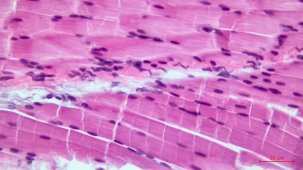
There has also been significant prior work showing how the mitochondria in muscle tissue behave. Mitochondria in muscle are not equal in their behaviors: the mitochondria closest to the blood-filled capillaries (subsarcolemmal mitochondria) bring energy to more centrally located ones (intermyofibrillar mitochondria) through an intracellular network [3]. Fragmentation of this network destroys this energy transfer but may also offer protection against damage being transferred as well [4].
Too much fragmentation and fission, however, causes muscle wasting in mice [5]; the opposite, mitochondrial fusion, causes muscles to grow in these animals [6]. The researchers’ previous work on healthy volunteers demonstrated that fragmentation begins to occur at day 6 of bed rest, while functional impairments were found to occur on day 55 [7]. However, that work did not prove one way or another whether mitochondrial fragmentation is a useful biomarker or warning sign for age-related muscle decline.
Decline begins before retirement
Wanting to avoid physical inactivity as a confounder and suspecting that this process may not be the same as actual sarcopenia, the researchers recruited a dozen young (average age 27) and ten middle-aged (average age 55) volunteers rather than significantly older people. The older group was slightly more overweight than the younger group.
Unsurprisingly, the younger people’s muscles used more oxygen to generate more power than the older people’s, according to multiple metrics of respiration and energy use. This was not linked to blood flow; instead, it was linked to how the muscles pull oxygen from the blood.
The researchers then examined the mitochondria more closely in biopsied muscle tissue. The total density of the intermyofibrillar mitochondria was the same between younger and older people; however, the older people had more, smaller mitochondria. While their shapes did not differ, markers of mitochondrial fragmentation were greater in this area in the older group.
In the subsarcolemmal area, however, the older people had approximately as many mitochondria as the younger people, which led to a reduction of density with age as these mitochondria were also smaller. Here, too, they were found to be significantly more fragmented. This fragmentation in both areas was associated with the accumulation of fat (lipid) droplets.
Looking ever closer
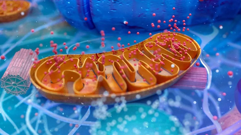
There were also differences involving the tiny folds inside mitochondria (cristae). Younger people’s mitochondria had regular and dense cristae, while those of older people were less regular, with some areas having no cristae at all. This, the researchers hold, represents “age-associated deterioration at the level of the individual mitochondrion.” Interestingly, however, further data suggests that the increased number of smaller mitochondria may have made up for this, restoring some of the lost function.
The authors then pivoted to the key thrust of their research: the connection between fragmentation and loss of capacity. Fragmentation in the intermyofibrillar mitochondria and a reduction in the cristae was found to be responsible for nearly all of the changes in the well-known metric of VO2max. Unsurprisingly, the density of the subsarcolemmal mitochondria was found to be associated with the muscles’ ability to extract oxygen from blood.
The researchers believe that their findings explain the basic reasons why people lose strength with age, even in the absence of defined sarcopenia. They also warn that this mitochondrial dysfunction only gets worse with aging. Furthermore, they hold that their findings “reflect an early ageing phenotype, making the mitochondrial changes observed herein strong candidates for intervention studies aiming to slow the progression of the effects of ageing on physical function.”
As exercise is associated with mitochondrial fusion [8] and one study had suggested that six months of endurance training can compensate for 30 years of aging [9], the authors suggest that further research on exercise in older people should be done with a close examination into mitochondrial changes.
Literature
[1] Gouspillou, G., Bourdel‐Marchasson, I., Rouland, R., Calmettes, G., Biran, M., Deschodt‐Arsac, V., … & Diolez, P. (2014). Mitochondrial energetics is impaired in vivo in aged skeletal muscle. Aging cell, 13(1), 39-48.
[2] Grevendonk, L., Connell, N. J., McCrum, C., Fealy, C. E., Bilet, L., Bruls, Y. M., … & Hoeks, J. (2021). Impact of aging and exercise on skeletal muscle mitochondrial capacity, energy metabolism, and physical function. Nature communications, 12(1), 4773.
[3] Glancy, B., Hartnell, L. M., Malide, D., Yu, Z. X., Combs, C. A., Connelly, P. S., … & Balaban, R. S. (2015). Mitochondrial reticulum for cellular energy distribution in muscle. Nature, 523(7562), 617-620.
[4] Glancy, B., Hartnell, L. M., Combs, C. A., Femnou, A., Sun, J., Murphy, E., … & Balaban, R. S. (2017). Power grid protection of the muscle mitochondrial reticulum. Cell reports, 19(3), 487-496.
[5] Romanello, V., Guadagnin, E., Gomes, L., Roder, I., Sandri, C., Petersen, Y., … & Sandri, M. (2010). Mitochondrial fission and remodelling contributes to muscle atrophy. The EMBO journal, 29(10), 1774-1785.
[6] Cefis, M., Dargegen, M., Marcangeli, V., Taherkhani, S., Dulac, M., Leduc‐Gaudet, J. P., … & Gouspillou, G. (2024). MFN2 overexpression in skeletal muscles of young and old mice causes a mild hypertrophy without altering mitochondrial respiration and H2O2 emission. Acta Physiologica, 240(5), e14119.
[7] Eggelbusch, M., Charlton, B. T., Bosutti, A., Ganse, B., Giakoumaki, I., Grootemaat, A. E., … & Wüst, R. C. (2024). The impact of bed rest on human skeletal muscle metabolism. Cell Reports Medicine, 5(1).
[8] Huertas, J. R., Ruiz‐Ojeda, F. J., Plaza‐Díaz, J., Nordsborg, N. B., Martín‐Albo, J., Rueda‐Robles, A., & Casuso, R. A. (2019). Human muscular mitochondrial fusion in athletes during exercise. The FASEB Journal, 33(11), 12087-12098.
[9] McGuire, D. K., Levine, B. D., Williamson, J. W., Snell, P. G., Blomqvist, C. G., Saltin, B., & Mitchell, J. H. (2001). A 30-year follow-up of the Dallas Bed Rest and Training Study: II. Effect of age on cardiovascular adaptation to exercise training. Circulation, 104(12), 1358-1366.


