Skin Aging and Cellular Mobility at the Protein Level
- The skeletons of skin cells control how they move.

In Aging, researchers have described how the changing production of skin cells’ proteins is a core part of their age-related decline.
Understanding why skin cells lose their function
In healthy skin, dermal fibroblasts produce the proteins needed to maintain the extracellular matrix (ECM), the network of collagen and elastin that holds soft tissues together [1]. When skin is damaged, they secrete matrix metalloproteinases (MMPs), which destroy the damaged tissue. They summon macrophages to damaged areas to clear away debris and bring in cells to repair the area [2].
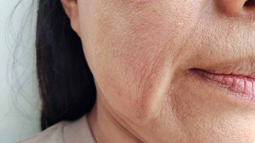
Unsurprisingly, with aging, this process is significantly impeded [3]. Aged skin has deteriorated ECM, which is the direct cause of wrinkles [4]. The destructive factors that young skin cells secrete as part of wound healing are constantly secreted in the aged [5]. Older people also have fewer dermal fibroblasts, and they are less able to migrate [6].
This paper takes a molecular look at the fibroblasts, focusing on what proteins they secrete and determining the role that cellular senescence plays in their deterioration.
Proteins and the cytoskeleton
This study compared skin cells derived from younger (under 35) and older (over 55) donors. While the researchers found nearly a thousand differences in secreted proteins (the secretome), under normal circumstances, only 16 of these secreted proteins were more than doubled or halved between the young and the old. When stimulated with the cytokine TGF-β, only 11 of these proteins met this threshold.
Many of the involved proteins were related to the ECM, senesence, and inflammation, and a particularly large fraction was related to enzymatic function. Some were related to actin, the protein responsible for the cells’ structure (cytoskeleton). The protein ACTC1, one of the six isoforms of actin, was very substantially decreased with age.
The total number of proteins (the proteome) that was different between the young and the old was more than four times as large as the secretome: over four thousand proteins were different, with 63 of them being doubled or halved under normal circumstances and 73 with the application of TGF-β. Like the secretome, many of these proteins were related to actin and to the ECM. Curiously, nearly a full quarter of the proteins found in older fibroblasts were ones for which the researchers did not know the functions, if any.
The researchers focused on four specific actin-related proteins. CFL1 was substantially decreased in older people, and this protein is also related to wound healing: removing CFL1 from younger fibroblasts sharply increases the amount of time it takes for these cells to engage in healing in vitro. CORO1C was multiplied substantially in older cells, and the researchers were unable to conclude a reason why this was the case: the related mRNA was not very different between younger and older cells. The same was found to be true for another actin-related protein, FLNB.
ARPC3 may be a key part of this puzzle. This protein was found to be necessary for fibroblast migration, and the researchers believe that it regulates CFL1 and CORO1C. They further hold that the combination of the three acts “together to regulate the actin cytoskeleton and the cytoskeleton regulation itself.” This protein is secreted less by aging cells.
In total, the researchers believe that the proteins involved in the cytoskeleton of skin cells, and their related ability to move, are a core part of skin aging and the related loss of wound healing ability. They hold that further study of these proteins might be productive in learning to better control skin aging.
Literature
[1] Braverman, I. M., & Fonferko, E. (1982). Studies in cutaneous aging: I. The elastic fiber network. Journal of Investigative Dermatology, 78(5), 434-443.
[2] Brun, C., Demeaux, A., Guaddachi, F., Jean-Louis, F., Oddos, T., Bagot, M., … & Michel, L. (2014). T-plastin expression downstream to the calcineurin/NFAT pathway is involved in keratinocyte migration. PloS one, 9(9), e104700.
[3] Brun, C., Jean‐Louis, F., Oddos, T., Bagot, M., Bensussan, A., & Michel, L. (2016). Phenotypic and functional changes in dermal primary fibroblasts isolated from intrinsically aged human skin. Experimental dermatology, 25(2), 113-119.
[4] Makrantonaki, E., & Zouboulis, C. C. (2008). Skin alterations and diseases in advanced age. Drug Discovery Today: Disease Mechanisms, 5(2), e153-e162.
[5] Lupa, D. M. W., Kalfalah, F., Safferling, K., Boukamp, P., Poschmann, G., Volpi, E., … & Krutmann, J. (2015). Characterization of skin aging–associated secreted proteins (SAASP) produced by dermal fibroblasts isolated from intrinsically aged human skin. Journal of Investigative Dermatology, 135(8), 1954-1968.
[6] Reed, M. J., Ferara, N. S., & Vernon, R. B. (2001). Impaired migration, integrin function, and actin cytoskeletal organization in dermal fibroblasts from a subset of aged human donors. Mechanisms of ageing and development, 122(11), 1203-1220.


