Affecting a Signaling Pathway Alleviates Alzheimer’s in Mice
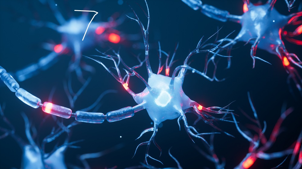
- Neurons and microglia chemically communicate.
- Neurons express somatostatin, a chemical signal that microglia receive.
- Alzheimer’s-prone mice engineered to express more somatostatin than normal have better outcomes, while similar mice that express less somatostatin have worse outcomes.
A new study shows that the overexpression of somatostatin (SST), a neuropeptide produced in neurons and acting mostly on microglia, lowers inflammation and amyloid β burden, improving cognitive abilities in a mouse model of Alzheimer’s. Drugs affecting this pathway are already available [1].
The unusual suspect
In Alzheimer’s disease, many signaling pathways in the brain become dysregulated. Since going after the main hallmarks of the disease (amyloid β and tau protein accumulation) has only yielded modest results so far, scientists are exploring various secondary targets whose levels correlate with the disease.
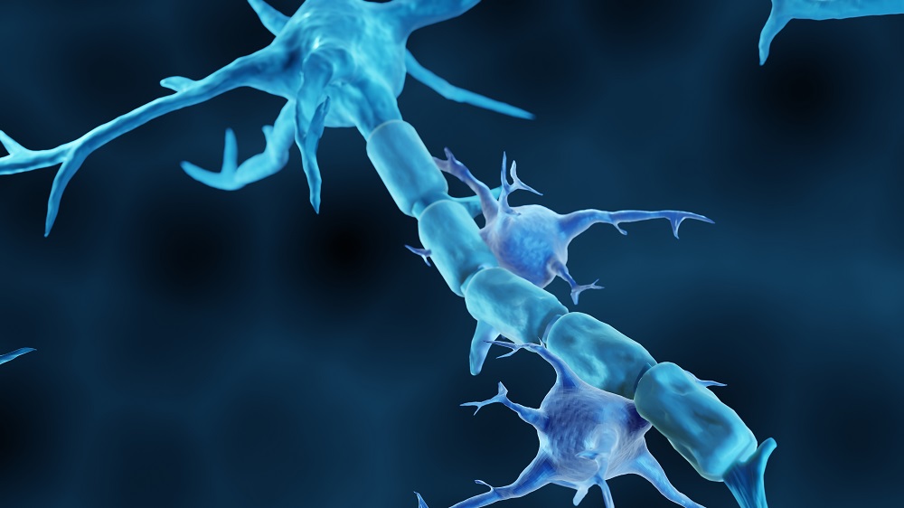
One such molecule is SST, a small signaling protein (a neuropeptide) released by a specific class of inhibitory neurons in the brain, which help regulate brain activity, mostly by calming it. SST binds to a family of five receptors called somatostatin receptors (SSTR1-5), and those are preferentially expressed by microglia, the brain’s resident immune cells. Microglia hyperactivation leads to chronic inflammatory states and has been linked to Alzheimer’s and other dementias [2].
Importantly, SST levels are lower in Alzheimer’s patients than in healthy people [3]. However, whether SST actually talks to microglia directly and whether the loss of SST in AD could be making microglial activation worse had never been systematically tested.
In a new study from Daegu Gyeongbuk Institute of Science and Technology in South Korea, published in Brain, Behavior, and Immunity, the authors hypothesized that SST normally keeps microglia in a healthier, more controlled state, and that its loss in Alzheimer’s contributes to the harmful microglial hyperactivation seen in the disease. They tested their idea first in isolated cells, and then in living mice.
Reduced microglial activation and inflammation
First, the researchers grew separate cultures of neurons, astrocytes (supportive brain cells), and microglia, and confirmed that microglia indeed express SSTR2, but not SST, which was expressed exclusively in neurons. Essentially, neurons have the key, and microglia have the lock.
They then treated primary microglia, isolated directly from mouse brains, with SST at various doses and time periods. Measurements of phagocytosis, the process when cells engulf and digest particles and the primary mechanism by which microglia clear amyloid β and debris, found that SST treatment indeed boosted phagocytosis in a dose-dependent manner, while blocking SST abolished the effect.
The researchers then treated microglia with SST for 48 hours and measured mRNA levels of a panel of signaling proteins that coordinate inflammatory responses (cytokines). They found that the treatment dampened the levels of the pro-inflammatory cytokine IL-12 and, conversely, elevated the levels of TGF-β1, a broadly immunosuppressive and tissue-remodeling cytokine associated with microglial homeostasis. Together, these shifts suggest that SST nudges microglia toward a less inflammatory state, hinting at a neuroprotective effect. However, the effect sizes were modest, and several cytokines tested showed no significant change.
What would happen to microglia if we manipulated SST levels in living animals? The researchers delivered an SST overexpression gene into the dentate gyrus neurons, the hippocampal region associated with memory and learning and heavily affected in Alzheimer’s, of healthy mice. Overexpressing SST did not affect microglial morphology and function, as the mice’s microglia were also mostly healthy and stayed that way following the treatment. Still, the treatment reduced markers of microglia activation. Conversely, knocking down endogenous SST led to microglia acquiring activation-associated morphology.
The team then moved to a mouse model of Alzheimer’s: 5xFAD, which shows extremely rapid Aβ plaque accumulation. The authors first confirmed that microglial activation in these mice is age-dependent and becomes pronounced around 5 months.
Overall microglial density was significantly reduced in SST-overexpressing 5xFAD mice compared to controls (a good sign), and microglial morphology was partially preserved. PCR analysis showed reversal of several activation-associated markers. Importantly, even at this early stage of the disease, there were positive signs with regard to Aβ accumulation, but overall plaque burden was not one of them.
Cognitive benefits in vivo
Two weeks after injection, the mice underwent a battery of cognitive and behavioral tests. Anxiety and recognition memory were unaffected, but SST-overexpressing 5xFAD mice eventually started showing significant improvements in spatial memory.
Finally, the researchers repeated the overexpression experiment in 10-month-old 5xFAD mice – a late-disease timepoint with extensive, well-established plaque burden. At this point, two weeks of SST overexpression clearly reduced microglial activation, Aβ plaque density, and average plaque size. This suggests that SST’s effects on overall plaque burden become evident when plaques are more established.
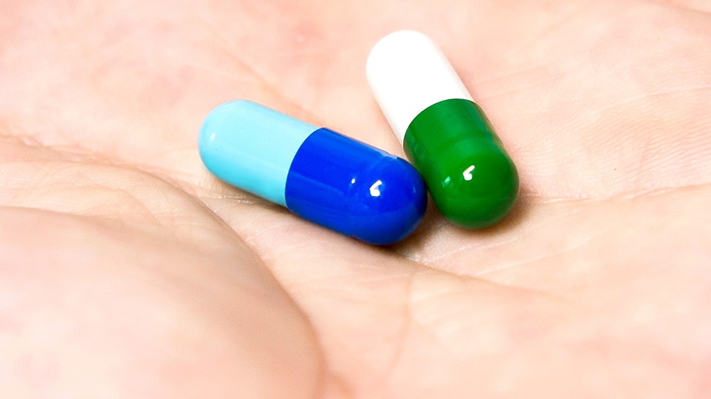
Importantly, approved drugs targeting SST receptors already exist, such as for treating acromegaly. This raises the possibility of repurposing them to treat Alzheimer’s patients, who currently have very few options.
Professor Jiwon Um from the Center for Synapse Diversity and Specificity at DGIST, the study’s lead author, said: “This study demonstrates for the first time that somatostatin, a brain neurotransmitter, can directly regulate the state of immune cells to alleviate dementia pathology and improve memory function. Previous clinical trials for dementia faced significant limitations. However, drugs already approved and used to treat other conditions now show new potential for application in treating dementia and neuroinflammation based on this newly discovered mechanism.”
Literature
[1] Jung, H., Hyun, G., Kim, S., Jeon, Y., Han, K. A., Lee, K. J., … & Um, J. W. (2026). Somatostatin-induced modulation of microglial activity contributes to mitigating Alzheimer’s disease pathology. Brain, Behavior, and Immunity, 106563.
[2] Lue, L. F., Kuo, Y. M., Beach, T., & Walker, D. G. (2010). Microglia activation and anti-inflammatory regulation in Alzheimer’s disease. Molecular neurobiology, 41(2), 115-128.
[3] Davis, K. L., Davidson, M., Yang, R. K., Davis, B. M., Siever, L. J., Mohs, R. C., … & Targum, S. D. (1988). CSF somatostatin in Alzheimer’s disease, depressed patients, and control subjects. Biological psychiatry.