Developing a Treatment for Arthritis from Stem Cell Signals
- This study focused on exosomes, a type of extracellular vesicle.

Resarchers publishing in Aging have found that extracellular vesicles (EVs) derived from human umbilical cord mesenchymal stem cells (MSCs) reduce inflammatory markers in chondrocytes, which are responsible for building and maintaining human cartilage.
An established approach
This is far from the first attempt at treating osteoarthritis using stem cells. We have previously reported on a study finding that injecting mesenchymal stem cells is effective for treating arthritis in guinea pigs, which naturally develop humanlike arthritis symptoms. However, transporting and storing these cells is not easy, and there is a risk of immune rejection [1].
However, previous research has also found that EVs, which are intercellular communication particles taken from these cells, are the driving factor behind their benefits. For example, EVs derived from MSCs have been reported to reduce senescence in mice. Their effects on chondrocytes have also been documented [2]. This research builds upon that work by focusing on the changes induced when these EVs are delivered.
Effective against inflammation
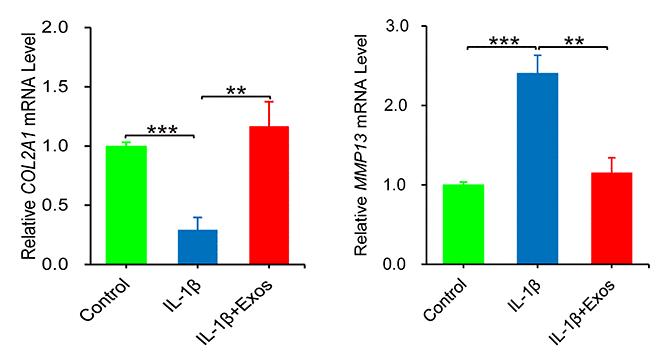
The interleukin IL-1ß is a known contributor to osteoarthritis, as it promotes inflammation in chondrocytes. Therefore, after deriving EVs from the human umbilical cord cells, the researchers administered them to chondrocytes exposed to IL-1ß.
They measured the RNA expression of two key genes: COL2A1, which is responsible for the extracellular matrix of cartilage, and MMP13, a matrix metalloproteinase that causes the extracellular matrix to degrade. They found that MSC-derived EVs almost perfectly counteracted the effects of IL-1ß.

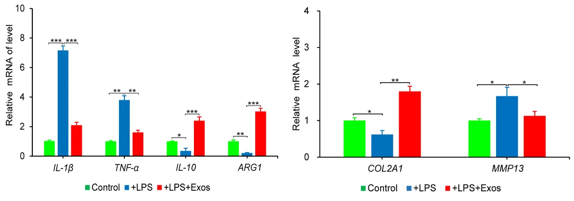
Next, the researchers introduced macrophages along with LPS, a compound that encourages them to exhibit the M1 inflammatory phenotype rather than the M2 phenotype that encourages long-term healing. M1 macrophages, as expected, upregulated inflammatory biomarkers in the chondrocytes, caused a decrease in COL2A1 and an increase in MMP13, and caused them to undergo the programmed death known as apoptosis. Adding MSC-derived EVs reversed this situation, similar to the chondrocytes exposed to IL-1ß.

The RNA in the EVs
The researchers then looked at the specific microRNA payloads that these EVs were carrying. They found high levels of miRNA-21-5p, which previous research has found to be responsible for many of the effects seen in this study [3]. They also found microRNAs that.were associated with inflammation along with the MAPK and PI3K-Akt signaling pathways, which have been implicated in osteoarthritis [4].
They were able to confirm that these EVs were having effects on these pathways in the chondrocytes themselves. RNA analysis showed that MAPK and PI3K-Akt pathways were indeed affected, as was the formation of cartilage, showing a direct relationship between EV exposure and potential benefits for osteoarthritis treatment.
However, despite these highly positive effects, this is a cellular study and was not conducted in a living animal model. It would be very informative to test if EVs derived from MSCs are effective in treating osteoarthritis in guinea pigs or other animals.
Literature
[1] He, L., Chen, Y., Ke, Z., Pang, M., Yang, B., Feng, F., … & Shu, T. (2020). Exosomes derived from miRNA-210 overexpressing bone marrow mesenchymal stem cells protect lipopolysaccharide induced chondrocytes injury via the NF-?B pathway. Gene, 751, 144764.
[2] Liu, Y., Lin, L., Zou, R., Wen, C., Wang, Z., & Lin, F. (2018). MSC-derived exosomes promote proliferation and inhibit apoptosis of chondrocytes via lncRNA-KLF3-AS1/miR-206/GIT1 axis in osteoarthritis. Cell cycle, 17(21-22), 2411-2422.
[3] Zhu, H., Yan, X., Zhang, M., Ji, F., & Wang, S. (2019). miR-21-5p protects IL-1ß-induced human chondrocytes from degradation. Journal of Orthopaedic Surgery and Research, 14, 1-9.
[4] Shi, J., Zhang, C., Yi, Z., & Lan, C. (2016). Explore the variation of MMP3, JNK, p38 MAPKs, and autophagy at the early stage of osteoarthritis. IUBMB life, 68(4), 293-302.


