How Parkinson’s Disease Perpetuates Itself
- Sometimes, the helper cells do more harm than good.

A new paper elaborates on how and why microglia fail to clean up the α-synuclein protein of Parkinson’s disease, gradually making the disease worse.

The aggregation of Parkinson’s
Parkinson’s disease, like Alzheimer’s, is characterized by protein aggregation. However, the affected neurons, the aggregates, and the pathology are all different. In Parkinson’s, aggregates of α-synuclein collect to form Lewy bodies in the brain stem [1] as motor function gradually declines.
While α-synuclein is expressed by neurons and propagates between them [2], the helper cells of the brain known as microglia play a role as well. Under normal circumstances, these cells consume α-synuclein, thus protecting the brain from Parkinson’s [3].
Both neurons and microglia have lysosomes, cellular garbage collectors whose purpose is to destroy unwanted proteins. Previous work has found that α-synuclein aggregates cause neuronal lysosomes to rupture [4], causing further aggregation of this protein in the cells and contributing to its spread [5]. Microglia that fail to destroy this protein in their lysosomes encapsulate it into exosomes instead, sending it back out into the brain [6].
When maintenance fails to maintain
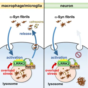
This paper, published in iScience, investigated how and why the microglia are perpetuating this dangerous protein. It focuses on the LRRK2/Rab10 pathway, which plays a major role in governing how these cells use lysosomes [7]. These researchers’ previous work demonstrated that this pathway can affect the release of lysosomal contents outside of cells [8].
In this paper, they began by introducing insoluble α-synuclein into various cell types from humans and mice, then stressed the lysosomes through chemicals such as chloroquine. Macrophages and microglia behaved as expected, sending aggregated α-synuclein out of themselves. Other cell types, however, do not excrete these aggregates in the same way.
Further experimentation, marking, and testing of the α-synuclein and other released proteins showed that the α-synuclein had, indeed, gone through lysosomes and had been incompletely digested in the process. However, it was still found to be pathogenic, capable of forming seeds around which other α-synuclein could form aggregates and thus contributing to Parkinson’s.
Silencing LRRK2 or Rab10 suppressed the release of exosomes, showing that this pathway is indeed responsible. Interestingly, inflammatory pathways, such as TLR2, were found to have no involvement. Instead, the internal α-synuclein, itself, was found to be the causative factor behind this pathway’s activation.
While this was a cellular study that only delved into causes, these findings offer a glimmer of hope for the development of future treatments. Ideally, being helper cells, microglia should never excrete dangerous proteins before digesting them. Treatments that encourage lysosomal clearance of α-synuclein, rather than its release, might be effective in ameliorating Parkinson’s.
Literature
[1] Baba, M., Nakajo, S., Tu, P. H., Tomita, T., Nakaya, K., Lee, V. M., … & Iwatsubo, T. (1998). Aggregation of alpha-synuclein in Lewy bodies of sporadic Parkinson’s disease and dementia with Lewy bodies. The American journal of pathology, 152(4), 879.
[2] Luk, K. C., Kehm, V., Carroll, J., Zhang, B., O’Brien, P., Trojanowski, J. Q., & Lee, V. M. Y. (2012). Pathological α-synuclein transmission initiates Parkinson-like neurodegeneration in nontransgenic mice. Science, 338(6109), 949-953.
[3] Choi, I., Zhang, Y., Seegobin, S. P., Pruvost, M., Wang, Q., Purtell, K., … & Yue, Z. (2020). Microglia clear neuron-released α-synuclein via selective autophagy and prevent neurodegeneration. Nature communications, 11(1), 1386.
[4] Flavin, W. P., Bousset, L., Green, Z. C., Chu, Y., Skarpathiotis, S., Chaney, M. J., … & Campbell, E. M. (2017). Endocytic vesicle rupture is a conserved mechanism of cellular invasion by amyloid proteins. Acta neuropathologica, 134, 629-653.
[5] Alvarez-Erviti, L., Seow, Y., Schapira, A. H., Gardiner, C., Sargent, I. L., Wood, M. J., & Cooper, J. M. (2011). Lysosomal dysfunction increases exosome-mediated alpha-synuclein release and transmission. Neurobiology of disease, 42(3), 360-367.
[6] Guo, M., Wang, J., Zhao, Y., Feng, Y., Han, S., Dong, Q., … & Tieu, K. (2020). Microglial exosomes facilitate α-synuclein transmission in Parkinson’s disease. Brain, 143(5), 1476-1497.
[7] Kuwahara, T., & Iwatsubo, T. (2020). The emerging functions of LRRK2 and Rab GTPases in the endolysosomal system. Frontiers in neuroscience, 14, 227.
[8] Eguchi, T., Kuwahara, T., Sakurai, M., Komori, T., Fujimoto, T., Ito, G., … & Iwatsubo, T. (2018). LRRK2 and its substrate Rab GTPases are sequentially targeted onto stressed lysosomes and maintain their homeostasis. Proceedings of the National Academy of Sciences, 115(39), E9115-E9124.


