Inflammation May Drive Fat Distribution in Older People
- Fat cells do not all behave the same way.

A new study published in Aging Cell has detailed what happens to individual fat cells in white adipose tissue (WAT) as they age.
When fat is more than just fat
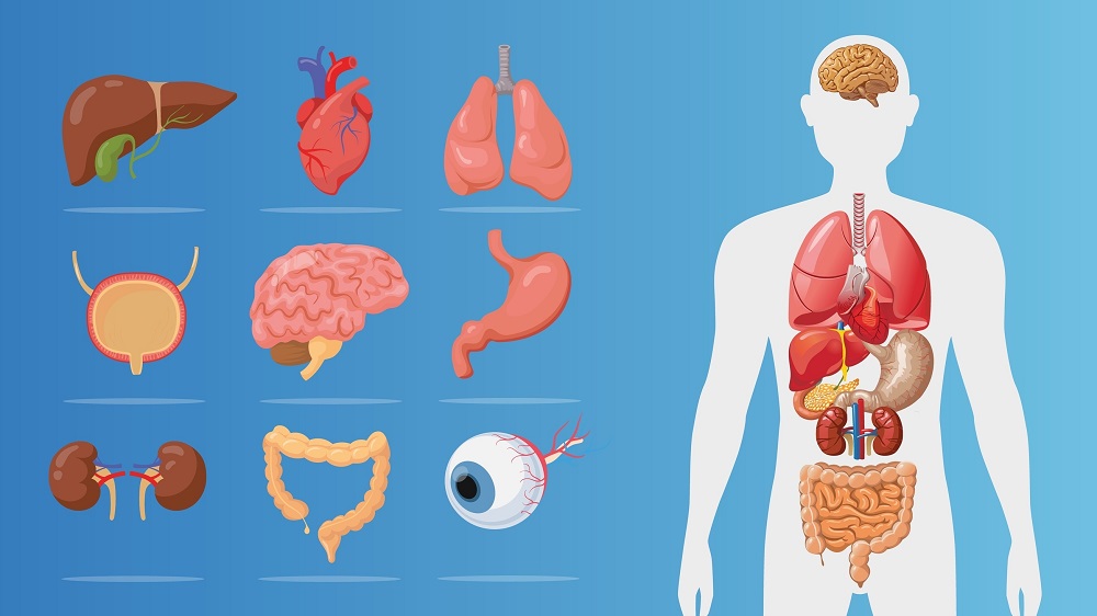
Previous research has described WAT as an organ in its own right, functioning not only as an energy storage source but as a regulator of metabolism [1]. If this function is impaired, adipose tissue moves towards the central abdomen [2]; causes fats to accumulate in other tissues, which leads to insulin resistance [3]; and leads to low-level chronic inflammation [4].
Like many other declines in function, this is related to aging. Cellular senescence [5], a lack of stem cell progenitors [6], and infiltration of immune cells into tissues [7] have all been pinpointed as potential causes.
However, those prior analyses were based on relatively primitive approaches, such as fluorescence imagery and relatively blunt cellular or tissue analysis. This research utilizes single-cell transcriptomics, a field that has recently been used to analyze human WAT [8]. As this team has recently used single-cell RNA sequencing on human fat cells [9], the researchers then chose to use that technique, in addition to others, in order to determine the effects of aging on these cells.
Fatter in the middle despite having the same BMI
This experiment drew samples from ten people who were at least 65 and ten more under the age of 30. Despite having similar metrics in insulin sensitivity and body mass, the older group had higher systolic blood pressure, greater waist circumference, and worse cholesterol measurements.
Analyzing the cells’ RNA to determine what genes were upregulated, the researchers initially found two groups of cells with differences that had little to do with aging. The first group, Adip_1, had upregulation of genes related to handling oxidation, while the second group, Adip_2, had upregulation of genes that were related to insulin responsiveness.
There were age-related differences in cell type and composition. Older people had more mast cells of connective tissue along with more macrophages associated with lipids, and these macrophages were of the M1 inflammatory type. Younger people’s stem cells produced more proteins related to the extracellular matrix, their vascular cells were more likely to create new blood vessels, and their Adip_2 cells had more active genes related to lipid metabolism.
Instead, in the older group, Adip_2 cells were more likely to have a gene expression profile associated with increased inflammation, and many cells in older people produced collagen that was associated with fibrosis and a lack of insulin sensitivity [10]. Fibrosis itself was, fortunately, not found to be increased with aging in WAT. Older people did, however, have more very large fat cells than younger people did.
Macrophages in the gut
Macrophage infiltration was visible under the microscope. In older males, macrophages attacking damaged or dying adipocytes would form crown-like structures in the process. This was very strongly associated with the accumulation of fat in the abdominal area. In particular, CXC14 was singled out as a key driver of inflammation and macrophage infiltration [11], and it was found to be upregulated in older people.
Unsurprisingly, older people had significantly increased levels of cellular senescence in WAT. Rather than being general among all cell types, though, pre-adipocytes, Adip_1 cells, and vascular tissues were noted as producing senescence-related proteins.
This study was illuminating for future work, associating previously unassociated biological metrics with inflammation and senescence. Fat accumulating in the gut is not just something that happens: it appears to be the result of the fat tissue, itself, suffering from inflammaging.
Literature
[1] Goodpaster, B. H., & Sparks, L. M. (2017). Metabolic flexibility in health and disease. Cell metabolism, 25(5), 1027-1036.
[2] Kuk, J. L., Saunders, T. J., Davidson, L. E., & Ross, R. (2009). Age-related changes in total and regional fat distribution. Ageing research reviews, 8(4), 339-348.
[3] Boren, J., Taskinen, M. R., Olofsson, S. O., & Levin, M. (2013). Ectopic lipid storage and insulin resistance: a harmful relationship. Journal of internal medicine, 274(1), 25-40.
[4] Starr, M. E., Evers, B. M., & Saito, H. (2009). Age-associated increase in cytokine production during systemic inflammation: adipose tissue as a major source of IL-6. Journals of Gerontology Series A: Biomedical Sciences and Medical Sciences, 64(7), 723-730.
[5] Justice, J. N., Gregory, H., Tchkonia, T., LeBrasseur, N. K., Kirkland, J. L., Kritchevsky, S. B., & Nicklas, B. J. (2018). Cellular senescence biomarker p16INK4a+ cell burden in thigh adipose is associated with poor physical function in older women. The Journals of Gerontology: Series A, 73(7), 939-945.
[6] Caso, G., McNurlan, M. A., Mileva, I., Zemlyak, A., Mynarcik, D. C., & Gelato, M. C. (2013). Peripheral fat loss and decline in adipogenesis in older humans. Metabolism, 62(3), 337-340.
[7] Trim, W. V., Walhin, J. P., Koumanov, F., Bouloumié, A., Lindsay, M. A., Chen, Y. C., … & Thompson, D. (2022). Divergent immunometabolic changes in adipose tissue and skeletal muscle with ageing in healthy humans. The Journal of physiology, 600(4), 921-947.
[8] Divoux, A., Whytock, K. L., Halasz, L., Hopf, M. E., Sparks, L. M., Osborne, T. F., & Smith, S. R. (2024). Distinct subpopulations of human subcutaneous adipose tissue precursor cells revealed by single-cell RNA sequencing. American Journal of Physiology-Cell Physiology, 326(4), C1248-C1261.
[9] Whytock, K. L., Divoux, A., Sun, Y., Hopf, M., Yeo, R. X., Pino, M. F., … & Sparks, L. M. (2023). Isolation of nuclei from frozen human subcutaneous adipose tissue for full-length single-nuclei transcriptional profiling. STAR protocols, 4(1), 102054.[10] Divoux, A., Tordjman, J., Lacasa, D., Veyrie, N., Hugol, D., Aissat, A., … & Clément, K. (2010). Fibrosis in human adipose tissue: composition, distribution, and link with lipid metabolism and fat mass loss. Diabetes, 59(11), 2817-2825.
[11] Lu, J., Chatterjee, M., Schmid, H., Beck, S., & Gawaz, M. (2016). CXCL14 as an emerging immune and inflammatory modulator. Journal of Inflammation, 13, 1-8.