
Researchers publishing in Nature Aging have described how mitochondrial stress is a key part of why senolytics are effective.
Finding targeted effectiveness

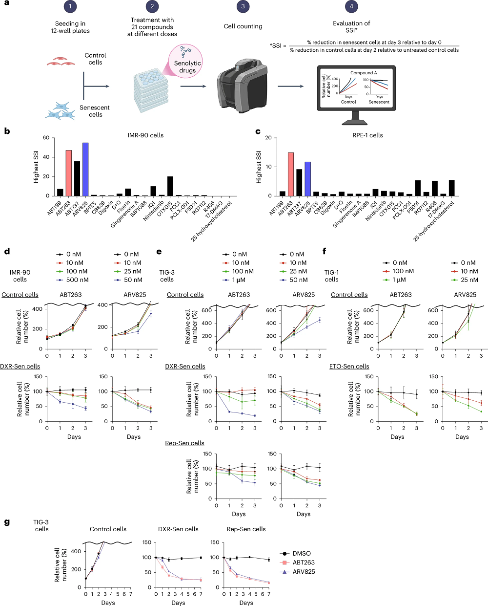
The researchers began this study by summarizing senescent cells and the senolytics created to eliminate them. They noted that few attempts have been made to determine which senolytics are the most broadly effective against senescent cells while having the least effects on non-senescent ones [1].
To that end, they created a senolytic specificity index (SSI), a simple metric that compares the number of senescent cells removed to the number of non-senescent cells removed. They tested 21 distinct agents, ranging from the well-known combination of dasatinib and quercetin (D+Q) to three different ABT compounds, one of which, ABT263 (Navitoclax), is well-known in the field as being an effective senolytic.
This researchers’ initial experiment confirmed that finding. Navitoclax was the most effective at selectively removing RPE-1 cells, which are human epithelial cells that are commonly used in senescence research; it was barely edged out in effectiveness by ARV825 at dealing with IMR-90, a line of human fibroblasts that serve the same purpose. Unfortunately, D+Q and fisetin performed very poorly on the SSI metric compared to these two compounds. Testing other types of senescent cells, and driving them senescent both replicatively and through toxin exposure, confirmed the broad effectiveness of both navitoclax and ARV825.
Some cells refuse to die
While these and similar compounds have advantages over other senolytics, such as not prompting suicidal apoptotic responses in non-senescent cells, they are not perfect. The researchers noted that previous work has found that BCL-2 inhibitors such as navitoclax are not effective against senescent preadipocytes [2] and that their own work has found imperfect clearance; roughly a quarter of the treated senescent cells survived navitoclax or ARV825, even after a week of senolytic treatment.
The researchers then took a step further, looking into why such strong senolytics failed against those particular cells. They found that the survivors had unusually high expressions of senescent cells’ characteristic SASP factors and that they fought more strongly against oxidative stress, decreasing the reactive oxygen species (ROS) that may have contributed to the other cells’ death.
Further analysis found that these cells were also better at clearing damaged mitochondria. One particular gene, ATP6V0E1, plays a key role in this process [3], and knocking this gene down greatly increased the effectiveness of navitoclax. The accumulation of damaged mitochondria is key to the effectiveness of both navitoclax and ARV825; cells with depleted mitochondria were significantly less likely to die to these senolytics.
Mitochondrial stress helps senolytics do their job
The researchers then experimented with various methods of imposing mitochondrial stress. First, they did so directly through gene silencing, finding that direct downregulation of mitochondrial maintenance functions causes senescent cells to die in the same way as when they are treated with these two senolytics. Directly interfering with mitochondrial DNA replication boosted their effects as well, and, critically, did not appear to kill off non-senescent cells.
The researchers switched cells from glycolysis to oxidative phosphorylation (OXPHOS) by reducing the amount of glucose that the cells received, simulating a low-carbohydrate diet and causing oxidative stress [4], but this had effects on normal cells as well as senescent ones. They then tested a GLUT1 inhibitor, BAY-876 [5], to force this shift; co-treating cells with BAY-876 along with navitoclax or ARV825 was found to increase the effectiveness of these senolytics while still sparing non-senescent cells from death.
These findings were replicated in mice. Older male mice were injected with melanoma cells that are known to co-locate with senescent cells, which fuel the growth of this cancer. Then, they were fed either navitoclax or ARV825 alongside either a normal or a low-carbohydrate ketogenic diet. The mice receiving the ketogenic diet had significantly stronger responses to senolytics; two key SASP factors that are known to attract this cancer were substantially reduced in the low-carb groups compared to the normal ones. While some previous work has linked ketogenic diets to cellular senescence [6], these researchers did not observe this in the lungs of their tested mice.
These findings are limited, and they present a conundrum to the field. The same basic stresses that prime senescent cells for removal by senolytics also affect how normal cells function. While these experiments showed benefits when stresses were combined with senolytics, it is still uncertain whether senolytics should be combined with physical interventions, such as low-carb diets or intensive exercise, for maximum effectiveness. Further work will need to be done on animals and people in order to determine if such combinations are helpful or harmful in the long run.
Literature
[1] Di Micco, R., Krizhanovsky, V., Baker, D., & d’Adda di Fagagna, F. (2021). Cellular senescence in ageing: from mechanisms to therapeutic opportunities. Nature reviews Molecular cell biology, 22(2), 75-95.
[2] Zhu, Y. I., Tchkonia, T., Fuhrmann‐Stroissnigg, H., Dai, H. M., Ling, Y. Y., Stout, M. B., … & Kirkland, J. L. (2016). Identification of a novel senolytic agent, navitoclax, targeting the Bcl‐2 family of anti‐apoptotic factors. Aging cell, 15(3), 428-435.
[3] Colacurcio, D. J., & Nixon, R. A. (2016). Disorders of lysosomal acidification—The emerging role of v-ATPase in aging and neurodegenerative disease. Ageing research reviews, 32, 75-88.
[4] Liu, Y., Song, X. D., Liu, W., Zhang, T. Y., & Zuo, J. (2003). Glucose deprivation induces mitochondrial dysfunction and oxidative stress in PC12 cell line. Journal of cellular and molecular medicine, 7(1), 49-56.
[5] Siebeneicher, H., Cleve, A., Rehwinkel, H., Neuhaus, R., Heisler, I., Müller, T., … & Buchmann, B. (2016). Identification and optimization of the first highly selective GLUT1 inhibitor BAY‐876. ChemMedChem, 11(20), 2261-2271.
[6] Wei, S. J., Schell, J. R., Chocron, E. S., Varmazyad, M., Xu, G., Chen, W. H., … & Gius, D. (2024). Ketogenic diet induces p53-dependent cellular senescence in multiple organs. Science advances, 10(20), eado1463.