The Blog

Building a Future Free of Age-Related Disease

Connected neurons

Tiny Brain Organoids Show Promise in Alzheimer’s Research

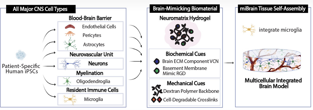
Scientists have developed a novel multicellular integrated brain model to study neurological diseases, incorporating all six major brain cell types derived from patient-specific induced pluripotent stem cells (iPSCs) [1].

Beyond organoids

Organoids, tiny lab-grown patches of tissue, are a promising tool for accelerating medical research [2]. They sit a step above cultured cells, being able to mimic the workings of a particular organ such as the liver. Organoids can often replace animal models as a cheaper and more humane alternative.

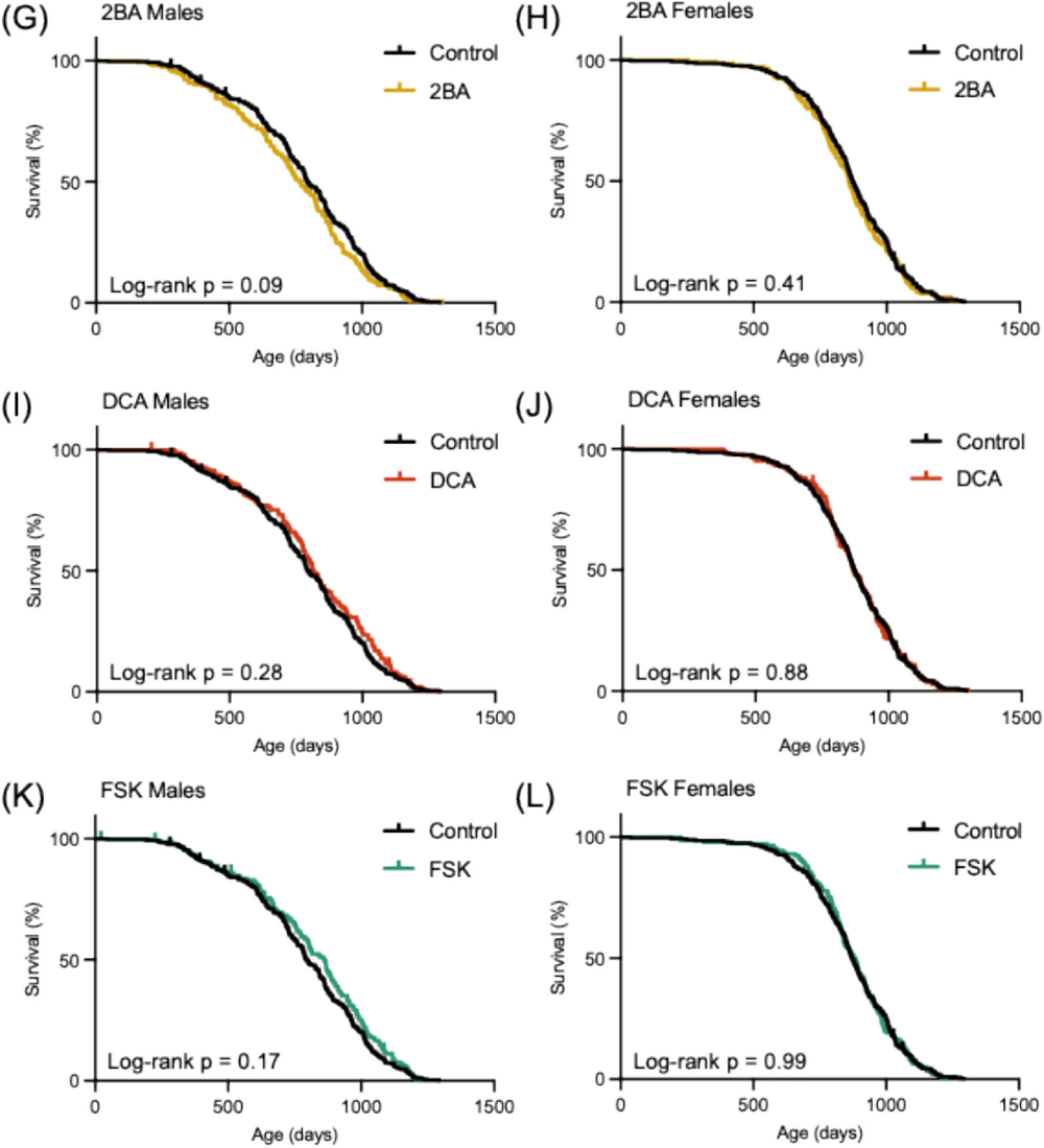
However, some organs and tissues are harder to replicate than others. Creating brain organoids, for instance, has been a challenge. In a new study published in the Proceedings of the National Academy of Science, MIT scientists have reported a breakthrough: they have created brain organoids called miBrains that contain all the essential brain cell types and even the blood-brain barrier.

Making a piece of brain

The scientists reprogrammed patient-derived induced pluripotent stem cells (iPSCs) into the required cell types (neurons, astrocytes, oligodendrocytes, microvascular endothelial cells, pericytes, and microglia), validating the transformation by measuring gene expression and cellular function. For instance, iPSC-derived neurons demonstrated robust expression of neuron-associated proteins, formed healthy networks, made abundant connections, fired on their own and when stimulated, and reacted to neurological drugs the way real neurons do.

The cells were mixed together inside a hydrogel environment called Neuromatrix, which closely matches the natural extracellular matrix (ECM), including ECM proteins and RGD, a basement membrane peptide mimic. The researchers experimented with several matrix variants until arriving at the one that the cells were willing to call home.

Vascularizing organoids presents a particular challenge. The brain completely relies on the blood-brain barrier, which consists of endothelial cells in the blood vessels that are tuned to only let certain molecules into the brain microenvironment. The researchers observed the self-assembly of microvessels and the appearance of tight junctions (protein “seals” between adjacent cells that block leaks and set cell polarity) – both crucial BBB features.

miBrain Structure

Interestingly, miBrain cells were more “lifelike” than their monocultured counterparts. For instance, these astrocytes exhibited enhanced identity and functional gene expression compared to monoculture conditions, and their gene expression was more similar to, but not quite the same as, astrocytes found in the human brain.

A research platform and more

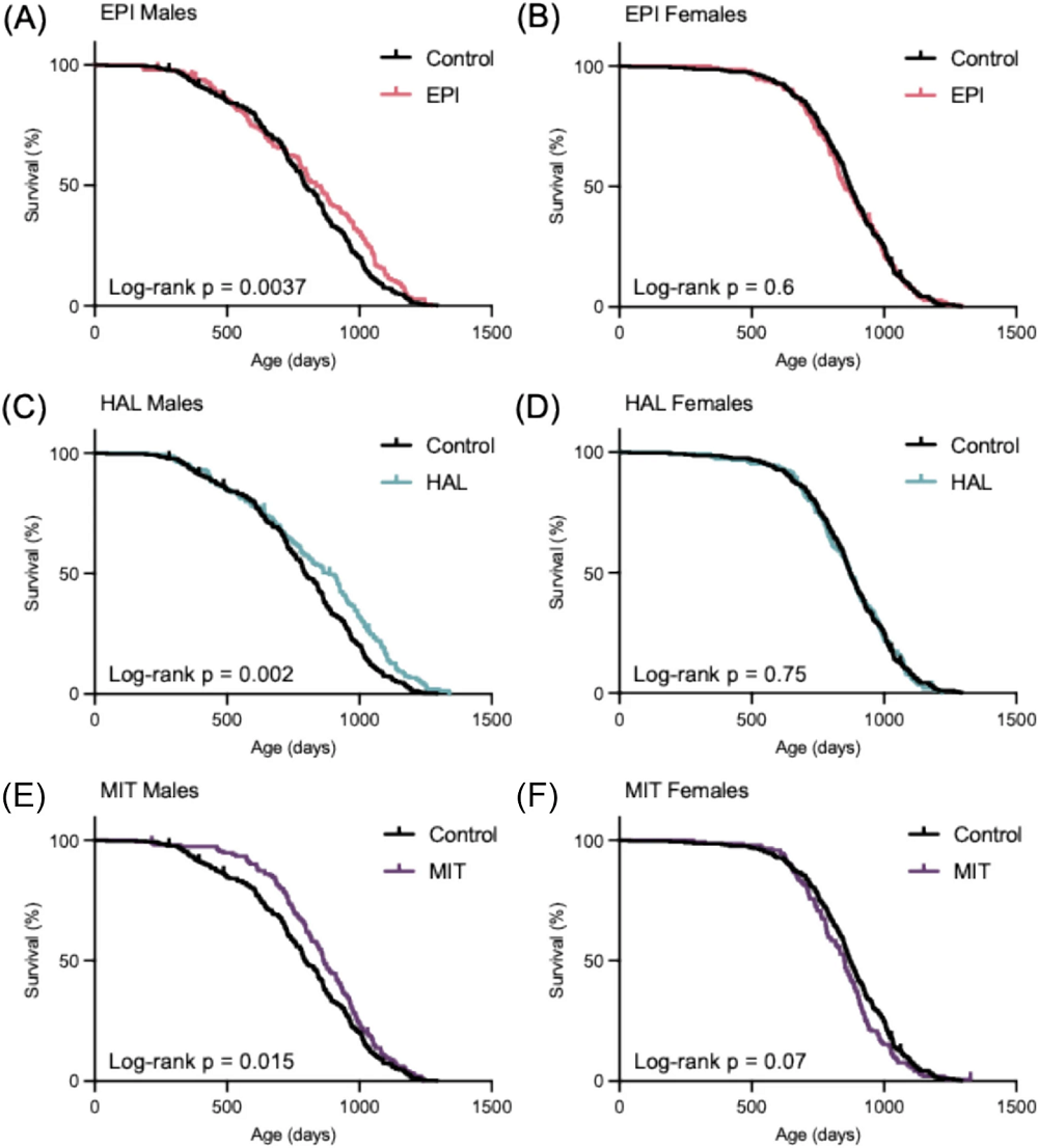
The researchers then conducted an experiment to demonstrate their model’s research value. APOE4, an allele of the gene APOE, is the strongest common genetic risk factor for late-onset Alzheimer’s [3]. Astrocytes make a lot of the APOE protein, but the extent to which astrocytic APOE4 drives pathology is unclear. With miBrain, the researchers were able to swap genotypes cell-by-cell to test whether APOE4 astrocytes are sufficient to trigger downstream problems, and whether this requires microglia crosstalk.

First, they confirmed that miBrains where all cell lineages were carrying APOE4 showed more amyloid aggregation and more tau phosphorylation than controls expressing APOE3 (the “neutral” allele). This signaled that the model naturally expresses APOE4-linked features seen in Alzheimer’s disease. When microglia were removed, the widespread neuronal tau phosphorylation largely dropped, implying the effect depends on astrocyte-to-microglia signaling.

The team then built APOE3 miBrains but replaced only the astrocytes with APOE4 astrocytes using CRISPR. The experiment showed that tau phosphorylation increased in this model, too, indicating that astrocytes can be sufficient to push neuronal tau changes.

“The miBrain is the only in vitro system that contains all six major cell types that are present in the human brain,” said Li-Huei Tsai, director of The Picower Institute of Learning and Memory and senior author of the study. “In their first application, miBrains enabled us to discover how one of the most common genetic markers for Alzheimer’s disease alters cells’ interactions to produce pathology. I’m most excited by the possibility to create individualized miBrains for different individuals. This promises to pave the way for developing personalized medicine.”

“Its highly modular design sets the miBrain apart, offering precise control over cellular inputs, genetic backgrounds, and sensors – useful features for applications such as disease modeling and drug testing,” said Alice Stanton, assistant professor at Harvard Medical School and Massachusetts General Hospital, who co-led the study. “Given its sophistication and modularity, there are limitless future directions. Among them, we would like to harness it to gain new insights into disease targets, advanced readouts of therapeutic efficacy, and optimization of drug delivery vehicles.”

Beyond their obvious uses in studying neurodegenerative diseases, miBrain-like technologies may one day power gradual brain tissue replacement in the longevity context, a project that Jean Hebert is working on at ARPA-H.

We would like to ask you a small favor. We are a non-profit foundation, and unlike some other organizations, we have no shareholders and no products to sell you. All our news and educational content is free for everyone to read, but it does mean that we rely on the help of people like you. Every contribution, no matter if it’s big or small, supports independent journalism and sustains our future.

Literature

[1] Stanton, A. E., Bubnys, A., Agbas, E., James, B., Park, D. S., Jiang, A., … & Tsai, L. H. (2025). Engineered 3D immuno-glial-neurovascular human miBrain model. Proceedings of the National Academy of Sciences, 122(42), e2511596122.

[2] Yao, Q., Cheng, S., Pan, Q., Yu, J., Cao, G., Li, L., & Cao, H. (2024). Organoids: development and applications in disease models, drug discovery, precision medicine, and regenerative medicine. MedComm, 5(10), e735.

[3] Belloy, M. E., Andrews, S. J., Le Guen, Y., Cuccaro, M., Farrer, L. A., Napolioni, V., & Greicius, M. D. (2023). APOE genotype and Alzheimer disease risk across age, sex, and population ancestry. JAMA neurology, 80(12), 1284-1294.

International Space Station

How Being in Orbit Affects Stem Cells

In Cell Stem Cell, researchers from the Cedars-Sinai Medical Center have published a review discussing what experiments conducted in space can do for stem cell research and medical research as a whole.

Cells behave differently in microgravity

The gravitational effects of freefalling in orbit are very difficult to properly simulate on Earth, making this the prime reason to conduct cellular research in space. Scientists working with NASA have found that cells cultured in such microgravity don’t need scaffolds to grow in three dimensions [1], but on the other hand, microgravity has been found to cause embryonic stem cells to fail to differentiate and regenerate tissues [3].

Other work conducted aboard the International Space Station (ISS) have found that neural organoids mature more quickly in microgravity [2]. This particular research may be strongly relevant to at least one age-related disease: the cells used in this experiment included dopaminergic neurons that deteriorate in Parkinson’s, and, unexpectedly, they had reduced signs of stress and grew readily even without close intervention. Future studies may model Alzheimer’s disease.

Microgravity’s effects on bone deterioration are well-known [4], and studies on bone-building cells have yielded surprising results. One study found that mice that spent time in space had stem cells that were better able to build bone after returning to Earth [5], which these reviewers describe as a “paradoxical” finding. Meanwhile, human bone marrow stem cells grown under these conditions had half the calcification, cell cycle arrest without the characteristic elements of senescence, and less stiffness of the extracellular matrix [6].

Mechanical forces control how cells grow

Physical forces having effects on cellular workings is known as mechanotransduction, and on Earth, cells are constantly exposed to such forces. For example, cells evolved to have their internal structures (cytoskeletons) handle Earth’s gravity. In its absence, these cytoskeletons grow differently [7]. One key pathway has been identified in this process: the Hippo-Yes-associated protein (YAP) signaling axis, which acutely responds to mechanical forces.

Cardiovascular progenitors grown in space upregulate YAP in a way that suggests increased regeneration [8], and using these progenitors to grow organoids in space creates spheres that are thrice the diameter, and contain roughly twenty times the cells, of Earth-bound organoids grown for the same amount of time, with sharp upregulations in genes related to profliferation and survival [9].

Overall, the research on cells grown in microgravity has discovered a complex combination of reactions, some of which appear to be beneficial and others that are not. The reviewers urge more epigenetic testing on these cells, seeking to discover what particular factors are responsible for these modifications. They also note a lack of immunological studies and studies related to vasculature in organoids, and they hold that deriving iPSCs from astronauts themselves may yield insights.

Engineering applications

The amount of energy needed to bring any payload into space, and bring it back safely, makes orbital manufacturing extraordinarily expensive and significantly increases the cost of any research that might be done there. However, pound for pound, creating functional cells is already one of the most expensive activities on Earth. The reviewers note that 3D printing of organoids, a difficult process on Earth because such things often collapse under their own weight, is far easier under conditions where that isn’t a factor, and bioprinting has been done on the ISS since 2019.

Cardiac organoids aren’t the only ones that can be grown rapidly in space; chondrocytes, the cells responsible for growing cartilage, grow up to twice as fast in microgravity conditions [11]. The reviewers suggest that such accelerated growth may make orbital production of these cells a viable prospect, but they also note that not being grown under Earth conditions may make them unable to properly handle shear stresses when they return here. Other work found that mesenchymal stem cells (MSCs) grown under microgravity secreted more anti-inflammatory factors than Earth-grown cells [12].

The researchers note that this work is in its very earliest stages and that stem cell production in orbit is still being investigated as research initatives rather than as production facilities meant to serve clinical patients. Scaling up these efforts will require significant future work and require the mitigation of serious technical hurdles, such as proper shielding against the intense radiation found outside of Earth’s atmosphere.

However, if these hurdles can be overcome, treating some age-related diseases here on Earth may possibly be done with cells grown in orbit.

We would like to ask you a small favor. We are a non-profit foundation, and unlike some other organizations, we have no shareholders and no products to sell you. All our news and educational content is free for everyone to read, but it does mean that we rely on the help of people like you. Every contribution, no matter if it’s big or small, supports independent journalism and sustains our future.

Literature

[1] Jogdand, A., Landolina, M., & Chen, Y. (2024). Organs in orbit: how tissue chip technology benefits from microgravity, a perspective. Frontiers in lab on a Chip Technologies, 3, 1356688.

[2] Marotta, D., Ijaz, L., Barbar, L., Nijsure, M., Stein, J., Pirjanian, N., … & Fossati, V. (2024). Effects of microgravity on human iPSC-derived neural organoids on the International Space Station. Stem Cells Translational Medicine, 13(12), 1186-1197.

[3] Blaber, E. A., Finkelstein, H., Dvorochkin, N., Sato, K. Y., Yousuf, R., Burns, B. P., … & Almeida, E. A. (2015). Microgravity reduces the differentiation and regenerative potential of embryonic stem cells. Stem cells and development, 24(22), 2605-2621.

[4] Grimm, D., Grosse, J., Wehland, M., Mann, V., Reseland, J. E., Sundaresan, A., & Corydon, T. J. (2016). The impact of microgravity on bone in humans. Bone, 87, 44-56.

[5] Blaber, E. A., Dvorochkin, N., Torres, M. L., Yousuf, R., Burns, B. P., Globus, R. K., & Almeida, E. A. C. (2014). Mechanical unloading of bone in microgravity reduces mesenchymal and hematopoietic stem cell-mediated tissue regeneration. Stem cell research, 13(2), 181-201.

[6] Bradamante, S., Rivero, D., Barenghi, L., Balsamo, M., Minardi, S. P., Vitali, F., & Cavalieri, D. (2018). SCD–stem cell differentiation toward osteoblast onboard the international space station. Microgravity Science and Technology, 30(5), 713-729.

[7] Wu, X. T., Yang, X., Tian, R., Li, Y. H., Wang, C. Y., Fan, Y. B., & Sun, L. W. (2022). Cells respond to space microgravity through cytoskeleton reorganization. The FASEB Journal, 36(2), e22114.

[8] Camberos, V., Baio, J., Bailey, L., Hasaniya, N., Lopez, L. V., & Kearns-Jonker, M. (2019). Effects of spaceflight and simulated microgravity on YAP1 expression in cardiovascular progenitors: implications for cell-based repair. International Journal of Molecular Sciences, 20(11), 2742.

[9] Rampoldi, A., Forghani, P., Li, D., Hwang, H., Armand, L. C., Fite, J., … & Xu, C. (2022). Space microgravity improves proliferation of human iPSC-derived cardiomyocytes. Stem Cell Reports, 17(10), 2272-2285.

[10] Jeyaraman, M., Ramasubramanian, S., Yadav, S., & Jeyaraman, N. (2024). Exploring New Horizons: Advancements in Cartilage Tissue Engineering Under Space Microgravity. Cureus, 16(8).

[11] Jeyaraman, M., Ramasubramanian, S., Yadav, S., & Jeyaraman, N. (2024). Exploring New Horizons: Advancements in Cartilage Tissue Engineering Under Space Microgravity. Cureus, 16(8).

[12] Huang, P., Russell, A. L., Lefavor, R., Durand, N. C., James, E., Harvey, L., … & Zubair, A. C. (2020). Feasibility, potency, and safety of growing human mesenchymal stem cells in space for clinical application. npj Microgravity, 6(1), 16.

Older woman in wheelchair

Female-Specific Mechanism of Neurodegeneration Discovered

Scientists have found that a gene on the X chromosome, which escapes silencing, promotes inflammation and neurodegeneration in a mouse model of multiple sclerosis, but the effects can be reversed with metformin. Human RNA sequencing data supports these findings [1].

Why are women more vulnerable?

Multiple sclerosis (MS) is an autoimmune and neurodegenerative disease that is most commonly diagnosed between the ages of 20 and 40. In MS, circulating immune cells, specifically lymphocytes and macrophages, infiltrate the central nervous system, causing various kinds of damage, such as a loss of myelin and the activation of microglia and astrocytes, the brain’s supporting cells. The resulting neuroinflammation, a common feature of MS and other age-related neurodegenerative diseases such as Alzheimer’s, is thought to be a major culprit [2].

Interestingly, females are more susceptible to MS, with a ratio of 3:1 compared to males [3], another aspect in which it resembles Alzheimer’s. Women are also more prone to relapses. The sex differences in the prevalence and development of these diseases have baffled researchers for decades. A new study from UCLA, published in Science Translational Medicine, yields interesting clues and a possible therapy.

An X chromosome gene that evades silencing

According to the paper, “sex differences in microglia can be caused by differential effects of sex hormones (estrogen versus testosterone), sex chromosomes (XX versus XY), or both.” Since males only have one X chromosome, the expression of the genes sitting on two chromosomes should be twice as high in women than in men. This is solved by the random inactivation of one X chromosome in females, but some genes escape silencing.

One such gene is Kdm6a (lysine demethylase 6A). Previous research has found that it has higher expression in females compared to males. Kdm6a encodes a protein which derepresses a particular region of chromatin via histone demethylation, allowing the translation of resident genes.

The researchers first studied brain-resident macrophages (microglia) derived from humans and detected high levels of Kdm6a, especially in cells from female donors. They then knocked out Kdm6a and observed a loss of its function in the cells.

The team then moved to an established mouse model of MS and again knocked out Kdm6a specifically in microglia. Female conditional knockout (cKO) mice exhibited much lower disease scores. Neuropathological assessments showed decreased axonal damage markers (APP) and increased intact axon markers (NF200) in cKO mice. More myelin was observed in cKO mice, indicating neuroprotective effects.

Kdm6a deletion in microglia also altered gene expression profiles. In healthy cKO mice, disease-associated microglial markers were indistinguishable from wild type mice. However, in MS cKO mice, resting microglial markers were slightly increased, suggesting a shift from disease-associated to resting microglial states. Gene ontology enrichment analysis indicated that several downregulated genes were involved in inflammatory responses.

The researchers repeated some of the experiments using both male and female mice. As expected, while female cKO mice received significant protective effects, males did not.

Female and male MS gene knockout effects

“Sex chromosomes and sex hormones achieve a balance through evolution,” said study lead author Dr. Rhonda Voskuhl, director of the Multiple Sclerosis Program at UCLA Health and lead neurologist for the UCLA Comprehensive Menopause Program. “There is a selection bias to do so. Females have a balance between X chromosome-driven inflammation that can be good to fight infections at child-bearing ages. This is held in check by estrogen, which is anti-inflammatory and neuroprotective. As women age, menopause causes loss of estrogen, unleashing the proinflammatory and neurodegenerative effects of this X chromosome in brain immune cells.”

Metformin provides protection

The researchers then gave mice metformin, a drug known in the longevity field for having possible geroprotective effects. Metformin is an anti-diabetes medication, whose mechanism of action is not fully understood, but it is thought to work via multiple pathways. One of its effects, which rarely gets attention, is to inhibit the histone demethylase activity of KDM6A.

Following this treatment, disease clinical scores improved in metformin-treated mice compared to vehicle-treated controls. Metformin reduced microglial activation and increased resting microglial markers. Gene expression changes mirrored those observed with Kdm6a deletion, indicating a reversal of disease-induced changes.

Due to metformin’s multiple mechanisms of action, it’s hard to ascribe the benefits solely to KDM6A inhibition. However, the treatment benefited almost exclusively females except for some limited early-disease benefits in males. This sex-specific effect is consistent with the KDM6A route.

Finally, the researchers examined the expression of KDM6A using human RNA sequencing data on people with MS. They found that in women, KDM6A expression was higher, and more microglial genes were dysregulated, supporting the findings in mouse models.

“It has long been known that there are sex differences in the brain,” said Voskuhl. “These can impact both health and neurological diseases. Multiple sclerosis and Alzheimer’s disease each affect women more often than men, about two to three times as often. Also, two-thirds of healthy women have ‘brain fog’ during menopause. These new findings explain why and point to a new treatment to target this.”

Voskuhl added that together, these findings may support the use of estrogens that target the brain to keep its balance, and thereby protect it, during menopause.

We would like to ask you a small favor. We are a non-profit foundation, and unlike some other organizations, we have no shareholders and no products to sell you. All our news and educational content is free for everyone to read, but it does mean that we rely on the help of people like you. Every contribution, no matter if it’s big or small, supports independent journalism and sustains our future.

Literature

[1] Yuichiro Itoh et al. Deletion of the X-chromosomal gene Kdm6a in microglia of female mice ameliorates neuroinflammation and restores translatome profiles. (2025) Sci. Transl. Med.17,eadq3401.

[2] Heneka, M. T., Carson, M. J., El Khoury, J., Landreth, G. E., Brosseron, F., Feinstein, D. L., … & Kummer, M. P. (2015). Neuroinflammation in Alzheimer’s disease. The Lancet Neurology, 14(4), 388-405.

[3] Portaccio, E., Magyari, M., Havrdova, E. K., Ruet, A., Brochet, B., Scalfari, A., … & Amato, M. P. (2024). Multiple sclerosis: emerging epidemiological trends and redefining the clinical course. The Lancet Regional Health–Europe, 44.

Infrared lamp

Infrared Lasers Clear Harmful Compounds in Mouse Brains

Researchers have discovered that infrared lasers promote the clearance of toxic metabolites from the brains of age-accelerated mice by improving lymphatic drainage.

Gunking up the works

Advanced glycation end-products (AGEs), like their acronym suggests, accumulate with age. These substances, which are formed when sugars bind to other molecules without the assistance of enzymes, drive multiple aspects of aging and can lead to metabolic diseases [1]. Introducing AGEs into the brain increases oxidation and leads to amyloid formation [2], and inhibiting the effects of AGEs is a potential treatment method [3].

Previous work has found that many other potentially hazardous, naturally produced substances, including amyloids, are drained by meningeal lymphatic vessels (MLVs) [4]. With aging, MLVs lose their youthful structure, becoming less able to carry away such wastes [5]. The only chemical method that has been found to expand MLVs, however, is vascular endothelial growth factor C, which must be injected directly into the brain in order to work [6].

As the meninges are on the brain’s surface, other treatment methods, such as near-infrared light, are feasible. Previous work has discovered that such phototherapy has beneficial effects in Alzheimer’s model mice [7], and improving MLV drainage also helps to heal brain injury in mice [8]. However, such previous work did not target AGEs, which are the focus of this study.

An age-accelerated model

This study began with ordinary, wild-type Black 6 mice that were exposed to D-galactose (D-gal), which is known to cause oxidative stress, mitochondrial dysfunction, and AGE accumulation [9]. At a 1275-nanometer wavelength, the near-infrared lasers used for the phototherapy were able to penetrate the skull. The researchers chose a laser dose of 10 milliwats per square centimeter, which was judged to be strong enough to provide benefits without excessively heating the brain.

Fluorescent imaging found that D-gal exposure did indeed AGEs in the brains of mice. Applying phototherapy moved these AGEs away from the cortex and into the deep cervical lymph nodes to be cleared. Reactive oxygen species, which cause oxidative damage, were also cleared. The treated mice had much less caspase-3, a compound that leads to cellular death by apoptosis.

The microglia were also significantly affected. Treated animals had considerable reductions in activated microglia, suggesting far less neuroinflammation, and the levels of related inflammatory cytokines were reduced. They also had significantly less astrocyte hypertrophy, signaling a significantly reduced effect of D-gal.

The researchers looked into potential mechanisms behind this clearance. They found that nitric oxide was a key mediator of the process; while MLVs treated with the lasers would expand and allow more clearance, inhibiting the function of nitric oxide prevented this from happening.

Behavioral benefits

Behavioral testing demonstrated that phototherapy had significant results on the mice’s cognition. In the well-known Morris water maze test, the treated animals’ ability to escape the maze was similar to that of animals that had never been exposed to D-gal at all. This was entirely due to cognition, not motor function. A correlation analysis found that the less oxidative stress and fewer inflammatory cytokines an animal had, the more likely it was to perform well in this test. There were also improvements in the treated mice’s ability to recognize novel objects.

Phototherapy Morris water maze

While there are obvious differences between humans and mice that might complicate such an approach, such as brain complexity and skull thickness, phototherapy has been used in human beings before. The researchers point to a study in which four football players with brain damage had been given such a therapy with positive results [10]. Therefore, they believe that this approach “may serve as a safe and effective intervention with high potential for rapid implementation into clinical antiaging applications.”

We would like to ask you a small favor. We are a non-profit foundation, and unlike some other organizations, we have no shareholders and no products to sell you. All our news and educational content is free for everyone to read, but it does mean that we rely on the help of people like you. Every contribution, no matter if it’s big or small, supports independent journalism and sustains our future.

Literature

[1] Chaudhuri, J., Bains, Y., Guha, S., Kahn, A., Hall, D., Bose, N., … & Kapahi, P. (2018). The role of advanced glycation end products in aging and metabolic diseases: bridging association and causality. Cell metabolism, 28(3), 337-352.

[2] Ko, S. Y., Lin, Y. P., Lin, Y. S., & Chang, S. S. (2010). Advanced glycation end products enhance amyloid precursor protein expression by inducing reactive oxygen species. Free Radical Biology and Medicine, 49(3), 474-480.

[3] Lu, J., Wu, D. M., Zheng, Y. L., Hu, B., Zhang, Z. F., Ye, Q., … & Wang, Y. J. (2010). Ursolic acid attenuates D-galactose-induced inflammatory response in mouse prefrontal cortex through inhibiting AGEs/RAGE/NF-κB pathway activation. Cerebral Cortex, 20(11), 2540-2548.

[4] Dupont, G., Iwanaga, J., Yilmaz, E., & Tubbs, R. S. (2020). Connections between amyloid beta and the meningeal lymphatics as a possible route for clearance and therapeutics. Lymphatic research and biology, 18(1), 2-6.

[5] Da Mesquita, S., Louveau, A., Vaccari, A., Smirnov, I., Cornelison, R. C., Kingsmore, K. M., … & Kipnis, J. (2018). Functional aspects of meningeal lymphatics in ageing and Alzheimer’s disease. Nature, 560(7717), 185-191.

[6] Da Mesquita, S., Papadopoulos, Z., Dykstra, T., Brase, L., Farias, F. G., Wall, M., … & Kipnis, J. (2021). Meningeal lymphatics affect microglia responses and anti-Aβ immunotherapy. Nature, 593(7858), 255-260.

[7] Li, D., Lin, H., Sun, S., Liu, S., Liu, Z., He, Y., … & Zhu, D. (2023). Photostimulation of lymphatic clearance of β-amyloid from mouse brain: a new strategy for the therapy of Alzheimer’s disease. Frontiers of optoelectronics, 16(1), 45.

[8] Dong, H., Dai, X., Zhou, Y., Shi, C., Bhuiyan, P., Sun, Z., … & Jin, W. (2024). Enhanced meningeal lymphatic drainage ameliorates lipopolysaccharide-induced brain injury in aged mice. Journal of Neuroinflammation, 21(1), 36.

[9] Azman, K. F., & Zakaria, R. (2019). D-Galactose-induced accelerated aging model: an overview. Biogerontology, 20(6), 763-782.

[10] Naeser, M. A., Martin, P. I., Ho, M. D., Krengel, M. H., Bogdanova, Y., Knight, J. A., … & Koo, B. (2023). Transcranial photobiomodulation treatment: significant improvements in four ex-football players with possible chronic traumatic encephalopathy. Journal of Alzheimer’s Disease Reports, 7(1), 77-105.

Interventions Testing Program Finds Three New Compounds

A new report from the Interventions Testing Program adds three new compounds to the list of lifespan-extending compounds confirmed during their experiments: epicatechin, halofuginone, and mitoglitazone [1].

Rigorous testing

The National Institute on Aging’s Interventions Testing Program (ITP) is designed to rigorously test potential lifespan-extending compounds with a protocol that ensures reproducibility. In a standard investigation, one lab usually conducts lifespan experiments, but the ITP instead designs an experiment and performs it simultaneously at three locations: the Jackson Laboratory, the University of Michigan, and the University of Texas Health Science Center at San Antonio. If all labs obtain the same results while using the agreed protocol, it strengthens the evidence and its trustworthiness [2]. The ITP also uses genetically heterogeneous mice rather than a common inbred strain, increasing the generalizability of the results.

The researchers of this study tested 2BAct, dichloroacetate, epicatechin, forskolin, halofuginone, and mitoglitazone for their lifespan-extending properties. These compounds were selected based on previous studies demonstrating that they either extended lifespans in simple organisms or had anti-aging effects.

Extending lifespan

The researchers analyzed and presented the data in the standard, consistent way that ITP experiments are conducted. They analyzed pooled data from all three sites and each site separately for both males and females. Their statistical analysis included, among others, median lifespan and the lifespan of the top 10% (maximal lifespan), which reflects the “drug’s potential to support exceptionally long survival.”

Accordingly to the data pooled from all sites, among the compounds that showed a positive effect on lifespan, epicatechin, a flavanol found in cocoa, increased median lifespan by ~5% and maximal lifespan by ~6%. However, this effect was only observed in males, not females. Theis effect was statistically significant only for data polled from all sites. When the data were analyzed separately, male median lifespan showed a trend towards increased lifespan in all sites but didn’t reach statistical significance.

Even though the researchers did not test any mechanism for epicatechin, based on previous research on model organisms and humans, they hypothesize that it could increase lifespan by improving mitochondrial function and angiogenesis.

The data were similar for halofuginone, which, in males, increased the median lifespan by ~9% and maximal lifespan by ~7% but did not impact female lifespan according to the pooled data. When the data were analyzed separately for each site, both median lifespan and survival showed a trend towards an increase, but it reached significance in only one of the sites.

The researchers discuss two major properties that may be responsible for halofuginone’s positive impacts on lifespan and survival. First, halofuginone has been reported to have anti-inflammatory properties and so may ameliorate age-related inflammation. Second, it activated a response that possibly mimics amino acid restriction, which can increase lifespan. However, these explanations remain to be tested.

Mitoglitazone’s effect on survival was different from those of epicatechin and halofuginone. While it also increased the median lifespan in males by ~9% according to the pooled data, it did not affect 90% survival in either sex. Additionally, while there was no statistically significant effect on the median lifespan in females, the researchers observed a trend toward a slightly shortened lifespan.

Based on previous studies, the researchers suggest a few molecular mechanisms that could contribute to this lifespan extension, such as reprogramming of mitochondrial metabolism, reduction of inflammation, and androgen suppression.

ITP Compounds 1

Impacts on early and late life

Epicatechin and halofuginone shared more similarities. Neither of them affects survival until midlife: for both compounds, there is no difference in survival until the latter half of the lifespan curve, suggesting that they have higher efficacy in animals of advanced age. This observation is not common among the compounds tested by the ITP, as so far only canagloflozin and 17α-E2 have been identified as male-specific agents that increase 90% survival. These four compounds are the exception as, as the authors wrote, “most male-specific drugs tested by the ITP affect median survival but fail to increase 90% survival, a measure of late life efficacy.” Mitoglitazone is one such compound that fails to increase 90% survival, as mitoglitazone’s impact on survival in males declines with age.

The researchers suggest that the mechanism underlying lifespan extension must differ between mitoglitazone and the two other compounds. They suggest that mitoglitazone might work by delaying illnesses that are more common in younger animals, while epicatechin and halofuginone impact an organism in the later stages of lifespan. They suggest other studies to test the plasma levels of drugs as a function of age, suggesting that age might impact the metabolism of those drugs, resulting in low mitoglitazone levels in later stages of life, while epicatechin and halofuginone levels increase.

Optimizing for increased survival

The remaining three tested compounds, 2BAct, dichloroacetate, and forskolin, didn’t show a statistically significant impact on the lifespan of either males or females. However, forskolin has demonstrated a trend toward increased median survival in males. While this result was not statistically significant, it suggests potential, and the authors advise that it would be wise to test different forskolin doses, as they might provide better results. Testing different doses for the compounds that have shown an increase in survival is also advisable, since these compounds have shown only a modest effect. Dose optimization might lead to more profound effects on survival.

ITP Compounds 2

Male-specific improvements

The study authors note that epicatechin, halofuginone, and mitoglitazone fall into a trend observed in the ITP program. During the two decades of testing different compounds, more compounds have been reported to have higher efficacy or be only efficacious in males. Specifically, among the 14 compounds shown to increase lifespan by the ITP researchers, eight have sex-specific effects and increase lifespan in males (astaxanthin, NDGA, 17a-estradiol, meclizine, protandim, epicatechin, halofuginone, and mitoglitazone) but not in females, and none of these agents extended only female lifespan.

This research adds to the growing evidence of the importance of sex-specific differences in aging research and the importance of including female subjects in the study (which was not a common practice in the past), as the results clearly show that male data doesn’t always translate to females. What’s more, some compounds might have the opposite effect. The authors also discuss that while the ITP program doesn’t focus on investigating the mechanisms behind the lifespan extension effects of its tested compounds, future research should explore them, including the reasons behind sex-dependent differences.

We would like to ask you a small favor. We are a non-profit foundation, and unlike some other organizations, we have no shareholders and no products to sell you. All our news and educational content is free for everyone to read, but it does mean that we rely on the help of people like you. Every contribution, no matter if it’s big or small, supports independent journalism and sustains our future.

Literature

[1] Strong, R., Nelson, J. F., Bogue, M. A., Colca, J. R., Denzel, M., Diaz, V., Finck, B. N., Gladyshev, V. N., Horvath, S., Jiang, N., Keller, T., Kletzien, R. F., Korstanje, R., Kumar, N., Leeuwenburgh, C., Fernandez, E., Galecki, A., Ginsburg, B., Han, M., Kaczorowski, C., … Harrison, D. E. (2025). Extension of lifespan by epicatechin, halofuginone and mitoglitazone in male but not female genetically heterogeneous mice. GeroScience, 10.1007/s11357-025-01881-6. Advance online publication.

[2] Nadon, N. L., Strong, R., Miller, R. A., Nelson, J., Javors, M., Sharp, Z. D., Peralba, J. M., & Harrison, D. E. (2008). Design of aging intervention studies: the NIA interventions testing program. Age (Dordrecht, Netherlands), 30(4), 187–199.

Naked mole rats

Why Naked Mole Rats Have Better DNA Repair

Scientists have found that the cGAS protein in naked mole rats, a famously long-lived species, boosts DNA repair, while the human version tends to hamper it. The difference boils down to just four amino acids [1].

DNA repair and cGAS

Despite their unassuming appearance, naked mole rats (NMRs) are stars in the longevity field. These animals can live up to about 40 years in captivity, which is ten times more than any comparably sized rodent. Scientists have been studying NMR biology for years, looking for the adaptations that drive their unique longevity. Previous studies have shown that NMRs produce unique (and apparently improved) versions of some proteins [2].

In this study, a team of scientists from China focused on how a protein called cGAS works in NMRs. This protein is most known as a part of the inflammatory cGAS-STING pathway. The role of cGAS is to sniff out foreign DNA in the cytosol, which can indicate viral or bacterial infection, and send the alarm signal down the chain, eventually triggering the expression of several inflammatory genes.

Interestingly, cGAS also accumulates in the nucleus, where it binds DNA wrapped around histones (nucleosomes). This likely serves to prevent cGAS from being triggered by the cell’s own DNA (autoreactivity), especially during cell division (mitosis) [3]

However, when in the nucleus, chromatin-bound cGAS influences DNA repair processes. In species like mice and humans, nuclear cGAS impairs homologous recombination, which is one of the cell’s ways to fix DNA breaks.

As slower DNA repair is linked to genomic instability, aging, and disease, this seems to pose a disadvantage. This might be partly a harmful byproduct of cGAS being stuck to the histones and partly a useful evolutionary adaptation. In a Perspective article, which accompanies the paper, John Martinez and colleagues note that “preliminary data from genetically modified mice lacking cGAS show derepression of transposable elements – ‘jumping genes’ that can move from one location of the genome to another – that ultimately leads to increased inflammatory signaling and a shortened life span.” [4]

The NMR version does the opposite

The discovery at the heart of this study is that in NMRs, nuclear cGAS has the opposite effect: it promotes homologous recombination by sticking to the chromatin for longer.

When DNA snaps, the cell needs to bring in the right repair crew in the proper order. Lingering around the break a little longer allows cGAS in NMRs to act as a temporary docking platform, facilitating the recruitment and anchoring of the proteins involved. By serving as a short-lived scaffold at the break, NMR cGAS facilitates accurate DNA repair.

The researchers discovered that the difference in function was due to just four amino acids near one of the protein’s tails. These four positions act like a toggle, which the team proved with a series of “mix-and-match” experiments. With the NMR version of the sequence installed, human cGAS stopped dampening repair. Conversely, replacing these four residues with the human version caused the pro-repair effect to disappear.

The team built transgenic fruit flies that express enzymatically inactive cGAS variants to isolate the repair role without the inflammatory part. In middle-aged flies, NMR cGAS improved gut barrier integrity, curbed age-related gut stem-cell overgrowth, boosted climbing ability, and extended lifespan. Swapping the four key residues erased these gains.

The researchers then delivered NMR cGAS, or a 4-residue mutant control, using viral vectors into 17-month-old male mice and checked outcomes two months later. The NMR cGAS group scored better on a standard frailty index, showed healthier hair and skin with fewer gray hairs, and had less of the circulating inflammation markers IgG and IL-6. Tissue analyses showed a similar picture, with lower senescence and DNA damage markers.

We’re not there yet

“The findings from Chen et al. describe an unexpected role for naked mole-rat cGAS in the nucleus that influences longevity,” wrote Martinez and colleagues. “Further research will be required to establish the roles that cGAS may play in the nucleus in other organisms, both short- and long-lived, but the answer may be substantially more complex than originally predicted.”

“This study underscores the importance of analyzing longevity adaptations in naturally long-lived animals,” Vera Gorbunova from the University of Rochester, a longtime NMR researcher and a co-author of the Perspective article, told Lifespan News. “Such studies reveal novel strategies that would not be found in short-lived laboratory species. Additionally, this study demonstrates that enhanced DNA double-strand break repair is required for longevity.”

While replacing human proteins with “superior” versions from long-lived species might seem enticing, scientists will probably have to proceed with caution, given how uniquely tuned every organism is. It might be a while before we have proteins borrowed from NMRs, bats, and bowhead whales in our bodies.

We would like to ask you a small favor. We are a non-profit foundation, and unlike some other organizations, we have no shareholders and no products to sell you. All our news and educational content is free for everyone to read, but it does mean that we rely on the help of people like you. Every contribution, no matter if it’s big or small, supports independent journalism and sustains our future.

Literature

[1] Chen, Y., Chen, Z., Wang, H., Cui, Z., Li, K. L., Song, Z., … & Mao, Z. (2025). A cGAS-mediated mechanism in naked mole-rats potentiates DNA repair and delays aging. Science, 390(6769), eadp5056.

[2] Zhang, Z., Tian, X., Lu, J. Y., Boit, K., Ablaeva, J., Zakusilo, F. T., … & Gorbunova, V. (2023). Increased hyaluronan by naked mole-rat Has2 improves healthspan in mice. Nature, 621(7977), 196-205.

[3] Volkman, H. E., Cambier, S., Gray, E. E., & Stetson, D. B. (2019). Tight nuclear tethering of cGAS is essential for preventing autoreactivity. Elife, 8, e47491.

[4] Martinez, J. C., Seluanov, A., & Gorbunova, V. (2025). Longevity steps on the cGAS. Science, 390(6769), 126-127.

DNA

Researchers Identify Key Age-Related Genes

In Aging Cell, researchers have described core genes that apply to a wide variety of species and appear to be causal drivers of aging.

Looking for signals in the noise

Introducing their study, the researchers note that gene expression changes are difficult to interpret because they occur across a panoply of genes and the strongest results are seen in downstream effects rather than upstream causes [1]. The increase in random noise with aging also obscures any particular signals that are occurring alongside it [2].

Many of these potential signals only occur in specific tissues, and others only occur in certain species, hampering their utility in research. Meta-analyses pioneered by luminaries in the field are useful for determining which genes are being more broadly upregulated and downregulated [3]. However, proving which of these genes are actually driving the processes of aging, rather than being downstream responses or compensatory effects, is difficult [4].

Studying the effects of these changes is one particular difficulty. Immortalized cell lines are, of course, useless for the purpose, and studying each of these changes in living organisms is uniquely difficult given the number of potential variables involved. Fortunately for this research, many of these genes are conserved between a very wide variety of organisms; orthologs in such short-lived species as C.elegans worms have similar effects in humans [5], even in the nervous system [6]. These nematodes live for days rather than years, making lifespan-related research much easier to iterate upon.

This study was conducted to mitigate the well-known problem of reasonably ascertaining that a worm gene has the same effect in a mammal, combining gene expression datasets with nematode experiments in order to find commonalities.

Finding the right data

These researchers used multiple mammalian gene expression datasets, including datasets that focused on aging and excluding those that involved illness, mutation, or very young organisms. Human studies were included as well, and no tissue types were excluded. All datasets came from the Gene Expression Omnibus of the National Center for Biotechnology Information, and the researchers counted mammalian homologs as their equivalent human genes. The researchers used 25 datasets in total.

Genes were considered based on how many different datasets that they were consistently upregulated or downregulated in. If a gene were consistently, significantly upregulated in nine datasets but downregulated in three, it would have a rank of six, which was the minimum criterion for pathway analysis. A rank of seven was required for inclusion in the nematode testing.

Because it was conducted over so many datasets with extremely variable goals and focuses, this screening yielded only genes that are consistently changed over a wide variety of tissues and species. More of these candidates were found to be upregulated with age than downregulated. Many of the downregulated genes were found to be involved in maintaining the extracellular matrix, and many of the upregulated ones involved the immune system.

Strong effects in worms

Knocking down these genes in C. elegans worms proved fruitful. In order to separate the effects of development and aging, bacteria containing RNA silencers of these genes were fed to the worms after they had finished growing. Some initial results were particularly strong, but confirmation experiments identified six orthologs that extended the worms’ lifespan by 9% to 15%. Very interestingly, it did not seem to matter whether these genes were downregulated or upregulated with age; if a change was age-related at all, knocking the relevant gene down had a life-extending effect in this worm model.

Of course, a worm study is a guide to future work and not a blueprint for interventions. The researchers note that many of the most promising genes have well-established effects on aging. For example, EFEMP1, which had very strong initial results, is associated with brain aging and macular degeneration [7]. CASP-1 already exists as a target for progeria and Alzheimer’s disease [8]. Antagonistic pleiotropy, the mechanism by which a gene is beneficial for young organisms but harmful for the old, is also a key factor. SPARC, which is necessary for bone development, is known to drive pathology in later life [9].

The researchers suggest that the lifespan-boosting effects of suppressing age-downregulated genes may be due to the downregulation being a compensatory mechanism, and completely suppressing such genes helps the worms compensate further. By demonstrating the effects of such genes, this work provides a list of key targets that may be applicable to many species, including people.

We would like to ask you a small favor. We are a non-profit foundation, and unlike some other organizations, we have no shareholders and no products to sell you. All our news and educational content is free for everyone to read, but it does mean that we rely on the help of people like you. Every contribution, no matter if it’s big or small, supports independent journalism and sustains our future.

Literature

[1] Hou, L., Wang, D., Cheng, H., Xian, B., & Han, J. D. J. (2016). Systems approaches to understanding aging. In Handbook of the Biology of Aging (pp. 241-261). Academic Press.

[2] Bahar, R., Hartmann, C. H., Rodriguez, K. A., Denny, A. D., Busuttil, R. A., Dollé, M. E., … & Vijg, J. (2006). Increased cell-to-cell variation in gene expression in ageing mouse heart. Nature, 441(7096), 1011-1014.

[3] Palmer, D., Fabris, F., Doherty, A., Freitas, A. A., & de Magalhães, J. P. (2021). Ageing transcriptome meta-analysis reveals similarities and differences between key mammalian tissues. Aging (Albany NY), 13(3), 3313.

[4] Perez-Gomez, A., Buxbaum, J. N., & Petrascheck, M. (2020). The aging transcriptome: read between the lines. Current opinion in neurobiology, 63, 170-175.

[5] Son, H. G., Altintas, O., Kim, E. J. E., Kwon, S., & Lee, S. J. V. (2019). Age‐dependent changes and biomarkers of aging in Caenorhabditis elegans. Aging cell, 18(2), e12853.

[6] Chen, C. H., Chen, Y. C., Jiang, H. C., Chen, C. K., & Pan, C. L. (2013). Neuronal aging: learning from C. elegans. Journal of Molecular Signaling, 8(1), 1-10.

[7] Cheng, L., Chen, C., Guo, W., Liu, K., Zhao, Q., Lu, P., … & Xu, X. (2020). EFEMP1 overexpression contributes to neovascularization in age-related macular degeneration. Front Pharmacol 11: 547436.

[8] Heneka, M. T., Kummer, M. P., Stutz, A., Delekate, A., Schwartz, S., Vieira-Saecker, A., … & Golenbock, D. T. (2013). NLRP3 is activated in Alzheimer’s disease and contributes to pathology in APP/PS1 mice. Nature, 493(7434), 674-678.

[9] Toba, H., & Takai, S. (2024). Exploring the roles of SPARC as a proinflammatory factor and its potential as a novel therapeutic target against cardiovascular disease. American Journal of Physiology-Heart and Circulatory Physiology, 327(5), H1174-H1186.

Nanoparticles

Nanoparticles Potently Reverse Alzheimer’s in Mice

Scientists have created polymersomes, a type of nanoparticle, that latch onto a master regulator of amyloid-beta clearance, diverting it towards a more efficient route. The treatment drained Aβ from mouse brains within hours and, after a short dosing regimen, restored cognition to near wild-type levels [1].

Get it out of your head

Since the discovery of Alzheimer’s disease, the bulk of scientific attention was devoted to the characteristic plaques that Alois Alzheimer saw in his microscope: large clumps of the misfolded protein amyloid beta (Aβ). Recent research, however, suggests that soluble Aβ monomers, oligomers, and small assemblies can be an even more important factor in the disease’s development [2].

Scientists have devised various ways to clear Aβ from the brain. Two antibody-based therapies have been approved for clinical use, but their efficacy remains limited. A new study from an international team co-led by the Institute for Bioengineering of Catalonia (IBEC) and the West China Hospital Sichuan University (WCHSU), published in Signal Transduction and Targeted Therapy Journal, suggests a new, surprisingly effective approach.

Opening the tunnel

Shuttling soluble Aβ from the brain’s interstitial liquid, through the blood-brain barrier (BBB), and into the bloodstream is regulated by the protein LRP1, which recognizes Aβ, binds to it, and ferries it further. It all, however, depends on avidity: the force with which ligands bind to LRP1 [3]. If avidity is low, little binding occurs.

In Alzheimer’s, however, the problem is the opposite: soluble Aβ aggregates tend to exhibit high avidity towards LRP1 (cling to it tightly). When this happens, the entire construct enters the endothelial cell, a building block of the blood-brain barrier, but never reaches the other side. Instead, both Aβ and the LRP1 molecules get degraded in lysosomes through the Rab5/lysosome pathway. This creates a shortage of available LRP1 molecules and further hampers Aβ removal.

Mid-avidity is the sweet spot: when this is the case, LRP1 recruits another protein, PACSIN2, which curves the endothelial cell’s membrane into a tubular “tunnel” that goes all the way through the cell. Both LRP1 and its toxic cargo then emerge on the other side, and Aβ is deposited into the bloodstream for degradation, while LRP1 returns to its “battle station,” keeping Aβ removal smooth and efficient.

To achieve this optimal state, the researchers created polymersomes studded with ligands for LRP1 and fine-tuned for mid-avidity. By clustering LRP1 just-right, they bias trafficking toward PACSIN2 tubules, restore LRP1 at the vessel wall, and boost Aβ efflux.

Fast-working, long-lasting

The researchers tested their invention in a mouse model of Alzheimer’s disease (APP/PS1). These mice’s brains quickly accumulate Aβ, causing significant cognitive decline. The nanoparticles reduced PET Aβ signal by ~46% at 12 hours and cut 3D-mapped brain Aβ volume by ~41% across 14 regions, while raising plasma/vascular Aβ, consistent with rapid brain-to-blood efflux.

After just three daily IV doses and a seven-day recovery, the treated mice showed drastic improvement on several cognitive tasks. On some of them, such as search efficiency in the Morris water maze, the results were largely indistinguishable from those of wild-type controls.

Nanoparticles Alzheimer's

The brain of a treated (left) and untreated (right) mouse 12 hours after an injection. Red dots show Aβ accumulation.

Importantly, the effect proved to be highly durable, indicating a successful reset of BBB function. At six months post-injection, treated APP/PS1 mice again found the platform in the water maze faster, stayed longer at the correct site in the probe, and their overall cognitive performance was similar to that of wild-type mice and significantly better than the control group’s.

The paper frames the platform as a shift from passive shuttles to “supramolecular regulators” that repair BBB transport – a blueprint for neurovascular medicine beyond Alzheimer’s. It is worth noting, however, that mouse models of Alzheimer’s are flawed and validation in humans is needed.

“The long-term effect comes from restoring the brain’s vasculature,” said Giuseppe Battaglia, ICREA Research Professor at IBEC, Principal Investigator of the Molecular Bionics Group and leader of the study. “We think it works like a cascade: when toxic species such as amyloid-beta accumulate, disease progresses. But once the vasculature is able to function again, it starts clearing Aβ and other harmful molecules, allowing the whole system to recover its balance. What’s remarkable is that our nanoparticles act as a drug and seem to activate a feedback mechanism that brings this clearance pathway back to normal levels.”

“Our study demonstrated remarkable efficacy in achieving rapid Aβ clearance, restoring healthy function in the blood–brain barrier and leading to a striking reversal of Alzheimer’s pathology,” added Lorena Ruiz Perez, researcher at the Molecular Bionics group from the Institute for Bioengineering of Catalonia (IBEC) and Serra Hunter Assistant Professor in the Faculty of Physics at the University of Barcelona (UB).

We would like to ask you a small favor. We are a non-profit foundation, and unlike some other organizations, we have no shareholders and no products to sell you. All our news and educational content is free for everyone to read, but it does mean that we rely on the help of people like you. Every contribution, no matter if it’s big or small, supports independent journalism and sustains our future.

Literature

[1] Chen, J., Xiang, P., Duro-Castano, A., Cai, H., Guo, B., Liu, X., Yu, Y., Lui, S., Luo, K., Ke, B., Ruiz-Pérez, L., Gong, Q., Tian, X., & Battaglia, G. (2025). Rapid amyloid-β clearance and cognitive recovery through multivalent modulation of blood-brain barrier transport. Signal transduction and targeted therapy, 10(1), 331.

[2] Benilova, I., Karran, E., & De Strooper, B. (2012). The toxic Aβ oligomer and Alzheimer’s disease: an emperor in need of clothes. Nature neuroscience, 15(3), 349-357.

[3] Tian, X., Leite, D. M., Scarpa, E., Nyberg, S., Fullstone, G., Forth, J., … & Battaglia, G. (2020). On the shuttling across the blood-brain barrier via tubule formation: Mechanism and cargo avidity bias. Science advances, 6(48), eabc4397.

NUS logo

NUS Medicine Launches New Clinical Trial Centre

Spanning 350 square metres, the new Centre brings together state-of-the art facilities within an integrated multidisciplinary framework to enable clinical research and implementation of gerodiagnostics and gerotherapeutics, enhancing and optimising healthspan throughout the adult lifespan and setting new benchmarks for healthy longevity research and innovation.

Singapore, 10 October 2025 — The Yong Loo Lin School of Medicine, National University of Singapore (NUS Medicine), today launched a new Clinical Trial Centre under its Academy for Healthy Longevity, a pioneering hub to advance clinical research and catalyse educational excellence in precision geromedicine. Spanning 350 square meters, the new Centre is poised to enhance research capabilities and accelerate the clinical translation of geroscience into real-world solutions.

Precision geromedicine entails the application of personalised, biomarker-driven strategies to optimise health, extend healthspan, prevent age-related diseases, and tailor interventions to an individual’s unique genetic, molecular, clinical, social, environmental, and behavioural profile. By integrating multi-omics data, digital health monitoring, and systems biology, precision geromedicine can predict ageing trajectories, detect early deviations from healthy ageing, and implement gerotherapeutics that enhance resilience and promote longevity throughout the adult lifespan.

The new Centre is purpose-built to accelerate precision geromedicine research. It supports multiple clinical trials in parallel, and enables on-site collection and immediate processing of biological samples, streamlining operations, and enhancing reproducibility. It houses a comprehensive suite of assessments to measure organ health across all physiological systems under one roof. New capabilities include advanced skin and scalp, ophthalmological and dental evaluations, as well as full-body bone mineral density scanning using Dual-Energy X-ray Absorptiometry (DEXA), sleep and behaviour monitoring, and an on-site investigative product repository. Together, these capabilities enable the most comprehensive evaluations available for healthy longevity clinical trials setting new benchmarks in global healthcare innovation.

Beyond clinical research, aligning with the highest standards of education, the Centre acts as the training ground for budding scientists, healthcare professionals, and educators. It anchors the Academy’s educational mission through programmes and courses that provide specific knowledge and skillsets for healthcare professionals looking to embark on their Healthy Longevity journey. New initiatives, that are currently in the planning phase, will immerse learners in real-world clinical trials by directly engaging with a team of multidisciplinary professionals and researchers, offering hands-on exposure to data collection, clinical assessments, and translational research processes designed to enhance expertise in this vital field. This integrated training model is essential for bridging the gap between theoretical knowledge and practical application.

Professor Andrea Maier, Oon Chiew Seng Professor in Medicine, Healthy Ageing and Dementia Research at NUS Medicine and Director of the NUS Academy for Healthy Longevity at NUS Medicine, explained, “At the core of the Academy’s approach is an interdisciplinary gerodiagnostics framework that assesses biological age and the function of multiple physiological and organ systems. The framework enables standardised evaluations across different levels, from molecular, clinical, psychological, behavioural, to social biomarkers of ageing, providing comprehensive, system wide analyses of participant health and intervention impact. With the Centre, we aim to generate high-quality evidence needed to extend healthspan, while training a new cadre of clinician-scientists to translate geroscience from bench to bedside.”

Pioneering multimodal clinical trials to optimise healthspan

Traditional clinical trials focus on a one factor (intervention, therapy, measurement method) to isolate the effect of that single modality on the targeted outcome. While that approach can yield important insights, it often fails to capture the complexity of ageing, where multiple biological systems interact and influence one another.

“Our approach to move towards multimodal trials combining interventions and measurements across multiple organ systems simultaneously allow us to understand and tailor interventions to achieve synergistic outcomes,” Professor Maier added.

Among the ongoing trials at the Academy is PROMETHEUS (PRecision gerOMedicinE: Tailored Healthy agEing with lifestyle, sUpplements and drugS), a Singapore-based feasibility trial that won the Milestone 1 Semi-finalist Award in the global XPRIZE Healthspan in May 2025. This 8-week programme, involving 20 participants aged 50 to 80 years, tests a personalised regimen that combines exercise, targeted supplements, and lifestyle coaching, with completion expected in March 2026. Its findings will guide Singapore’s submission to the XPRIZE Healthspan Finals in April 2026.

XPRIZE Healthspan, widely regarded as the world’s largest competition to restore years of health in ageing adults, offers a combined US$101 million in prize funding to identify accessible therapies that reverse aspects of ageing by restoring physical function, cognitive performance, and immune resilience in people aged 50 to 80.

Other ongoing trials include the CEDIRA trial, a 12-month, double-blind study that investigates the impact of daily multivitamin and mineral supplementation on biological age in 400 relatively healthy adults aged 40 to 60 years whose biological age exceeds chronological age. The SIRT6 Activator trial examines the potential of Fucoidan, a natural compound extracted from brown seaweed that activates the SIRT6 protein to enhance DNA repair and improve markers of biological age. Initiated in September 2025, the trial involves 60 pre-frail adults aged 50-80 years and will continue until October 2026.

The 3-in-1 multi-nutrient trial is a 12-week exploratory study evaluating the synergistic effects of a novel synbiotics nutritional formulation combined with structured exercise in 40 pre-frail adults aged 50 to 80 years. It investigates the effects of the intervention on gut microbiome composition, muscle strength, immune and cognitive function, and biological age as measured by epigenetic clocks. Initiated in October 2025, the first phase of the trial will be completed by February 2026.

Collectively, these trials showcase Singapore’s leadership in real-world clinical integration of advanced diagnostics and personalised interventions to optimise healthspan. Professor Chong Yap Seng, Dean, NUS Medicine, added, “Ageing is one of the greatest challenges confronting the world today. While Singaporeans are living longer, the last 10 years of life are often spent in ill health. The Clinical Trial Centre strengthens our ability to translate our research into real-world solutions that improve care, policy, and population health. By integrating geroscience research and education into one facility, we are building the talent and the evidence base required to benefit our ageing communities here and around the world.”

Forging global partnerships to accelerate healthy longevity research

In tandem with the launch of the Clinical Trial Centre, the Academy also announced the signing of three new strategic partnerships; it formalised collaborations with the State University of Makassar in Indonesia to advance education in healthy longevity; with the Royal Melbourne Institute of Technology (RMIT) to foster cross-border academic and research initiatives; and with the Alliance of Patients’ Organisations Singapore (APOS) to strengthen patient-powered collaboration in healthy longevity medicine.

The Academy also engages in research collaboration with global leaders such as Abbott, Haleon, L’Oréal, and Danone, alongside national and international start-ups like AMILI and DoNotAge. Its multi-trial partnership with L’Oréal underscores the role of skin as both a shield and signal of ageing for innovations that advance healthy longevity. The Academy’s partnerships extend to organisations including IQVIA for healthcare data technology in clinical trials, the Lifespan Research Institute for scientific communication and public engagement, and Science Exploration Press for publishing Geromedicine, an open-access journal advancing precision geromedicine.

The Academy also spearheads the Global Epigenetic Age Systematic Review Consortium, a pioneering effort to rigorously evaluate the validity and real-world application of epigenetic clocks, and the HELO Survey Consortium, a multi-nation study examining public awareness and motivation toward healthy longevity.

“The launch of the Centre marks a pivotal step forward in the pursuit of sustainable solutions for advancing precision geromedicine. Recognising that extending healthspan requires coordinated efforts across diverse fields, the Academy brings together the expertise of geroscientists, innovative industry partners, public and patient advocacy organisations, regulatory bodies, and healthcare professionals. This collaborative synergy underscores the Academy’s leadership in establishing new standards for geroscience and precision medicine. By fostering a global network dedicated to advancing clinical research and integrating comprehensive educational approaches, the Academy is well positioned to shape the future of healthy longevity worldwide,” added Professor Chong.

For media enquiries, please contact:

Shaun YEE

Executive, Communications

Yong Loo Lin School of Medicine

National University of Singapore

DID: +65 9012 1928

Email

About National University of Singapore (NUS)

The National University of Singapore (NUS) is Singapore’s flagship university, which offers a global approach to education, research and entrepreneurship, with a focus on Asian perspectives and expertise. We have 15 colleges, faculties and schools across three campuses in Singapore, with more than 40,000 students from 100 countries enriching our vibrant and diverse campus community. We have also established more than 20 NUS Overseas Colleges entrepreneurial hubs around the world.

Our multidisciplinary and real-world approach to education, research and entrepreneurship enables us to work closely with industry, governments and academia to address crucial and complex issues relevant to Asia and the world. Researchers in our faculties, research centres of excellence, corporate labs and more than 30 university-level research institutes focus on themes that include energy; environmental and urban sustainability; treatment and prevention of diseases; active ageing; advanced materials; risk management and resilience of financial systems; Asian studies; and Smart Nation capabilities such as artificial intelligence, data science, operations research and cybersecurity.

About the NUS Yong Loo Lin School of Medicine (NUS Medicine)

The NUS Yong Loo Lin School of Medicine is Singapore’s first and largest medical school. Our enduring mission centres on nurturing highly competent, values-driven and inspired healthcare professionals to transform the practice of medicine and improve health around the world.

Through a dynamic and future-oriented five-year curriculum that is inter-disciplinary and inter-professional in nature, our students undergo a holistic learning experience that exposes them to multiple facets of healthcare and prepares them to become visionary leaders and compassionate doctors and nurses of tomorrow. Since the School’s founding in 1905, more than 12,000 graduates have passed through our doors.

In our pursuit of health for all, our strategic research programmes focus on innovative, cutting- edge biomedical research with collaborators around the world to deliver high impact solutions to benefit human lives. The School is the oldest institution of higher learning in the National University of Singapore and a founding institutional member of the National University Health System. It is one of the leading medical schools in Asia and ranks among the best in the world (Times Higher Education World University Rankings 2025 by subject and the Quacquarelli Symonds (QS) World University Rankings by subject 2025).

Drug evaluation

A Public Website for Evaluating Potential Anti-Aging Drugs

Researchers publishing in the International Journal of Molecular Sciences have showcased their creation of PASS GERO, a public-facing application that allows researchers to evaluate potential anti-aging compounds.

Taking a PASS at aging

This paper begins with a discussion of how aging is currently treated, with a focus on lifespan and chronic conditions. The researchers echo the common refrain that such conditions, including cancer and neurological problems are only treated after they have already occurred, with little attention paid to potential methods of preventing them. Such compounds, known as geroprotectors, have proven to be difficult to test in humans, and so bringing them into the clinic is challenging [1].

In order to make the earliest stages of drug development easier, these researchers have developed an application based on Prediction of Activity Spectra for Substances (PASS) software, which estimates a potential drug’s predicted biological activity based on its structure [2]. Currently, PASS encompasses over 9,000 biological activity types with a training set of nearly 1.5 million compounds. PASS has already been used in the development of potential drugs for the treatment of age-related diseases [3].

PASS GERO is a specialized version of PASS, with an additional 117 mechanisms related to anti-aging activities. These mechanisms directly address hallmarks such as mitochondrial dysfunction, cellular senescence, and epigenetic alterations. Its creators found PASS GERO to be even more accurate than the already highly accurate PASS, and they further found that it correctly identifies the mechanisms of action of well-known and commonly taken drugs such as metformin.

A wide variety of targets

The particular categories that PASS GERO identifies include antioxidant activity, including compounds that scavenge free radicals along with those that chelate metals and inhibit certain avenues of oxidation. Specific mitochondrial functions are considered, and the software looks for compounds that enhance the cellular maintenance process of autophagy. Proteostasis is addressed, includiing compounds that inhibit the production of amyloid protein aggregates.

Telomerase stimulants, compounds with anti-mutagenic effects, and various regulators of specific epigenetic effects are included. As expected, the mTOR and AMPK pathways are also targeted, as is the NF-κB inflammatory pathway and MMP-9, which is associated with inflammation and fibrosis. Fibrosis is also its own target in PASS GERO.

The software also looks for compounds that may support neurons, including potential memory drugs, along with compounds that benefit the thymus, the organ that trains T cells. Finally, the software targets multiple miscellaneous age-related alterations, including some that are related to diabetes.

Free to the public

Unlike many other databases in this industry, these researchers have chosen to make their tool free and available to the public. The website encourages visitors to import SMILES molecular data or use a site-based tool to create compounds or modify existing ones. The site assesses the probability of any compound’s potential activity, and it offers thresholds such as its probability being over a certain percentage or simply being above its probability of inactivity.

PASS GERO Urolithin A

While this is a potentially useful tool, all in silico compound analysis does come with some caveats. There is, of course, always the potential for toxicity and off-target effects, compounds created within a web-based tool may be impossible to create in practice, and such a database cannot determine if any particular drug works in some populations but not others. However, researchers seeking to determine if any given substance may or may not be viable as a treatment for a specific age-related disease can first experiment with this tool, potentially finding promising candidates or discarding unpromising ones before serious effort is expended or the first animal is dosed.

We would like to ask you a small favor. We are a non-profit foundation, and unlike some other organizations, we have no shareholders and no products to sell you. All our news and educational content is free for everyone to read, but it does mean that we rely on the help of people like you. Every contribution, no matter if it’s big or small, supports independent journalism and sustains our future.

Literature

[1] Moskalev, A. A. (2023). Potential geroprotectors–from bench to clinic. Biochemistry (Moscow), 88(11), 1732-1738.

[2] Mervin, L. H., Afzal, A. M., Drakakis, G., Lewis, R., Engkvist, O., & Bender, A. (2015). Target prediction utilising negative bioactivity data covering large chemical space. Journal of cheminformatics, 7(1), 51.

[3] Corominas-Faja, B., Santangelo, E., Cuyàs, E., Micol, V., Joven, J., Ariza, X., … & Menendez, J. A. (2014). Computer-aided discovery of biological activity spectra for anti-aging and anti-cancer olive oil oleuropeins. Aging (Albany NY), 6(9), 731.

ALSAE logo

New Foundation Unites Hollywood and the Arts Against Aging

The Alliance for Longevity Science, Arts & Entertainment (ALSAE)—pronounced “all say”—today announced its official launch, pioneering a new nonprofit effort to accelerate progress against the chronic diseases of aging by bridging breakthroughs in longevity science with the transformative influence of arts and entertainment.

Longevity science (sometimes called geroscience) marks a revolutionary shift—moving beyond traditional biomedical research that treats the diseases of aging one by one, to targeting the root biological processes that drive them all. By addressing mechanisms like cellular senescence, DNA damage, stem cell exhaustion, and immune decline, longevity science holds the potential to delay, prevent, and in some cases reverse multiple chronic and ultimately fatal diseases at once—including Alzheimer’s, cancer, cardiovascular disease, diabetes, and other diseases driven by aging.

“After two decades helping build this field through philanthropic fundraising and outreach, I believe the obstacle is no longer the science itself, but whether society is ready to embrace it,” said Maria Entraigues Alan, co-founder of ALSAE and a lifelong performing artist. “Art moves people and shapes culture, and culture defines society. ALSAE exists to ignite that cultural shift, educating the public about the science that is shaping a new story of aging—one where growing older no longer means getting sick.”

ALSAE launches with a growing coalition of extraordinary creators, scientists, and industry leaders. Cultural Ambassadors include music legend Herbie Hancock and acclaimed actor/director Edward James Olmos. Scientific Ambassadors include Eric Verdin, MD, President and CEO of the Buck Institute for Research on Aging, and Jamie Justice, PhD, Executive Vice President of Health at XPRIZE, and Executive Director of XPRIZE Healthspan.

Founding partners include XPRIZE Healthspan, the Aoki Foundation, and the Buck Institute for Research on Aging. Founding Vision Circle members include Life Extension® group, Kimera Labs, Cyclarity Therapeutics, Open Cures, Intervene Immune, and Methuselah Foundation—a long-standing leader in the longevity field that also serves as ALSAE’s fiscal sponsor, ensuring that all donations are fully tax-deductible.

“ALSAE exists to do more than just talk about the promise of longevity science—we’re creating spaces where science and culture collide,” said Gary J. Alan, co-founder of ALSAE. “We’re curating salons, immersive lab visits, and collaborations with the arts and entertainment industry. Our goal is to inspire creators to imagine stories that reveal the power of breakthrough science to free us from the devastating diseases of aging.”

About ALSAE Foundation

The Alliance for Longevity Science, Arts & Entertainment (ALSAE) is a pioneering nonprofit uniting longevity science and the arts to inspire a cultural shift that accelerates progress against age-related diseases. By bringing together visionary researchers with influential creators, ALSAE sparks a global conversation that embraces the promise of extended healthspan—longer, healthier, more vital lives for all.

Learn more at alsae.org

Media Contacts

press@alsae.org

We would like to ask you a small favor. We are a non-profit foundation, and unlike some other organizations, we have no shareholders and no products to sell you. All our news and educational content is free for everyone to read, but it does mean that we rely on the help of people like you. Every contribution, no matter if it’s big or small, supports independent journalism and sustains our future.
Rapamycin

Impact of Off-Label Low-Dose Rapamycin on Healthy Adults

A team of scientists has reviewed the clinical data regarding low-dose rapamycin therapy in healthy adults. They concluded that, while there is plenty of preclinical data regarding the impact of rapamycin, there is no sufficient evidence that low-dose rapamycin use can extend healthspan and lifespan in healthy humans [1].

An old friend

Rapamycin is a well-known compound in the longevity field. It has anti-fungal, anti-tumorigenic, and immunomodulatory properties. Its name is included in the mechanistic target of rapamycin (mTOR), which is a regulator of cell growth, autophagy, and division, and plays a role in age-related diseases. The results of preclinical studies, including data from the Interventions Testing Program (ITP), suggest that rapamycin can be used to delay age-related diseases and extend mean and maximal lifespan in model systems.

Some people, encouraged by such preclinical data, use rapamycin off-label to achieve the same benefits; however, it is unclear if this promising data translates to humans. The authors of this paper reviewed the current evidence for using low-dose rapamycin and rapalog therapies in healthy human adults. They note that such evidence is minimal, with only a handful of trials that assessed different biomarkers.

They list several reasons why there is such a scarcity of data. First, human longevity studies are challenging and require a long time and resources. Second, since rapamycin is a generic drug, there is no financial incentive for private companies to invest in studying its different applications. Nevertheless, a few studies have been performed, and the authors discuss their results.

The dose makes a difference

The first study the reviewers discussed was conducted in 2014, and it investigated markers of immune function in 218 older adults. Those researchers used everolimus (RAD001) therapy, an mTOR complex 1 (mTORC1) rapalog, which was followed by an influenza vaccination [2]. The everolimus treatment, rather than suppressing the immune system, resulted in a stronger and more youthful response of the immune system, with some side effects such as benign mouth ulcers.

The same group conducted a phase 2 trial of 264 healthy adults [3] using everolimus combined with an ATP-competitive kinase inhibitor, which displays secondary mTOR inhibition effects (RTB101), claiming that it reduced respiratory tract infections.

Further clinical trials at phases 2b and 3 demonstrated that 10 mg/day of RTB101 reduced respiratory tract infections compared to controls without significant side effects. This positive effect was not observed for different doses of RTB101 or the 10mg/day RTB101 + everolimus combination [4]. Further analysis also showed improved immune functions.

The authors note that there is probably a threshold at which low mTOR inhibition obtained with low doses of inhibitors is beneficial to immune system function. However, higher levels of mTOR inhibition lead to immunosuppression.

They also note some caveats and limitations of these studies, such as an increase in respiratory tract infections in smokers and the fact that not all results were replicable between phases 2b and 3; there were some complications with endpoint alterations, which could limit the statistical power to observe differences. Taking into account the obtained results and limitations, they summarize that the evidence “is compelling but not convincing at present.“

Not a clear answer

A study from a different group reported on people who use rapamycin for longevity purposes. They observed that they had decreased incidences of COVID infection and long COVID. Those people also self-report improved well-being and physical stamina along with improvements in abdominal cramps, depression, abdominal pain, muscle tightness, anxiety, and eye pain, without any side effects, compared to non-users; however, caution should be taken as this cohort might have experienced a placebo effect, since this was not a blinded study. Also, this study doesn’t show effects on lifespan [5].

Another 2020 study investigated 15-day everolimus use in 22 healthy young men, who experienced reduced levels of two interleukins, IL-2 and IL-10, one of which is an anti-inflammatory cytokine associated with increased longevity. Additionally, lower and medium doses of everolimus led to an increase in self-reported anxiety and increased noradrenaline [6].

A study of 25 healthy older adults (aged 70-95 years) who continuously used 1 mg/day sirolimus for 8 weeks didn’t show significant improvements in various metabolic parameters but showed some changes in hematologic parameters, which didn’t provide clear answers. The rapamycin-treated group had reduced body weight, but no change in handgrip strength was observed, and walking speed did not slow down in the rapamycin-treated group, while it slowed in the control group [7]. While there may have been small changes, measurements of inflammatory and immune system markers were not significantly different between groups due to the small number of trial participants. Overall, the study’s authors concluded that “rapamycin therapy did not demonstrate any significant adverse outcome in the short term, nor was a signal of clear benefit identified.“

The review authors used the data from this study and entered it into a biological clock, PhenoAge. When comparing the change from baseline to the end of the study, they observed 78.32 years and 78.47 years, respectively, for the placebo group (+0.15 years). For the rapamycin-treated group, the baseline and end-of-study PhenoAge were 81.34 years and 77.38 years (-3.96 years), suggesting a reduced biological age. However, this should be interpreted cautiously as not all necessary biomarkers were available, and the sample was small.

Theoretical studies that used Mendelian randomization suggest that mTOR inhibition can help to achieve longevity, and mTOR therapy is associated with a lower incidence of Parkinson’s and Alzheimer’s diseases. Still, such associations were not identified for many other age-related diseases [8-10].

Many unanswered questions

When the researchers evaluated the impact of rapamycin on cardiovascular health, they noted that while numerous studies in animal models and cell cultures suggest varying outcomes “across vascular territories and cardiac function,” human data is scarce and limited to biomarkers. Similarly, no data investigated the impact of low-dose rapamycin in healthy, non-immunocompromised people regarding cancer incidence. Additionally, investigations into rapamycin’s effect on muscle protein synthesis did not give clear answers.

No trials have been completed on cognitive functions. However, two Phase 2 ongoing trials will evaluate structural changes in the brain, cerebral glucose metabolism, and various markers of Alzheimer’s disease burden.

The authors of this review summarize that “Despite extensive preclinical evidence supporting sirolimus and other mTOR inhibitors as potential gero-therapeutics, human data have yet to demonstrate that rapamycin can extend mean or maximal lifespan or delay the onset of age-related diseases” and call for large clinical trials to answer those questions. They list three main issues that they believe should be addressed by future research. Efficacy and clinical endpoints in healthy adults should be developed, therapeutic dose-response curves should be developed, and interactions with other gerotherapeutics should be investigated.

We would like to ask you a small favor. We are a non-profit foundation, and unlike some other organizations, we have no shareholders and no products to sell you. All our news and educational content is free for everyone to read, but it does mean that we rely on the help of people like you. Every contribution, no matter if it’s big or small, supports independent journalism and sustains our future.

Literature

[1] Hands, J. M., Lustgarten, M. S., Frame, L. A., & Rosen, B. (2025). What is the clinical evidence to support off-label rapamycin therapy in healthy adults?. Aging, 17(8), 2079–2088.

[2] Mannick, J. B., Del Giudice, G., Lattanzi, M., Valiante, N. M., Praestgaard, J., Huang, B., Lonetto, M. A., Maecker, H. T., Kovarik, J., Carson, S., Glass, D. J., & Klickstein, L. B. (2014). mTOR inhibition improves immune function in the elderly. Science translational medicine, 6(268), 268ra179.

[3] Mannick, J. B., Morris, M., Hockey, H. P., Roma, G., Beibel, M., Kulmatycki, K., Watkins, M., Shavlakadze, T., Zhou, W., Quinn, D., Glass, D. J., & Klickstein, L. B. (2018). TORC1 inhibition enhances immune function and reduces infections in the elderly. Science translational medicine, 10(449), eaaq1564.

[4] Mannick, J. B., Teo, G., Bernardo, P., Quinn, D., Russell, K., Klickstein, L., Marshall, W., & Shergill, S. (2021). Targeting the biology of ageing with mTOR inhibitors to improve immune function in older adults: phase 2b and phase 3 randomised trials. The lancet. Healthy longevity, 2(5), e250–e262.

[5] Kaeberlein, T. L., Green, A. S., Haddad, G., Hudson, J., Isman, A., Nyquist, A., Rosen, B. S., Suh, Y., Zalzala, S., Zhang, X., Blagosklonny, M. V., An, J. Y., & Kaeberlein, M. (2023). Evaluation of off-label rapamycin use to promote healthspan in 333 adults. GeroScience, 45(5), 2757–2768.

[6] Hörbelt, T., Kahl, A. L., Kolbe, F., Hetze, S., Wilde, B., Witzke, O., & Schedlowski, M. (2020). Dose-Dependent Acute Effects of Everolimus Administration on Immunological, Neuroendocrine and Psychological Parameters in Healthy Men. Clinical and translational science, 13(6), 1251–1259.

[7] Kraig, E., Linehan, L. A., Liang, H., Romo, T. Q., Liu, Q., Wu, Y., Benavides, A. D., Curiel, T. J., Javors, M. A., Musi, N., Chiodo, L., Koek, W., Gelfond, J. A. L., & Kellogg, D. L., Jr (2018). A randomized control trial to establish the feasibility and safety of rapamycin treatment in an older human cohort: Immunological, physical performance, and cognitive effects. Experimental gerontology, 105, 53–69.

[8] Sobczyk, M.K., Gaunt, T.R. (2023) Evaluating the life-extending potential and safety profile of rapamycin: a Mendelian Randomization study of the mTOR pathway. medRxiv.10.02.23296427.

[9] Cai, H. Y., Hou, S. J., Wen, R., Feng, Q. F., Xi, Y. J., Zhang, S. X., Qiao, J., & Wu, M. N. (2023). Causal Association Between mTOR-Dependent Protein Levels and Alzheimer’s Disease: A Mendelian Randomization Study. Journal of Alzheimer’s disease : JAD, 94(4), 1477–1485.

[10] Tan, C., Ai, J., & Zhu, Y. (2023). mTORC1-Dependent Protein and Parkinson’s Disease: A Mendelian Randomization Study. Brain sciences, 13(4), 536.

ALSAE

Meet ALSAE: Improving the Cultural Image of Longevity

To challenge cultural stereotypes and misconceptions about longevity, a new organization aims to engage people who create culture. Its list of “cultural ambassadors” includes Oscar- and Grammy-winning artists.

Reframing the case for longevity

Even as geroscience is becoming a respected field, producing breakthroughs and spawning dozens of startups, the public’s attitudes toward longevity still largely range from indifference to scorn, fueled by anti-longevity cultural norms and insidious misconceptions. This crucial bottleneck prevents geroscience from getting more funding, both public and private, and hampers its progress.

Organizations such as lifespan.io, which last year merged with SENS Research Foundation to form Lifespan Research Institute (LRI), have engaged in public advocacy for years. However, it’s always good to have new allies.

Changing cultural norms by engaging people who shape culture is the driving principle behind a new organization founded by a lifelong artist and leading advocate for longevity research, Maria Entraigues Alan, and creative producer Gary J. Alan, who also happen to be husband and wife. Over the past 20 years, Maria has raised money for longevity research, created several partnerships, and organized events and conferences.

ALSAE Founders

Alliance for Longevity Science, Arts & Entertainment, or ALSAE (pronounced “all-say”), aims to bridge the worlds of geroscience and popular culture. By engaging directly with creators, artists, and storytellers, ALSAE seeks to dismantle the negative stereotypes and counter the misinformation that have long clouded the field of longevity, reframing the quest for longer, healthier lives as a positive and urgent humanitarian goal.

“After over 20 years of working to build and shape this field,” said Maria, “I realized that the science is finally moving, but the problem is the public’s perception. There’s a lack of information, and on the other hand, there are misconceptions. When people know something about the field, they very often have it wrong.”

Culture meets science

As an artist, Maria believes that there’s no better vehicle than the arts and entertainment to convey a message and inspire people. “The idea of our foundation,” she said, “is to work with creators, storytellers, and artists to make them advocates so they can send this message to their followers. We’re not going to be working directly with the public; we’re going to work more behind the scenes, educating and inspiring creators so they feel excited about this.”

“A big factor is the vacuum of awareness about the current state of longevity science,” Gary added. “Most of the public, who are unaware that this field exists, assume it’s in the realm of eternal, unrealized promises of curing diseases that have always been intractable. So, there’s a natural tendency to cast anyone trying to find solutions as an immoral person, because they’re selling you something you can never have. As a result, characters throughout history who have pursued more life or more youth have been significantly demonized.”

The portrayal of life extension in popular culture has indeed never been positive. One study from 2013, analyzing 19 movies shot between 1973 and 2011, found that pursuing life extension was almost universally portrayed as unnatural, arrogant, selfish, reckless, godless, heretical, and, in one instance, satanic. Even when the scientists in such movies are driven by the purest intentions, the pursuit of longevity does not end well, teaching the audience the lesson not to perturb “the natural way of things.”

ALSAE’s answer to this is to inspire a cultural change by directly engaging creators and turning them into “cultural ambassadors.” The current list on the organization’s website includes such notable names as renowned musician Herbie Hancock, actor and director Edward James Olmos, and Grammy winner Cheche Alara.

There are also “scientific ambassadors” to keep the message scientifically sound and disseminate basic knowledge about the science of longevity; these include Dr. Eric Verdin, President and CEO of the Buck Institute for Research on Aging; several other Buck researchers; Drs. Amit Sharma and Amutha Boominathan, scientists from LRI; and others. Finally, the industry leaders listed on the website include Reason, CEO of Repair Biotechnologies; David Gobel, CEO of Methuselah Foundation; and Keith Comito, President of LRI.

Through this network, ALSAE plans to foster a more accurate and compelling story about longevity science, one that emphasizes extending healthspan and rallies support for the research that can make it a reality for everyone rather than just a select few billionaires, as a popular stereotype suggests. “They will learn that it’s a positive thing about keeping ourselves healthy and adding life to years, not just years to life. If they get excited, they will hopefully impart this to their followers,” Maria said.

Events and collaborations

ALSAE is already planning two events in the next couple of months. “We are going to take between 10 to 12 top Hollywood showrunners, the people who create the TV shows we all watch, to one of the top research centers in California,” Maria said. “They’re going to spend the night, have a tour, see presentations, and attend a reception. In the beginning, we’re talking about curated, invitation-only salons.”

ALSAE is quickly integrating into the existing longevity advocacy infrastructure. This newborn organization is a member of Healthspan Action Coalition and collaborates with the Public Longevity Group (PLG), LRI’s recently announced initiative, which aims to build the world’s first AI-based “cultural intelligence system” for longevity by tracking real-time public sentiment and testing messages and narratives.

“We’re excited to support the Public Longevity Group’s initiative,” Maria confirmed. “It will be a great example of how AI can be applied within our field. We expect this to become a valuable resource for the longevity ecosystem, and a natural complement to ALSAE’s mission of uniting science and the arts to emotionally connect with the public and shift culture at large.”

“We’re proud to contribute to LRI’s crowdfunding campaign for PLG,” Gary added. “Not just because we support it, but because we want to see it thrive. We’re also happy to welcome PLG as an ALSAE partner alongside other like-minded organizations united by shared purpose. And we’re pleased to join LRI’s Lifespan Alliance, standing in solidarity with our aligned missions to cure age-related diseases. Together with LRI and PLG, we see the beginning of a strong and dynamic collaboration to ensure our shared mission gains the societal support it truly deserves.”

We would like to ask you a small favor. We are a non-profit foundation, and unlike some other organizations, we have no shareholders and no products to sell you. All our news and educational content is free for everyone to read, but it does mean that we rely on the help of people like you. Every contribution, no matter if it’s big or small, supports independent journalism and sustains our future.
Functional neurons

Partial Reprogramming Enhances Nerve Repair in Rats

In Advanced Science, a team of researchers has explained how partial cellular reprogramming through the OSKM factors restores nerve repair ability to older animals.

Stress as a signal

This paper focuses on Schwann cells, glial cells that are often responsible for maintaining the protective sheaths of myelin around neuronal axons and help peripheral nerves to regenerate [1]. However, as these cells age, these abilities diminish, leading to reduced regeneration after injuries [2] even while the neurons themselves have the same amounts of regenerative factors [3].

The researchers chose to investigate this aging in the context of stress granules (SGs), which occur when ribosomes bound to mRNA (polysomes) become unbound, leaving the mRNA free to bind to other proteins [4]. SGs often prevent cellular senescence by sequestering core senescence-related proteins [5]. Under normal circumstances, SGs form under stress conditions and then are disassembled when the stress is alleviated; however, with aging, cells fail both to assemble [6] and disassemble [7] SGs. Treatments to reduce SGs in axons themselves have been found to aid in regeneration [8].

As partial cellular reprogramming through OSKM has been found to assist in nerve repair [9], the researchers decided to take a closer look at its effects on Schwann cells and their responses to nerve injury.

Older repair cells become stuck

The researchers’ first experiment involved a crush injury to the sciatic nerves of 3-month-old (young) and 24-month-old (aged) rats. As expected, the young rats recovered much more completely and quickly than the aged rats; the aged rats’ local muscles began to deteriorate while the younger rats’ did not, they did not recover ankle flexion nearly as quickly, and their nerves healed far more slowly. Senescence markers increased in both groups, but they increased particularly strongly in the aged group, and even more in Schwann cells compared to neurons. These included both markers of DNA damage and increases in p16 and p21.

A single-cell analysis of gene expression in Schwann cells provided some insight as to why. These cells’ gene expression was highly perturbed by nerve injury; notably more than many other types of cells. Schwann cells were found to dedifferentiate into repair-related states three days after the injury, while two weeks afterwards, they redifferentiated into myelin-producing cells. In aged animals, however, many of the cells failed to redifferentiate; these cells, identified by their expresion of Runx2, became stuck in an intermediate state and were unable to remyelinate neurons.

Reprogramming unsticks cells and reduces senescence

The researchers then looked into partial reprogramming as a potential method of solving this problem. Mice were engineered to produce the OSKM reprogramming factors when doxycycline was administered, and these mice were then aged for 20 months and compared to a young control group. Inducing OSKM expression for two weeks after a sciatic nerve injury had a moderate effect, somewhat lengthening axons compared to untreated aged mice, but inducing it for four weeks made the older mice’s axons even longer and their regeneration much more like that of the young mice. As expected, the OSKM-induced groups had fewer Schwann cells stuck at the intermediate state represented by Runx2.

Also as expected, the reprogrammed Schwann cells had a significant decrease in inflammatory, pro-senescence factors and a significant increase in pro-regeneration factors. These changes came alongside an increased homeostasis of SGs; the reprogrammed cells were found to be much more effective in both creating and dismantling SGs than their unreprogrammed counterparts. Much of this effect was found to be due to eIF2, a protein signaling pathway that governs the regulation of SGs through G3bp1, which governs the production of Runx2. The increase in SG dismantling was also found to be improved by an increase in autophagy, the maintenance process by which cells consume their own organelles.

This research clearly shows one way that epigenetic reprogramming can be used to improve cellular functionality and regeneration. Translating this reprogramming into a therapy for human use, however, is particularly difficult, and there is no way yet known to precisely reprogram cells within a human being. The researchers suggest that it may be possible to target the Runx2-positive population of Schwann cells. It may also be possible to introduce iPSC-generated or other Schwann cells to better repair damaged nerves in older people in order to restore function and motion.

We would like to ask you a small favor. We are a non-profit foundation, and unlike some other organizations, we have no shareholders and no products to sell you. All our news and educational content is free for everyone to read, but it does mean that we rely on the help of people like you. Every contribution, no matter if it’s big or small, supports independent journalism and sustains our future.

Literature

[1] Bosch-Queralt, M., Fledrich, R., & Stassart, R. M. (2023). Schwann cell functions in peripheral nerve development and repair. Neurobiol Dis, 176(105952), 10-1016.

[2] Painter, M. W., Lutz, A. B., Cheng, Y. C., Latremoliere, A., Duong, K., Miller, C. M., … & Woolf, C. J. (2014). Diminished Schwann cell repair responses underlie age-associated impaired axonal regeneration. Neuron, 83(2), 331-343.

[3] Chen, W. A., Luo, T. D., Barnwell, J. C., Smith, T. L., & Li, Z. (2017). Age-dependent schwann cell phenotype regulation following peripheral nerve injury. The Journal of Hand Surgery (Asian-Pacific Volume), 22(04), 464-471.

[4] Ma, Y., & Farny, N. G. (2023). Connecting the dots: Neuronal senescence, stress granules, and neurodegeneration. Gene, 871, 147437.

[5] Omer, A., Patel, D., Lian, X. J., Sadek, J., Di Marco, S., Pause, A., … & Gallouzi, I. E. (2018). Stress granules counteract senescence by sequestration of PAI‐1. EMBO reports, 19(5), e44722.

[6] Lindström, M., Chen, L., Jiang, S., Zhang, D., Gao, Y., Zheng, J., … & Liu, B. (2022). Lsm7 phase-separated condensates trigger stress granule formation. Nature Communications, 13(1), 3701.

[7] Wu, H., Wang, L. C., Sow, B. M., Leow, D., Zhu, J., Gallo, K. M., … & Li, R. (2024). TDP43 aggregation at ER-exit sites impairs ER-to-Golgi transport. Nature communications, 15(1), 9026.

[8] van Erp, S., van Berkel, A. A., Feenstra, E. M., Sahoo, P. K., Wagstaff, L. J., Twiss, J. L., … & Eva, R. (2021). Age-related loss of axonal regeneration is reflected by the level of local translation. Experimental Neurology, 339, 113594.

[9] Tamanini, S., Comi, G. P., & Corti, S. (2018). In vivo transient and partial cell reprogramming to pluripotency as a therapeutic tool for neurodegenerative diseases. Molecular Neurobiology, 55(8), 6850-6862.

Agentic AI Against Aging

Agentic AI Against Aging Hackathon

HackAging.ai is the global online hackathon at the intersection of Agentic AI and longevity science, bringing together researchers, founders, and engineers to accelerate solutions that extend healthy human lifespan. Turn two weeks into a job, a useful tool, a collaboration, or a company.

The event is sponsored by Retro.bio, Gero, Bio Protocol, VitaDAO, AthenaDAO, Immortal Dragons and Open Longevity.

Registration Deadline: October 5, 2025, 11-59 pm PT

Dates: October 7–20, 2025

Prize Pool: $20,000

Offline Finals (Optional): San Francisco

Tracks:

  • Fundamental Track — applied, well-scoped challenges with measurable KPIs curated by Retro.bio, Gero.ai, and leading aging researchers
  • Rapid Adoption Track (sponsored by VitaDAO & BIO.XYZ) — build tools that can immediately deliver value to the industry as products or startups including Female Longevity challenge.

Not an engineer? No problem — researchers, entrepreneurs, designers, and visionaries are all welcome.

Register here: hackaging.ai

We would like to ask you a small favor. We are a non-profit foundation, and unlike some other organizations, we have no shareholders and no products to sell you. All our news and educational content is free for everyone to read, but it does mean that we rely on the help of people like you. Every contribution, no matter if it’s big or small, supports independent journalism and sustains our future.