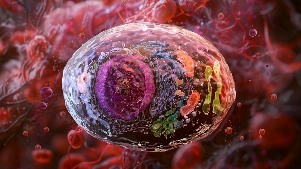
Using Placental Cells to Test Anti-Aging Compounds
- These human cells naturally age quickly when differentiated.
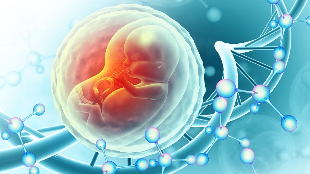
- The human placenta, which only lasts for 10 months, ages relatively quickly.
- Placental cells can be grown and differentiated in vitro, after which they exhibit familiar signs of aging, including cellular senescence and genomic instability.
- Due to their short lifespan, these cells may potentially be of use in rapidly testing potential anti-aging treatments.
Researchers publishing in Aging Cell have discovered that cells derived from the human placenta may be useful in estimating the effects of potential anti-aging treatments.
A seemingly odd choice
Of all the organs in the body, the placenta may be the least concerning with regards to aging; it only exists for at most 10 months, after which it is discharged as part of the birthing process. The researchers openly admit that this lifespan difference may make placenta-related aging processes distinct from those in other tissues, which harms translation and generalizability.
However, it is this limited lifespan that makes placental tissue potentially desirable for study. Its lifespan is under half that of mice, and it is made of human cells rather than murine ones. Just like in other organs, senescence and other core aging processes happen in the placenta as well, and unsurprisingly, accelerated placental senescence is linked to preterm birth and other problems [1]. There is also some evidence that the placenta may affect the rest of the body’s aging [2], and placenta-specific genes have been found to be activated in unrelated cells during senescence [3].
These researchers focus on three core placental cell types: cytotrophoblasts (CTBs), which differentiate into multinucleated syncytiotrophoblasts (STBs) and extravillous trophoblasts (EVTs). Differentiating CTBs into STBs can be done within a week in vitro and has been done since 2018 [4]. These researchers focus on this process, determining how similar it is to natural placental aging and how it can be harnessed to study aging more broadly.
Differentiated placental cells age quickly
The researchers first did a thorough examination of STBs compared to CTBs. Tthe differentiated CTBs had significantly more signs of senescence and across-the-board decreases in gene expression related to DNA maintenance, and there were increases in telomere attrition and metabolic differences as well. EVTs were found to have similar differences. Tthe researchers also discovered that results derived from the CTB to STB transition matched well with the epigenetic clock results of stem cells derived from the same donors, leading them to conclude that “cellular aging features observed in other tissue contexts can, therefore, be effectively modeled by the CTB-STB system.”
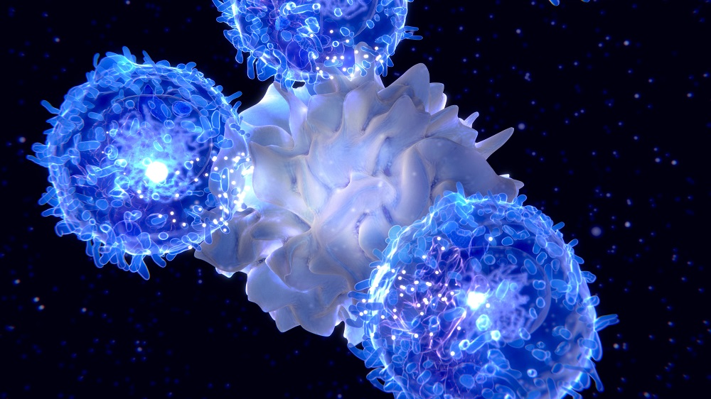
In their next experiment, the authors used human trophoblast stem cells (hTSCs), which themselves came from human expanded potential stem cells (hEPSCs), in order to ultimately differentiate them into STBs. As expected, these cells were pushed towards senescence as well, exhibiting familiar signs of resistance towards death by apoptosis while becoming more susceptible to death by necroptosis. Other molecular hallmarks of cellular senescence, including p16, were more prevalent as well.
Highly relevant aging features
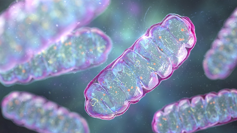
Very unsurprisingly, this increase in senescence coincided with an increase in the inflammatory senescence-associated secretory phenotype (SASP). Along with telomere maintenance failures, deregulated nutrient sensing, and mitochondrial dysfunction, STBs were far more likely than hTSCs to exhibit SASP upregulation. Interestingly, however, the results between mRNA expression and actual proteins did not match as expected, suggesting a possible age-related change in protein function.
The researchers then tested how well the cells resisted DNA damage. Undifferentiated hTSCs, which contained plenty of active DNA repair pathways as well as protective lamins, were strongly resistant to etoposide, a chemical that damages DNA. STBs, however, had fewer lamin protections and less repair capability; they were far more susceptible, exhibiting signs of double-strand breaks along with the DNA damage marker γH2AX.
This loss of protection also extended to the epigenome. Compared to hTSCs, STBs had significant alterations in histones and histone regulators, which are at the core of epigenetic alterations. H3K9me3 and H3K27me3 were downregulated, H3K4me3 was increased, and total histones and methylation were both decreased; all of these results are in line with those of other aging tissues.
Transposable elements normally found in the human genome, which come loose and become expressed during aging, lead to systemic inflammation [5]. They were far more expressed in STBs than in hTSCs. Knocking down some of these retrotransposons (HERVK) partially suppressed some features of senescence and inflammation in STBs.
Intended for practical use
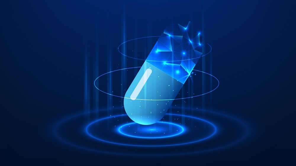
The researchers then moved on to the possibly most critical portion of their experiment: testing anti-aging molecules against STBs. They began with a screen of the most well-known interventions, including rapamycin, fisetin, NMN, quercetin, resveratrol, and navitoclax. All of these compounds were found to have effects, to varying extents, on the aging of STBs.
While these results are entirely preliminary, this study is meant to, and does, serve as a proof of concept. These are cells that can be rapidly produced and tested to determine the effects of potential anti-aging treatments. While they may not apply to every potential treatment, these cells may be used to accelerate the verification, and thus development, of promising interventions.
Literature
[1] Kajdy, A., Sys, D., Modzelewski, J., Bogusławska, J., Cymbaluk-Płoska, A., Kwiatkowska, E., … & Kwiatkowski, S. (2023). Evidence of placental aging in late SGA, fetal growth restriction and stillbirth—a systematic review. Biomedicines, 11(7), 1785.
[2] Pham, H., Thompson-Felix, T., Czamara, D., Rasmussen, J. M., Lombroso, A., Entringer, S., … & O’Donnell, K. J. (2024). The effects of pregnancy, its progression, and its cessation on human (maternal) biological aging. Cell metabolism, 36(5), 877-878.
[3] Bi, S., Jiang, X., Ji, Q., Wang, Z., Ren, J., Wang, S., … & Qu, J. (2024). The sirtuin-associated human senescence program converges on the activation of placenta-specific gene PAPPA. Developmental Cell, 59(8), 991-1009.
[4] Okae, H., Toh, H., Sato, T., Hiura, H., Takahashi, S., Shirane, K., … & Arima, T. (2018). Derivation of human trophoblast stem cells. Cell stem cell, 22(1), 50-63.
[5] Liu, X., Liu, Z., Wu, Z., Ren, J., Fan, Y., Sun, L., … & Liu, G. H. (2023). Resurrection of endogenous retroviruses during aging reinforces senescence. Cell, 186(2), 287-304.