The Blog

Building a Future Free of Age-Related Disease

Vincere

Vincere Biosciences Awarded $5 Million Grant

Cambridge, MA, USA – November 18, 2025: Vincere Biosciences today announced the receipt of a $5 million grant from The Michael J. Fox Foundation for Parkinson’s Research (MJFF) through its Therapeutics Pipeline Program, which supports the advancement of promising therapies through preclinical and clinical stages. The initiative focuses on candidates with strong potential to slow or halt disease progression or alleviate burdensome symptoms for those living with Parkinson’s disease. The funding accelerates Vincere’s lead USP30 inhibitor, a potentially first-in-class therapeutic designed to modify the course of Parkinson’s disease, through IND-enabling studies toward a 2026 clinical trial initiation. In parallel, the grant will also fund Vincere’s biomarker development efforts to evaluate target engagement and guide clinical translation, strengthening the foundation for future human studies.

“The Michael J. Fox Foundation remains steadfast in our mission to accelerate the development of transformative treatments and, ultimately, a cure for Parkinson’s disease,” said Jessica Tome Garcia, PhD, MJFF’s lead scientific program manager. “Our collaboration with Vincere Biosciences over the years has supported the advancement of research targeting mitochondrial dysfunction, a key driver of Parkinson’s pathology. This next phase of work builds on that foundation and represents important progress toward disease-modifying therapies that could meaningfully improve patients’ lives.”

Targeting the Root Cause of Neuronal Vulnerability

Mitochondrial dysfunction and impaired mitophagy are central features of PD pathophysiology. Mitophagy, the selective recycling of damaged mitochondria, is essential for maintaining neuronal health. In PD, this process is often compromised, leading to toxic buildup of defective mitochondria that accelerates neurodegeneration. Vincere aims to restore mitochondrial quality control and prevent the progression of neuronal injury by selectively inhibiting USP30, a mitochondrial deubiquitinating enzyme that acts as a negative regulator of mitophagy. By targeting the cellular processes at the root of Parkinson’s, Vincere’s approach has the potential to not only slow disease progression but also improve quality of life for millions living with PD.

“Mitochondria sit at the crossroads of Parkinson’s and aging (the biggest risk factor for Parkinson’s),” says Dr. Spring Behrouz, co-founder and CEO of Vincere, “fix the mitochondria and you strike at the root of the disease. MJFF’s support helps us move that science from the lab to the clinic.”

Transitioning from Discovery to Clinic

MJFF’s award will fund critical IND-enabling studies, including pharmacology, toxicology, and regulatory workstreams. These studies will establish the foundation required for a successful Investigational New Drug (IND) application with the U.S. Food and Drug Administration (FDA), marking the transition of Vincere’s program from preclinical development to the clinic. This award builds on prior MJFF support, which continues to move Vincere’s USP30 inhibitor program forward.

“It’s been exciting to see growing enthusiasm for USP30 since our AI platform prioritized this target in 2018. The new support from MJFF positions the company well for ongoing partnering discussions with larger organizations who may accelerate clinical development of this promising approach,” says Andy Lee, co-founder and CBO of Vincere.

About Vincere Biosciences Inc.

https://www.vincerebio.com

Boston-based Vincere Biosciences develops innovative, mechanism-driven therapeutics that target the intersection of aging and disease. Using proprietary computational tools and cutting-edge biology, Vincere is dedicated to discovering and developing small molecules that address the root causes of conditions like Parkinson’s and other age-related disorders.

We would like to ask you a small favor. We are a non-profit foundation, and unlike some other organizations, we have no shareholders and no products to sell you. All our news and educational content is free for everyone to read, but it does mean that we rely on the help of people like you. Every contribution, no matter if it’s big or small, supports independent journalism and sustains our future.
Cynomolgus monkey

Improved Stem Cells Rejuvenate the Brains of Monkeys

Scientists have genetically modified human mesenchymal progenitor cells to express a more potent version of the “longevity gene” FOXO3, producing rejuvenative effects in monkeys, mice, and human cells [1].

Making aging-resistant cells

Stem cell exhaustion is one of the mechanisms of aging. Replenishing the aging stem cell pool with new exogenous cells sounds like a good idea, but when new cells interact with an aging organism, they often experience accelerated aging and senescence [2]. To overcome this problem, in a new study published in Cell, scientists from China created genetically modified stem cells and injected them into cynomolgus monkeys.

The team started with wild-type human mesenchymal progenitor cells (MPCs), stem cells that can differentiate into several cell types and have a low immunogenic risk: they express few surface proteins that can be recognized by the immune system and secrete factors that actually calm immunity around them.

The MPCs were differentiated from human embryonic stem cells modified by a CRISPR-based editing system, which tweaked FOXO3, a transcription factor that orchestrates stress resistance and repair. FOXO3 is considered a promising “longevity gene,” since it extends lifespan and healthspan in animal models [3] and is associated with human longevity [4]. In stem cells, FOXO3 helps keep stem cells quiescent but competent, and it modulates inflammatory signaling.

In more technical terms, the researchers replaced two serine residues (Ser253 and Ser315) with alanine, eliminating the ability of these sites to be phosphorylated. Because phosphorylation at these sites normally promotes FOXO3 nuclear export and inactivation, this mutation keeps FOXO3 active for longer.

In vitro, the researchers confirmed that their senescence-resistant MPCs (SRCs) had a more youthful phenotype than wild-type MPCs (WTCs), exhibiting lower senescence and inflammation markers (SA-β-gal, IL-6, IL-8), longer telomeres, and more stable heterochromatin. SRCs were more competent at differentiation and more resistant to stressors, and they had their proliferation and tumor-suppressor programs upregulated.

Significant effects on monkeys

In the main experiment, cynomolgus monkeys were divided into four age groups. The three younger groups were used for studying natural aging in these animals and creating several bespoke clocks, which combined gene expression and DNA methylation patterns across multiple tissues to estimate each animal’s biological age. The last and oldest group (19-23 years) formed the intervention cohort and was further divided into subgroups that received a sham treatment, human WTCs, or human SRCs.

The treatment groups received bi-weekly IV infusions for 44 weeks, and upon the completion of the treatment, a large battery of tests was performed. The first part of it focused on the brain. Functionally, on a delayed-reward memory task, the SRC group performed significantly better than old controls, while the WTC group’s results were not clearly distinguishable from this control group’s.

SRCs preserved or even partially restored cortical thickness and volume in several brain regions, relative to age-matched controls. WTCs had weaker effects. Diffusion MRI revealed improved structural connectivity.

SRCs also reduced age-related myelin thinning and the levels of amyloid-β aggregates and p-Tau aggregates, both of which indicate Alzheimer’s disease. A single-cell clock showed an average reduction of biological age by about 2.5 years across hippocampal cell types following the SRC treatment.

This and other biological age measurements were compared to similarly aged untreated monkeys rather than the animals’ own baselines. In other words, the net age reversal, as measured by these metrics, was approximately 0.9 years less, as this is how long the experiment took to conduct.

The researchers also profiled 13 immune cell types. SRCs reversed more aging-related changes in gene expression than WTCs. Both treatments downregulated cellular senescence and apoptosis while upregulating DNA damage repair, autophagy, and lymphocyte differentiation and function. Both treatments also reduced the inflammation markers IL-6 and TNF-α in the plasma and CHIT1, a marker of microglial activation and neuroinflammation, in the cerebrospinal fluid (CSF).

Exosomes recapitulate many of the changes

In total, the team applied their bulk-tissue RNA-seq-based clock to 61 tissues from 10 organ systems. Generally, both WTCs and SRCs attenuated age-related trajectories, but SRCs had larger effects in most tissues, with an average biological age reduction of 3.3 years by SRCs compared to 2.8 years by WTCs.

Interestingly, the strongest effects were observed in reproductive tissues (ovary, testis, uterus, prostate, seminal vesicle, and epididymis). In tissues where transcriptomic aging was reversed, the methylation-based clock agreed with the transcriptomic one: according to it, SRCs made brains 5 years younger and skeletal muscle 4 years younger on average.

Because MPCs are thought to act largely via secreted factors, the researchers zoomed in on exosomes. Both in mice and in human cells, SRC-derived exosomes produced significant rejuvenation, lowering senescence markers and shifting gene expression profiles towards younger phenotypes.

We would like to ask you a small favor. We are a non-profit foundation, and unlike some other organizations, we have no shareholders and no products to sell you. All our news and educational content is free for everyone to read, but it does mean that we rely on the help of people like you. Every contribution, no matter if it’s big or small, supports independent journalism and sustains our future.

Literature

[1] Lei, J., Xin, Z., Liu, N., Ning, T., Jing, Y., Qiao, Y., … & Liu, G. H. (2025). Senescence-resistant human mesenchymal progenitor cells counter aging in primates. Cell.

[2] Liu, J., Gao, J., Liang, Z., Gao, C., Niu, Q., Wu, F., & Zhang, L. (2022). Mesenchymal stem cells and their microenvironment. Stem cell research & therapy, 13(1), 429.

[3] Flachsbart, F., Dose, J., Gentschew, L., Geismann, C., Caliebe, A., Knecht, C., … & Nebel, A. (2017). Identification and characterization of two functional variants in the human longevity gene FOXO3. Nature communications, 8(1), 2063.

[4] Willcox, B. J., Donlon, T. A., He, Q., Chen, R., Grove, J. S., Yano, K., … & Curb, J. D. (2008). FOXO3A genotype is strongly associated with human longevity. Proceedings of the National Academy of Sciences, 105(37), 13987-13992.

Frailty in older age

A Sarcopenia-Related MicroRNA May Help Pinpoint Its Origin

In Aging Cell, researchers have discovered a potential way to use a microRNA to diagnose sarcopenia, the age-related loss of muscle.

Primary and secondary sarcopenia

Previous research has been able to distinguish sarcopenia by its sources. Primary sarcopenia directly comes from the processes of aging, while secondary sarcopenia is a side effect of such things as poor nutrition, atrophy from disuse, and both acute and chronic diseases [1]. The researchers of this study note that the difference is difficult to distinguish in the clinic, where these two categories heavily overlap.

Heart failure, which is defined here as a progressive clinical syndrome [2] rather than an effect of acute causes such as heart attack, is heavily associated with sarcopenia [3]. A substantial amount of other work has shone some light on this relationship, and these problems share many of the same causes [4].

Looking for a way to diagnose the true causes of sarcopenia in the context of heart failure, these researchers have turned to microRNAs, non-coding RNA molecules that affect gene transcription and have been used to investigate the sources of diseases, including sarcopenia in the context of obesity [5]. One of these microRNAs is microRNA-22-3p (miR-22), which is found in many species and is expressed predominantly in muscle tissue and nerves [6]. This suggests a possible use in diagnostics and therapies, and work has been done in using it as a treatment for heart attack [7].

The biological effects of miR-22 vary by tissue. Some work has found that it blocks calcium uptake in a way that leads to diminished heart contraction ability [8]. Other work has found that it suppresses proliferation while encouraging differentiation in skeletal muscle cells, and inhibiting it does the opposite [9]. Work in mice has revealed that miR-22 in exosomes, which cells use to send signals to one another, leads to insulin resistance [10]. These researchers also performed their own in silico pathway analysis, which confirmed that it impacts an enormous network of metabolic and age-related pathways, many of which are related to aging.

Two studies create a combined picture

These researchers evaluated data from the SPRINTT study, which recruited 61 people aged 70 or older. Half of the participants showed signs of sarcopenia, which SPRINTT defined as a pre-disability state defined by gradual muscle failure and measured using a significant absence of lean mass. People with advanced heart conditions and other serious age-related disorders, such as cancer and dementia, were excluded. While that study focused on technological and nutritional interventions rather than microRNAs, it still took biomarker data from its participants. SPRINTT was intended to evaluate people with primary sarcopenia.

Other data came from SICA-HF, which recruited 176 people with heart failure of all ages. Like SPRINTT, its basis for a sarcopenia diagnosis was the absence of sufficient muscle mass. The reearchers used SICA-HF as their basis for secondary sarcopenia.

Using data from the SPRINTT study, the researchers found that people with primary sarcopenia have significantly more miR-22 than people without it. This difference was not found in other microRNAs that the researchers analyzed. Using this data, they determined that it was possible to develop a miR-22-based test that could be used to screen for sarcopenia, although such a test would not be completely perfect.

Interestingly, however, the opposite was true for secondary sarcopenia, with people with sarcopenia in the SICA-HF study being more likely to have less miR-22. While this data was somewhat weaker than the results derived from SPRINTT, it still reached the level of statistical significance.

The combination of these results leads these researchers to believe that miR-22 is a potentially useful diagnostic tool for the evaluation of primary versus secondary sarcopenia. They offer a few plausible reasons why this may be the case, suggesting that the source of the circulating miR-22 (cardiac or skeletal muscle) may be the primary difference [11] and that miR-22 may play different roles in these muscle types. The researchers also suggest that the effects of miR-22 on differentiation and proliferation, leading to changes in its regulation, may be involved. Furthermore, the correlation between miR-22 and sarcopenia in the cardiac patients may be due to an upstream cause rather than a direct relationship.

These researchers further acknowledge that there are limitations in using two separate studies to create a conclusion based on both of them. Those studies did not define sarcopenia in exactly the same way, and there is still the issue of overlapping causes. Therefore, substantial further work must be done in order to determine how microRNAs relate to this muscle-wasting condition.

We would like to ask you a small favor. We are a non-profit foundation, and unlike some other organizations, we have no shareholders and no products to sell you. All our news and educational content is free for everyone to read, but it does mean that we rely on the help of people like you. Every contribution, no matter if it’s big or small, supports independent journalism and sustains our future.

Literature

[1] Cruz-Jentoft, A. J., Bahat, G., Bauer, J., Boirie, Y., Bruyère, O., Cederholm, T., … & Zamboni, M. (2019). Sarcopenia: revised European consensus on definition and diagnosis. Age and ageing, 48(1), 16-31.

[2] Tomasoni, D., Adamo, M., Lombardi, C. M., & Metra, M. (2019). Highlights in heart failure. ESC heart failure, 6(6), 1105-1127.

[3] Fülster, S., Tacke, M., Sandek, A., Ebner, N., Tschöpe, C., Doehner, W., … & von Haehling, S. (2013). Muscle wasting in patients with chronic heart failure: results from the studies investigating co-morbidities aggravating heart failure (SICA-HF). European heart journal, 34(7), 512-519.

[4] Sato, R., Vatic, M., Peixoto da Fonseca, G. W., Anker, S. D., & von Haehling, S. (2024). Biological basis and treatment of frailty and sarcopenia. Cardiovascular research, 120(9), 982-998.

[5] Dowling, L., Duseja, A., Vilaca, T., Walsh, J. S., & Goljanek‐Whysall, K. (2022). MicroRNAs in obesity, sarcopenia, and commonalities for sarcopenic obesity: a systematic review. Journal of Cachexia, Sarcopenia and Muscle, 13(1), 68-85.

[6] Huang, Z. P., & Wang, D. Z. (2018). miR-22 in smooth muscle cells: a potential therapy for cardiovascular disease. Circulation, 137(17), 1842-1845.

[7] Gupta, S. K., Foinquinos, A., Thum, S., Remke, J., Zimmer, K., Bauters, C., … & Thum, T. (2016). Preclinical development of a microRNA-based therapy for elderly patients with myocardial infarction. Journal of the American College of Cardiology, 68(14), 1557-1571.

[8] Gurha, P., Wang, T., Larimore, A. H., Sassi, Y., Abreu-Goodger, C., Ramirez, M. O., … & Rodriguez, A. (2013). microRNA-22 promotes heart failure through coordinate suppression of PPAR/ERR-nuclear hormone receptor transcription. PLoS One, 8(9), e75882.

[9] Wang, S., Cao, X., Ge, L., Gu, Y., Lv, X., Getachew, T., … & Sun, W. (2022). MiR-22-3p inhibits proliferation and promotes differentiation of skeletal muscle cells by targeting IGFBP3 in Hu sheep. Animals, 12(1), 114.

[10] Zhang, H., Zhang, X., Wang, S., Zheng, L., Guo, H., Ren, Y., … & Yan, Y. (2023). Adipocyte-derived exosomal miR-22-3p modulated by circadian rhythm disruption regulates insulin sensitivity in skeletal muscle cells. Journal of Biological Chemistry, 299(12).

DNA Closeup

NAD+ Rescues Mouse Tauopathy by Fixing Alternative Splicing

A new study reveals a surprising mechanism that might be behind the beneficial effects of NAD+ in preclinical models of Alzheimer’s [1].

Which way to splice it?

Not every part of a DNA sequence gets translated into a protein. Each sequence consists of exons, which are included in the final RNA transcript, and introns, which are thrown away.

However, nothing is that simple in biology. Exons and introns can be combined in various ways to create several versions of the protein, with different and sometimes opposite qualities. This is known as alternative RNA splicing. It gets increasingly dysregulated as we age [2] and has been implicated in various diseases, including Alzheimer’s disease (AD) [3].

NAD+ is a key metabolite that also modulates RNA processing. It slows Alzheimer’s disease progression in preclinical models [4], but the mechanism behind this effect is not well understood. In a new study from the University of Oslo published in Science Advances, the researchers asked whether this could be related to alternative splicing.

“Preliminary studies have shown that supplementation with NAD⁺ precursors, such as nicotinamide riboside (NR) or nicotinamide mononucleotide (NMN), can offer therapeutic benefits in AD animal models and early clinical trials. However, the molecular mechanisms behind these benefits remain largely unclear,” first author Alice Ruixue Ai said.

From worms to mice

First, the researchers used C. elegans nematode worms engineered with two key features: neurons that express a human P301L tau variant (a model of tauopathy, which is a hallmark of Alzheimer’s) and a fluorescent reporter that changes green/red signal depending on how a particular exon is spliced. In aging tau worms, the reporter showed that splicing becomes gradually more error-prone. The worms also developed memory-like defects and shorter lifespans.

Treating worms from hatching with nicotinamide riboside (NR), a NAD+ precursor, altered the neuronal splicing fidelity, with strong effects early in life, suggesting that NAD+ can modulate splicing machinery during development. It also partially rescued lifespan and memory.

In a mouse model of tauopathy (Tau.P301S mice, with human P301S tau expressed in neurons), the researchers saw a similar picture: splicing in tau mice was disrupted and partially normalized by NR. This time, however, they focused on the mechanism.

Hippocampal RNA sequencing found 509 differentially expressed genes compared to wild-type mice, with many affected genes involved in RNA processing and splicing. The gene Eva1c, which codes for a protein involved in neuronal development and activity, was both significantly upregulated and abnormally spliced in tauopathic mice compared to controls.

NR supplementation significantly reversed both expression and splicing changes, which made Eva1c stand out. An AI-based analysis suggested that the key effects were less caused by the overall levels of this protein and more by the proportions of its various isoforms.

The researchers then manipulated the gene in mice and its homolog (eva-1) in worms. In C. elegans, eva-1 knockdown abolishes NR’s benefits on lifespan and memory-like behavior in tau worms, showing that those benefits indeed were eva-1-dependent.

Restoring the balance

In a slightly different mouse model with tauopathy induced by AAV-based tau overexpression, experiments involved Eva1c manipulation alongside supplementation with NMN, another NAD+ precursor. The researchers changed the Eva1c isoform mix by overexpressing the one isoform that was most robustly altered by NR in previous experiments.

Tau.P301S AAV mice showed memory deficits in the novel object recognition test, but NMN treatment brought the readouts back to control levels. This effect disappeared with the Eva1c knockdown. Overexpressing the most NR-responsive isoform partly mimicked the NMN effect.

A similar pattern was seen for total and phosphorylated tau levels. Together, this shows that NMN’s anti-tau and pro-memory effects in this mouse model require intact Eva1c and can be mimicked by restoring the Eva1c isoform balance, such as by rescuing alternative splicing.

“Notably, we found that when the EVA1C gene was knocked down, these benefits were lost, confirming that EVA1C is essential for NAD⁺-mediated neuroprotection,” Associate Professor Evandro Fei Fang-Stavem said.

Finally, mining an extensive RNA-seq dataset of Alzheimer’s and non-Alzheimer’s hippocampal tissue and analyzing post-mortem samples, the researchers found that EVA1C expression is altered in Alzheimer’s brains compared with controls, consistent with EVA1C being involved in human tau pathology as well.

“We propose that maintaining NAD⁺ levels could help preserve neuronal identity and delay cognitive decline, paving the way for combination treatments to enhance RNA splicing,” Ai said.

We would like to ask you a small favor. We are a non-profit foundation, and unlike some other organizations, we have no shareholders and no products to sell you. All our news and educational content is free for everyone to read, but it does mean that we rely on the help of people like you. Every contribution, no matter if it’s big or small, supports independent journalism and sustains our future.

Literature

[1] Ai, R., Mao, L., Jin, X., Campos-Marques, C., Zhang, S. Q., Pan, J., … & Fang, E. F. (2025). NAD+ reverses Alzheimer’s neurological deficits via regulating differential alternative RNA splicing of EVA1C. Science Advances, 11(45), eady9811.

[2] Bhadra, M., Howell, P., Dutta, S., Heintz, C., & Mair, W. B. (2020). Alternative splicing in aging and longevity. Human genetics, 139(3), 357-369.

[3] Nikom, D., & Zheng, S. (2023). Alternative splicing in neurodegenerative disease and the promise of RNA therapies. Nature Reviews Neuroscience, 24(8), 457-473.

[4] Hou, Y., Lautrup, S., Cordonnier, S., Wang, Y., Croteau, D. L., Zavala, E., … & Bohr, V. A. (2018). NAD+ supplementation normalizes key Alzheimer’s features and DNA damage responses in a new AD mouse model with introduced DNA repair deficiency. Proceedings of the National Academy of Sciences, 115(8), E1876-E1885.

Scientific questions

The Key Questions of Longevity Research

In GeroScience, a large team of researchers, including João Pedro de Magalhães, has described a hundred currently unsolved problems in the field.

Finding the right questions

Understanding the fundamental nature of aging has been a problem since people first endeavored to live longer, to the point that finding the right questions can matter more than finding the right answers. Nearly fifty years ago, B.L. Strehler attempted to tackle this in Time, Cells, and Aging [1], and there has been skepticism as to whether it can be done at all [2]. As the field has completely changed between now and Strehler’s time, these researchers chose to revisit the topic, using modern analysis techniques the most frequently asked questions in research.

These questions came from a few sources. First, they sought comments from the research community, collecting a total of 160 open problems and adding them to their database. Additionally, they held a 3-day workshop with 24 scientists in order to gain a better view of the field, creating groups and brainstorming unsolved problems, gaining another 130.

Then, the researchers filtered these problems for similarity and relevance, reducing the number to 204. They quested for how frequently these problems were mentioned in article abstracts, using a natural language processing algorithm and multiple checks to ensure that the articles and problems matched. Of these 204 problems, 100 were selected by Prof. de Magalhães for inclusion into the OpenLongevity database.

The biggest question is why

Unsurpisingly, the biggest overall question is “Why do we age?”, with 10,808 scientific articles asking this question. This was followed by a question about somatic mutation accumulation, which had 5,977. Questions about cellular processes and species longevity followed, as were questions involving fundamental aging processes in animals and whether or not those processes translate to humans. Biomarkers were also major aspects of concern. A broad question about interventions involving cellular function was less commonly asked than questions about these fundamental questions of aging.

Intervention-related questions, however, were the most commonly found on the list. These questions involved targeting inflammation, utilizing embryogenetic processes to slow aging, and employing partial reprogramming techniques. Many other questions involved the amount of overall damage that originates from particular aspects of aging, such as the secretions of senescent cells.

Open Longevity Problems

There were also questions that were almost never asked, mostly involving very little-researched, very particular aspects of the field. For example, only one paper asked about the homeodynamic space, which refers to organizational stability. Only a few papers asked about prioritizing interventions, and mitochondrial citrate and immune responses to mutated cells were also lightly explored topics.

The field has come a long way, but there is still much to do

There are similarities between the questions of today and the questions asked by Strehler in 1977, although he focused more on central nervous system disorders and some questions regarding inter-species differences have been fairly well resolved. Modern research is still exploring the roles of mitochondria, although the modern focus is more on such cellular dynamics than on the fundamental aspects of biology, such as enzymes and ribosomes, that were of scientific interest at the time and are largely well-understood today. However, how these biological components are changed during aging is not always certain.

Answering questions in science, especially the sorts of complex questions involved in biology, often leads to more questions. These 100 questions are intended to be fundamental to the field, and while answering them may lead to more questions, the process may lead to interventions that extend healthy human lifespan.

We would like to ask you a small favor. We are a non-profit foundation, and unlike some other organizations, we have no shareholders and no products to sell you. All our news and educational content is free for everyone to read, but it does mean that we rely on the help of people like you. Every contribution, no matter if it’s big or small, supports independent journalism and sustains our future.

Literature

[1] Strehler, B. (1977). Some unexplored avenues of cellular aging—current and future research. Time, Cells and Aging, edited by Strehler, B, 374-418.

[2] Partridge, L. (2010). The new biology of ageing. Philosophical Transactions of the Royal Society B: Biological Sciences, 365(1537), 147-154.

If Death Were Optional, Would You Still Choose It?

The idea of living longer, healthier lives thanks to rejuvenation biotechnology has steadily become more common. Gradually increasing numbers of articles are discussing this idea, especially as science is starting to catch up and may eventually even deliver on it.

With that in mind, I was pleasantly surprised this week to be greeted by an interesting questionnaire from YouGovChat. YouGov PLC is an international, Internet-based market research and data analytics firm that has a headquarters in the UK.

Public sentiment on living forever or at least indefinitely

The question that caught my attention was: “If you could live forever, staying healthy and young, would you do it?”

The important part here is that this question includes the words healthy and young. Previous surveys have often asked this question without those important words, and that changes the response.

People don’t generally want to live longer if that additional time means being sick and frail. Simply asking people if they want to live another 20+ years often conjures up images of being in a care home. They think of having no independence or quality of life accompanying those extra years.

This is understandable. The modern healthcare system has done well in keeping people alive longer, but often without an accompanying quality of life. The idea of effectively losing what makes life worth living and prolonging suffering isn’t something desirable.

However, phrasing the question with such qualifiers can lead to a more accurate and honest answer. So, how did people respond when the question was framed with the understanding that health and youthfulness are included?

YouGov Questionnaire 1

The key thing to note here is that most people in the poll were open to living long and healthy lives. However, they wanted the choice to end their life if they so desired.

In my decade as a rejuvenation biotech journalist, I have heard many reasons not to develop the technology. One of those objections is that people will be forced to take therapies and made to live longer or even indefinitely against their will.

Some of that concern may be playing into the responses here. People are likely to prefer to have such an option than not, and no one likes the idea of having no choice. We cannot truly know why people responded this way, as it was beyond the scope of the poll. On the plus side, the majority don’t appear to be against the idea.

The meaning of life

The next question got into the philosophical side of things and touched upon something often used to justify death.

YouGov Questionnaire 2

The idea that death makes life meaningful has long been used to rationalize death. It is also used as a reason for not trying to reverse biological aging to prevent age-related diseases.

There is nothing at all wrong with growing chronologically older and hopefully wiser, but getting biologically older is the primary risk factor for a myriad of such diseases. Our field is focused on slowing down and even reversing the damage that aging causes to organs and tissues. The goal is to keep people biologically younger and healthy for longer.

It is good to see that these responses largely reject the idea that death is needed to make life meaningful. Unfortunately, the set answers focused on how we live and not how long, which kind of defeats the point of the original question, especially since most of the respondents had expressed positive attitudes towards indefinite lifespans.

Feeling a bit emotional about longevity

The next question delved into the emotional side of the human experience.

YouGov Questionnaire 3

While it is not possible to know how longer lives might affect human emotions, we have explored some related concerns. We covered the idea that longevity might lead to eternal boredom and why this is unlikely. The concept that society would stagnate if people lived longer is something that we have also written about. We have also touched upon lost motivation due to increased lifespans.

The truth is that we do not know if or how living indefinitely might change the human experience, assuming it would, but I don’t agree that it’s a reason to let people die from potentially curable age-related diseases.

I prefer to think that given more time, people would use that extra time to focus on long-term plans and ambitions. If you have more years of health and youth, it’s easy to picture having many careers, hobbies, and interests.

We still have a long way to go to get the public on board

The penultimate question itself was reasonable, as it asked about the support of research into indefinitely increasing human lifespans. The issue here is that the set answers included the very loaded “Yes, humans should aim to conquer death”.

YouGov Questionnaire 4

The problem is that this isn’t what our field of science is trying to do. Let’s be clear here, rejuvenation biotechnology is not immortality. The goal of the field is not to conquer death. It is to prevent or reverse age-related diseases by targeting the causes of aging.

Rejuvenation won’t stop you from dying in a car crash. It can’t save you from falling off a tall building. While it may improve the immune system, it won’t necessarily prevent death from an infectious disease. There are many ways to die, and only age-related diseases are covered by our field, nothing else.

Even if people have an indefinite lifespan it does not equate to conquering death or immortality. This is why I think this particular question is flawed, because the responses miss the point of rejuvenation biotechnology. As a result, the answers people give here are based on that poor framing.

This is why there is a contradiction in their responses. They are mostly fine with living indefinitely according to their responses earlier in the poll, but they are against it later. It would be great to see these questions asked again with better framing for this particular question. I am willing to bet that the answers would be more in favour of supporting research if the question and answers are framed properly.

Back to the meaning of life again

The poll already touched upon the old idea that death gives life meaning, but for some reason, the poll makers chose to return to the same question in a different way, despite the fact that the majority had already rejected the notion that death made life meaningful.

YouGov Questionnaire 5

However, we see a shift and somewhat of a contradiction, again because the framing changed. Phrased this way, more people think life has meaning because it ends, so that presumably means death makes it meaningful.

Ultimately, these results suggest that how a question is asked, along with the possible answers, is really important. I believe the main point is that the poll shows the topic is reaching more public audiences. A decade ago, I would never have dreamed to see something like this, but here we are now with the subject being discussed.

An important caveat

While this is interesting, there are important caveats here. The UK government and other organisations make use of the data collected by YouGovChat, although the extent to which this information is used is unknown.

In other words, while this is interesting, the data should be taken with a pinch of salt. Polls such as this have known limitations. They do not represent the entire population, they are only suggestive of the general sentiment.

If you want to take part and are from the UK, you can join the “Would you choose to live forever if you could?” poll.

Help us to build better advocacy tools and ask the right questions

Public trust is the key that opens all other doors. For a long time, longevity research has needed better tools to understand public opinion. This will help to build trust.

YouGov Questionnaire 6

To achieve this, we are supporting the development of a cultural intelligence platform by the Public Longevity Group (PLG). The objective is to develop tools for assessing public opinion by evaluating media coverage and analyzing social media engagement in the area and to formulate effective communication methods to connect with previously overlooked audiences.

Thanks to your support, we are more than halfway to the fundraiser goal, but there’s still time to make an even greater impact!

The LRI Board of Directors is matching all donations up to $25,000 to push us over the finish line. Every dollar you give will be doubled. This will help launch the first data-driven sentiment analysis engine for longevity science.

Donate today, and join us in building the tools for effective longevity advocacy!

Multiple languages

Multilingualism Is Associated With Delayed Aging

A recent study of over 80,000 Europeans concluded that speaking more than one language is associated with delayed aging. Further analysis suggested that the protective effect of speaking one foreign language diminished with age, while the protective effect of speaking two or more foreign languages was more robust with aging [1].

Beyond communication

Learning a foreign language and maintaining this knowledge in the long term is not an easy endeavor. However, as research suggests, it can bring benefits that go beyond simply communication and cultural enrichment.

Research on multilingualism (“the regular use of more than one language”) suggests that it has a protective role in delaying cognitive decline and age-associated neurodegenerative diseases. [2-4]. However, those studies have some caveats, such as investigating people with cognitive decline rather than healthy people or having small sample sizes.

In a recent study, researchers sought to address these shortcomings and investigate whether multilingualism influences aging. They utilized data from a large population of 86,149 individuals with a mean age of 66.55 years (age range: 51-90 years) from 27 European countries, excluding people with a diagnosis of dementia.

In this analysis, multilingualism was assessed as an aggregate, country-level percentage of people speaking one, two, three, or more languages. At the same time, individual data was used to estimate the aging rate.

The authors measured this rate by calculating biobehavioral age gaps (BAGs), which represent the difference between an individual’s chronological age and the age predicted by a model. This measure is more sensitive than the indirect markers used in previous studies on this topic. The model that calculates BAG considers both positive and adverse risk factors to which the individual was exposed. Positive BAG values indicate accelerated aging, while negative values suggest delayed aging.

Multilingual aging 1

The more the merrier

The researchers performed two types of analysis: cross-sectional analysis, which analyzes data at a single time point, and longitudinal analysis, which allows for the analysis of a population over a period of time. While the results differed slightly, they both point to multilingualism having positive effects.

According to the cross-sectional analysis, people who speak only one language (monolinguals) are 2.11 times more likely to experience accelerated aging. In comparison, people who speak at least one more language are 2.17 times less likely to experience accelerated aging. The exact odds differ depending on the number of languages spoken, with bilinguals 1.3 times less likely to experience accelerated aging, trilinguals 1.96 times less likely, and polyglots who speak four or more languages 1.56 times less likely.

Analyzing this population over time through longitudinal analysis suggests a similar protective effect of language learning, with monolinguals having 1.4 times higher chances of experiencing accelerated aging over time, while the risk is 1.43 times lower for multilinguals, and as previously depends on the number of languages spoken and showing a dose-dependent effect. The results of this analysis showed that bilinguals were 1.11 times less likely to develop accelerated aging, trilinguals 1.25 times, and polyglots speaking four or more languages 1.41 times less likely.

An additional analysis that grouped participants by age suggested the same protective effects. However, it also indicated that the protective effect of speaking one foreign language diminished with age. In contrast, the protective effect of speaking two or more foreign languages remained more robust with aging.

Other factors

The authors of this study noted that many previous studies did not account for confounding factors and exposure to various lifestyle-related and socioeconomic factors, which can all lead to inconsistent results and improper data interpretation. To correct that, they used aggregate country-level data to adjust their analysis for different linguistic, physical, social, and sociopolitical conditions. They observed only minor changes in the magnitude of the protective effect of multilingualism.

They only noted the effects of two factors. The positive impact of delayed aging in polyglots was lost in the cross-sectional analysis following adjustments for immigration status, and in the longitudinal analysis, a positive effect in the bilingual population was lost when the researchers controlled for gender equality.

The authors speculate that since migration is often linked to different stressors, it “can lead to stressful multilingualism,” where acquisition of other languages is done out of necessity and pressure. Such stress and pressure might diminish the positive effect of language learning. However, as the authors discuss, their analysis is lacking a significant amount of data regarding migration, such as whether it was forced or voluntary, the length of stay, migration history, and other relevant details. Therefore, this result should be interpreted with caution.

Their results also suggest that similar negative factors, as in the case of migration, can be present in environments lacking gender equality, thereby contributing to the limited positive effects of multilingualism for people in those environments, suggesting that factors beyond individual lifestyle choices might have a profound impact on aging.

Multilingual aging 2

Not the first evidence, but strong evidence

Overall, this study revealed a strong association between multilingualism and a reduced risk of accelerated aging, whereas monolingualism was associated with an increased risk of accelerated aging.

While this study was not the first to show the positive effects of speaking multiple languages, it addressed many shortcomings of previous studies, thereby strengthening the evidence and adding additional support that multilingualism, along with other lifestyle factors, can be incorporated into public health guidelines as a protective factor against accelerated aging.

Although this study draws conclusions from a robust dataset, it cannot establish causality between the observed associations. Further studies, based on experimental or intervention-based designs, are needed to establish causality.

The researchers also point out that their “measures were coarse, and participants likely represented mixed multilingual profiles.” Still, the strength of the observed associations suggests that the observations can be generalized, as they are derived from the broad variability of profiles. However, to identify differences in the level of protection between various multilingual profiles, there is a need for additional studies that incorporate individual-level multilingual metrics such as age of acquisition and proficiency level, as this analysis relied on country-level data, which limited the authors’ conclusions and ability to analyze the impact of those individual factors on aging trajectories.

We would like to ask you a small favor. We are a non-profit foundation, and unlike some other organizations, we have no shareholders and no products to sell you. All our news and educational content is free for everyone to read, but it does mean that we rely on the help of people like you. Every contribution, no matter if it’s big or small, supports independent journalism and sustains our future.

Literature

[1] Amoruso, L., Hernandez, H., Santamaria-Garcia, H., Moguilner, S., Legaz, A., Prado, P., Cuadros, J., Gonzalez, L., Gonzalez-Gomez, R., Migeot, J., Coronel-Oliveros, C., Cruzat, J., Carreiras, M., Medel, V., Maito, M. A., Duran-Aniotz, C., Tagliazucchi, E., Baez, S., García, A. M., & Ibanez, A. (2025). Multilingualism protects against accelerated aging in cross-sectional and longitudinal analyses of 27 European countries. Nature aging, 10.1038/s43587-025-01000-2. Advance online publication.

[2] Alladi, S., Bak, T. H., Duggirala, V., Surampudi, B., Shailaja, M., Shukla, A. K., Chaudhuri, J. R., & Kaul, S. (2013). Bilingualism delays age at onset of dementia, independent of education and immigration status. Neurology, 81(22), 1938–1944.

[3] Craik, F. I., Bialystok, E., & Freedman, M. (2010). Delaying the onset of Alzheimer disease: bilingualism as a form of cognitive reserve. Neurology, 75(19), 1726–1729.

[4] Venugopal, A., Paplikar, A., Varghese, F. A., Thanissery, N., Ballal, D., Hoskeri, R. M., Shekar, R., Bhaskarapillai, B., Arshad, F., Purushothaman, V. V., Anniappan, A. B., Rao, G. N., & Alladi, S. (2024). Protective effect of bilingualism on aging, MCI, and dementia: A community-based study. Alzheimer’s & dementia : the journal of the Alzheimer’s Association, 20(4), 2620–2631.

CRISPR Editing

New Gene Therapy Robustly Lowers LDL and Triglycerides

A new Phase 1 trial produced encouraging safety and efficacy results for a CRISPR-based gene therapy that silences a gene important for lipid regulation. This therapy might increase adherence and reduce side effects [1].

Addressing the adherence problem

High levels of LDL cholesterol (LDL-C) and triglycerides are a major risk factor for cardiovascular disease and mortality. They can be caused by genetic variations, other diseases like hypothyroidism and diabetes, and environmental factors such as modern eating habits [2]. This dyslipidemia is also age-related, with LDL-C levels tending to rise in older people [3].

Effective therapies, especially against high LDL-C, exist. Statins, a class of cholesterol-lowering drugs that lead to robust reductions in cardiovascular events and related mortality, are a go-to first-line option. However, low adherence and side effects remain a problem, with many patients discontinuing treatment within a year.

In this trial, the company CRISPR Therapeutics tested an experimental gene therapy agent, CTX310, against abnormally high LDL-C and triglyceride levels. The therapy, based on the CRISPR platform, silences the production of ANGPTL3, a protein that is secreted primarily by the liver and plays a key role in regulating blood lipid levels. This protein inhibits the enzymes lipoprotein lipase (LPL) and endothelial lipase, which are crucial for breaking down triglycerides and remodeling high-density lipoprotein (HDL). As a result, higher ANGPTL3 levels lead to higher levels of circulating triglycerides and cholesterol in the blood.

LDL and triglycerides halved

This multicenter, open-label, single-ascending-dose study was conducted in Australia, New Zealand, and the UK and involved 15 adults with uncontrolled hypercholesterolemia, hypertriglyceridemia, or mixed dyslipidemia. The participants’ median age was 53 years with a range of 31 and 68. 87% were male, 40% had a history of atherosclerotic cardiovascular disease, and 40% had familial hypercholesterolemia, with 33% carrying confirmed genetic mutations. Background lipid-lowering therapies included statins (60%), ezetimibe (53%), and PCSK9 monoclonal antibodies (40%).

The elements of the CRISPR system (SpCas9 mRNA + a single guide RNA targeting ANGPTL3) were encapsulated in lipid nanoparticles (LNPs). The treatment was administered once via IV infusion, and the follow-up period prior to publication of the results was 60 days with a one-year continuation.

The treatment caused a drastic decline in the average ANGPTL3 level at higher doses: up to -79.7% for the 0.7 mg/kg dose. The average level was actually slightly, but not significantly, higher for 0.8 mg/kg: -73.2%. Most importantly, the treatment significantly lowered mean LDL-C and triglyceride levels at 0.8 mg/kg; LDL-C was down by 48.9% and triglycerides by 55.2% at this dose.

Why do we need ANGPTL3, if silencing it seems to benefit us? Like some other genes, it might have lost its evolutionary sense as food became more abundant. By shunting post-meal triglycerides to fat tissue for storage, ANGPTL3 might have helped our ancestors to eat large meals when food was available without burdening the heart and muscle with excess lipid.

Waiting for more robust studies

The trial reported minimal adverse events, indicating a favorable safety profile for CTX310. No dose-limiting toxic effects were observed. However, variability in lipid-lowering effects was noted among participants receiving the same CTX310 dose.

According to the researchers, this variability was not simply dose-related: editing efficacy and lipid responses may be influenced by hepatic steatosis, inflammation, and pre-existing genetic/metabolic profiles, and the cohort itself had mixed phenotypes, as some were primarily high LDL-C, and others had high triglycerides. This likely contributed to differences in lipid-lowering effects even at the same dose. The team calls for more rigorous studies to identify patient-specific predictors and to optimize dosing.

The sample size was also extremely small: only 2-4 patients received each dose, resulting in low statistical power for each cohort. The primary outcome of this Phase 1 trial was safety, and subsequent phases will provide more robust efficacy results.

The cohort was heavily male, and the authors themselves note that few women were enrolled, limiting subgroup assessment. Known sex differences exist in lipid biology, such as menopause-related shifts [4], and they affect responses to biological interventions and in the way people age, so generalizability from this sample is uncertain.

Finally, the effect size was not necessarily larger than that of some existing therapies. However, those require constant administration and can cause considerable side effects.

“Adherence to cholesterol-lowering therapy is one of the biggest challenges in preventing heart disease,” said Steven E. Nissen, M.D., FAHA, a co-author of the study and chief academic officer at the Cleveland Clinic Heart, Vascular and Thoracic Institute. “Many patients stop taking their cholesterol medications within the first year. The possibility of a one-time treatment with lasting effects could be a major clinical advance.”

We would like to ask you a small favor. We are a non-profit foundation, and unlike some other organizations, we have no shareholders and no products to sell you. All our news and educational content is free for everyone to read, but it does mean that we rely on the help of people like you. Every contribution, no matter if it’s big or small, supports independent journalism and sustains our future.

Literature

[1] Laffin, L. J., Nicholls, S. J., Scott, R. S., Clifton, P. M., Baker, J., Sarraju, A., … & Nissen, S. E. (2025). Phase 1 Trial of CRISPR-Cas9 Gene Editing Targeting ANGPTL3. New England Journal of Medicine.

[2] Yanai, H., & Yoshida, H. (2021). Secondary dyslipidemia: its treatments and association with atherosclerosis. Global health & medicine, 3(1), 15-23.

[3] Liu, H. H., & Li, J. J. (2015). Aging and dyslipidemia: a review of potential mechanisms. Ageing research reviews, 19, 43-52.

[4] Palmisano, B. T., Zhu, L., Eckel, R. H., & Stafford, J. M. (2018). Sex differences in lipid and lipoprotein metabolism. Molecular metabolism, 15, 45-55.

Fibrous foods

High-Fiber Foods May Fight T Cell Senescence

Researchers have discovered that butyrate, a short-chain fatty acid with well-documented gut benefits, fights senescence in T cells.

Immune senescence drives inflammaging

Senescence of the immune system (immunosenescence) is a problem that drives many others. In particular, T cells are known to secrete inflammatory SASP compounds [1] and drive the constant, age-related inflammation known as inflammaging [2]; in essence, the aged immune system overactivates itself. However, instead of being more effective against pathogens, this overactivated system has a degraded ability to effectively respond to threats [3], which is part of why older people have suffered from significantly greater COVID-related mortality [4].

An increase in T cell senescence has been found to drive pathologies in other systems in mice, including the muscles, vasculature, and cognition [5]. In people, this immunosenescence has been linked to arthritis [6] and acute heart failure [7]; unsurprisingly, there is an possible Alzheimer’s link as well [8].

The authors of this paper focus on butyrate as a potential method of mitigating T cell senescence, pointing to previous papers that suggest that it has beneficial influences in this area [9] along with benefits for B cells, another immune cell type [10]. However, while the gut microbiome has been heavily investigated in the context of aging and a link between butyrate and decreased T cell senescence has been found [11], these researchers stated that “no studies have systematically investigated how butyrate influences the function of aged immune cells.”

Butyrate is correlated with less senescence in people

This study’s first experiment used blood and stool samples from 40 healthy people over the age of 60 and 40 more between the ages of 18 and 37. Slightly more women than men participated in this study. None of the participants had any known chronic infections or any immunological problems.

The researchers first noted that older people have significantly less butyrate in their feces than younger people do; they also reported an age-related decrease in blood butyrate, which they noted to be never previously reported. Furthermore, they found that blood butyrate was significantly negatively associated with the prevalence of senescent T cells in the older group.

Cells and mice suggest a causal link

Encouraged, the researchers performed an in vitro experiment, introducing butyrate to T cells that had been driven senescent through a chemical regimen particular to these cells. While they found no change in the cells’ viability, there was a significant decrease in the cells’ production of IL-6, a major component of the SASP. This suggests that butyrate is a senomorphic; while it cannot reverse senescence on its own, it affects how senescent cells behave and may reduce the rate at which they drive other cells senescent through the SASP. This effect was stronger on younger cells than older cells.

This diminishment of the senescent phenotype was accompanied by other decreases in senescence-related features. The treated cells had decreased levels of the tumor suppressor p53, and they had fewer DNA strand breaks as measured by the marker γH2AX. While p38 was unaffected, there was a significant decrease in the key inflammatory factor NFκB. CD8-expressing T cells had fewer reactive oxygen species (ROS) after butyrate administration, but CD4-expressing T cells did not. These findings were buttressed by a gene expression analysis, which found that the butyrate-exposed senescent cells express less of a compound that prevents death by apoptosis, and they had reductions in the expressions of other inflammatory compounds.

These findings were accompanied by a mouse study. After treatment with antibiotics to purge their gut microbiomes, older mice were given fecal filtrations rich in butyrate derived from younger mice. The treatment group had significantly fewer p53-expressing T cells and significantly less IL-6.

The researchers note that butyrate cannot be the sole arbiter of the senescence-related behavior of T cells, and they also note that the concentrations used in cellular experiments may not always reflect those in people, particularly because there is a large difference between gut and serum levels. However, the problems with accurate dosing are less relevant in this case, as this is one of the few interventions that could be entirely conducted through diet alone; butyrate is a naturally occurring short-chain fatty acid, and fibrous fruits and vegetables are well-known to be rich in it. These researchers suggest a clinical trial using butyrate supplements or precursors to confirm their findings.

We would like to ask you a small favor. We are a non-profit foundation, and unlike some other organizations, we have no shareholders and no products to sell you. All our news and educational content is free for everyone to read, but it does mean that we rely on the help of people like you. Every contribution, no matter if it’s big or small, supports independent journalism and sustains our future.

Literature

[1] Callender, L. A., Carroll, E. C., Beal, R. W., Chambers, E. S., Nourshargh, S., Akbar, A. N., & Henson, S. M. (2018). Human CD 8+ EMRA T cells display a senescence‐associated secretory phenotype regulated by p38 MAPK. Aging cell, 17(1), e12675.

[2] Dugan, B., Conway, J., & Duggal, N. A. (2023). Inflammaging as a target for healthy ageing. Age and ageing, 52(2), afac328.

[3] Duggal, N. A. (2018). Reversing the immune ageing clock: lifestyle modifications and pharmacological interventions. Biogerontology, 19(6), 481-496.

[4] Covre, L. P., De Maeyer, R. P., Gomes, D. C., & Akbar, A. N. (2020). The role of senescent T cells in immunopathology. Aging cell, 19(12), e13272.

[5] Desdín-Micó, G., Soto-Heredero, G., Aranda, J. F., Oller, J., Carrasco, E., Gabandé-Rodríguez, E., … & Mittelbrunn, M. (2020). T cells with dysfunctional mitochondria induce multimorbidity and premature senescence. Science, 368(6497), 1371-1376.

[6] Raza, K., Sharma-Oates, A., Padyukov, L., van der Helm-van Mil, A., Pratt, A. G., Jones, S. W., … & A Duggal, N. Specific Features of Immune Ageing are Detected in the Earliest Stages in Rheumatoid Arthritis Development. Arthur G. and Jones, Simon W. and Filer, Andrew and Lord, Janet and A Duggal, Niharika, Specific Features of Immune Ageing are Detected in the Earliest Stages in Rheumatoid Arthritis Development.

[7] Youn, J. C., Jung, M. K., Yu, H. T., Kwon, J. S., Kwak, J. E., Park, S. H., … & Shin, E. C. (2019). Increased frequency of CD4+ CD57+ senescent T cells in patients with newly diagnosed acute heart failure: exploring new pathogenic mechanisms with clinical relevance. Scientific reports, 9(1), 12887.

[8] Gate, D., Saligrama, N., Leventhal, O., Yang, A. C., Unger, M. S., Middeldorp, J., … & Wyss-Coray, T. (2020). Clonally expanded CD8 T cells patrol the cerebrospinal fluid in Alzheimer’s disease. Nature, 577(7790), 399-404.

[9] Bachem, A., Makhlouf, C., Binger, K. J., de Souza, D. P., Tull, D., Hochheiser, K., … & Bedoui, S. (2019). Microbiota-derived short-chain fatty acids promote the memory potential of antigen-activated CD8+ T cells. Immunity, 51(2), 285-297.

[10] Kim, M., Qie, Y., Park, J., & Kim, C. H. (2016). Gut microbial metabolites fuel host antibody responses. Cell host & microbe, 20(2), 202-214.

[11] Monaghan, T. M., Duggal, N. A., Rosati, E., Griffin, R., Hughes, J., Roach, B., … & Kao, D. H. (2021). A multi-factorial observational study on sequential fecal microbiota transplant in patients with medically refractory clostridioides difficile infection. Cells, 10(11), 3234.

Astrocytes

Mice With Reduced Astrocytic Oxidative Stress Live Longer

Scientists have discovered that directly reducing the production of reactive oxygen species (ROS) at their source in astrocytes, mitochondrial complex III, improves neuronal health and significantly increases lifespan in a mouse model of Alzheimer’s [1].

Dangerous species

Reactive oxygen species (ROS) are short-lived, highly reactive oxygen-containing molecules such as superoxide and hydrogen peroxide that are formed as byproducts of normal metabolism, particularly in mitochondria. In small numbers, ROS can be important signaling molecules, but excessive ROS levels trigger oxidative stress, which damages proteins, lipids, and DNA. Oxidative stress has been found to play a major role in age-related conditions, including dementia [2].

Scientists have proposed various antioxidant strategies, and some antioxidants have become popular supplements. The human body also produces natural antioxidants, such as glutathione. However, those scavengers cannot neutralize ROS immediately at the sites of production, so some damage is inevitable [3]. In a new study published in Nature Metabolism, researchers from Weill Cornell Medical College and other institutions attempted a novel, highly targeted approach to brain ROS.

“Decades of research implicate mitochondrial ROS in neurodegenerative diseases,” said Dr. Adam Orr, an assistant professor of research in neuroscience at the Feil Family Brain and Mind Research Institute at Weill Cornell, who co-led this study. “But most antioxidants tested in clinical studies have failed. That lack of success might be related to the inability of antioxidants to block ROS at their source and do so selectively without altering cell metabolism.”

Patching the leak at the source

The same team previously identified small molecules that can target mitochondrial ROS right where they are generated. They call these molecules site-selective electron-leak suppressors (SELs): S3QELs (pronounced “sequels”) for the complex III site and S1QELs (pronounced “cycles”) for the complex I site.

Trying to understand the role of astrocytes, a type of supportive brain cell, in brain pathologies, the researchers exposed them to disease-relevant cues, such as the inflammatory cytokine IL-1α and oligomeric amyloid-β (Aβ), a hallmark of Alzheimer’s disease. Both of these compounds increased mitochondrial hydrogen peroxide (H₂O₂), a major ROS, indicating stimulus-dependent ROS generation at complex III. S3QELs blunted these increases while preserving ATP production.

Importantly, the researchers mapped specific protein cysteine oxidations, showing that ROS produced at complex III serve as signaling inputs. Under pathological cues, however, this signaling becomes overactive, amplifying disease-associated transcriptional changes in astrocytes.

In neuron-astrocyte co-cultures, astrocytes primed to produce complex-III ROS made nearby neurons fare worse than neurons paired with quiescent astrocytes. Crucially, applying S3QELs to the astrocytes (not the neurons) attenuated neuronal harm. The effect also held in a conditioned medium, meaning that neuronal injury was largely driven by the molecules that astrocytes secrete downstream of complex-III ROS.

Interestingly, results differed when neurons were co-cultured with microglia, the brain’s resident macrophages. Primed microglia could also harm neurons, but dialing down complex-III ROS with S3QELs did not rescue neurons the way it did in the astrocyte–neuron setup. The authors suggest that in microglia, neuronal injury depends less on complex-III ROS and more on other pathways.

Longer lifespan in an Alzheimer’s model

Finally, the researchers moved to an in vivo tauopathy model: PS19 mice expressing human tauP301S. In pharmacokinetics experiments, S3QEL2 crossed the blood-brain barrier and was generally well tolerated.

Mice given a six-week bolus regimen (yielding higher brain exposure) showed significantly lower hippocampal phosphorylated tau and reductions in several inflammation markers compared to controls. With chronic chow dosing, pathology effects were minimal and attributed to much lower brain levels compared with bolus feeding. However, in a lifespan cohort, chow-dosed mice still lived longer than controls: median lifespan increased by about 17%, and the oldest ages reached increased by 19.9%; of course, PS19 mice have markedly shorter lifespans than wild-type mice.

The idea of suppressing ROS production at the source may have broader applications. “I’m really excited about the translational potential of this work,” said Dr. Anna Orr, the Nan and Stephen Swid Associate Professor of Frontotemporal Dementia Research at the Feil Family Brain and Mind Research Institute and member of the Appel Alzheimer’s Disease Research Institute at Weill Cornell, who co-led this research. “We can now target specific mechanisms and go after the exact sites that are relevant for disease.”

We would like to ask you a small favor. We are a non-profit foundation, and unlike some other organizations, we have no shareholders and no products to sell you. All our news and educational content is free for everyone to read, but it does mean that we rely on the help of people like you. Every contribution, no matter if it’s big or small, supports independent journalism and sustains our future.

Literature

[1] Barnett, D., Zimmer, T.S., Booraem, C. et al. (2025). Mitochondrial complex III-derived ROS amplify immunometabolic changes in astrocytes and promote dementia pathology. Nat Metab.

[2] Perluigi, M., Di Domenico, F., & Butterfield, D. A. (2023). Oxidative damage in neurodegeneration: roles in the pathogenesis and progression of Alzheimer disease. Physiological Reviews.

[3] Watson, M. A., Wong, H. S., & Brand, M. D. (2019). Use of S1QELs and S3QELs to link mitochondrial sites of superoxide and hydrogen peroxide generation to physiological and pathological outcomes. Biochemical Society Transactions, 47(5), 1461-1469.

Insilico Medicine

Insilico Unveils Portfolio of Unique Cardiometabolic Assets

Nov 7, 2025 – Insilico Medicine (“Insilico”), a clinical-stage generative artificial intelligence (AI)-driven drug discovery and development company, today announced the launch of its innovative cardiometabolic disease portfolio of unique highly-differentiated molecules discovered using generative AI.

Powered by Insilico’s proprietary end-to-end Pharma.AI platform, the portfolio covers a range of diverse mechanisms and stages from early discovery to preclinical development. It consists of eight oral small molecules with unique properties designed to unlock the full potential of established high-confidence targets such as GLP-1R, GIPR, Amylin, APJ, and Lp(a), as well as moderate-novelty targets such as NLRP3 and NR3C1. The two novel GLP-1RAs are designed for improved safety and pharmacokinetics at low dose to allow for multi-pill combinations with the other molecules in the portfolio. One of the GLP-1RAs is designed to sustain once-weekly (QW) dosing.

“Cardiometabolic and antiobesity drugs such as GLP-1RAs may be the first wave of longevity therapeutics increasing both healthspan and lifespan in a large population. With our cardiometabolic strategy we decided to choose several established and moderate-novelty targets but deliver a high level of differentiation through novelty in chemistry focusing on properties to help unlock the full potential of the targets in a variety of diseases and biologic processes and allow for combinations at low dose. Our multi-parameter optimized GLP-1RAs are oral small molecules with very high level of preclinical safety tested in multiple species and unique molecular structure that can potentially sustain once a week (QW) dosing” said Alex Zhavoronkov, PhD, founder and CEO of Insilico Medicine.

Insilico’s Diverse AI-Powered Pipeline for Cardiometabolic Disease Innovation

The introduction of Insilico’s new cardiometabolic portfolio, powered by its Pharma.AI platform, features eight programs targeting seven distinct targets, spanning from lead optimization to IND-enabling stages.

At the center of the portfolio’s lineup are two oral small-molecule candidate agonists targeting the GLP-1 receptor (GLP-1RAs), which have reached preclinical candidate (PCC) stage and form the basis of the portfolio’s metabolic effects designed for mono- and combination therapy with the other molecules. Also nearing the PCC stage, a small molecule targeting NR3C1 is designed to treat hypercortisolism-associated metabolic diseases.

In the early stages of the R&D pipeline, four additional programs targeting GIPR, Amylin, APJ, and Lp(a) are currently in the lead optimization phase. AI-driven development has led to significant differentiation and enhanced properties compared to existing therapies.

Rounding out the newly disclosed programs, ISM8969 stands out as the most advanced candidate, having recently completed IND-enabling studies. ISM8969 is a potentially best-in-class, highly selective, orally available, and brain-penetrant small molecule inhibitor of NLRP3, designed primarily for the treatment of inflammatory and central nervous system (CNS) diseases.

Insilico Cardiometabolic

For more details, please contact: BD@insilico.com

The portfolio is centered around the following targets:

GLP-1RA (QD); Stage: IND-enabling

This is a fully biased once-daily oral GLP-1R agonist that combines low microsomal clearance with minimal CYP inhibition, favorable cross-species pharmacokinetics, delivering favorable long-term anti-obesity efficacy alongside acute food-intake and glucose-control effects.

GLP-1R (QW); Stage: Pre-Developmental Candidate

This is a fully biased oral GLP-1R agonist engineered for once-weekly dosing, offering high solubility, extremely low metabolic and systemic clearance, long predicted half-life supporting the potential for weekly dosing with weight-loss efficacy comparable or superior to daily existing oral GLP-1R agonist.

NR3C1; Stage: Close to Preclinical Candidate

Insilico’s NR3C1 antagonist is a proprietary, selective glucocorticoid receptor blocker that delivers strong solubility, permeability, and no CYP3A4 inhibition. Improved pharmacokinetic properties compared to other NR3C1 inhibitors, with significantly higher exposure and improved in vivo efficacy, translate into stronger low-dose efficacy in reversing cortisol-induced metabolic dysfunction and enhancing semaglutide-mediated weight loss, positioning it for orphan-eligible Cushing’s syndrome and broader hypercortisolism-linked metabolic diseases.

NLRP3; Stage: IND-enabling

This is an orally bioavailable, brain-penetrant small molecule that selectively and potently inhibits NLRP3, shows a promising in vitro safety/PK profile, and delivers robust efficacy across preclinical models of Parkinson’s disease, peritonitis, pancreatitis, multiple sclerosis, collectively giving it a differentiated edge over largely peripherally restricted competitor compounds.

Dual Amylin and Calcitonin Receptor Agonist; Stage: Lead Identification

This program features a potent dual amylin and calcitonin receptor agonist (DACRA) designed to deliver synergistic metabolic benefits by enhancing satiety, reducing food intake, and improving glycemic control. The molecule exhibits novel structure and high receptor potency.

GIPR; Stage: Lead Optimization

The next-generation GIP receptor (GIPR) antagonist is designed to complement GLP-1–based therapies by enhancing insulin secretion, improving lipid metabolism, and amplifying weight-loss efficacy. Leveraging Insilico’s generative AI design capabilities, the molecule exhibits high predicted receptor selectivity, optimized metabolic stability, and favorable oral bioavailability. It is being advanced to achieve synergistic efficacy and improved tolerability in multi-drug combinations, positioning it as a differentiated candidate for obesity and type 2 diabetes treatment.

APJ; Stage: Lead Optimization

Selective activation of the APJ G-protein signaling pathway yields cardiovascular benefits, reduces inflammation, and improves metabolic profiles. In contrast, simultaneous activation of both G protein and β-arrestin pathways may cause cardiac hypertrophy and increased inflammation, while receptor desensitization can compromise long-term efficacy. Utilizing Insilico’s generative AI and multi-parameter optimization framework, the ISM molecule was designed as a potent and highly biased APJ agonist with novel scaffold, showing promise for enhanced safety and therapeutic outcomes. The lead compound lowered blood glucose, promoted body weight loss, and improved lean mass ratio in DIO mice following oral administration, overcoming limitations associated with non-biased agonists.

Lp(a); Stage: Lead Optimization

This molecule features a novel structure which exhibits superior in vivo pharmacokinetic profiles, with higher AUC and longer half-life in animal studies. It demonstrates comparable in vivo efficacy with the clinically validated Lp(a) lowering candidate achieving a reduction in Lp(a) levels in transgenic mouse models, equivalent to the clinically validated candidate. The safety profile is improved, showing significantly less off-target plasminogen inhibition after 5-day BID dosing.

“The AI revolution in drug discovery is no longer theoretical, as demonstrated by our clinical-stage programs in fibrosis, oncology, and inflammation,” said Feng Ren, PhD, Co-CEO and Chief Scientific Officer of Insilico Medicine. “By harnessing our proven generative AI platform to address the complexity of cardiometabolic disease, we are committed to accelerating the discovery and development of differentiated therapies that will benefit millions of patients worldwide and help people live longer and healthier.”

Setting the Benchmark: Insilico’s Breakthroughs in AI-Powered Therapeutics

Since pioneering next-generation AI in drug discovery, Insilico Medicine has built an extensive therapeutic portfolio across high-demand therapeutic areas, rapidly advancing its internal R&D pipeline and setting a new industry benchmark for efficiency. Traditionally, early-stage drug discovery can take 2.5 to 4 years, while Insilico has nominated 22 preclinical candidates at an average pace of just 12 to 18 months per program, synthesizing and testing only 60 to 200 molecules each, which highlights the exceptional capabilities of its AI-driven platform and expert multidisciplinary validation teams.

Within the company’s clinical-stage programs, Rentosertib, the world’s first AI-discovered novel-mechanism anti-fibrotic candidate, has completed Phase 2a proof-of-concept clinical trial, demonstrating promising efficacy trends and a favorable safety profile. ISM5411, the PHD1/2 inhibitor with best-in-class potential for treating inflammatory bowel disease (IBD) has completed two Phase I trials, showing good safety and a gut-restricted PK profile. Additionally, three of Insilico’s anti-tumor programs have now reached the first-in-patient dosing stage, with interim results expected in the near future.

In recent years, Insilico has steadily increased its investment in cutting-edge research, remaining dedicated to sharing its discoveries through peer-reviewed publications. Since the beginning of 2024, the company has published 6 papers in the Nature portfolio:

In March 2024, Insilico published in Nature Biotechnology the journey of the Rentosertib program from inception to Phase 1 clinical trials, along with part of experimental data. In December 2024, Insilico published in Nature Biotechnology the AI-enabled preclinical research journey and partial experimental data for the gut-restricted PHD1/2 inhibitor ISM5411. In January 2025, Insilico, together with partners including the University of Toronto, published research in Nature Biotechnology on exploring generative AI with a quantum-classical hybrid model to design novel KRAS inhibitors. In May 2025, Insilico published collaborative research in Nature Communications on AI-enabled development of pan-coronavirus inhibitors. In the same month, Insilico published another Nature Communications paper on AI-enabled development of next-generation ENPP1 inhibitors for innate immune modulation. Most recently, in June 2025, Insilico reported the Phase IIa clinical result of Rentosertib in a Nature medicine paper.

Leveraging sustained scientific breakthroughs at the intersection of biotechnology, artificial intelligence, and automation, Insilico ranked Top 100 global corporate institutions in Nature Index‘s “2025 Research Leaders: global corporate institutions for biological sciences and natural sciences publications”.

About Insilico Medicine

Insilico Medicine, a leading and global AI-driven biotech company, utilizes its proprietary Pharma.AI platform and cutting-stage automated laboratory to accelerate drug discovery and advance innovations in life sciences research. By integrating AI and automation technologies and deep in-house drug discovery capabilities, Insilico is delivering innovative drug solutions for unmet needs including fibrosis, oncology, immunology, pain, and obesity and metabolic disorders. Additionally, Insilico extends the reach of Pharma.AI across diverse industries, such as advanced materials, agriculture, nutritional products and veterinary medicine.

For more information, visit www.insilico.com.

Media Contact: media@insilicomedicine.com.

We would like to ask you a small favor. We are a non-profit foundation, and unlike some other organizations, we have no shareholders and no products to sell you. All our news and educational content is free for everyone to read, but it does mean that we rely on the help of people like you. Every contribution, no matter if it’s big or small, supports independent journalism and sustains our future.
Finding a specific gene

EDA2R May Be an Aging Biomarker and Inflammaging Target

A review in Aging Cell has cataloged the harmful effects of EDA2R, a protein that affects three distinct inflammation-related pathways.

A necessary protein gone bad

Like nearly every other protein with documented harmful effects, this one is required for certain systems to function properly. The EDA gene is needed for proper skin development [1] and hair follicle creation [2], and animals with mutated forms of this gene suffer from ectodermal dysplasia, which causes severe malformations in these areas [3].

However, this gene is a member of the tumor necrosis factor (TNF) superfamily [4], which provides a hint as to its potential undesirable effects; TNF-α is a well-known inflammatory biomarker. As previous work has linked EDA2R to muscle atrophy in cancer [5] and some evidence has already suggested that it may be a biomarker of aging [6], these reviewers have gone through the literature to determine what this gene and its protein are doing.

Beyond the skin

As expected, initial work on EDA2R has found that this gene is expressed in the hair and skin [7]. Further work found that it is expressed in the fat, vasculature, and immune system [8], and a comprehensive tissue expression database found that it is highly expressed in the reproductive and endocrine systems along with many other organs. This gene is located close to androgen-related genes on the X chromosome, so it is unsurprising that it has related effects involving male pattern baldness [9] and that its tissue expression varies between men and women [10]; that particular study also pinpointed EDA2R as a potential biomarker of aging. Furthermore, and perhaps more crucially, its genetic locus has been found to be associated with lipid and lipoprotein metabolism [11].

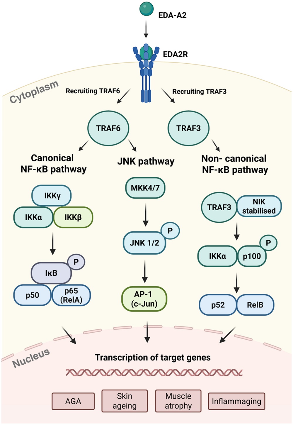
There are three different pathways by which EDA2R appears to lead to inflammaging. Through TRAF6, the well-known canonical NF-κB pathway and the JNK pathway can be activated, but a different ligand, TRAF3, activates the non-canonical NF-κB pathway. All three of these pathways have been documented to be involved in inflammation [12-14].

EDA2R pathways

There have been several studies suggesting a direct relationship between EDA2R and both inflammatory and age-related diseases. Overexpression of EDA2R is associated with an overexpression of lipids related to acne [15], and it is increased in progeria as well [10]. Another study found that its overexpression is related to many other lipid-related disorders, including characteristics of obesity along with metabolic issues; that study also found a related increase in inflammation [16]. There has also been a documented, significant relationship between EDA2R and frailty [17].

The researchers sum up these findings: “Collectively, the evidence from diverse cohorts, animal models and clinical studies underscores EDA2R gene expression as a robust and versatile biomarker of multiple interconnected pathophysiological processes.”

Further work appears to agree with this assessment. One study that used UK Biobank data found that the EDA2R protein is associated with premature aging, including both frailty and multiple epigenetic aging clocks, such as PhenoAge, along with having a negative healthspan association [18]. A different study found it to be associated with a broad range of cancers [19], another study found it to be linked to metabolic disorders such as diabetes [20], and yet another found a link between EDA2R and dementia [21].

A potentially difficult target

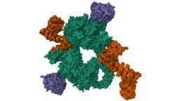
The reviewers took some time to discuss the physical intricacies of EDA2R as a protein, including its shape and the protein it binds to. They note its similarities to EDAR and that there are only two amino acid residues that distinguish the two, which may make it more difficult to develop a drug that targets EDA2R without affecting EDAR. The protein’s folded structure has also not been conclusively determined, although AI-based systems such as AlphaFold have made good guesses and it is possible to use expensive but slow microscopy techniques to have a definitive answer.

However, there are some existing techniques that appear to decrease EDA2R. A mouse study found that ginkgolide B decreases the expression of the gene’s murine counterpart [22], while human studies have found that excessive bed rest increases it [23] while fasting decreases it [24]. This suggests that it may be modulated by the same general treatment recommended everywhere: diet and exercise.

We would like to ask you a small favor. We are a non-profit foundation, and unlike some other organizations, we have no shareholders and no products to sell you. All our news and educational content is free for everyone to read, but it does mean that we rely on the help of people like you. Every contribution, no matter if it’s big or small, supports independent journalism and sustains our future.

Literature

[1] Mikkola, M. L. (2008). TNF superfamily in skin appendage development. Cytokine & growth factor reviews, 19(3-4), 219-230.

[2] Lee, J., & Tumbar, T. (2012, October). Hairy tale of signaling in hair follicle development and cycling. In Seminars in cell & developmental biology (Vol. 23, No. 8, pp. 906-916). Academic Press.

[3] Katthika, V. K., & Auerkari, E. I. (2018, May). Ectodermal Dysplasia. In 11th International Dentistry Scientific Meeting (IDSM 2017) (pp. 230-238). Atlantis Press.

[4] Dostert, C., Grusdat, M., Letellier, E., & Brenner, D. (2019). The TNF family of ligands and receptors: communication modules in the immune system and beyond. Physiological reviews, 99(1), 115-160.

[5] Bilgic, S. N., Domaniku, A., Toledo, B., Agca, S., Weber, B. Z., Arabaci, D. H., … & Kir, S. (2023). EDA2R–NIK signalling promotes muscle atrophy linked to cancer cachexia. Nature, 617(7962), 827-834.

[6] Arif, M., Lehoczki, A., Haskó, G., Lohoff, F. W., Ungvari, Z., & Pacher, P. (2025). Global and tissue-specific transcriptomic dysregulation in human aging: Pathways and predictive biomarkers. GeroScience, 1-20.

[7] Bergqvist, C., Ramia, P., Abbas, O., & Kurban, M. (2017). Genetics of syndromic and non‐syndromic hereditary nail disorders. Clinical Genetics, 91(6), 813-823.

[8] Kanoni, S., Graham, S. E., Wang, Y., Surakka, I., Ramdas, S., Zhu, X., … & Leonard, H. L. (2022). Implicating genes, pleiotropy, and sexual dimorphism at blood lipid loci through multi-ancestry meta-analysis. Genome biology, 23(1), 268.

[9] Prodi, D. A., Pirastu, N., Maninchedda, G., Sassu, A., Picciau, A., Palmas, M. A., … & Pirastu, M. (2008). EDA2R is associated with androgenetic alopecia. Journal of Investigative Dermatology, 128(9), 2268-2270.

[10] Barbera, M. C., Guarrera, L., Re Cecconi, A. D., Cassanmagnago, G. A., Vallerga, A., Lunardi, M., … & Bolis, M. (2025). Increased ectodysplasin-A2-receptor EDA2R is a ubiquitous hallmark of aging and mediates parainflammatory responses. Nature Communications, 16(1), 1898.

[11] Zoodsma, M., Beuchel, C., Yasmeen, S., Kohleick, L., Nepal, A., Koprulu, M., … & Langenberg, C. (2025). A genetic map of human metabolism across the allele frequency spectrum. Nature Genetics, 1-11.

[12] Moneva-Sakelarieva, M., Kobakova, Y., Konstantinov, S., Momekov, G., Ivanova, S., Atanasova, V., … & Atanasov, P. (2025). The role of the transcription factor NF-kB in the pathogenesis of inflammation and carcinogenesis. Modulation capabilities. Pharmacia, 72, 1-13.

[13] Wu, N., Wang, S., Zhang, Y., & Wang, S. (2025). Research Progress on Anti-Inflammatory Mechanism of Inula cappa. International Journal of Molecular Sciences, 26(5), 1911.

[14] Kaltschmidt, C., Greiner, J. F., & Kaltschmidt, B. (2021). The transcription factor NF-κB in stem cells and development. Cells, 10(8), 2042.

[15] Kwack, M. H., Hamida, O. B., Lee, W. J., & Kim, M. K. (2024). EDA-A2 increases lipid production in EDA2R-expressing human sebocytes. Journal of Dermatological Science, 113(1), 34-37.

[16] Arif, M., Lehoczki, A., Haskó, G., Lohoff, F. W., Ungvari, Z., & Pacher, P. (2025). Global and tissue-specific transcriptomic dysregulation in human aging: Pathways and predictive biomarkers. GeroScience, 1-20.

[17[ Perez, K., Ciotlos, S., McGirr, J., Limbad, C., Doi, R., Nederveen, J. P., … & Melov, S. (2022). Single nuclei profiling identifies cell specific markers of skeletal muscle aging, frailty, and senescence. Aging (Albany NY), 14(23), 9393.

[18] Ma, L. Z., Liu, W. S., He, Y., Zhang, Y., You, J., Feng, J. F., … & Yu, J. T. (2025). Plasma proteomics identify novel biomarkers and dynamic patterns of biological aging. Journal of Advanced Research.

[19] Papier, K., Atkins, J. R., Tong, T. Y., Gaitskell, K., Desai, T., Ogamba, C. F., … & Travis, R. C. (2024). Identifying proteomic risk factors for cancer using prospective and exome analyses of 1463 circulating proteins and risk of 19 cancers in the UK Biobank. Nature Communications, 15(1), 4010.

[20] Qian, H., Wu, C., Li, B., Rosenzweig, A., & Wang, M. (2025). Plasma Proteomics Linking Primary and Secondary diseases: Insights into Molecular Mediation from UK Biobank Data. medRxiv, 2025-08.

[21] Gong, J., Williams, D. M., Scholes, S., Assaad, S., Bu, F., Hayat, S., … & Steptoe, A. (2025). Unraveling the role of proteins in dementia: insights from two UK cohorts with causal evidence. Brain Communications, 7(2), fcaf097.

[22] Lee, C. W., Wang, B. Y. H., Wong, S. H., Chen, Y. F., Cao, Q., Hsiao, A. W. T., … & Lee, O. K. S. (2025). Ginkgolide B increases healthspan and lifespan of female mice. Nature aging, 5(2), 237-258.

[23] Fernandez‐Gonzalo, R., Tesch, P. A., Lundberg, T. R., Alkner, B. A., Rullman, E., & Gustafsson, T. (2020). Three months of bed rest induce a residual transcriptomic signature resilient to resistance exercise countermeasures. The FASEB Journal, 34(6), 7958-7969.

[24] Pietzner, M., Uluvar, B., Kolnes, K. J., Jeppesen, P. B., Frivold, S. V., Skattebo, Ø., … & Langenberg, C. (2024). Systemic proteome adaptions to 7-day complete caloric restriction in humans. Nature metabolism, 6(4), 764-777.

IVF

Rapamycin May Delay Age-Related Fertility Decline

In a recent study, researchers identified that an increase in the expression of ribosome-related genes and a loss of protein homeostasis contribute to the age-related decline in female fertility. Rapamycin restored this balance and increased fertility rates in a human trial [1].

The first system to age

The loss of fertility is one of the first signs of aging in women, with the first signs of fertility decline occurring around the mid-thirties. This fertility decline is primarily attributed to decreased oocyte quality and quantity. However, many more molecular changes occur in the oocyte and the surrounding cells, such as cumulus and granulosa cells.

In this study, the researchers investigated molecular changes in aging oocytes and cumulus cells and tested a possible intervention for improving clinical fertility outcomes.

Loss of balance

Analyzing the gene expression of the oocytes and cumulus cell samples donated for this study, along with an independent cohort dataset of oocytes from women of different ages, showed age-related changes with a “distinct shift in oocyte gene expression profiles” that “emerged around the age of 34.” Among the various pathways that were impacted by age, the researchers specifically noted that increased age was correlated with an enrichment in ribosome signaling and increased expression of ribosome-related genes, suggesting a link between ribosome dysregulation and oocyte aging.

Cumulus cells showed even more age-related changes in gene expression. Among many affected processes, those related to ribosomes were also increased in aging cumulus cells.

Ribosomes are cell structures where the translation of RNA into protein occurs; therefore, disruptions in ribosome-related processes might suggest disrupted protein homeostasis, which is exactly what researchers have observed in the cumulus cells. Specifically, those cells showed the presence of protein aggregates. The authors suggest that such aggregates can be there for two reasons: an increase in synthesis or a decrease in degradation.

The researchers observed both problems. There was decreased gene expression relating to lysosomes and proteasomes, both of which play a role in protein degradation, and a decrease in the activity of lysosomes with age. 18S and 28S rRNAs, key structures in ribosomes, were increased with age in cumulus cells, which suggests a related increase in translation and protein synthesis. This was further confirmed by additional tests.

However, the researchers also noted that rapamycin, a well-known drug in the aging field, reduced the increase in age-related protein synthesis in cumulus cells, helping restore the disrupted protein homeostasis.

Regulating gene accessibility

Changes in gene expression can result from changes in DNA methylation. The researchers observed that DNA methylation levels were modestly increased in aging oocytes, and the pattern of changes suggests that methylation might be responsible for the age-related changes in gene expression in oocytes, at least for some genes.

Cumulus cells showed minimal changes in DNA methylation; however, the changes that were present suggested that they might impact the expression of ribosome-related genes.

Along the same lines, the researchers observed changes in chromatin composition. Chromatin is a DNA and protein complex. Changes in its composition can make genes more accessible (euchromatin) or less accessible (heterochromatin) for transcription.

The heterochromatin levels in cumulus cells differed in young and old cells, showing an overall decrease in the level of heterochromatin with age. In young cells, heterochromatin-rich areas were associated with genes related to ribosome biogenesis, while in the old cells, heterochromatin-rich regions were associated with negative regulation of translation. Additionally, heterochromatin levels related to ribosome-related genes were decreased in old cumulus cells. All this suggests that the transcription of ribosomal biogenesis- and translation-related genes might be inhibited at the chromatin level in young cells, but aging reduces this inhibition, resulting in increased transcription of ribosomal genes.

Such regulation on the chromatin composition level was also observed for the lysosome-related genes, and it was correlated with a decrease in the expression of lysosome-related genes.

Delaying fertility loss in mice

Since the researchers observed an increase in ribosome-related gene expression, which suggests an increase in translation in aged oocytes and cumulus cells, they hypothesized that decreasing these processes might help delay the reduction in fertility. They first tested this assumption in mice, which showed similarities in gene expression changes with humans.

Comparison of rapamycin-treated ovaries from young (2-month-old) and old (10-month-old) mice showed that rapamycin reduced translation and ribosome biogenesis. In old cells, rapamycin inhibited the activity of senescence-related markers and decreased reactive oxygen species (ROS) levels. Rapamycin also reduced chromosomal abnormalities that are increased in aged oocytes, which suggests that rapamycin helps to delay oocyte aging.

More babies

Most importantly, the researchers continued by conducting a controlled clinical trial with 100 women undergoing in vitro fertilization (IVF). Half of the participants received only the standardized GnRH agonist long protocol, a standard IVF treatment; the other half received the same treatment, combined with 1mg rapamycin treatment for 21-28 days.

The authors note that “significantly more zygotes, embryos, and good-quality embryos were obtained in the rapamycin group than in the control group.” Comparing oocyte retrieval and embryo development between the groups, the researchers concluded that rapamycin treatment “can effectively improve oocyte quality and subsequent embryo development following the fertilization of retrieved oocytes.”

They also observed higher rates of pregnancy in females who took rapamycin compared to controls (50.0% vs. 28.2%) without a negative impact on live birth. However, they note that when they compared data regarding different stages of embryo transfer, they observed that it might be more beneficial for patients who take rapamycin to transfer their embryos at the 5- to 6-day stage instead of the 3-day stage.

Real-life benefits

All in all, in this study, the researchers identified that increased levels of ribosome-related genes and disruptions in proteostasis have a negative impact on oocytes and surrounding cells, contributing to age-related fertility problems.

Using rapamycin to remedy those issues resulted in improved ovarian functioning and increased fertility not only in mouse models but in human females undergoing IVF. While the trial results are encouraging, the researchers suggest optimization of dosage and duration of treatment might yield even better effects; however, for this, a trial with a higher number of participants might be required.

We would like to ask you a small favor. We are a non-profit foundation, and unlike some other organizations, we have no shareholders and no products to sell you. All our news and educational content is free for everyone to read, but it does mean that we rely on the help of people like you. Every contribution, no matter if it’s big or small, supports independent journalism and sustains our future.

Literature

[1] Li, J., Wang, H., Zhu, P., Chen, H., Zuo, H., Liu, C., Liu, L., Ye, X., Feng, G., Wu, Y., Liu, Q., Yang, T., Keefe, D. L., Bai, X., Shang, W., Wu, X., & Liu, L. (2025). Ribosome dysregulation and intervention in age-related infertility. Cell reports. Medicine, 102424. Advance online publication.

Skin capillaries

Skin Aging Underlined by Loss of Capillary Macrophages

A new study ties the disappearance of capillary-associated macrophages to age-related vascular degeneration in the skin. Boosting their function with a growth factor offers a possible avenue for anti-aging interventions [1].

Macrophages, capillaries, and skin aging

Skin aging might not be the most dangerous aspect of aging, but it certainly is one of the most conspicuous. It also provides a valuable model for studying aging as a whole.

Skin thinning and pesky wrinkles have mutiple origins, including diminished blood supply from the capillaries that permeate the skin [2]. In this study published in Nature, researchers from New York University School of Medicine investigated the role of capillary-associated macrophages (CAMs): immune cells that reside near the capillaries, where they clean debris, fight pathogens, and facilitate tissue repair.

Aging tissues lose resident macrophages [3] and microvascular function, but how these are linked in living organisms is not clear. This study aimed to determine if CAMs in skin decline with age, if such a loss impairs capillary perfusion and repair, and if they can be restored.

In vivo imaging reveals waning function

The researchers used an ingenious technique for in vivo imaging, which allowed them to analyze blood flow and skin condition of live mice longitudinally, from 1 to 18 months of age. “Older tissues show fewer blood vessels,” said Kailin R. Mesa, currently assistant professor at Princeton and the study’s corresponding author. “To understand how these changes develop and lead to age-related dysfunction, we built a multiphoton light microscopy imaging system to track tissue aging in living mice.”

Time-lapse imaging showed that upper-dermal CAMs decline faster than epidermal or lower-dermal macrophages and faster than capillary rarefaction itself. Thus, “macrophage-deficient” vascular niches are created, with capillaries devoid of CAMs. These capillaries without nearby CAMs experience higher rates of obstructed red blood cell (RBC) flow.

Human skin samples analyzed by the researchers showed the same pattern: CAM decline outpaced capillary loss, implying diminished coverage with age. Acute macrophage depletion reduced blood flow even more. According to this paper, aging leads CAMs to significantly skew towards loss rather than proliferation, contributing to their decline.

Injury and a growth factor boost macrophage recruitment

The study also revealed that CAMs are required for capillary repair and preservation. Following precise laser clotting of single capillaries, nearby CAMs were rapidly recruited within about two days and engulfed RBC debris. Capillaries with local CAMs were significantly better at re-establishing blood flow after injury than CAM-devoid ones. Ablation of CAMs prior to clot induction impaired capillary repair, indicating their critical role in vascular recovery.

The researchers then applied larger laser wounds to the upper dermis or the overlying epidermis to test whether more serious tissue damage triggers CAM replenishment, which it did. “We found that large laser-induced epidermal damage resulted in a lasting increase in CAMs below the damaged regions compared with in the neighboring control regions,” the paper says. Basically, while CAMs hardly renew on their own in aging skin, the study suggests that environmental changes, such as injury, can stimulate CAM replenishment and enhance vascular function in older mice.

Colony-stimulating factor 1 (CSF1) is a growth cue that tells macrophages to survive and divide. The team used CSF1-Fc, an enhanced version that makes the signal hang around longer in tissue to test whether it can improve CAM function by micro-injecting it into the skin of old mice (20-24 months) once a day for four days. This treatment resulted in more capillary-associated macrophages (CAMs) lining the tiny vessels without noticeably changing the balance between long-lived resident cells and freshly recruited ones, which are derived from monocytes.

CSF1-Fc reduced the number of capillary segments with sluggish or blocked red blood cell flow at baseline, and when the researchers created pinpoint clots, treated areas cleared debris and reperfused more reliably over the next week. In other words, topping up the resident macrophage niche in old skin rejuvenated repair capacity. “We also found that dermal macrophage self-renewal and vascular support could be acutely enhanced in aged mice through local CSF1 therapeutic treatment,” the paper says.

The importance of this discovery goes beyond skin, since dense capillary beds in the brain, heart, kidney, and skeletal muscle are also maintained by perivascular macrophages. Their age-related decline probably produces the same pattern seen in skin: sluggish flow, failed micro-repairs, and gradual pruning of the network. Improving things in the macrophage niche might provide a general route to preserving microvascular health in multiple tissues and organs. “When macrophages disappear, skin blood vessels age. Loss of skin macrophages with age blocks blood vessel flow and clot repair, revealing a new cellular trigger of tissue aging,” said Mesa.

We would like to ask you a small favor. We are a non-profit foundation, and unlike some other organizations, we have no shareholders and no products to sell you. All our news and educational content is free for everyone to read, but it does mean that we rely on the help of people like you. Every contribution, no matter if it’s big or small, supports independent journalism and sustains our future.

Literature

[1] Mesa, K. R., O’Connor, K. A., Ng, C., Salvatore, S. P., Dolynuk, A., Lomeli, M. R., & Littman, D. R. (2025). Niche-specific dermal macrophage loss promotes skin capillary ageing. Nature, 1-9.

[2] Bentov, I., & Reed, M. J. (2015). The effect of aging on the cutaneous microvasculature. Microvascular research, 100, 25-31.

[3] Park, M. D., Yatim, N., Zhang, J., Cho, B. A., Yoo, S. K., Schaefer, M. M., … & Merad, M. (2025). Restoring resident tissue macrophages to combat aging and cancer. Nature Aging, 5(8), 1383-1392.

Rejuvenation Roundup October 2025

Rejuvenation Roundup October 2025

The trick-or-treaters have all gone home as the leaves have fallen to the ground, but rejuvenation biotech attempts to prevent human beings from fading away. Here’s what’s been done to fight aging in October.

Team and activities

Public Longevity GroupOur Campaign for Public Longevity: Public trust is the breakthrough that unlocks all others. The field of longevity science has long needed better tools to measure public sentiment in order to understand what drives that trust, and how to build it.

Thanks to your support, the Public Longevity Group (PLG) campaign is more than halfway to its goal, but there’s still time to make an even greater impact. We’re extending the campaign by two more weeks, with the LRI Board of Directors matching all donations up to $25,000 to push us across the finish line. That means every dollar you give will be doubled to support the launch of the first data-driven sentiment analysis engine for longevity science.

Interviews

Joe Betts-Lacroix InterviewJoe Betts-Lacroix on Retro Bio and Its Recent AI Advancement: Retro Biosciences was founded about five years ago by the tech entrepreneur Joe Betts-Lacroix with a $180 million investment from his friend Sam Altman, the CEO of OpenAI.

Sam Sharifi on Fixing Our DNA: Behind the vision of Sam Sharifi, PhD and Matter Bio’s Chief Scientific Officer, stands some serious science. Matter Bio has matured into a company with a pipeline and a cash flow.

Andrea Maier InterviewAndrea Maier on Longevity Medicine for All: Dr. Andrea Maier, Oon Chiew Seng Professor in Medicine at the National University of Singapore, is a veteran geroscientist and one of the most familiar faces in the rising field of longevity medicine. Andrea has been working on bringing longevity medicine to the public.

Advocacy and Analysis

Meet ALSAE: Improving the Cultural Image of Longevity: To challenge cultural stereotypes and misconceptions about longevity, a new organization aims to engage people who create culture. Its list of “cultural ambassadors” includes Oscar- and Grammy-winning artists.

Research Roundup

Dividing cellsCell Cycle Stage Impacts the Efficacy of Senotherapeutics: A recent study reported that the effectiveness of the senotherapeutic drug ABT-263 depends on the cell’s DNA content, which is based on the cell cycle phase at which the senescent cell was arrested.

Partial Reprogramming Enhances Nerve Repair in Rats: In Advanced Science, a team of researchers has explained how partial cellular reprogramming through the OSKM factors restores nerve repair ability to older animals.

RapamycinImpact of Off-Label Low-Dose Rapamycin on Healthy Adults: A team of scientists has concluded that there is no sufficient evidence that low-dose rapamycin use can extend healthspan and lifespan in healthy humans.

A Public Website for Evaluating Potential Anti-Aging Drugs: Researchers publishing in the International Journal of Molecular Sciences have showcased their creation of PASS GERO, a public-facing application that allows researchers to evaluate potential anti-aging compounds.

NanoparticlesNanoparticles Potently Reverse Alzheimer’s in Mice: Scientists have created polymersomes, a type of nanoparticle, that latch onto a master regulator of amyloid-beta clearance, diverting it towards a more efficient route.

Researchers Identify Key Age-Related Genes: In Aging Cell, researchers have described core genes that apply to a wide variety of species and appear to be causal drivers of aging.

Naked mole ratsWhy Naked Mole Rats Have Better DNA Repair: Scientists have found that the cGAS protein in naked mole rats, a famously long-lived species, boosts DNA repair, while the human version tends to hamper it. The difference boils down to just four amino acids.

Interventions Testing Program Finds Three New Compounds: A new report from the Interventions Testing Program adds three new compounds to the list of lifespan-extending compounds confirmed during their experiments: epicatechin, halofuginone, and mitoglitazone.

Infrared lampInfrared Lasers Clear Harmful Compounds in Mouse Brains: Researchers have discovered that infrared lasers promote the clearance of toxic metabolites from the brains of age-accelerated mice by improving lymphatic drainage.

Female-Specific Mechanism of Neurodegeneration Discovered: Scientists have found that a gene on the X chromosome, which escapes silencing, promotes inflammation and neurodegeneration in a mouse model of multiple sclerosis, but the effects can be reversed with metformin.

International Space StationHow Being in Orbit Affects Stem Cells: In Cell Stem Cell, researchers from the Cedars-Sinai Medical Center have published a review discussing what experiments conducted in space can do for stem cell research and medical research as a whole.

Tiny Brain Organoids Show Promise in Alzheimer’s Research: Scientists have developed a novel multicellular integrated brain model to study neurological diseases, incorporating all six major brain cell types derived from patient-specific induced pluripotent stem cells.

Macular degenerationAddressing Age-Related Vision Decline With Lipid Supplements: Researchers have partially reversed age-related vision decline in mice by injecting lipids directly into the retina.

Measuring Dog Aging With Protein Metabolism: Researchers working with data from the Dog Aging Project have found that post-translationally modified amino acids, which are generated from protein metabolism, are useful in determining the biological ages of dogs.

Neural plaquesAmyloid-Fibrinogen Complexes Exacerbate Alzheimer’s in Mice: A study from Rockefeller University suggests that an unholy duo of two proteins causes harm in much lower concentrations than either of them alone.

Stiff Collagen May Lead to Senescent Cells: A preprint study has found that the stiffness of the extracellular matrix encourages cells to undergo senescence. These researchers placed cells into a hydrogel scaffold that can have its stiffness changed on the fly.

Bacteria under microscopeReview Links Lingering Infections to Senescence: In a comprehensive review, scientists discuss the various mechanisms by which chronic infections drive cellular senescence and aging.

Organ Fat Is Associated With Carotid Atherosclerosis: A recent study discovered an association between visceral and, to a lesser extent, hepatic fat with cardiovascular risk factors and carotid atherosclerosis.

Maintenance Stays Efficient in Healthy Older People’s Cells: Researchers publishing in Aging Cell have found that the efficiency of autophagy, a cellular maintenance process, increases rather than decreases in some T cells derived from healthy older people.

Men Might Need More Exercise to Lower Cardiovascular Risks: A new observational study suggests that men need more than twice as much exercise as women do to achieve the same level of reduction in the risk of cardiovascular heart disease.

Relationship between weekends catch-up sleep and risk of aging: Stratifying by bedtime, the relationship between catch-up sleep and reduced aging risk is only observed in individuals who usually go to sleep before midnight and have catch-up seep less than 2 hours.

Dose-response relationship of resistance training and the effects on circulating biomarkers of inflammation or neuroplasticity in older adults: These findings demonstrate that resistance training can significantly improve circulating biomarkers of neuroplasticity and inflammation and highlight key dose-response relationships critical for optimizing such programs.

An Ad Libitum-Fed Diet That Matches the Beneficial Lifespan Effects of Caloric Restriction: Ad libitum-fed diets can be designed to replicate, and potentially enhance, the geroprotective benefits of caloric restriction, albeit via different mechanisms, potentially offering a more sustainable dietary approach to longevity extension.

α-Lipoic acid mitigates age-related cognitive decline by modulating PPARγ/NF-κB-mediated neuroinflammation: Administration demonstrated significant cognitive-enhancing effects, accompanied by suppression of both microgliosis and astrocytosis, as well as a reduction in pro-inflammatory cytokines in the hippocampus of aged mice.

Effects of lifetime supplementation with ubiquinol 10 on the lifespan and progression of aging in female C57BL/6 mice: Maximum lifespans and survival curves did not significantly differ between the ubiquinol 10 and control groups.

Enhanced Selenium Supplement Extends Lifespan and Delays Multi-Organs Aging by Regulating the Sik1 Pathway Through Maintaining Calcium Homeostasis: This systematic approach provides a solid theoretical foundation and clinical evidence for the application of nano-selenium in anti-aging research.

Glycation-lowering compounds inhibit ghrelin signaling to reduce food intake, lower insulin resistance, and extend lifespan: As a late-life intervention, Gly-Low slows hypothalamic aging signatures, improves glucose homeostasis and motor coordination, and increases lifespan, suggesting its potential benefits in ameliorating age-associated decline.

Effect of the mitophagy inducer urolithin A on age-related immune decline: a randomized, placebo-controlled trial: These findings indicate that short-term urolithin A supplementation modulates human immune cell composition and function, supporting its potential to counteract age-related immune decline and inflammaging.

Combined creatine and HMB co-supplementation improves functional strength independent of muscle mass in physically active older adults: This combined approach represents a promising strategy for preserving functional capacity and promoting healthy ageing.

Pharmacokinetic analysis of intermittent rapamycin administration in early-stage Alzheimer’s Disease: These findings suggest that weekly rapamycin administration in Alzheimer’s patients results in acceptable pharmacokinetic variability, supporting fixed-dose regimens in future trials.

Mesenchymal stem cell derived extracellular vesicles reverses neural aging via OSKM modulation: This study represents a pioneering achievement in regenerative medicine, demonstrating the potential of microRNAs in extracellular vesicles to stimulate OSKM, a significant stride forward in addressing neural aging.

Dual-ligand engineered exosome regulates WNT signaling activation to promote liver repair and regeneration: Collectively, this work revealed the broad therapeutic effects of WNT signaling activation in the liver through the dual-ligand-carrying exosomes.

Effects of low-intensity pulsed ultrasound on muscle mass and Fndc5 mRNA expression in aged male mice: These findings suggest that this technique may serve as a safe, non-invasive intervention to counteract sarcopenia by promoting muscle growth and mitochondrial gene activation in aged skeletal muscle.

Effects of Computerized Cognitive Training on Vesicular Acetylcholine Transporter Levels in Healthy Older Adults: These findings clarify the neurochemical basis of cognitive training benefits, showing that speed training upregulates binding in networks that support attention, memory, and executive function.

The dissipation theory of aging: a quantitative analysis using a cellular aging map: These results provide a novel perspective on aging as a dissipative process and introduce a computational framework that enables measuring age-related changes with molecular resolution.

News Nuggets

Agentic AI Against AgingAgentic AI Against Aging Hackathon: HackAging.ai is the global online hackathon at the intersection of Agentic AI and longevity science, bringing together researchers, founders, and engineers to accelerate solutions that extend healthy human lifespan.

New Foundation Unites Hollywood and the Arts Against Aging: The Alliance for Longevity Science, Arts & Entertainment announced its official launch, pioneering a new nonprofit effort to accelerate progress against the chronic diseases of aging.

NUS_Logo-Stacked-Centered-LineNUS Medicine Launches New Clinical Trial Centre: Spanning 350 square metres, the new Centre brings together state-of-the art facilities within an integrated multidisciplinary framework to enable clinical research and implementation of gerodiagnostics and gerotherapeutics.

We would like to ask you a small favor. We are a non-profit foundation, and unlike some other organizations, we have no shareholders and no products to sell you. All our news and educational content is free for everyone to read, but it does mean that we rely on the help of people like you. Every contribution, no matter if it’s big or small, supports independent journalism and sustains our future.