The Blog

Building a Future Free of Age-Related Disease

Rejuvenation Roundup October 2025

Rejuvenation Roundup October 2025

The trick-or-treaters have all gone home as the leaves have fallen to the ground, but rejuvenation biotech attempts to prevent human beings from fading away. Here’s what’s been done to fight aging in October.

Team and activities

Public Longevity GroupOur Campaign for Public Longevity: Public trust is the breakthrough that unlocks all others. The field of longevity science has long needed better tools to measure public sentiment in order to understand what drives that trust, and how to build it.

Thanks to your support, the Public Longevity Group (PLG) campaign is more than halfway to its goal, but there’s still time to make an even greater impact. We’re extending the campaign by two more weeks, with the LRI Board of Directors matching all donations up to $25,000 to push us across the finish line. That means every dollar you give will be doubled to support the launch of the first data-driven sentiment analysis engine for longevity science.

Interviews

Joe Betts-Lacroix InterviewJoe Betts-Lacroix on Retro Bio and Its Recent AI Advancement: Retro Biosciences was founded about five years ago by the tech entrepreneur Joe Betts-Lacroix with a $180 million investment from his friend Sam Altman, the CEO of OpenAI.

Sam Sharifi on Fixing Our DNA: Behind the vision of Sam Sharifi, PhD and Matter Bio’s Chief Scientific Officer, stands some serious science. Matter Bio has matured into a company with a pipeline and a cash flow.

Andrea Maier InterviewAndrea Maier on Longevity Medicine for All: Dr. Andrea Maier, Oon Chiew Seng Professor in Medicine at the National University of Singapore, is a veteran geroscientist and one of the most familiar faces in the rising field of longevity medicine. Andrea has been working on bringing longevity medicine to the public.

Advocacy and Analysis

Meet ALSAE: Improving the Cultural Image of Longevity: To challenge cultural stereotypes and misconceptions about longevity, a new organization aims to engage people who create culture. Its list of “cultural ambassadors” includes Oscar- and Grammy-winning artists.

Research Roundup

Dividing cells
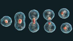
Cell Cycle Stage Impacts the Efficacy of Senotherapeutics: A recent study reported that the effectiveness of the senotherapeutic drug ABT-263 depends on the cell’s DNA content, which is based on the cell cycle phase at which the senescent cell was arrested.

Partial Reprogramming Enhances Nerve Repair in Rats: In Advanced Science, a team of researchers has explained how partial cellular reprogramming through the OSKM factors restores nerve repair ability to older animals.

RapamycinImpact of Off-Label Low-Dose Rapamycin on Healthy Adults: A team of scientists has concluded that there is no sufficient evidence that low-dose rapamycin use can extend healthspan and lifespan in healthy humans.

A Public Website for Evaluating Potential Anti-Aging Drugs: Researchers publishing in the International Journal of Molecular Sciences have showcased their creation of PASS GERO, a public-facing application that allows researchers to evaluate potential anti-aging compounds.

Nanoparticles
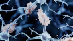
Nanoparticles Potently Reverse Alzheimer’s in Mice: Scientists have created polymersomes, a type of nanoparticle, that latch onto a master regulator of amyloid-beta clearance, diverting it towards a more efficient route.

Researchers Identify Key Age-Related Genes: In Aging Cell, researchers have described core genes that apply to a wide variety of species and appear to be causal drivers of aging.

Naked mole rats
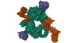
Why Naked Mole Rats Have Better DNA Repair: Scientists have found that the cGAS protein in naked mole rats, a famously long-lived species, boosts DNA repair, while the human version tends to hamper it. The difference boils down to just four amino acids.

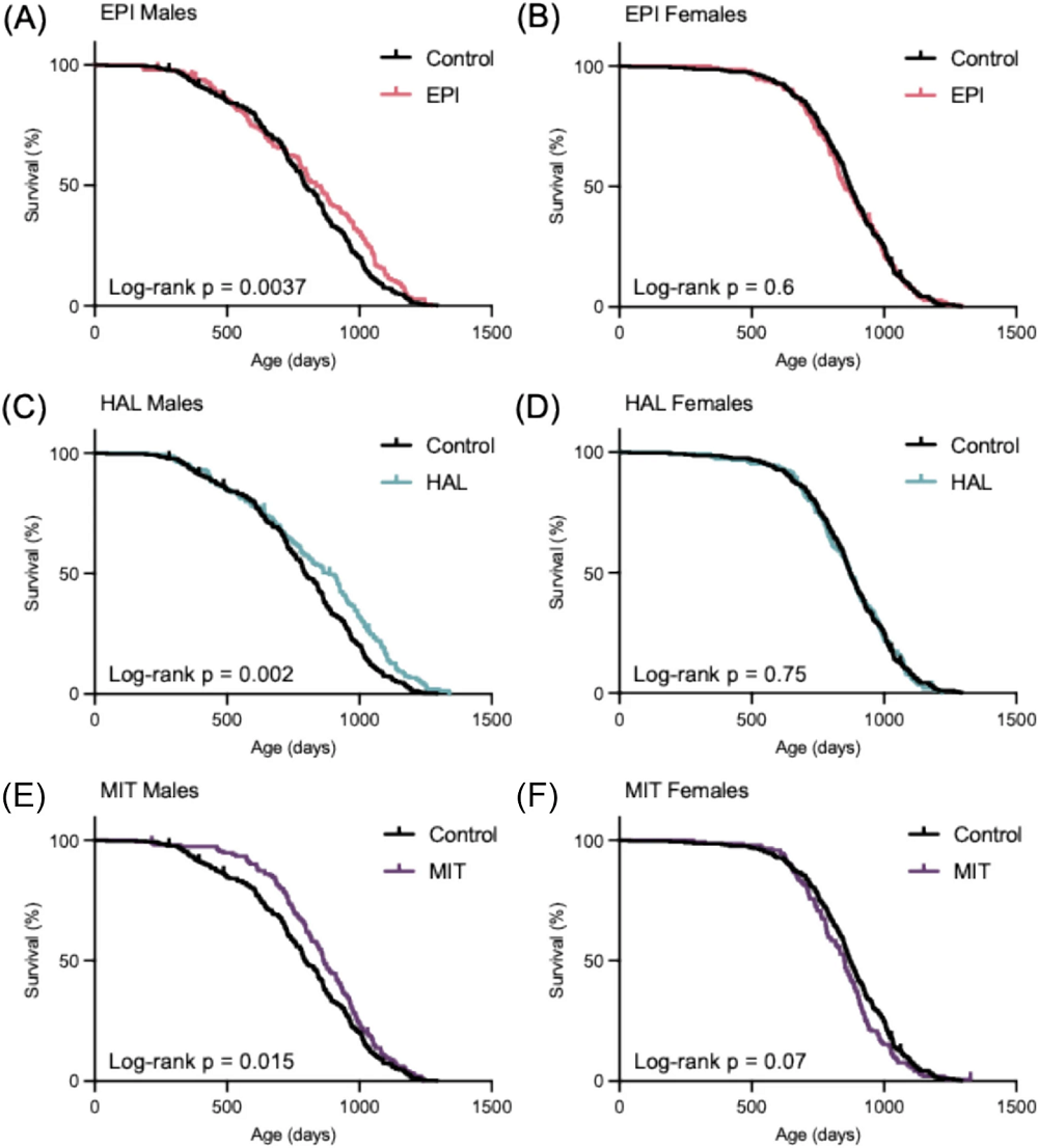
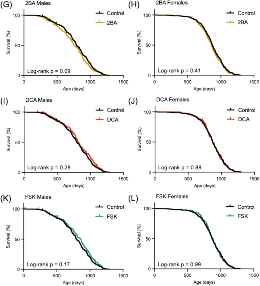
Interventions Testing Program Finds Three New Compounds: A new report from the Interventions Testing Program adds three new compounds to the list of lifespan-extending compounds confirmed during their experiments: epicatechin, halofuginone, and mitoglitazone.

Infrared lamp
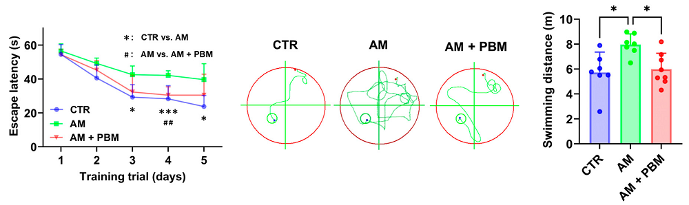
Infrared Lasers Clear Harmful Compounds in Mouse Brains: Researchers have discovered that infrared lasers promote the clearance of toxic metabolites from the brains of age-accelerated mice by improving lymphatic drainage.

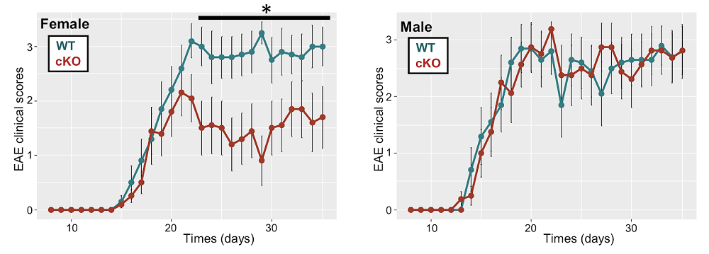
Female-Specific Mechanism of Neurodegeneration Discovered: Scientists have found that a gene on the X chromosome, which escapes silencing, promotes inflammation and neurodegeneration in a mouse model of multiple sclerosis, but the effects can be reversed with metformin.

International Space StationHow Being in Orbit Affects Stem Cells: In Cell Stem Cell, researchers from the Cedars-Sinai Medical Center have published a review discussing what experiments conducted in space can do for stem cell research and medical research as a whole.

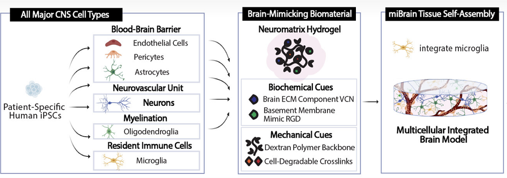
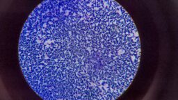
Tiny Brain Organoids Show Promise in Alzheimer’s Research: Scientists have developed a novel multicellular integrated brain model to study neurological diseases, incorporating all six major brain cell types derived from patient-specific induced pluripotent stem cells.

Macular degenerationAddressing Age-Related Vision Decline With Lipid Supplements: Researchers have partially reversed age-related vision decline in mice by injecting lipids directly into the retina.

Measuring Dog Aging With Protein Metabolism: Researchers working with data from the Dog Aging Project have found that post-translationally modified amino acids, which are generated from protein metabolism, are useful in determining the biological ages of dogs.

Neural plaquesAmyloid-Fibrinogen Complexes Exacerbate Alzheimer’s in Mice: A study from Rockefeller University suggests that an unholy duo of two proteins causes harm in much lower concentrations than either of them alone.

Stiff Collagen May Lead to Senescent Cells: A preprint study has found that the stiffness of the extracellular matrix encourages cells to undergo senescence. These researchers placed cells into a hydrogel scaffold that can have its stiffness changed on the fly.

Bacteria under microscope
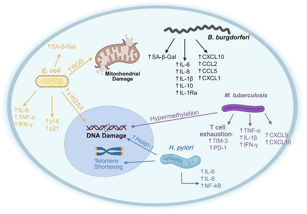
Review Links Lingering Infections to Senescence: In a comprehensive review, scientists discuss the various mechanisms by which chronic infections drive cellular senescence and aging.

Organ Fat Is Associated With Carotid Atherosclerosis: A recent study discovered an association between visceral and, to a lesser extent, hepatic fat with cardiovascular risk factors and carotid atherosclerosis.

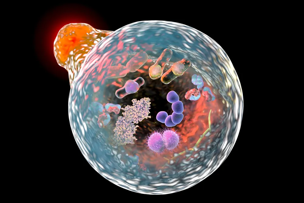
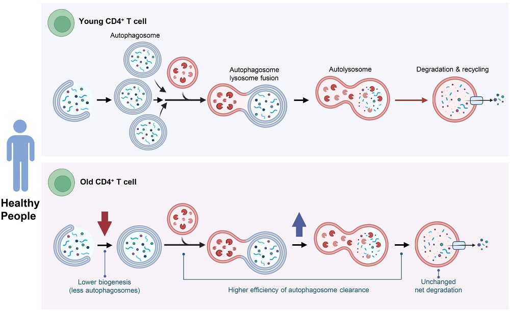
Maintenance Stays Efficient in Healthy Older People’s Cells: Researchers publishing in Aging Cell have found that the efficiency of autophagy, a cellular maintenance process, increases rather than decreases in some T cells derived from healthy older people.

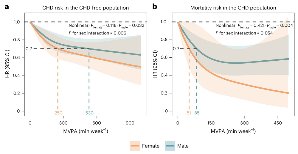
Men Might Need More Exercise to Lower Cardiovascular Risks: A new observational study suggests that men need more than twice as much exercise as women do to achieve the same level of reduction in the risk of cardiovascular heart disease.

Relationship between weekends catch-up sleep and risk of aging: Stratifying by bedtime, the relationship between catch-up sleep and reduced aging risk is only observed in individuals who usually go to sleep before midnight and have catch-up seep less than 2 hours.

Dose-response relationship of resistance training and the effects on circulating biomarkers of inflammation or neuroplasticity in older adults: These findings demonstrate that resistance training can significantly improve circulating biomarkers of neuroplasticity and inflammation and highlight key dose-response relationships critical for optimizing such programs.

An Ad Libitum-Fed Diet That Matches the Beneficial Lifespan Effects of Caloric Restriction: Ad libitum-fed diets can be designed to replicate, and potentially enhance, the geroprotective benefits of caloric restriction, albeit via different mechanisms, potentially offering a more sustainable dietary approach to longevity extension.

α-Lipoic acid mitigates age-related cognitive decline by modulating PPARγ/NF-κB-mediated neuroinflammation: Administration demonstrated significant cognitive-enhancing effects, accompanied by suppression of both microgliosis and astrocytosis, as well as a reduction in pro-inflammatory cytokines in the hippocampus of aged mice.

Effects of lifetime supplementation with ubiquinol 10 on the lifespan and progression of aging in female C57BL/6 mice: Maximum lifespans and survival curves did not significantly differ between the ubiquinol 10 and control groups.

Enhanced Selenium Supplement Extends Lifespan and Delays Multi-Organs Aging by Regulating the Sik1 Pathway Through Maintaining Calcium Homeostasis: This systematic approach provides a solid theoretical foundation and clinical evidence for the application of nano-selenium in anti-aging research.

Glycation-lowering compounds inhibit ghrelin signaling to reduce food intake, lower insulin resistance, and extend lifespan: As a late-life intervention, Gly-Low slows hypothalamic aging signatures, improves glucose homeostasis and motor coordination, and increases lifespan, suggesting its potential benefits in ameliorating age-associated decline.

Effect of the mitophagy inducer urolithin A on age-related immune decline: a randomized, placebo-controlled trial: These findings indicate that short-term urolithin A supplementation modulates human immune cell composition and function, supporting its potential to counteract age-related immune decline and inflammaging.

Combined creatine and HMB co-supplementation improves functional strength independent of muscle mass in physically active older adults: This combined approach represents a promising strategy for preserving functional capacity and promoting healthy ageing.

Pharmacokinetic analysis of intermittent rapamycin administration in early-stage Alzheimer’s Disease: These findings suggest that weekly rapamycin administration in Alzheimer’s patients results in acceptable pharmacokinetic variability, supporting fixed-dose regimens in future trials.

Mesenchymal stem cell derived extracellular vesicles reverses neural aging via OSKM modulation: This study represents a pioneering achievement in regenerative medicine, demonstrating the potential of microRNAs in extracellular vesicles to stimulate OSKM, a significant stride forward in addressing neural aging.

Dual-ligand engineered exosome regulates WNT signaling activation to promote liver repair and regeneration: Collectively, this work revealed the broad therapeutic effects of WNT signaling activation in the liver through the dual-ligand-carrying exosomes.

Effects of low-intensity pulsed ultrasound on muscle mass and Fndc5 mRNA expression in aged male mice: These findings suggest that this technique may serve as a safe, non-invasive intervention to counteract sarcopenia by promoting muscle growth and mitochondrial gene activation in aged skeletal muscle.

Effects of Computerized Cognitive Training on Vesicular Acetylcholine Transporter Levels in Healthy Older Adults: These findings clarify the neurochemical basis of cognitive training benefits, showing that speed training upregulates binding in networks that support attention, memory, and executive function.

The dissipation theory of aging: a quantitative analysis using a cellular aging map: These results provide a novel perspective on aging as a dissipative process and introduce a computational framework that enables measuring age-related changes with molecular resolution.

News Nuggets

Agentic AI Against AgingAgentic AI Against Aging Hackathon: HackAging.ai is the global online hackathon at the intersection of Agentic AI and longevity science, bringing together researchers, founders, and engineers to accelerate solutions that extend healthy human lifespan.

New Foundation Unites Hollywood and the Arts Against Aging: The Alliance for Longevity Science, Arts & Entertainment announced its official launch, pioneering a new nonprofit effort to accelerate progress against the chronic diseases of aging.

NUS_Logo-Stacked-Centered-LineNUS Medicine Launches New Clinical Trial Centre: Spanning 350 square metres, the new Centre brings together state-of-the art facilities within an integrated multidisciplinary framework to enable clinical research and implementation of gerodiagnostics and gerotherapeutics.

We would like to ask you a small favor. We are a non-profit foundation, and unlike some other organizations, we have no shareholders and no products to sell you. All our news and educational content is free for everyone to read, but it does mean that we rely on the help of people like you. Every contribution, no matter if it’s big or small, supports independent journalism and sustains our future.

Older man and woman exercising

Men Might Need More Exercise to Lower Cardiovascular Risks

A new observational study suggests that men need more than twice as much exercise as women do to achieve the same level of reduction in the risk of cardiovascular heart disease [1].

Understanding sex differences

In recent years, scientists have questioned how much exercise is needed for tangible health benefits. Current guidelines from the American Heart Association, the European Society of Cardiology, and the World Health Organization suggest that everyone should be getting at least 150 minutes per week of moderate-to-vigorous physical activity (MVPA) to significantly lower the risk of cardiovascular heart disease (CHD).

Numerous studies have added layers of complexity to these assessments, such as by showing diminishing returns, with the “sweet spot” lying around 7,000 daily steps [2] or even a U-shaped relationship, in which too much exercise can actually be harmful [3]. On many of the questions, the jury is still out.

One important piece of the puzzle, however, remains understudied: the effects of sexual differences on physical activity. Work in animal models, demographic studies, and human studies all point to the two sexes experiencing aging and disease differently. Yet, this difference is rarely accounted for in health recommendations. In this new study, scientists used data from UK Biobank (UKB), a repository of health information on about half a million British citizens, to shed some light on this question.

Men need to work twice as hard

Over 80,000 UKB participants wore wrist accelerometers for one week to record their physical activity. This created a trove of data that many scientists have used. The sample included 5,169 people with CHD at baseline and 3,764 incident CHD events with a median follow-up of about 8 years.

The study’s central finding was that, while women showed less adherence to the guidelines (150 minutes or above), it was also much easier for them to reach an equal reduction in CHD risk. For a 30% risk reduction, women needed about 250 min/week of MVPA, while men needed about 530 min/week: more than twice as much.

Among patients with CHD, physically active females experienced much greater mortality risk reduction than males (70% vs. only 19% in men). However, this finding, impressive as it is, was based on a far smaller CHD mortality sample (6 deaths among 340 adherent women), so estimates are imprecise even if statistically significant. The researchers urge caution and call for validation in larger CHD cohorts with wearable data.

Men women exercise differences

The research went to great lengths to control for possible confounding factors. These included age, geography (England, Scotland, or Wales), ethnicity, education, Townsend deprivation index, BMI, smoking and alcohol consumption status (never/ever/current), sleep duration, a dietary health score, as well as such medical conditions and treatments as diabetes, hypertension, dyslipidemia, use of cholesterol-lowering drugs, blood-pressure medication, insulin therapy, and Charlson comorbidity index.

The researchers also controlled for actual physical activity intensity inside the relatively wide MVPA range by measuring average acceleration. Other important variables taken into account were the polygenic risk score for CHD in incidence models and CHD treatment medications (antiplatelet, antianginal) in the mortality models. People who are already sick are likely to undergo less physical activity, but this reverse causality was mitigated by excluding participants who had events that occurred in the first year after accelerometer wear.

The team ran multiple model specifications with different covariate sets to confirm that the sex-interaction findings were not model-dependent. This did not fundamentally change the results: women benefited more per unit of MVPA across all these specifications, time scales, and strategies.

Possible explanations

The authors also hypothesized possible biological underpinnings of their results. For instance, higher circulating estrogen in women can shift fuel use toward greater lipid oxidation during exercise, which is linked to better CHD outcomes. Sex differences in fiber-type composition (women have more type I fibers, while men have more type II) and oxidative capacity could make women more “PA-sensitive,” yielding larger clinical benefit per minute.

The study’s observational design means that it cannot establish causality, only correlation, and causality and mechanisms will have to be elucidated by future research. Still, the pattern is consistent with prior accelerometer studies in older women [4].

The take-home message is important both for men and women. Since adherence is generally lower in women, knowing that they need much less effort to achieve the same CHD risk reduction may be encouraging. On the other hand, men should know that adhering to the current “sex-blind” guidelines might not be enough.

We would like to ask you a small favor. We are a non-profit foundation, and unlike some other organizations, we have no shareholders and no products to sell you. All our news and educational content is free for everyone to read, but it does mean that we rely on the help of people like you. Every contribution, no matter if it’s big or small, supports independent journalism and sustains our future.

Literature

[1] Chen, J., Wang, Y., Zhong, Z., Chen, X., Zhang, L., Jie, L., … & Wang, Y. (2025). Sex differences in the association of wearable accelerometer-derived physical activity with coronary heart disease incidence and mortality. Nature Cardiovascular Research, 1-11.

[2] ing, D., Nguyen, B., Nau, T., Luo, M., del Pozo Cruz, B., Dempsey, P. C., … Owen, K. (n.d.). Daily steps and health outcomes in adults: a systematic review and dose-response meta-analysis. The Lancet Public Health.

[3] Schnohr, P., O’Keefe, J. H., Marott, J. L., Lange, P., & Jensen, G. B. (2015). Dose of jogging and long-term mortality: the Copenhagen City Heart Study. Journal of the American College of Cardiology, 65(5), 411-419.

[4] LaMonte, M. J., Buchner, D. M., Rillamas‐Sun, E., Di, C., Evenson, K. R., Bellettiere, J., … & LaCroix, A. Z. (2018). Accelerometer‐measured physical activity and mortality in women aged 63 to 99. Journal of the American Geriatrics Society, 66(5), 886-894.

Maintenance Stays Efficient in Healthy Older People’s Cells

Researchers publishing in Aging Cell have found that the efficiency of autophagy, a cellular maintenance process, increases rather than decreases in some T cells derived from healthy older people.

Keeping cells healthy

The researchers introduce their paper by discussing autophagy and its decline in aging. They focus specifically on its effects on T cells, noting that a decline in autophagy causes mitochondrial dysfunction in CD4+ T cells due to old mitochondria not being cleared [1] and is associated with DNA damage in CD8+ T cells [2].

This paper focuses specifically on CD4+ T cells, building upon previous work demonstrating that an age-related deficiency of a glycolytic enzyme prevents these cells from using autophagy as an energy source [3] and that better autophagy in these cells is hereditary and associated with longer lifespans [4].

This study began by measuring the base rate of autophagy in CD4+ T cells derived from one group of 28- to 35-year-olds and another group of 67- to 93-year-olds. The first analysis was of compartments (puncta) that tested positive for microtubule-associated protein 1A/1B-light chain 3 (LC3), a marker of autophagosomes.

No autophagic decline in cells from healthy people

Interestingly, while the number of LC3+ puncta trended towards being lower in the older group, this did not reach the level of statistical significance. The number of protein-destroying lysosomes was also similar, as was the number of autolysosomes, which are combinations of autophagosomes and lysosomes that actually destroy unwanted mitochondria and organelles.

Experimentation with an inhibitor of autophagic degradation found that there is an increase in autophagic flux with age. This led to the researchers’ surprising conclusion: in CD4+ T cells, a reduction in the number of lysosomes is compensated for by an increase in efficiency, allowing these cells to maintain themselves through autophagy. The researchers suggest that “when autophagy is stimulated, the greater increase in older adults may be the result of a much larger amount of damaged material that needs to be processed.”

Autophagy in older people

There was also some evidence that the autolysosomes of older CD4+ T cells are more heterogenous and possibly less stable, and introducing a molecule that affects autophagy through stress revealed that these cells may have a reduced ability to respond to this stress.

Unexpected results

Overall, these findings ran counter to the researchers’ assumptions, who noted that previous work has found a decline in autophagy with age in various models, including human cells, and that autophagy has been repeatedly found to play a role in aging [5]. However, the researchers also suggest that animal models may not translate well to humans in this respect, as it is plausible that increased autophagic maintenance is one of the reasons we live longer. Furthermore, some previous work has found that certain conditions, such as Type 2 diabetes, have been found to increase rather than decrease autophagic flux [6].

These experiments were performed only on a certain subset of T cells, and these findings may not be universal across cell types. Additionally, this study included only cells derived from healthy donors, and the researchers note that this may have played a large role in its negative results: “A burning question is whether the preservation of effective autophagy in these healthy individuals free of chronic diseases is one of the reasons for the maintenance of their healthy status.” They recommend further studies that compare health status to autophagic maintenance.

We would like to ask you a small favor. We are a non-profit foundation, and unlike some other organizations, we have no shareholders and no products to sell you. All our news and educational content is free for everyone to read, but it does mean that we rely on the help of people like you. Every contribution, no matter if it’s big or small, supports independent journalism and sustains our future.

Literature

[1] Bektas, A., Schurman, S. H., Gonzalez-Freire, M., Dunn, C. A., Singh, A. K., Macian, F., … & Ferrucci, L. (2019). Age-associated changes in human CD4+ T cells point to mitochondrial dysfunction consequent to impaired autophagy. Aging (Albany NY), 11(21), 9234.

[2] Phadwal, K., Alegre-Abarrategui, J., Watson, A. S., Pike, L., Anbalagan, S., Hammond, E. M., … & Simon, A. K. (2012). A novel method for autophagy detection in primary cells: impaired levels of macroautophagy in immunosenescent T cells. autophagy, 8(4), 677-689.

[3] Yang, Z., Fujii, H., Mohan, S. V., Goronzy, J. J., & Weyand, C. M. (2013). Phosphofructokinase deficiency impairs ATP generation, autophagy, and redox balance in rheumatoid arthritis T cells. Journal of Experimental Medicine, 210(10), 2119-2134.

[4] Raz, Y., Guerrero-Ros, I., Maier, A., Slagboom, P. E., Atzmon, G., Barzilai, N., & Macian, F. (2017). Activation-induced autophagy is preserved in CD4+ T-cells in familial longevity. Journals of Gerontology Series A: Biomedical Sciences and Medical Sciences, 72(9), 1201-1206.

[5] Aman, Y., Schmauck-Medina, T., Hansen, M., Morimoto, R. I., Simon, A. K., Bjedov, I., … & Fang, E. F. (2021). Autophagy in healthy aging and disease. Nature aging, 1(8), 634-650.

[6] Bensalem, J., Teong, X. T., Hattersley, K. J., Hein, L. K., Fourrier, C., Liu, K., … & Sargeant, T. J. (2023). Basal autophagic flux measured in blood correlates positively with age in adults at increased risk of type 2 diabetes. Geroscience, 45(6), 3549-3560.

Fatty liver

Organ Fat Is Associated With Carotid Atherosclerosis

A recent study discovered an association between visceral and, to a lesser extent, hepatic fat with cardiovascular risk factors and carotid atherosclerosis. This association persists even after adjusting for cardiovascular risk factors [1].

Beyond BMI

Body mass index (BMI) is a metric that compares height to total weight, including fat located under the skin (subcetaneous fat). It is easy to calculate, which is part of why it is a standard measurement of health. Some scientific studies use BMI in the assessment of health and disease risk, and they associate high BMI with many health risks, including cardiometabolic risk and various diseases [2].

However, there might be better metrics than BMI for assessing health-related risk. The researchers find that accumulation of fat around the visceral organs (visceral adipose tissue) and fat within the liver (hepatic fat) are both related to cardiometabolic risk and arteriosclerosis and can be a better reflection of those risks than BMI. Visceral fat is also linked to multiple conditions, such as type 2 diabetes mellitus (T2DM), hypertension, elevated cholesterol, increased inflammation, reduced cognitive function, and cancers [3, 4].

Large cohorts with precise data

In a recent study, the researchers set out to investigate whether visceral adipose tissue and hepatic fat are associated with carotid atherosclerosis, a condition defined as a buildup of plaques in the carotid arteries that supply blood to the brain.

They used the data from two cohorts: The Canadian Alliance for Healthy Hearts and Minds (CAHHM), which included 6,760 Canadians with a mean age of 57.1, and the UK Biobank (UKB), which included 26,547 participants with a mean age of 54.7. While the number of analyzed participants makes it a large sample size, which adds to the strength of the analysis, the cohort was mainly of European heritage, which limits the generalizability of the results to other races and ethnicities.

The datasets included information regarding health, demographics, and lifestyle. To measure carotid atherosclerosis, they used an MRI scan of the abdomen and carotid arteries (CAHHM) and an ultrasound of carotid intima-media thickness (CIMT), which is the thickness of the inner two layers of the carotid arterial wall (UKB). The authors note that the MRI assessment yields the most accurate data; it is more sensitive than an ultrasound of carotid intima-media thickness and is much better than some indirect measures, such as waist circumference or elevated liver function tests.

Positive associations

The Canadian cohort reported that visceral adipose tissue and hepatic fat fraction were associated with higher cardiovascular risk factor burden and higher odds of hypertension, diabetes mellitus, and dyslipidemia. They also noted that an increase in visceral adipose tissue volume, but not hepatic fat fraction, was associated with a higher carotid wall volume, which persisted after adjustment for cardiovascular risk factors.

The United Kingdom cohort showed a positive association between visceral adipose tissue volume and carotid-intima media thickness, as well as between hepatic fat fraction and carotid-intima media thickness, even after adjustment for cardiovascular risk factors.

The researchers also pooled the data from both studies. This analysis showed a positive association of visceral adipose tissue and hepatic fat fraction with metrics of carotid atherosclerosis, even after adjustment for cardiovascular risk factors. However, authors advise caution when interpreting those results, as the two polled studies used different measurements of carotid atherosclerosis.

Adding to the evidence

“This study shows that even after accounting for traditional cardiovascular risk factors like cholesterol and blood pressure, visceral and liver fat still contribute to artery damage,” said Russell de Souza, co-lead author of the study and a faculty member in the Mary Heersink School of Global Health and Social Medicine, and member of the Centre for Metabolism, Obesity and Diabetes Research (MODR) and at McMaster. “The findings are a wake-up call for clinicians and the public alike.”

This study adds to the growing body of evidence about visceral fat’s impact on health and cardiovascular disease risks. Previous studies on hepatic fat are less clear about its relationship to the development of cardiovascular diseases; some show the association between hepatic fat and the risk of such diseases, while others don’t [5-8].

Visceral fat is emerging as an important biomarker for cardiovascular conditions. As the authors wrote, “the International Atherosclerosis Society and International Chair on Cardiometabolic Risk Working Group on Visceral Obesity hold the position that among adiposity measures, visceral fat is the strongest predictor of adverse CV risk and is a better predictor of subclinical atherosclerosis than waist circumference” [3].

Visceral or liver “fat is metabolically active and dangerous; it’s linked to inflammation and artery damage even in people who aren’t visibly overweight. That’s why it’s so important to rethink how we assess obesity and cardiovascular risk,” said Sonia Anand, the corresponding author of the study, a vascular medicine specialist at Hamilton Health Sciences and a professor in the Department of Medicine at McMaster. “You can’t always tell by looking at someone whether they have visceral or liver fat.”

As of now, the primary way to reduce visceral and hepatic fat is through changing behavior, and in this study, the authors recommend exercising, maintaining a healthy body weight, and changing to a Mediterranean diet while avoiding food generally considered ‘unhealthy’, such as foods that are fried, heavily processed, or have dded sugar. They also suggest that time-restricted eating, very low-calorie ketogenic diets, and low-fat vegan diets may have a positive effect.

We would like to ask you a small favor. We are a non-profit foundation, and unlike some other organizations, we have no shareholders and no products to sell you. All our news and educational content is free for everyone to read, but it does mean that we rely on the help of people like you. Every contribution, no matter if it’s big or small, supports independent journalism and sustains our future.

Literature

[1] de Souza, R. J., Pigeyre, M. E., Schulze, K. M., Lamri, A., Al-Khazraji, B. K., Awadalla, P., Beyene, J., Desai, D., Despres, J. P., Dummer, T. J. B., Friedrich, M. G., Hicks, J., Ho, V., LaRose, É., Lear, S. A., Lee, D. S., Leipsic, J. A., Lettre, G., Moody, A. R., Noseworthy, M. D., … Anand, S. S. (2025). Visceral adipose tissue and hepatic fat as determinants of carotid atherosclerosis. Communications medicine, 5(1), 424.

[2] Brixner, D., Ghate, S. R., McAdam-Marx, C., Ben-Joseph, R., & Said, Q. (2008). Association between cardiometabolic risk factors and body mass index based on diagnosis and treatment codes in an electronic medical record database. Journal of managed care pharmacy : JMCP, 14(8), 756–767.

[3] Neeland, I. J., Ross, R., Després, J. P., Matsuzawa, Y., Yamashita, S., Shai, I., Seidell, J., Magni, P., Santos, R. D., Arsenault, B., Cuevas, A., Hu, F. B., Griffin, B., Zambon, A., Barter, P., Fruchart, J. C., Eckel, R. H., International Atherosclerosis Society, & International Chair on Cardiometabolic Risk Working Group on Visceral Obesity (2019). Visceral and ectopic fat, atherosclerosis, and cardiometabolic disease: a position statement. The lancet. Diabetes & endocrinology, 7(9), 715–725.

[4] Anand, S. S., Friedrich, M. G., Lee, D. S., Awadalla, P., Després, J. P., Desai, D., de Souza, R. J., Dummer, T., Parraga, G., Larose, E., Lear, S. A., Teo, K. K., Poirier, P., Schulze, K. M., Szczesniak, D., Tardif, J. C., Vena, J., Zatonska, K., Yusuf, S., Smith, E. E., … Canadian Alliance of Healthy Hearts and Minds (CAHHM) and the Prospective Urban and Rural Epidemiological (PURE) Study Investigators (2022). Evaluation of Adiposity and Cognitive Function in Adults. JAMA network open, 5(2), e2146324.

[5] Al Rifai, M., Silverman, M. G., Nasir, K., Budoff, M. J., Blankstein, R., Szklo, M., Katz, R., Blumenthal, R. S., & Blaha, M. J. (2015). The association of nonalcoholic fatty liver disease, obesity, and metabolic syndrome, with systemic inflammation and subclinical atherosclerosis: the Multi-Ethnic Study of Atherosclerosis (MESA). Atherosclerosis, 239(2), 629–633.

[6] Mellinger, J. L., Pencina, K. M., Massaro, J. M., Hoffmann, U., Seshadri, S., Fox, C. S., O’Donnell, C. J., & Speliotes, E. K. (2015). Hepatic steatosis and cardiovascular disease outcomes: An analysis of the Framingham Heart Study. Journal of hepatology, 63(2), 470–476.

[7] Pisto, P., Santaniemi, M., Bloigu, R., Ukkola, O., & Kesäniemi, Y. A. (2014). Fatty liver predicts the risk for cardiovascular events in middle-aged population: a population-based cohort study. BMJ open, 4(3), e004973.

[8] Kotronen, A., & Yki-Järvinen, H. (2008). Fatty liver: a novel component of the metabolic syndrome. Arteriosclerosis, thrombosis, and vascular biology, 28(1), 27–38.

Bacteria under microscope

Review Links Lingering Infections to Senescence

In a comprehensive review, scientists discuss the various mechanisms by which chronic infections drive cellular senescence and aging [1].

Lurking in the body

People are mostly aware of acute infections, such as the common cold, COVID-19, and malaria. Science has made great strides against these kinds of infectious diseases, making them less deadly and even eradicating some of the most dangerous ones.

Chronic infections, on the other hand, continue to fly under the radar. Most people would be generally surprised to learn how many different pathogens call their body their permanent home.

Some pathogens are surprisingly prevalent [2]. 90-95% of adults worldwide have been infected at least once with the Epstein-Barr virus (EBV), which then lingers in the body indefinitely; the prevalence of cytomegalovirus (CMV) is thought to exceed 80%; the bacterium Helicobacter pylori can be found in almost half of the population; and there are many other examples. In short, it would be safe to say that nobody is safe.

Since many chronic infections do not cause acute symptoms, conventional medicine rarely bothers treating or even diagnosing them. However, there is increasing evidence that chronic infections can be extremely harmful, including the pre-existing infections that play a role in syndromes such as “long COVID” [3].

Some of the most important aspects of aging, such as chronic inflammation and immunosenescence, might also be driven by chronic infections. Essentially, even if pathogens do not trigger acute conditions, that doesn’t mean they are harmless.

Causing senescence via multiple routes

This new review published in Immunity and Ageing seeks to address one aspect of this relationship: the ways in which chronic infections drive cellular senescence. As the introduction puts it, “while senescence is traditionally associated with aging, growing evidence reveals that chronic infections such as viral, bacterial, and protozoan parasites can serve as powerful inducers of senescence, contributing to premature aging and long-term tissue damage.”

The researchers start with viruses. “Persistent viral infections,” they note, “have been shown to promote cellular aging through mechanisms such as the induction of cell cycle arrest, accumulation of DNA damage, and sustained secretion of proinflammatory cytokines.”

For example, hepatitis C virus (HCV) upregulates several senescence markers, such as p16, p21, p27, and γ-H2AX, in hepatocytes and promotes senescence in T cells by shortening their telomeres. Hepatic senescence-associated beta galactosidase (SA-β-gal) activity is increased in approximately 50% of patients with chronic HCV.

Human cytomegalovirus (HCMV) induces senescence in fibroblasts and renal cells, increasing pro-inflammatory senescence-associated secretory phenotype (SASP) factors like IL-6 and IL-8. Human immunodeficiency virus (HIV) promotes senescence through viral proteins like Tat, which upregulate senescence markers and disrupt mitochondrial function.

Bacterial infections can trigger cellular senescence, too. Some strains of Escherichia coli produce the toxin colibactin, which has been shown to cause senescence by inducing cell cycle arrest via DNA damage. E. coli-mediated senescence is associated with elevated levels of proinflammatory cytokines such as IL-6, TNF-α, and IFN-γ, which can promote tumor growth in colorectal cancer models.

Helicobacter pylori, another ubiquitous bacterium, causes telomere shortening and genomic instability through inflammation-induced oxidative stress. Here, evidence shows that senescent phenotypes persist post-eradication and extend beyond the gut. Chronic Mycobacterium tuberculosis infection results in persistent inflammation; elevated SASP proteins including TNF-α, CXCL9, and CXCL10; T-cell senescence and exhaustion; and an average increase in epigenetic age of 12-14 years.

Colonies of Borrelia burgdorferi (the parasite behind Lyme disease), the authors argue, “profoundly alter host cellular processes, causing persistent symptoms in patients and creating a physiological state reminiscent of inflammaging”, the simmering inflammation that gets stronger as we age. They cite increased levels of SA-β-gal in chronic Lyme infection, molecules found in bacterial cell walls (peptidoglycans) lingering in the liver and causing immune stimulation, and a broad inflammatory response from astrocytes in the brain triggered by B. burgdorferi’s basic membrane protein A (BmpA).

Long-Term Bacterial Effects

Treating chronic infections for longevity

The researchers round their review up with protozoans, single-celled eukaryotes that invade other living cells. Protozoan parasites largely follow the same “playbook.” Toxoplasma gondii causes oxidative stress, which leads to increases in the senescence markers p16 and p21, loss of the crucial structural protein Lamin B1, another hallmark of senescence, and more SASP in gut tissue.

Plasmodium infection shortens telomeres and elevates p16 and inflammatory cytokines in humans, while cerebral malaria features senescent astrocytes. Trypanosoma cruzi induces SA-β-gal, oxidative stress, DNA damage, and contractile decline in cardiomyocytes, eventually leading to cardiomyopathy in about one-third of infected patients.

This review raises important questions about the role of persistent pathogens in aging. While cellular senescence and inflammaging have multiple causes, chronic infections might be the most overlooked among them. Many chronic infections can be cured or at least reigned in with antibiotics. Doing so might slow aging, although this demands further investigation.

We would like to ask you a small favor. We are a non-profit foundation, and unlike some other organizations, we have no shareholders and no products to sell you. All our news and educational content is free for everyone to read, but it does mean that we rely on the help of people like you. Every contribution, no matter if it’s big or small, supports independent journalism and sustains our future.

Literature

[1] Johnson, A., Rought, T., Aronov, J., Pokharel, P., Chiu, A., & Nasuhidehnavi, A. (2025). The impacts of chronic infections on shaping cellular senescence. Immunity & Ageing, 22(1), 37.

[2] Naghavi, M., Mestrovic, T., Gray, A., Hayoon, A. G., Swetschinski, L. R., Aguilar, G. R., … & Murray, C. J. (2024). Global burden associated with 85 pathogens in 2019: a systematic analysis for the Global Burden of Disease Study 2019. The Lancet Infectious Diseases, 24(8), 868-895.

[3] Peluso, M. J., Deveau, T. M., Munter, S. E., Ryder, D., Buck, A., Beck-Engeser, G., … & Henrich, T. J. (2023). Chronic viral coinfections differentially affect the likelihood of developing long COVID. The Journal of clinical investigation, 133(3).

Blood vessels

Stiff Collagen May Lead to Senescent Cells

A preprint study has found that the stiffness of the extracellular matrix (ECM) itself encourages cells to undergo senescence.

An unexplored relationship

ECM stiffness and cellular senescence are both well-known aspects of aging. The cross-linked collagens, such as glucosepane, that lead to a less-flexible ECM accumulate with time and have long been the subject of aging research efforts. Senescent cells, which have lost their capacity to divide and do not always perform their proper functions, are among the most studied subjects in aging.

However, despite ECM stiffness being known to affect cellular function [1], particularly in the vasculature [2], the idea that ECM stiffness may be a direct contributor to senescence has gone almost entirely unexplored. This is not an easy relationship to study, as specialized modeling systems are required to properly isolate stiffness from confounding stimuli [3].

To that end, these researchers placed vascular endothelial cells (ECs) into a hydrogel scaffold that can have its stiffness changed on the fly without impacting ECs in unrelated ways. This allowed them to mimic different ECM conditions while keeping biochemical cues constant, thus ensuring that stiffness is the only variable being tested. The hydrogel stiffnesses used in this study were similar to the ECM stiffnesses of naturally aging mice.

When vascular cells stop forming vasculature

In their first experiment, the researchers cultivated colony-forming ECs for 48 hours in their hydrogel substrate, then altered the stiffnesses of different groups for another 48 hours. In a hydrogel where the ECM remained soft, the ECs continued to proliferate and form blood vessel networks; however, once the stiffness was increased to moderate or severe levels, vessel formation dropped dramatically, becoming minimal in the moderate group and nearly nonexistent in the severe group.

This change in vessel formation was matched by increases in gene expression levels of CDKN1A, which produces p21, and CDKN2A, which produces p16. Their proteins were similarly increased along with the well-known senescence marker SA-β-gal. However, they did not secrete the same phenotype as many other senescent cells do (the SASP); the SASP cytokines IL-6, IL-8, and CXCL1 were notably downregulated, while IL-33, IL-1α, and IFN-γ were upregulated instead. The researchers hypothesized that this suggests a “divergent SASP trajectory under mechanical stress” that uses different pathways.

Further experimentation confirmed this hypothesis. One of these alternative pathways involves Notch signaling, which was found to be increased with increasing ECM stiffness. Notch is associated with several other senesence-related pathways, including JNK; however, a different senescence pathway, c-JUN, was unaffected, which explains the lack of IL-6 and IL-8. Inhibiting Notch by administering nirogacestat mitigated stiffness-related senescence.

Similar effects in humans

Synthetic breast implants often cause localized areas of fibrosis, a stiff tissue that contains substantial quantities of senescent cells. RNA sequencing of this tissue found that Notch and senescence-related signaling were upregulated compared to the surrounding tissue. Perhaps most concerning was the finding that fibrosis-related genes were upregulated in the local ECs; while these cells are not the primary cause of fibrosis, this suggests that ECM stiffness may lead such cells to encourage its progression.

The researchers hold that ECM stiffness plays an upstream role in the progression of cellular senescence, at least in this circumstance. This means that addressing molecular cross-links through glucosepane breakers and other treatments may be more vital than previously anticipated; if these findings are validated, not only are such cross-links leading to the physical problems associated with tissue stiffness, they are also contributing to even more long-term damage in the process.

We would like to ask you a small favor. We are a non-profit foundation, and unlike some other organizations, we have no shareholders and no products to sell you. All our news and educational content is free for everyone to read, but it does mean that we rely on the help of people like you. Every contribution, no matter if it’s big or small, supports independent journalism and sustains our future.

Literature

[1] Selman, M., & Pardo, A. (2021). Fibroageing: An ageing pathological feature driven by dysregulated extracellular matrix-cell mechanobiology. Ageing Research Reviews, 70, 101393.

[2] Schnellmann, R., Ntekoumes, D., Choudhury, M. I., Sun, S., Wei, Z., & Gerecht, S. (2022). Stiffening matrix induces age‐mediated microvascular phenotype through increased cell contractility and destabilization of adherens junctions. Advanced Science, 9(22), 2201483.

[3] Wang, E. Y., Zhao, Y., Okhovatian, S., Smith, J. B., & Radisic, M. (2022). Intersection of stem cell biology and engineering towards next generation in vitro models of human fibrosis. Frontiers in Bioengineering and Biotechnology, 10, 1005051.

Neural plaques

Amyloid-Fibrinogen Complexes Exacerbate Alzheimer’s in Mice

A study from Rockefeller University suggests that an unholy duo of two proteins causes harm in much lower concentrations than either of them alone. This points to a possible route for intervention [1].

When amyloid met fibrinogen

Amyloid beta (Aβ) and tau proteins are the usual suspects in Alzheimer’s disease. However, focusing on either of them has not been enough to explain all the intricacies of this deadly illness. This group has previously demonstrated that such complexes might be relevant to its progression, and this study expands the search by investigating the complexes that Aβ42, the most damaging type of amyloid peptide, forms with another protein, fibrinogen.

Fibrinogen is the precursor to fibrin, the protein that regulates blood clotting. Normally, fibrinogen should not be present in brain tissue. However, as the blood-brain barrier (BBB) deteriorates, which is one of the hallmarks of Alzheimer’s, fibrinogen seeps through it [2] and interacts with Aβ peptides. The question the researchers asked was, if Aβ and fibrinogen bind to each other, does their partnership actively damage synapses and the BBB?

Wreaking more havoc together

The team started with an ex vivo mouse model: organotypic hippocampal slice cultures (OHCs), which are living 400-µm slices that preserve local circuitry. The researchers treated the slices with Aβ42 and/or fibrinogen in various concentrations, or their pre-assembled complexes, measuring the levels of two proteins indicative of synaptic activity, SYP and PSD-95.

A low dose of Aβ42 (150 nM) by itself or fibrinogen (50 nM) by itself left SYP and PSD-95 levels unchanged, while a high dose of Aβ42 (500 nM) depressed both, as expected. However, when the same low doses (150 nM Aβ42 + 50 nM fibrinogen) were administered as complexes, synaptic proteins plummeted as much as with the high Aβ dose.

“It takes a larger amount of Aβ or fibrinogen alone to cause serious damage in the Alzheimer’s brain,” said Erin Norris, research associate professor in the laboratory of Sidney Strickland at Rockefeller. “But when the two complex together, you only need very small amounts of each to cause damage. There’s a synergistic effect with Aβ and fibrinogen.”

When the researchers pre-incubated Aβ42 with a molecule that blocks the fibrinogen-binding site, the synaptic loss was prevented. This pins the effect on Aβ-fibrinogen binding, not merely on having both molecules present.

The team moved on to in vivo experiments in mice. After an intracerebroventricular (ICV, directly into the brain) infusion, mice that received pre-formed complexes showed reduced hippocampal SYP and PSD-95. Conversely, equal or even higher single-agent doses did not cause this effect. Blocking complex formation erased the synaptic damage signal, just as in slices.

The damage to the brain caused by the Aβ42/fibrinogen complexes went beyond synapses. The researchers also examined phosphorylated tau181 (p-tau181), a widely used early Alzheimer’s biomarker and a biochemical sign that tau is entering a pathogenic state. Just like in the previous experiments, only the complex triggered an increase in hippocampal p-tau181, while neither Aβ42 nor fibrinogen alone did.

Microglial activation occurs when microglia, the brain’s supporting cells, enter a pro-inflammatory state. Two of its markers, CD68 and GFAP, showed a similar picture in that they were boosted by the complex but not by Aβ42 or fibrinogen alone.

A vicious circle?

Finally, the same happened with four key markers of BBB integrity. Consistent with a BBB breach, the endogenous mouse fibrinogen and albumin, two blood proteins that should not be in brain parenchyma, appeared at elevated levels in the hippocampus after complex infusion.

“We wanted to really show the damage – to zoom in on exactly how pre- and post-synaptic terminals were being harmed,” said Research Associate Elisa Nicoloso Simões-Pires. “We showed that the complex actually induces blood-brain barrier leakage, when the proteins alone did not. Disruption of the blood-brain barrier allows for blood proteins to cross into the brain, which leads to additional harm.”

“Only recently, with a number of breakthroughs in the field, did people begin to believe that the vascular system is involved in AD pathogenesis,” said Norris. “Since our initial findings, we’ve been focused on studying the mechanisms that explain how a dysfunctional vascular system impacts AD.”

The researchers also ruled out a different known mechanism of fibrinogen, which binds the protein Mac-1 on microglia to drive harmful activation and dendritic spine loss. In Mac-1 knock-out mice, Aβ42/fibrinogen complexes were just as harmful, suggesting that the effect was Mac-1-independent.

These findings could offer new directions for the study of Alzheimer’s, which has not been particularly successful in the last decades. They also underscore the importance of early intervention when Aβ levels are still low but the cardiovascular damage is already happening. Interstingly, previous work by the same group shows that lecanemab, an approved drug for Alzheimer’s, acts on Aβ/fibrinogen complexes [3], but it is probably not the optimal way to tackle them.

“It’s not a simple disease,” Simões-Pires said. “A lot of other factors can induce neurotoxicity, and we certainly do not propose that inhibiting this complex formation would cure AD. But perhaps targeting this complex would alleviate some of the pathologies and be even more effective in combination with other therapies.”

We would like to ask you a small favor. We are a non-profit foundation, and unlike some other organizations, we have no shareholders and no products to sell you. All our news and educational content is free for everyone to read, but it does mean that we rely on the help of people like you. Every contribution, no matter if it’s big or small, supports independent journalism and sustains our future.

Literature

[1] Simões‐Pires, E. N., Torrente, D., Singh, P., Strickland, S., & Norris, E. H. (2025). Synergistic effects of the Aβ/fibrinogen complex on synaptotoxicity, neuroinflammation, and blood–brain barrier damage in Alzheimer’s disease models. Alzheimer’s & Dementia, 21(5), e70119.

[2] Sweeney, M. D., Sagare, A. P., & Zlokovic, B. V. (2018). Blood–brain barrier breakdown in Alzheimer disease and other neurodegenerative disorders. Nature Reviews Neurology, 14(3), 133-150.

[3] Singh, P. K., Simões-Pires, E. N., Chen, Z. L., Torrente, D., Calvano, M., Sharma, A., … & Norris, E. H. (2024). Lecanemab blocks the effects of the Aβ/fibrinogen complex on blood clots and synapse toxicity in organotypic culture. Proceedings of the National Academy of Sciences, 121(17), e2314450121.

Old and young dogs

Measuring Dog Aging With Protein Metabolism

Researchers working with data from the Dog Aging Project have found that post-translationally modified amino acids (ptmAAs), which are generated from protein metabolism, are useful in determining the biological ages of dogs.

The Dog Aging Project

With enrollment beginning in 2020, the Dog Aging Project (DAP) is an initiative to study canine aging [1], and its well-known co-director Matt Kaeberlein sits on our Scientific Advisory Board. Its purpose is twofold: to see how aging and age-related diseases affect these companion animals and to give researchers useful data that may be applied to human beings. Unlike laboratory mice in controlled conditions, the dogs enrolled in the DAP live with humans, so environmental effects are similar. The DAP is akin to human cohorts such as UK Biobank, although follow-ups can be conducted much more rapidly due to dogs’ naturally shorter lifespans.

There has been previous work done on analyzing the changes in canine metabolism with age, and it revealed substantial differences associated with age, diet, sex, and breed [2]. Breed is a major factor in analyzing dog aging; some breeds live twice as long as others [3].

This study builds on that work by using data from the DAP’s precision cohort, which initially consisted of 784 dogs that were specifically selected for deep molecular screening [4]. These dogs represent a cross-section of canine companions in the United States; five-sixths of them were neutered or spayed, the sex ratio was balanced, and there was a representative mix of breeds, including mixed-breed dogs. These dogs were also healthier overall than the dogs in the main DAP, a much larger cohort that contains roughly 50,000 animals.

A broad base of metabolites

A total of 133 metabolites was analyzed in this study, including carbohydrates, fatty acids and fatty esters, phosphate molecules, and amino acid derivatives. A principal component analysis found that, unsurprisingly, many of these metabolites varied strongly by factors other than age; sex, breed, weight, and sterilization status had notable effects, and genetic relatedness had even stronger effects than breed.

Age, however, did affect 48 of the 133 metabolites. Two groups were particularly enriched with age: carnitines and ptmAAs. Many other metabolites decreased with age, and not all ptmAAs were increased, either; methionine sulfoxide and hydroxyproline decreased, while tryptophan, glutamine, alanine, and N-terminally acetylated phenylalanine were increased. There were also “ambiguous” amino acids that are not consistently post-translationally modified, and the researchers omitted them from further analysis.

The metabolic connection

The changes to the abundance of ptmAAs with aging was found to be uncorrelated with changes to the abundance of their precursors. Therefore, these changes must result from either changes in how they are formed or changes in how they are removed. The researchers found evidence that these abundance changes were caused by a common source that affects all of these amino acids at the same time.

Further work found that these changes were not related to diet; being fed by ordinary people, some of these dogs had diets that varied from the common baseline of dry kibble, but dietary differences did not affect the age-related ptmAA differences.

Instead, these changes were found to be most likely due to changes in kidney function and protein catabolism. BUN, a well-known marker of kidney health, was strongly correlated with changes to ptmAAs, as was creatinine, a marker of protein turnover. In the case of hydroxyproline, there was a strong association with urine specific gravity (uSG), another marker of kidney health.

While relatively limited in scope, this study lays the groundwork for better understanding at least one key aspect of canine aging. In human beings, these proteins have been found to be useful as biomarkers [5]; using them in dogs is a step towards more completely assessing how they age.

We would like to ask you a small favor. We are a non-profit foundation, and unlike some other organizations, we have no shareholders and no products to sell you. All our news and educational content is free for everyone to read, but it does mean that we rely on the help of people like you. Every contribution, no matter if it’s big or small, supports independent journalism and sustains our future.

Literature

[1] Creevy, K. E., Akey, J. M., Kaeberlein, M., & Promislow, D. E. (2022). An open science study of ageing in companion dogs. Nature, 602(7895), 51-57.

[2] Puurunen, J., Ottka, C., Salonen, M., Niskanen, J. E., & Lohi, H. (2022). Age, breed, sex and diet influence serum metabolite profiles of 2000 pet dogs. Royal Society Open Science, 9(2), 211642.

[3] Yordy, J., Kraus, C., Hayward, J. J., White, M. E., Shannon, L. M., Creevy, K. E., … & Boyko, A. R. (2020). Body size, inbreeding, and lifespan in domestic dogs. Conservation genetics, 21(1), 137-148.

[4] Prescott, J., Keyser, A. J., Litwin, P., Dunbar, M. D., McClelland, R., Ruple, A., … & Promislow, D. E. (2025). Rationale and design of the Dog Aging Project precision cohort: a multi-omic resource for longitudinal research in geroscience. GeroScience, 1-24.

[5] Johnson, L. C., Martens, C. R., Santos-Parker, J. R., Bassett, C. J., Strahler, T. R., Cruickshank-Quinn, C., … & Seals, D. R. (2018). Amino acid and lipid associated plasma metabolomic patterns are related to healthspan indicators with ageing. Clinical science, 132(16), 1765-1777.

Andrea Maier Interview

Andrea Maier on Longevity Medicine for All

Dr. Andrea Maier, Oon Chiew Seng Professor in Medicine at the National University of Singapore, is a veteran geroscientist and one of the most familiar faces in the rising field of longevity medicine, which aims to translate the early successes of geroscience into clinical practice. Parallel to her scientific career, Andrea runs her own longevity medicine company, Chi Longevity, and is the founding president of the Healthy Longevity Medicine Society.

However, where others are perfectly happy providing lucrative concierge services to wealthy customers, Andrea has been working on bringing longevity medicine to the masses. A few days ago, NUS announced the launch of the Clinical Trial Centre of the NUS Academy for Healthy Longevity “to enhance research capabilities and accelerate the clinical translation of geroscience into real-world solutions,” and we decided it was a great moment to catch up with Andrea and talk about precision geromedicine and longevity for all.

What was your personal journey to the longevity field, and what does human longevity mean to you?

I love the human body, as well as those of animals and plants. I love creatures – everything that grows and has the capacity to rejuvenate. I think I am simply in love with the biology of aging. That’s why I really want to maintain whatever has been built, understand why it’s deteriorating, and what consequences that has.

In terms of my journey, I was always good at biology and mathematics. Don’t ask me to speak multiple languages, though. I think I finished high school with the lowest possible marks in Latin and Greek. As you know, I studied medicine. I love being an internal medicine specialist and geriatrician, but psychologically, I’m more of a surgeon. I love making decisions, I love the knife, I love action. So, I think I’m a mixture of loving biology, making decisions, and being a bit of a politician.

I didn’t know that. Did you have a career as a surgeon?

No, but as a student, I did three different jobs to finance my medical studies. One was working night shifts in a bar – I’m very good at making cocktails. The second, if any of the readers drives an old VW Passat, the back of the car might have been built by me. I worked in the car industry for a long time to earn money. And the third was helping out in the surgical departments on Saturdays and Sundays, especially in the operating room.

I saw many appendectomies and colectomies, and I loved working with my hands in people’s abdomens. I discovered that surgery is wonderful because you can touch the organs, and it’s a sort of art. However, it’s also very repetitive. I love to work with my hands, but not with that much repetition.

So, like some other people in the field, you were trained as an MD but ended up doing geroscience.

When I was in my second year of medical studies, I started doing research on monocytes, macrophages, and lung diseases because I wanted to discover why people get COPD. Then, in my third and fourth years, I did research on cytomegalovirus (CMV) and aging in Groningen, so I was already moving between countries. From the beginning of my medical studies, I was involved in research, and from the third or fourth year on, I was specifically involved in aging research.

Later, I did research in a psychiatric medical units and started to study traditional Chinese medicine. I also went to China to study further, and I think that’s where it all clicked. I remember one morning at 4:00 AM – far too early – when I had to do Tai Chi with a very old lady. She seemed very young to me, although she was over 90 years old. And I thought, “I want to discover why.” I can still see her in front of me. I wanted to understand why she was so healthy, lean, and flexible. And I decided then that I wanted to do geriatrics, because at that time, we thought geriatrics was the aging field.

I still see myself in 1999, trying to write an email from a small town in China to Groningen, which was the place to do geriatrics at the time, to see if I could do research with them. They accepted me. So, I’m a bit of a dinosaur in this field.

That’s an amazing story. Are you still practicing Tai Chi?

I was sort of forced to do Tai Chi every morning, but if you give me the opportunity, I’ll choose kickboxing. I’m much better at that. So, it’s absolutely not for me, but I do see its strengths – the balance, the relaxation component. It’s quite rewarding. But I prefer kickboxing.

Today, you are at the National University of Singapore (NUS), which has become a huge longevity hub. How did you get there, and what is Singapore’s place in the current longevity landscape?

I first went to Melbourne. While I was already considering that move, because I got a headhunting offer to manage the non-surgical part of the Royal Melbourne Hospital and lead the internal medicine research at the University of Melbourne, Singapore also knocked on my door. I initially said no because I had signed my contract in Melbourne.

But the attractiveness of Singapore was its real dedication to making healthy longevity work. There’s a dedication to change, to having a long-term impact, and to bringing evidence-based science and clinical care to the table. This is driven by the fact that Singapore will have a super-aged population in the coming years. There’s a recognized need to change, and there’s a long-term vision, which makes it very attractive.

Of course, it’s also a hub in the APAC region. If you want to start public-private partnerships, it’s much easier here than in Australia. Asia is the new hub for longevity investment, and Singapore is partly driving that.

The university is superb, great partnerships are already in place, and – most importantly – there is a driver for the entire field, which is the dean, Professor Chong Yap Seng. He is an unbelievable driver of this ecosystem. Twelve years ago, he was already thinking about what we now call healthy longevity medicine and how to make it happen.

He was revolutionary, studying children even before they were born to understand the early determinants of aging. This is all captured in the “human potential” programs. Human potential applies to every age group, and I now use that term very often. We’re talking about fulfilling human potential at every life stage.

Maximizing longevity as fulfilling human potential, I like it. Is the university the central player, or is there also government support, like in Saudi Arabia?

Oh yes, the government is hugely invested. A couple of years ago, the government launched “Healthier SG” [Healthier Singapore]. This program is embedded in the public health structure, where every Singaporean has a primary family physician. That GP is tasked with bringing preventative care into that individual’s life. It’s a huge initiative.

We are starting rigorous, science-based screening programs from the age of 40 onwards. But it’s not just about screening; it’s about helping families make lifestyle changes and focusing on the early detection and prevention of age-related diseases.

What we see now is a fusion of these ideas. You have a strong public health sector, we are discovering how to apply healthy longevity medicine in clinical practice, and now we’re investigating how to merge the two. We are figuring out how to bring the academic thought of healthy longevity medicine into the public health sector. That’s the stage we are at right now.

That brings us to the new Centre for Healthy Longevity Clinical Trials. The press release says its purpose is to “accelerate precision geroscience medicine research.” What does that mean?

Healthy longevity medicine is the public-facing term. Precision geroscience medicine is the academic term we use in the specialized field. Healthy longevity medicine is defined as optimizing health and healthspan by targeting aging processes across the lifespan. Precision geroscience medicine is simply precision medicine for the aging field.

Everyone now knows what precision medicine is – it’s taking individual characteristics into account, looking at the individual level rather than the group level. In oncology, we have precision oncology. In endocrinology, adapting insulin levels based on a continuous glucose monitor is precision medicine.

My colleague Guido Kroemer and I had many conversations about how we should term our field, and we came up with “precision geromedicine.” We are also introducing terms like “gerodiagnostics” and “gerotherapeutics” because we need our own grown-up vocabulary that everyone understands.

To establish this field, we need trials. That’s why we started the NUS Academy for Healthy Longevity Clinical Trials Unit. We’ve been operational for about four years and have already finished trials, but we didn’t have the capacity to run parallel trials and truly grow the space. Now we do.

We have developed standardized operating procedures for measuring things like women’s health, oral health, and cognition, especially for middle-aged individuals. This is a sector where we often lack the necessary diagnostic tools; everything either comes from geriatrics, where you have ceiling and flooring effects, or from primary care, where tools lack sensitivity.

We have established biological, clinical, and digital biomarkers of aging, and we are matchmaking them with therapeutics in both unimodal and multimodal interventions. Think about nutraceutical trials, drug trials, or combinations of lifestyle with nutraceuticals. This is what our field needs. We know one supplement or one drug might work, but that’s not real life. In real life, people do several things, and the effects might depend on sleep or physical exercise. We now have the capacity and infrastructure to study that.

Multimodal trials are tricky, and you also have the challenge of finding good endpoints and biomarkers. What is your solution?

It’s actually not that hard. I think we are in a space where we can allow ourselves to try and to make errors. We are toddlers in this field, not yet grown-ups. We don’t know what really works yet.

So, we take a combined de-risked and high-risk approach. Our trials always have primary outcomes that are known by the FDA. This could be something like VO2 max, where we know its predictive value for long-term outcomes, or HbA1c, a marker everyone understands. We borrow sophisticated clinical or biological parameters from other fields where I, as the principal investigator, know how hard or easy it is to change them.

We mix that conventional trial design with innovative elements. For example, in our ABLE study, we tested calcium alpha-ketoglutarate (AKG) for six months. We only included 40- to 60-year-olds whose biological age was greater than their chronological age, which we measured by combining four different epigenetic clocks. At the time, three or four years ago, we combined two first-generation and two second-generation clocks because we had no idea which was better. Of course, this was super risky. Now we know more about their accuracy, especially the second-generation clocks. But the idea was to only include individuals who might actually need a molecule that interferes with epigenetic changes.

For the first time, the primary outcome parameter in a trial like this was the change in those same epigenetic clocks. The secondary outcomes were all the conventional clinical parameters we already know.

I think that’s the way we should go. We have the safety and recognition from the broader field of trialists, showing that we know how to randomize, we use the right software, and we apply tests properly. Then, as the cherry on top, we add all our other biomarkers to get a better understanding of how these interventions act. In other trials, we might use immune parameters or HbA1c as the primary outcome, and then new biomarkers of aging – based on the microbiome or epigenetics – as secondary outcomes.

Let’s talk about one of your most interesting trials, PROMETHEUS, which reached the semi-finals in the XPRIZE Healthspan competition. The idea is to build personalized regimens. What kind of signal can you get from regimens that are different for each person, and what type of functional decline are you trying to reverse, given that recovery of function is the XPRIZE criterion?

We are using the XPRIZE outcomes, which focus on immune, cognitive, and musculoskeletal function. We are measuring everything they require: CD4/CD8 ratio, muscle mass, muscle strength, and brain function using the NIH toolbox. Alongside that, we are including many other parameters like proteomics, epigenetics, microbiome data, and digital biomarkers to track changes in health.

The PROMETHEUS intervention is based on “gerotypes.” We only include individuals who are between the 25th and 75th percentile for these three functions. We exclude those who are doing very poorly and those who are doing very well. We are including the average person, like you and me.

Based on these three areas – cognition, immune function, and muscle – we build a personalized regimen that includes lifestyle interventions, dietary recommendations, nutraceuticals, and, in the next stage, drugs targeting the individual’s specific gerotype. For example, if someone is low in muscle strength and mass, we would add urolithin A to their regimen, in addition to protein and creatine. We wouldn’t do that for someone who doesn’t have that specific weakness.

We built grids and flowcharts to standardize this personalized approach. It’s a huge list of nutraceuticals, including urolithin A, NMN, and ergothioneine – compounds where we have at least some human evidence of a signal and a very low likelihood of harm.

It sounds like a proof of concept for longevity medicine as a field, where you’re throwing all your knowledge at several cases to see what the best result you can get is.

Exactly. When we finish this trial, which is running now and is very intense – it’s like a boot camp for the participants – we won’t be able to disentangle exactly what worked for any single person. But we will know which combinations we prescribed based on their gerotype. Then, in the post-hoc analysis, we can see what was most likely to help.

But most importantly, we also measure compliance. We have a psychologist in the group who uses nudging techniques and helps us understand what people want to achieve. We are combining diagnostics from psychology to biology to the social environment to give our intervention the highest chance of working.

If it doesn’t work, we have to discover why. It could be because of the multimodal design, or because we don’t know if the effects are additive or if they cancel each other out. However, this is how we think in clinical practice. This is the first time we are strategically bringing that mindset into a clinical trial design.

You have several other trials running with interesting molecules such as AKG, fucoidan, rapamycin, and NMN. Do you have any juicy preliminary results you can share?

Yes. The ABLE study, which I’ve presented at a few conferences, really shows that we can’t just put longevity supplements on a shelf and let people decide for themselves whether to buy them. We do a lot of responder/non-responder analysis, and I think that’s the key issue for our field. We shouldn’t underestimate how heterogeneous the aging process is. Some people might have an epigenetic reprogramming problem, others a DNA damage problem, or issues with proteostasis.

Our AKG trial shows that we find significant epigenetic changes towards a lower biological age in individuals who were already biologically older, just by giving them one gram of calcium AKG for six months. But we find the signal is especially strong in individuals who are more physically active. Here you see that the combination of lifestyle and a supplement is very important. We also see better results in certain demographics. And we only applied it to individuals who were already epigenetically older – why would you charge your phone if it’s already full?

It’s the same for the NMN study we did a couple of years ago. After doing all the post-hoc analysis, we showed that the only individuals with a moderate NAD+ level increase showed clinical benefits from taking the NMN.

For me, it doesn’t matter if a trial’s primary outcome is positive or negative. The most important outcome is that we learn from the data. We have many non-responders in these trials, but we also have an equal number of people who are huge responders. The challenge for us in the coming years is to understand this responsiveness—who are the responders? That will show us who should actually be taking certain interventions in clinical practice.

I think your collaboration with the Alliance of Patient Organizations is particularly interesting. I’ve always thought that geroscience should get more support from patient advocacy groups. After all, we’re all patients when it comes to aging. Is this connection finally happening?

It is. It’s super active right now, though it only started six months ago. I was always looking for patient alliances, but it was hard because they were very specific: autism, menopause, colon cancer, and so on.

I have now found the overarching alliance of all patient organizations [Alliance of Patients Organizations Singapore – APOS]. We are now looking forward to building registries, giving good information to individuals, and recruiting from their members. It’s a hugely active and proactive partnership. I am so happy that we finally have the consumer at the table, in addition to healthcare professionals, industry partners, governments, and policymakers.

As someone who’s spearheading the longevity medicine movement, where does it stand today? I think it’s in an interesting spot. We don’t have many proven interventions, yet longevity clinics are popping up everywhere. And you are trying to build public longevity medicine, which seems critically important.

Healthy longevity medicine should grow both publicly and privately. However, what we really need is a very strong academic basis for it. If I could dream – and it seems the dream is coming true – we will have an alliance of academic clinics working together to build standards. We can test what works and what doesn’t in a non-private environment, from implementation strategies to cost-effectiveness to running randomized controlled trials together.

The reason we need academic centers to stand up is that we currently have zero capacity to teach and train the healthcare professionals who should be in this field. How can we grow private clinics if we have no capacity in normal teaching hospitals for people to be trained? It’s crazy.

We need to do the studies, but we also need teaching, teaching, and more teaching. Every medical specialty grew out of normal teaching capacities. If you go to a surgeon, you want that surgeon to be trained to do the job. Right now, we are just exposing ourselves to physicians who are not properly trained. I can’t exclude myself – I think I know what I’m doing, but I can’t prove that I’m a good physician in this field because there’s no one to certify me.

That’s what we have to solve now. Otherwise, this can become a very dangerous field. There’s a lot of ‘snake oil’ out there. We must establish a rigorous academic basis and have teaching hospitals that can offer rotations of six months, twelve months, or two years. We would never accept this lack of education and certification for oncologists. We would never accept it for surgeons. But we are accepting it at this moment for anyone who calls themselves a “longevity physician.” We have to do better.

We would like to ask you a small favor. We are a non-profit foundation, and unlike some other organizations, we have no shareholders and no products to sell you. All our news and educational content is free for everyone to read, but it does mean that we rely on the help of people like you. Every contribution, no matter if it’s big or small, supports independent journalism and sustains our future.
Macular degeneration

Addressing Age-Related Vision Decline With Lipid Supplements

Researchers have partially reversed age-related vision decline in mice by injecting lipids directly into the retina [1].

The lipids in the retina

The membranes that surround our cells are made of lipids and proteins. The composition of lipids, which changes as we age, impacts many biophysical properties of these membranes. In this study, the researchers focused explicitly on the age-related changes in the lipid composition of the retina and their link to age-related macular degeneration (AMD) [2].

AMD was previously linked to the age-related decrease in retinal long- and very long-chain polyunsaturated fatty acids (LC-PUFAs and VLC-PUFAs) [3]. Preserving or replenishing proper lipid composition in the aging retina could be used as a strategy to preserve its proper functioning, and one way to achieve this would be to supplement PUFAs in the diet. However, studies investigating dietary supplementation of different forms of PUFAs have given contradicting results [4-6].

The lack of other therapeutic approaches to prevent the age-related decrease in VLC-PUFAs is partly caused by a lack of understanding of its mechanism. However, this lab had shed some light on this mechanism in its previous studies. These researchers have linked the activity of an enzyme, elongation of very long chain fatty acids protein 2 (ELOVL2) to aging processes in the eye [7]. ELOVL2 is an enzyme that plays an essential function in elongating LC-and VLC-PUFAs. Specifically, ELOVL2 elongates docosapentaenoic acid (DPA) from 22:5n-3 to 24:5n-3, which is further converted to VLC-PUFAs and 22:6n-3 (DHA). Methylation of the ELOVL2 regulatory region is a biomarker of chronological aging [8].

In this study, the researchers delved deeper into the impact of ELOVL2 on the aging retina membranes and their lipid composition and proposed “a potential new therapy to reverse the symptoms of aging in the eye and prevent age-related eye diseases.”

The essential gene

Investigating key enzymes in the PUFA elongation pathway, this study found that Elovl2 expression decreases in the aged mouse retina compared to young mice. To investigate its role in age-related vision loss, the researchers used genetically engineered mice that lacked ELOVL2 enzymatic activity and measured lipid profiles in their retinas. Lower levels of PUFAs were synthesized from ELOVL2 in the 18-month-old genetically engineered mice compared to the age-matched wild-type retinas. The authors also observed a decrease in several metrics of visual function compared with age-matched wild-type animals.

A gene expression analysis found that 18-month-old wild-type mice and 12-month-old genetically engineered mice had similarities in their retinas, while the retinas of 12-month-old wild-type mice were different in gene expression from those two groups.

Lipid injection treatment

Following these observations, the researchers hypothesized that “the lack of direct ELOVL2 product, 24:5n-3, in the aging retina is one of the main culprits of age-related visual decline and that supplementation with this fatty acid may improve vision in aged animals.” To test this, they supplemented these lipids by directly injecting them into the retina (intravitreal injection), which allowed for precise administration.

They found the optimal dose and tested several different PUFAs on aged mice. Treatment with 24:5n-3 improved vision parameters and lipid composition in older mice while not causing any side effects or inflammation.

Comparing 18-month-old control retinas to 4-month-old retinas showed an increased inflammatory response. 24:5n-3 supplementation decreased the upregulation of several of those pathways. The treatment also reduced immune and oxidative stress response levels, which contribute to age-related vision loss.

“It’s a proof-of-concept for turning lipid injection into a possible therapy,” says Skowronska-Krawczyk, PhD, the corresponding author in the study. “We have also shown on a molecular level that it actually reverses the aging features.”

Single vs. repeated

While most previous experiments assess the impact of such treatments relatively shortly after they are administered, for a treatment to be applicable in a clinic, it needs to have a long-term effect. Therefore, the researchers tested this effect in two ways. First, they applied long-term follow-ups at 2 and 4 weeks after the initial injection into 18-month-old mice. Second, they used a repeated injection strategy, with injections every 3 weeks, starting at 16 months old. They collected the retina for analysis after the third injection.

The first strategy showed visual improvement, such as rod and cone photoreceptor function, and improvements in the brain’s visual cortex function were still observed at 2 weeks following the injection. At 4 weeks, improvements in rod and cone photoreceptor function were still observed. For the repeated strategy, the researchers observed some improvements after the first two injections but not after the third one.

An analysis of lipid profiles showed a modest increase in VLC-PUFA-containing phospholipids following the first strategy, which was observed even after long-term follow-up. Five days after the first injection, the researchers also noted an accumulation of multiple DHA-containing fatty acids, but only one of them remained increased after the 4-week follow-up, suggesting that the benefits of the treatment might be limited beyond this point.

The authors suggest two paths that supplemented 24:5n-3 might follow. First, 24:5n-3 is elongated into VLC-PUFAs and incorporated into phospholipids, which can be incorporated into membranes, restoring age-related VLC-PUFA loss and supporting visual improvement. Second, 24:5n-3 is converted into 22:6n-3 and stored in triglycerides.

The repeated injection approach led to the accumulation of several DHA-containing triglycerides and free VLC-PUFA, with no vision improvement. The researchers discuss that this “suggested that excessive lipid supplementation may diminish the beneficial effects of treatment” since no vision improvements were also observed with single injections at higher doses.

Identifying people at higher risk

The demonstrated importance of ELOVL2 in mice prompted the researchers to see whether human data might suggest its importance as well.

Investigation into the association between genetic variants within the ELOVL2 gene and the age of onset of intermediate AMD in the human population showed that two of the ELOVL2 gene variants were correlated with almost 5 months of earlier earlier AMD onset.

“Now we actually have a genetic connection to the disease and its aging aspect,” said Skowronska-Krawczyk, “so we could potentially identify people at higher risk for vision loss progression.”

Optimization needed

Overall, this therapeutic approach reversed several functional and structural signs of aging and restored a more youthful retinal gene expression profile.

However, the authors are also aware of some shortcomings of their approach, and they discuss that if their approach were to be used as a therapy, it would need to be optimized. Their research showed the importance of choosing a proper dose and treatment frequency. The mode of administration would also need to be adjusted to be more feasible, for example, eye drops instead of injection, and most importantly, the results would need to be confirmed in humans.

We would like to ask you a small favor. We are a non-profit foundation, and unlike some other organizations, we have no shareholders and no products to sell you. All our news and educational content is free for everyone to read, but it does mean that we rely on the help of people like you. Every contribution, no matter if it’s big or small, supports independent journalism and sustains our future.

Literature

[1] Gao, F., Tom, E., Rydz, C., Cho, W., Kolesnikov, A. V., Sha, Y., Papadam, A., Jafari, S., Joseph, A., Ahanchi, A., Balalaei Someh Saraei, N., Lyon, D. C., Foik, A., Nie, Q., Grassmann, F., Kefalov, V. J., & Skowronska-Krawczyk, D. (2025). Retinal polyunsaturated fatty acid supplementation reverses aging-related vision decline in mice. Science translational medicine, 17(817), eads5769.

[2] Gordon, W. C., Kautzmann, M. I., Jun, B., Cothern, M. L., Fang, Z., & Bazan, N. G. (2023). Rod-specific downregulation of omega-3 very-long-chain polyunsaturated fatty acid pathway in age-related macular degeneration. Experimental eye research, 235, 109639.

[3] Skowronska-Krawczyk, D., & Chao, D. L. (2019). Long-Chain Polyunsaturated Fatty Acids and Age-Related Macular Degeneration. Advances in experimental medicine and biology, 1185, 39–43.

[4] Gorusupudi, A., Rallabandi, R., Li, B., Arunkumar, R., Blount, J. D., Rognon, G. T., Chang, F. Y., Wade, A., Lucas, S., Conboy, J. C., Rainier, J. D., & Bernstein, P. S. (2021). Retinal bioavailability and functional effects of a synthetic very-long-chain polyunsaturated fatty acid in mice. Proceedings of the National Academy of Sciences of the United States of America, 118(6), e2017739118.

[5] Yang, Z. H., Gorusupudi, A., Lydic, T. A., Mondal, A. K., Sato, S., Yamazaki, I., Yamaguchi, H., Tang, J., Rojulpote, K. V., Lin, A. B., Decot, H., Koch, H., Brock, D. C., Arunkumar, R., Shi, Z. D., Yu, Z. X., Pryor, M., Kun, J. F., Swenson, R. E., Swaroop, A., … Remaley, A. T. (2023). Dietary fish oil enriched in very-long-chain polyunsaturated fatty acid reduces cardiometabolic risk factors and improves retinal function. iScience, 26(12), 108411.

[6] Age-Related Eye Disease Study 2 Research Group (2013). Lutein + zeaxanthin and omega-3 fatty acids for age-related macular degeneration: the Age-Related Eye Disease Study 2 (AREDS2) randomized clinical trial. JAMA, 309(19), 2005–2015.

[7] Chen, D., Chao, D. L., Rocha, L., Kolar, M., Nguyen Huu, V. A., Krawczyk, M., Dasyani, M., Wang, T., Jafari, M., Jabari, M., Ross, K. D., Saghatelian, A., Hamilton, B. A., Zhang, K., & Skowronska-Krawczyk, D. (2020). The lipid elongation enzyme ELOVL2 is a molecular regulator of aging in the retina. Aging cell, 19(2), e13100.

[8] Garagnani, P., Bacalini, M. G., Pirazzini, C., Gori, D., Giuliani, C., Mari, D., Di Blasio, A. M., Gentilini, D., Vitale, G., Collino, S., Rezzi, S., Castellani, G., Capri, M., Salvioli, S., & Franceschi, C. (2012). Methylation of ELOVL2 gene as a new epigenetic marker of age. Aging cell, 11(6), 1132–1134.

Connected neurons

Tiny Brain Organoids Show Promise in Alzheimer’s Research

Scientists have developed a novel multicellular integrated brain model to study neurological diseases, incorporating all six major brain cell types derived from patient-specific induced pluripotent stem cells (iPSCs) [1].

Beyond organoids

Organoids, tiny lab-grown patches of tissue, are a promising tool for accelerating medical research [2]. They sit a step above cultured cells, being able to mimic the workings of a particular organ such as the liver. Organoids can often replace animal models as a cheaper and more humane alternative.

However, some organs and tissues are harder to replicate than others. Creating brain organoids, for instance, has been a challenge. In a new study published in the Proceedings of the National Academy of Science, MIT scientists have reported a breakthrough: they have created brain organoids called miBrains that contain all the essential brain cell types and even the blood-brain barrier.

Making a piece of brain

The scientists reprogrammed patient-derived induced pluripotent stem cells (iPSCs) into the required cell types (neurons, astrocytes, oligodendrocytes, microvascular endothelial cells, pericytes, and microglia), validating the transformation by measuring gene expression and cellular function. For instance, iPSC-derived neurons demonstrated robust expression of neuron-associated proteins, formed healthy networks, made abundant connections, fired on their own and when stimulated, and reacted to neurological drugs the way real neurons do.

The cells were mixed together inside a hydrogel environment called Neuromatrix, which closely matches the natural extracellular matrix (ECM), including ECM proteins and RGD, a basement membrane peptide mimic. The researchers experimented with several matrix variants until arriving at the one that the cells were willing to call home.

Vascularizing organoids presents a particular challenge. The brain completely relies on the blood-brain barrier, which consists of endothelial cells in the blood vessels that are tuned to only let certain molecules into the brain microenvironment. The researchers observed the self-assembly of microvessels and the appearance of tight junctions (protein “seals” between adjacent cells that block leaks and set cell polarity) – both crucial BBB features.

miBrain Structure

Interestingly, miBrain cells were more “lifelike” than their monocultured counterparts. For instance, these astrocytes exhibited enhanced identity and functional gene expression compared to monoculture conditions, and their gene expression was more similar to, but not quite the same as, astrocytes found in the human brain.

A research platform and more

The researchers then conducted an experiment to demonstrate their model’s research value. APOE4, an allele of the gene APOE, is the strongest common genetic risk factor for late-onset Alzheimer’s [3]. Astrocytes make a lot of the APOE protein, but the extent to which astrocytic APOE4 drives pathology is unclear. With miBrain, the researchers were able to swap genotypes cell-by-cell to test whether APOE4 astrocytes are sufficient to trigger downstream problems, and whether this requires microglia crosstalk.

First, they confirmed that miBrains where all cell lineages were carrying APOE4 showed more amyloid aggregation and more tau phosphorylation than controls expressing APOE3 (the “neutral” allele). This signaled that the model naturally expresses APOE4-linked features seen in Alzheimer’s disease. When microglia were removed, the widespread neuronal tau phosphorylation largely dropped, implying the effect depends on astrocyte-to-microglia signaling.

The team then built APOE3 miBrains but replaced only the astrocytes with APOE4 astrocytes using CRISPR. The experiment showed that tau phosphorylation increased in this model, too, indicating that astrocytes can be sufficient to push neuronal tau changes.

“The miBrain is the only in vitro system that contains all six major cell types that are present in the human brain,” said Li-Huei Tsai, director of The Picower Institute of Learning and Memory and senior author of the study. “In their first application, miBrains enabled us to discover how one of the most common genetic markers for Alzheimer’s disease alters cells’ interactions to produce pathology. I’m most excited by the possibility to create individualized miBrains for different individuals. This promises to pave the way for developing personalized medicine.”

“Its highly modular design sets the miBrain apart, offering precise control over cellular inputs, genetic backgrounds, and sensors – useful features for applications such as disease modeling and drug testing,” said Alice Stanton, assistant professor at Harvard Medical School and Massachusetts General Hospital, who co-led the study. “Given its sophistication and modularity, there are limitless future directions. Among them, we would like to harness it to gain new insights into disease targets, advanced readouts of therapeutic efficacy, and optimization of drug delivery vehicles.”

Beyond their obvious uses in studying neurodegenerative diseases, miBrain-like technologies may one day power gradual brain tissue replacement in the longevity context, a project that Jean Hebert is working on at ARPA-H.

We would like to ask you a small favor. We are a non-profit foundation, and unlike some other organizations, we have no shareholders and no products to sell you. All our news and educational content is free for everyone to read, but it does mean that we rely on the help of people like you. Every contribution, no matter if it’s big or small, supports independent journalism and sustains our future.

Literature

[1] Stanton, A. E., Bubnys, A., Agbas, E., James, B., Park, D. S., Jiang, A., … & Tsai, L. H. (2025). Engineered 3D immuno-glial-neurovascular human miBrain model. Proceedings of the National Academy of Sciences, 122(42), e2511596122.

[2] Yao, Q., Cheng, S., Pan, Q., Yu, J., Cao, G., Li, L., & Cao, H. (2024). Organoids: development and applications in disease models, drug discovery, precision medicine, and regenerative medicine. MedComm, 5(10), e735.

[3] Belloy, M. E., Andrews, S. J., Le Guen, Y., Cuccaro, M., Farrer, L. A., Napolioni, V., & Greicius, M. D. (2023). APOE genotype and Alzheimer disease risk across age, sex, and population ancestry. JAMA neurology, 80(12), 1284-1294.

International Space Station

How Being in Orbit Affects Stem Cells

In Cell Stem Cell, researchers from the Cedars-Sinai Medical Center have published a review discussing what experiments conducted in space can do for stem cell research and medical research as a whole.

Cells behave differently in microgravity

The gravitational effects of freefalling in orbit are very difficult to properly simulate on Earth, making this the prime reason to conduct cellular research in space. Scientists working with NASA have found that cells cultured in such microgravity don’t need scaffolds to grow in three dimensions [1], but on the other hand, microgravity has been found to cause embryonic stem cells to fail to differentiate and regenerate tissues [3].

Other work conducted aboard the International Space Station (ISS) have found that neural organoids mature more quickly in microgravity [2]. This particular research may be strongly relevant to at least one age-related disease: the cells used in this experiment included dopaminergic neurons that deteriorate in Parkinson’s, and, unexpectedly, they had reduced signs of stress and grew readily even without close intervention. Future studies may model Alzheimer’s disease.

Microgravity’s effects on bone deterioration are well-known [4], and studies on bone-building cells have yielded surprising results. One study found that mice that spent time in space had stem cells that were better able to build bone after returning to Earth [5], which these reviewers describe as a “paradoxical” finding. Meanwhile, human bone marrow stem cells grown under these conditions had half the calcification, cell cycle arrest without the characteristic elements of senescence, and less stiffness of the extracellular matrix [6].

Mechanical forces control how cells grow

Physical forces having effects on cellular workings is known as mechanotransduction, and on Earth, cells are constantly exposed to such forces. For example, cells evolved to have their internal structures (cytoskeletons) handle Earth’s gravity. In its absence, these cytoskeletons grow differently [7]. One key pathway has been identified in this process: the Hippo-Yes-associated protein (YAP) signaling axis, which acutely responds to mechanical forces.

Cardiovascular progenitors grown in space upregulate YAP in a way that suggests increased regeneration [8], and using these progenitors to grow organoids in space creates spheres that are thrice the diameter, and contain roughly twenty times the cells, of Earth-bound organoids grown for the same amount of time, with sharp upregulations in genes related to profliferation and survival [9].

Overall, the research on cells grown in microgravity has discovered a complex combination of reactions, some of which appear to be beneficial and others that are not. The reviewers urge more epigenetic testing on these cells, seeking to discover what particular factors are responsible for these modifications. They also note a lack of immunological studies and studies related to vasculature in organoids, and they hold that deriving iPSCs from astronauts themselves may yield insights.

Engineering applications

The amount of energy needed to bring any payload into space, and bring it back safely, makes orbital manufacturing extraordinarily expensive and significantly increases the cost of any research that might be done there. However, pound for pound, creating functional cells is already one of the most expensive activities on Earth. The reviewers note that 3D printing of organoids, a difficult process on Earth because such things often collapse under their own weight, is far easier under conditions where that isn’t a factor, and bioprinting has been done on the ISS since 2019.

Cardiac organoids aren’t the only ones that can be grown rapidly in space; chondrocytes, the cells responsible for growing cartilage, grow up to twice as fast in microgravity conditions [11]. The reviewers suggest that such accelerated growth may make orbital production of these cells a viable prospect, but they also note that not being grown under Earth conditions may make them unable to properly handle shear stresses when they return here. Other work found that mesenchymal stem cells (MSCs) grown under microgravity secreted more anti-inflammatory factors than Earth-grown cells [12].

The researchers note that this work is in its very earliest stages and that stem cell production in orbit is still being investigated as research initatives rather than as production facilities meant to serve clinical patients. Scaling up these efforts will require significant future work and require the mitigation of serious technical hurdles, such as proper shielding against the intense radiation found outside of Earth’s atmosphere.

However, if these hurdles can be overcome, treating some age-related diseases here on Earth may possibly be done with cells grown in orbit.

We would like to ask you a small favor. We are a non-profit foundation, and unlike some other organizations, we have no shareholders and no products to sell you. All our news and educational content is free for everyone to read, but it does mean that we rely on the help of people like you. Every contribution, no matter if it’s big or small, supports independent journalism and sustains our future.

Literature

[1] Jogdand, A., Landolina, M., & Chen, Y. (2024). Organs in orbit: how tissue chip technology benefits from microgravity, a perspective. Frontiers in lab on a Chip Technologies, 3, 1356688.

[2] Marotta, D., Ijaz, L., Barbar, L., Nijsure, M., Stein, J., Pirjanian, N., … & Fossati, V. (2024). Effects of microgravity on human iPSC-derived neural organoids on the International Space Station. Stem Cells Translational Medicine, 13(12), 1186-1197.

[3] Blaber, E. A., Finkelstein, H., Dvorochkin, N., Sato, K. Y., Yousuf, R., Burns, B. P., … & Almeida, E. A. (2015). Microgravity reduces the differentiation and regenerative potential of embryonic stem cells. Stem cells and development, 24(22), 2605-2621.

[4] Grimm, D., Grosse, J., Wehland, M., Mann, V., Reseland, J. E., Sundaresan, A., & Corydon, T. J. (2016). The impact of microgravity on bone in humans. Bone, 87, 44-56.

[5] Blaber, E. A., Dvorochkin, N., Torres, M. L., Yousuf, R., Burns, B. P., Globus, R. K., & Almeida, E. A. C. (2014). Mechanical unloading of bone in microgravity reduces mesenchymal and hematopoietic stem cell-mediated tissue regeneration. Stem cell research, 13(2), 181-201.

[6] Bradamante, S., Rivero, D., Barenghi, L., Balsamo, M., Minardi, S. P., Vitali, F., & Cavalieri, D. (2018). SCD–stem cell differentiation toward osteoblast onboard the international space station. Microgravity Science and Technology, 30(5), 713-729.

[7] Wu, X. T., Yang, X., Tian, R., Li, Y. H., Wang, C. Y., Fan, Y. B., & Sun, L. W. (2022). Cells respond to space microgravity through cytoskeleton reorganization. The FASEB Journal, 36(2), e22114.

[8] Camberos, V., Baio, J., Bailey, L., Hasaniya, N., Lopez, L. V., & Kearns-Jonker, M. (2019). Effects of spaceflight and simulated microgravity on YAP1 expression in cardiovascular progenitors: implications for cell-based repair. International Journal of Molecular Sciences, 20(11), 2742.

[9] Rampoldi, A., Forghani, P., Li, D., Hwang, H., Armand, L. C., Fite, J., … & Xu, C. (2022). Space microgravity improves proliferation of human iPSC-derived cardiomyocytes. Stem Cell Reports, 17(10), 2272-2285.

[10] Jeyaraman, M., Ramasubramanian, S., Yadav, S., & Jeyaraman, N. (2024). Exploring New Horizons: Advancements in Cartilage Tissue Engineering Under Space Microgravity. Cureus, 16(8).

[11] Jeyaraman, M., Ramasubramanian, S., Yadav, S., & Jeyaraman, N. (2024). Exploring New Horizons: Advancements in Cartilage Tissue Engineering Under Space Microgravity. Cureus, 16(8).

[12] Huang, P., Russell, A. L., Lefavor, R., Durand, N. C., James, E., Harvey, L., … & Zubair, A. C. (2020). Feasibility, potency, and safety of growing human mesenchymal stem cells in space for clinical application. npj Microgravity, 6(1), 16.

Older woman in wheelchair

Female-Specific Mechanism of Neurodegeneration Discovered

Scientists have found that a gene on the X chromosome, which escapes silencing, promotes inflammation and neurodegeneration in a mouse model of multiple sclerosis, but the effects can be reversed with metformin. Human RNA sequencing data supports these findings [1].

Why are women more vulnerable?

Multiple sclerosis (MS) is an autoimmune and neurodegenerative disease that is most commonly diagnosed between the ages of 20 and 40. In MS, circulating immune cells, specifically lymphocytes and macrophages, infiltrate the central nervous system, causing various kinds of damage, such as a loss of myelin and the activation of microglia and astrocytes, the brain’s supporting cells. The resulting neuroinflammation, a common feature of MS and other age-related neurodegenerative diseases such as Alzheimer’s, is thought to be a major culprit [2].

Interestingly, females are more susceptible to MS, with a ratio of 3:1 compared to males [3], another aspect in which it resembles Alzheimer’s. Women are also more prone to relapses. The sex differences in the prevalence and development of these diseases have baffled researchers for decades. A new study from UCLA, published in Science Translational Medicine, yields interesting clues and a possible therapy.

An X chromosome gene that evades silencing

According to the paper, “sex differences in microglia can be caused by differential effects of sex hormones (estrogen versus testosterone), sex chromosomes (XX versus XY), or both.” Since males only have one X chromosome, the expression of the genes sitting on two chromosomes should be twice as high in women than in men. This is solved by the random inactivation of one X chromosome in females, but some genes escape silencing.

One such gene is Kdm6a (lysine demethylase 6A). Previous research has found that it has higher expression in females compared to males. Kdm6a encodes a protein which derepresses a particular region of chromatin via histone demethylation, allowing the translation of resident genes.

The researchers first studied brain-resident macrophages (microglia) derived from humans and detected high levels of Kdm6a, especially in cells from female donors. They then knocked out Kdm6a and observed a loss of its function in the cells.

The team then moved to an established mouse model of MS and again knocked out Kdm6a specifically in microglia. Female conditional knockout (cKO) mice exhibited much lower disease scores. Neuropathological assessments showed decreased axonal damage markers (APP) and increased intact axon markers (NF200) in cKO mice. More myelin was observed in cKO mice, indicating neuroprotective effects.

Kdm6a deletion in microglia also altered gene expression profiles. In healthy cKO mice, disease-associated microglial markers were indistinguishable from wild type mice. However, in MS cKO mice, resting microglial markers were slightly increased, suggesting a shift from disease-associated to resting microglial states. Gene ontology enrichment analysis indicated that several downregulated genes were involved in inflammatory responses.

The researchers repeated some of the experiments using both male and female mice. As expected, while female cKO mice received significant protective effects, males did not.

Female and male MS gene knockout effects

“Sex chromosomes and sex hormones achieve a balance through evolution,” said study lead author Dr. Rhonda Voskuhl, director of the Multiple Sclerosis Program at UCLA Health and lead neurologist for the UCLA Comprehensive Menopause Program. “There is a selection bias to do so. Females have a balance between X chromosome-driven inflammation that can be good to fight infections at child-bearing ages. This is held in check by estrogen, which is anti-inflammatory and neuroprotective. As women age, menopause causes loss of estrogen, unleashing the proinflammatory and neurodegenerative effects of this X chromosome in brain immune cells.”

Metformin provides protection

The researchers then gave mice metformin, a drug known in the longevity field for having possible geroprotective effects. Metformin is an anti-diabetes medication, whose mechanism of action is not fully understood, but it is thought to work via multiple pathways. One of its effects, which rarely gets attention, is to inhibit the histone demethylase activity of KDM6A.

Following this treatment, disease clinical scores improved in metformin-treated mice compared to vehicle-treated controls. Metformin reduced microglial activation and increased resting microglial markers. Gene expression changes mirrored those observed with Kdm6a deletion, indicating a reversal of disease-induced changes.

Due to metformin’s multiple mechanisms of action, it’s hard to ascribe the benefits solely to KDM6A inhibition. However, the treatment benefited almost exclusively females except for some limited early-disease benefits in males. This sex-specific effect is consistent with the KDM6A route.

Finally, the researchers examined the expression of KDM6A using human RNA sequencing data on people with MS. They found that in women, KDM6A expression was higher, and more microglial genes were dysregulated, supporting the findings in mouse models.

“It has long been known that there are sex differences in the brain,” said Voskuhl. “These can impact both health and neurological diseases. Multiple sclerosis and Alzheimer’s disease each affect women more often than men, about two to three times as often. Also, two-thirds of healthy women have ‘brain fog’ during menopause. These new findings explain why and point to a new treatment to target this.”

Voskuhl added that together, these findings may support the use of estrogens that target the brain to keep its balance, and thereby protect it, during menopause.

We would like to ask you a small favor. We are a non-profit foundation, and unlike some other organizations, we have no shareholders and no products to sell you. All our news and educational content is free for everyone to read, but it does mean that we rely on the help of people like you. Every contribution, no matter if it’s big or small, supports independent journalism and sustains our future.

Literature

[1] Yuichiro Itoh et al. Deletion of the X-chromosomal gene Kdm6a in microglia of female mice ameliorates neuroinflammation and restores translatome profiles. (2025) Sci. Transl. Med.17,eadq3401.

[2] Heneka, M. T., Carson, M. J., El Khoury, J., Landreth, G. E., Brosseron, F., Feinstein, D. L., … & Kummer, M. P. (2015). Neuroinflammation in Alzheimer’s disease. The Lancet Neurology, 14(4), 388-405.

[3] Portaccio, E., Magyari, M., Havrdova, E. K., Ruet, A., Brochet, B., Scalfari, A., … & Amato, M. P. (2024). Multiple sclerosis: emerging epidemiological trends and redefining the clinical course. The Lancet Regional Health–Europe, 44.

Infrared lamp

Infrared Lasers Clear Harmful Compounds in Mouse Brains

Researchers have discovered that infrared lasers promote the clearance of toxic metabolites from the brains of age-accelerated mice by improving lymphatic drainage.

Gunking up the works

Advanced glycation end-products (AGEs), like their acronym suggests, accumulate with age. These substances, which are formed when sugars bind to other molecules without the assistance of enzymes, drive multiple aspects of aging and can lead to metabolic diseases [1]. Introducing AGEs into the brain increases oxidation and leads to amyloid formation [2], and inhibiting the effects of AGEs is a potential treatment method [3].

Previous work has found that many other potentially hazardous, naturally produced substances, including amyloids, are drained by meningeal lymphatic vessels (MLVs) [4]. With aging, MLVs lose their youthful structure, becoming less able to carry away such wastes [5]. The only chemical method that has been found to expand MLVs, however, is vascular endothelial growth factor C, which must be injected directly into the brain in order to work [6].

As the meninges are on the brain’s surface, other treatment methods, such as near-infrared light, are feasible. Previous work has discovered that such phototherapy has beneficial effects in Alzheimer’s model mice [7], and improving MLV drainage also helps to heal brain injury in mice [8]. However, such previous work did not target AGEs, which are the focus of this study.

An age-accelerated model

This study began with ordinary, wild-type Black 6 mice that were exposed to D-galactose (D-gal), which is known to cause oxidative stress, mitochondrial dysfunction, and AGE accumulation [9]. At a 1275-nanometer wavelength, the near-infrared lasers used for the phototherapy were able to penetrate the skull. The researchers chose a laser dose of 10 milliwats per square centimeter, which was judged to be strong enough to provide benefits without excessively heating the brain.

Fluorescent imaging found that D-gal exposure did indeed AGEs in the brains of mice. Applying phototherapy moved these AGEs away from the cortex and into the deep cervical lymph nodes to be cleared. Reactive oxygen species, which cause oxidative damage, were also cleared. The treated mice had much less caspase-3, a compound that leads to cellular death by apoptosis.

The microglia were also significantly affected. Treated animals had considerable reductions in activated microglia, suggesting far less neuroinflammation, and the levels of related inflammatory cytokines were reduced. They also had significantly less astrocyte hypertrophy, signaling a significantly reduced effect of D-gal.

The researchers looked into potential mechanisms behind this clearance. They found that nitric oxide was a key mediator of the process; while MLVs treated with the lasers would expand and allow more clearance, inhibiting the function of nitric oxide prevented this from happening.

Behavioral benefits

Behavioral testing demonstrated that phototherapy had significant results on the mice’s cognition. In the well-known Morris water maze test, the treated animals’ ability to escape the maze was similar to that of animals that had never been exposed to D-gal at all. This was entirely due to cognition, not motor function. A correlation analysis found that the less oxidative stress and fewer inflammatory cytokines an animal had, the more likely it was to perform well in this test. There were also improvements in the treated mice’s ability to recognize novel objects.

Phototherapy Morris water maze

While there are obvious differences between humans and mice that might complicate such an approach, such as brain complexity and skull thickness, phototherapy has been used in human beings before. The researchers point to a study in which four football players with brain damage had been given such a therapy with positive results [10]. Therefore, they believe that this approach “may serve as a safe and effective intervention with high potential for rapid implementation into clinical antiaging applications.”

We would like to ask you a small favor. We are a non-profit foundation, and unlike some other organizations, we have no shareholders and no products to sell you. All our news and educational content is free for everyone to read, but it does mean that we rely on the help of people like you. Every contribution, no matter if it’s big or small, supports independent journalism and sustains our future.

Literature

[1] Chaudhuri, J., Bains, Y., Guha, S., Kahn, A., Hall, D., Bose, N., … & Kapahi, P. (2018). The role of advanced glycation end products in aging and metabolic diseases: bridging association and causality. Cell metabolism, 28(3), 337-352.

[2] Ko, S. Y., Lin, Y. P., Lin, Y. S., & Chang, S. S. (2010). Advanced glycation end products enhance amyloid precursor protein expression by inducing reactive oxygen species. Free Radical Biology and Medicine, 49(3), 474-480.

[3] Lu, J., Wu, D. M., Zheng, Y. L., Hu, B., Zhang, Z. F., Ye, Q., … & Wang, Y. J. (2010). Ursolic acid attenuates D-galactose-induced inflammatory response in mouse prefrontal cortex through inhibiting AGEs/RAGE/NF-κB pathway activation. Cerebral Cortex, 20(11), 2540-2548.

[4] Dupont, G., Iwanaga, J., Yilmaz, E., & Tubbs, R. S. (2020). Connections between amyloid beta and the meningeal lymphatics as a possible route for clearance and therapeutics. Lymphatic research and biology, 18(1), 2-6.

[5] Da Mesquita, S., Louveau, A., Vaccari, A., Smirnov, I., Cornelison, R. C., Kingsmore, K. M., … & Kipnis, J. (2018). Functional aspects of meningeal lymphatics in ageing and Alzheimer’s disease. Nature, 560(7717), 185-191.

[6] Da Mesquita, S., Papadopoulos, Z., Dykstra, T., Brase, L., Farias, F. G., Wall, M., … & Kipnis, J. (2021). Meningeal lymphatics affect microglia responses and anti-Aβ immunotherapy. Nature, 593(7858), 255-260.

[7] Li, D., Lin, H., Sun, S., Liu, S., Liu, Z., He, Y., … & Zhu, D. (2023). Photostimulation of lymphatic clearance of β-amyloid from mouse brain: a new strategy for the therapy of Alzheimer’s disease. Frontiers of optoelectronics, 16(1), 45.

[8] Dong, H., Dai, X., Zhou, Y., Shi, C., Bhuiyan, P., Sun, Z., … & Jin, W. (2024). Enhanced meningeal lymphatic drainage ameliorates lipopolysaccharide-induced brain injury in aged mice. Journal of Neuroinflammation, 21(1), 36.

[9] Azman, K. F., & Zakaria, R. (2019). D-Galactose-induced accelerated aging model: an overview. Biogerontology, 20(6), 763-782.

[10] Naeser, M. A., Martin, P. I., Ho, M. D., Krengel, M. H., Bogdanova, Y., Knight, J. A., … & Koo, B. (2023). Transcranial photobiomodulation treatment: significant improvements in four ex-football players with possible chronic traumatic encephalopathy. Journal of Alzheimer’s Disease Reports, 7(1), 77-105.

Interventions Testing Program Finds Three New Compounds

A new report from the Interventions Testing Program adds three new compounds to the list of lifespan-extending compounds confirmed during their experiments: epicatechin, halofuginone, and mitoglitazone [1].

Rigorous testing

The National Institute on Aging’s Interventions Testing Program (ITP) is designed to rigorously test potential lifespan-extending compounds with a protocol that ensures reproducibility. In a standard investigation, one lab usually conducts lifespan experiments, but the ITP instead designs an experiment and performs it simultaneously at three locations: the Jackson Laboratory, the University of Michigan, and the University of Texas Health Science Center at San Antonio. If all labs obtain the same results while using the agreed protocol, it strengthens the evidence and its trustworthiness [2]. The ITP also uses genetically heterogeneous mice rather than a common inbred strain, increasing the generalizability of the results.

The researchers of this study tested 2BAct, dichloroacetate, epicatechin, forskolin, halofuginone, and mitoglitazone for their lifespan-extending properties. These compounds were selected based on previous studies demonstrating that they either extended lifespans in simple organisms or had anti-aging effects.

Extending lifespan

The researchers analyzed and presented the data in the standard, consistent way that ITP experiments are conducted. They analyzed pooled data from all three sites and each site separately for both males and females. Their statistical analysis included, among others, median lifespan and the lifespan of the top 10% (maximal lifespan), which reflects the “drug’s potential to support exceptionally long survival.”

Accordingly to the data pooled from all sites, among the compounds that showed a positive effect on lifespan, epicatechin, a flavanol found in cocoa, increased median lifespan by ~5% and maximal lifespan by ~6%. However, this effect was only observed in males, not females. Theis effect was statistically significant only for data polled from all sites. When the data were analyzed separately, male median lifespan showed a trend towards increased lifespan in all sites but didn’t reach statistical significance.

Even though the researchers did not test any mechanism for epicatechin, based on previous research on model organisms and humans, they hypothesize that it could increase lifespan by improving mitochondrial function and angiogenesis.

The data were similar for halofuginone, which, in males, increased the median lifespan by ~9% and maximal lifespan by ~7% but did not impact female lifespan according to the pooled data. When the data were analyzed separately for each site, both median lifespan and survival showed a trend towards an increase, but it reached significance in only one of the sites.

The researchers discuss two major properties that may be responsible for halofuginone’s positive impacts on lifespan and survival. First, halofuginone has been reported to have anti-inflammatory properties and so may ameliorate age-related inflammation. Second, it activated a response that possibly mimics amino acid restriction, which can increase lifespan. However, these explanations remain to be tested.

Mitoglitazone’s effect on survival was different from those of epicatechin and halofuginone. While it also increased the median lifespan in males by ~9% according to the pooled data, it did not affect 90% survival in either sex. Additionally, while there was no statistically significant effect on the median lifespan in females, the researchers observed a trend toward a slightly shortened lifespan.

Based on previous studies, the researchers suggest a few molecular mechanisms that could contribute to this lifespan extension, such as reprogramming of mitochondrial metabolism, reduction of inflammation, and androgen suppression.

ITP Compounds 1

Impacts on early and late life

Epicatechin and halofuginone shared more similarities. Neither of them affects survival until midlife: for both compounds, there is no difference in survival until the latter half of the lifespan curve, suggesting that they have higher efficacy in animals of advanced age. This observation is not common among the compounds tested by the ITP, as so far only canagloflozin and 17α-E2 have been identified as male-specific agents that increase 90% survival. These four compounds are the exception as, as the authors wrote, “most male-specific drugs tested by the ITP affect median survival but fail to increase 90% survival, a measure of late life efficacy.” Mitoglitazone is one such compound that fails to increase 90% survival, as mitoglitazone’s impact on survival in males declines with age.

The researchers suggest that the mechanism underlying lifespan extension must differ between mitoglitazone and the two other compounds. They suggest that mitoglitazone might work by delaying illnesses that are more common in younger animals, while epicatechin and halofuginone impact an organism in the later stages of lifespan. They suggest other studies to test the plasma levels of drugs as a function of age, suggesting that age might impact the metabolism of those drugs, resulting in low mitoglitazone levels in later stages of life, while epicatechin and halofuginone levels increase.

Optimizing for increased survival

The remaining three tested compounds, 2BAct, dichloroacetate, and forskolin, didn’t show a statistically significant impact on the lifespan of either males or females. However, forskolin has demonstrated a trend toward increased median survival in males. While this result was not statistically significant, it suggests potential, and the authors advise that it would be wise to test different forskolin doses, as they might provide better results. Testing different doses for the compounds that have shown an increase in survival is also advisable, since these compounds have shown only a modest effect. Dose optimization might lead to more profound effects on survival.

ITP Compounds 2

Male-specific improvements

The study authors note that epicatechin, halofuginone, and mitoglitazone fall into a trend observed in the ITP program. During the two decades of testing different compounds, more compounds have been reported to have higher efficacy or be only efficacious in males. Specifically, among the 14 compounds shown to increase lifespan by the ITP researchers, eight have sex-specific effects and increase lifespan in males (astaxanthin, NDGA, 17a-estradiol, meclizine, protandim, epicatechin, halofuginone, and mitoglitazone) but not in females, and none of these agents extended only female lifespan.

This research adds to the growing evidence of the importance of sex-specific differences in aging research and the importance of including female subjects in the study (which was not a common practice in the past), as the results clearly show that male data doesn’t always translate to females. What’s more, some compounds might have the opposite effect. The authors also discuss that while the ITP program doesn’t focus on investigating the mechanisms behind the lifespan extension effects of its tested compounds, future research should explore them, including the reasons behind sex-dependent differences.

We would like to ask you a small favor. We are a non-profit foundation, and unlike some other organizations, we have no shareholders and no products to sell you. All our news and educational content is free for everyone to read, but it does mean that we rely on the help of people like you. Every contribution, no matter if it’s big or small, supports independent journalism and sustains our future.

Literature

[1] Strong, R., Nelson, J. F., Bogue, M. A., Colca, J. R., Denzel, M., Diaz, V., Finck, B. N., Gladyshev, V. N., Horvath, S., Jiang, N., Keller, T., Kletzien, R. F., Korstanje, R., Kumar, N., Leeuwenburgh, C., Fernandez, E., Galecki, A., Ginsburg, B., Han, M., Kaczorowski, C., … Harrison, D. E. (2025). Extension of lifespan by epicatechin, halofuginone and mitoglitazone in male but not female genetically heterogeneous mice. GeroScience, 10.1007/s11357-025-01881-6. Advance online publication.

[2] Nadon, N. L., Strong, R., Miller, R. A., Nelson, J., Javors, M., Sharp, Z. D., Peralba, J. M., & Harrison, D. E. (2008). Design of aging intervention studies: the NIA interventions testing program. Age (Dordrecht, Netherlands), 30(4), 187–199.