Reprogrammed Cardiomyocytes Soften the Blow in Heart Attack
- This approach unlocks complete division in heart cells.
- Adult heart cells frequently divide their chromosomes and nuclei but cannot divide their whole bodies.
- OSK, which initiates partial reprogramming, allowed these cells to divide fully and reduced heart scarring in mice.
A new study has found that partial reprogramming mitigates the damage of myocardial infarction in mice by helping heart muscle cells to complete division [1].
When heart cells get stuck
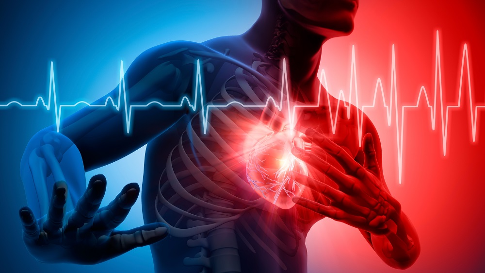
When a heart attack (myocardial infarction, MI) kills a patch of heart muscle, the adult mammalian heart cannot replace it, since the lost contractile muscle cells (cardiomyocytes, CMs) do not meaningfully regenerate. Instead, the heart heals with scar tissue, which, over time, leads to heart failure [2].
Why do adult cardiomyocytes lose this regenerative capacity? One piece of the puzzle is that mature CMs acquire a rigid, highly organized internal scaffold of contractile machinery called the sarcomere: the repeating protein units that generate force when the heart beats. This is great for pumping blood but terrible for cell division, because dividing requires the cell to dismantle its internal structure.
Another piece is that adult CMs often carry more than the normal two sets of chromosomes (polyploid) or have more than one nucleus per cell (multinucleated). This happens because they can enter the cell cycle and replicate DNA but then fail to complete the final step: cytokinesis, the physical splitting of one cell into two daughter cells [3].
The authors of a new study published in the Journal of Molecular and Cellular Cardiology liken this to turning on the tap without unblocking the drain: CMs are pushed into DNA replication, but if they cannot actually divide, no new heart cells are produced. To try and “unblock” the process, the researchers induced partial reprogramming of CMs using three of the four classic “Yamanaka factors”: OCT4, SOX2, and KLF4 (OSK). The idea was that this would help the cells dismantle their sarcomeres and complete a full division.
Unblocking the drain
OSK overexpression in neonatal and adult mouse CMs reduced the expression of cardiac troponin T, a sarcomere protein that marks mature CM identity, and disrupted the striped, organized sarcomere architecture. The gene expression profile shifted from that of an adult heart cell back toward an embryonic heart cell, suggesting dedifferentiation without loss of cellular identity.
The full four-factor cocktail, OSKM (M stands for c-Myc), had the same effect, but, unlike OSK, it also produced clonal clusters of rapidly dividing cells that lost their CM markers. The authors interpret this as dysregulated proliferation approaching a pre-tumorigenic state, as opposed to the controlled dedifferentiation seen with OSK.
Interestingly, c-Myc is a well-known oncogene; it was already recognized as cancer-causing before Yamanaka’s team included it in the original cocktail, where it acts by dampening the cellular brakes that normally prevent runaway division. It is required for full reprogramming and the production of induced pluripotent stem cells (iPSCs), but not for partial reprogramming.
Since dedifferentiation is thought to be a prerequisite for heart regeneration – as seen in zebrafish and neonatal mice – does OSK also drive cell cycle re-entry? OSKM and c-Myc alone both strongly drove cells into the cycle, confirming c-Myc’s classic proliferation-boosting effect, but OSK did not. So, the central question of the paper became: If OSK does not make cells divide more often, how can it help the heart regenerate?
As the researchers found out, while OSK did not increase the number of cells “attempting” division, far more of the cells that did try completed the split successfully, as if OSK solved the “blocked drain” problem. Supporting this hypothesis, OSK-treated cultures had more single-nucleus cells and fewer two-nucleus cells, a result that is consistent with successful division.
Rescue in vivo
To see if this would work in living animals, the researchers delivered OSK to newborn mouse hearts using a virus (AAV) targeted to heart cells. Two weeks later, the hearts showed the same pattern seen in vitro: dedifferentiated cells, disassembled sarcomeres, and more completed cytokinesis events.
However, when OSK expression was allowed to continue for four weeks, the hearts developed a combination of thin walls, enlarged chambers, and weakened pumping (dilated cardiomyopathy), suggesting that prolonged dedifferentiation becomes harmful. Interestingly, in adult mice, the same duration of OSK caused no obvious damage: it seems that adult hearts tolerate OSK better than developing ones. This might also mean that treatment timing and duration will be important for clinical translation.
For the final and crucial test, the researchers induced heart attacks and injected OSK at the same time. Over the following month, OSK-treated mice, compared to controls, showed better blood pumping (higher ejection fractions) at 14 and 28 days, less scarring (fibrosis), and more dividing heart cells, especially near the injury site. They also had smaller individual heart cells, suggesting less compensatory enlargement; in other words, new cells were sharing the load.
Harvard geroscientist David Sinclair, who was not involved in this new study but whose team has used OSK to successfully restore vision in animal models, discussed the results in an X post: “Why is this such a big deal? Because adult heart cells do not meaningfully divide, which is why the heart heals with scar tissue rather than regeneration. This fundamental limitation has defined cardiology for decades. In 2020, OSK restored function in damaged neurons. Now the same principle is being explored in the heart, building on results already seen in eye, brain, liver, and skin.”
Dr. Sinclair added that the findings were consistent with his Information Theory of Aging, which postulates that cells have a “backup copy” of their youthful epigenetic makeup that can be restored by cellular reprogramming. “Cells retain the instructions for repair; aging is a loss of access to them. Restore that information, and regeneration follows.”
Literature
[1] Yan, Y., Huang, Y., Cao, C., Li, D., Che, Y., Wang, Q., … & Wang, L. (2026). OSK-mediated partial reprogramming induces cardiomyocyte dedifferentiation, overcomes cytokinesis barriers, and promotes post-MI endogenous cardiac regeneration. Journal of Molecular and Cellular Cardiology.
[2] Frangogiannis, N. G. (2015). Pathophysiology of myocardial infarction. Comprehensive physiology, 5(4), 1841-1875.
[3] Derks, W., & Bergmann, O. (2020). Polyploidy in cardiomyocytes: roadblock to heart regeneration?. Circulation research, 126(4), 552-565.