The Blog

Building a Future Free of Age-Related Disease

X-ray of fibrosis

Towards a Telomerase Gene Therapy for Pulmonary Fibrosis

Back in January 2018, researchers at CNIO reversed lung fibrosis in mice using a single gene therapy. Today, we want to spotlight a new study that builds on the positive results of the 2018 research, taking us another step closer to telomerase gene therapy in humans to reverse pulmonary fibrosis.

The road to a gene therapy for pulmonary fibrosis 

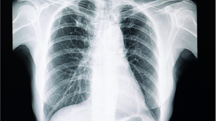
Idiopathic pulmonary fibrosis is an age-related disease that currently has no cure and very limited treatment options. Pulmonary fibrosis is a disease of the lung tissue that causes it to become stiff and scarred, leading to a progressive shortness of breath.

This disease seems to develop due to a combination of genetic and environmental factors. While exposure to toxic substances can play a role in its development, the disease typically appears during advanced age or due to an underlying genetic condition, and it is linked to telomere dysfunction.

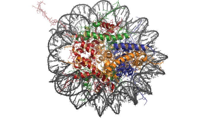
Telomeres are located at the ends of our chromosomes, which store our genetic information. A telomere contains a specific DNA sequence that is repeated thousands of times and serves as a protective cap, and it is sometimes likened to the aiglet on the end of a shoelace, preventing it from unraveling.

This sequence has two functions: it protects the coding regions of the chromosomes and prevents them from being damaged, and it acts as a clock that controls the number of replications that a cell can make. The telomeres shorten with each cell division, and this erosion is thought to be one of the reasons we age.

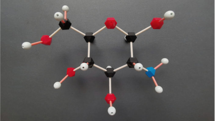
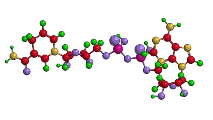
Telomerase gene therapy activates the production of the telomerase enzyme, which repairs the telomeres located at the ends of our chromosomes, restoring the sequence and building the protective cap back up. The telomerase therapy used by the Spanish National Cancer Research Centre (CNIO) is based on activating telomerase expression transiently. To achieve this, a virus is used as a telomerase gene carrier and is injected intravenously into the mice.

Telomere dysfunction leads to pulmonary fibrosis

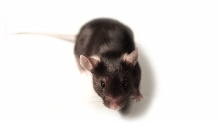
Previous studies by the CNIO team had shown that there are genetic factors linked to the dysfunction of telomeres. In 2015, the team developed a superior mouse model of pulmonary fibrosis that lacked the telomerase gene, which prevented those mice’s cells from expressing telomerase.

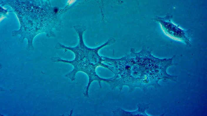
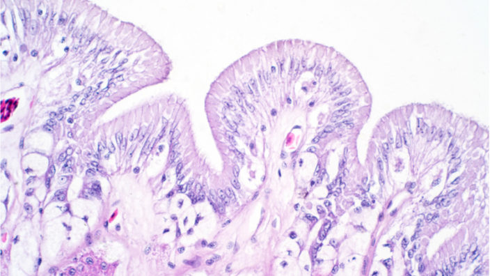
This mouse model works because alveolar type II cells, which play a role in lung tissue regeneration, experience telomere dysfunction and die. This results in the mice developing pulmonary fibrosis as the lung tissue is unable to regenerate and repair itself periodically, resulting in the formation of fibrotic scar tissue.

In 2018, the CNIO team published a study using their superior mouse model of fibrosis and demonstrated that telomerase gene therapy was able to reverse that fibrosis [1].

A new study published by Dr. Maria Blasco and her research team at the Telomeres and Telomerase Group at the CNIO, in collaboration with researchers from the Complutense University of Madrid and the Autonomous University of Barcelona, shows that the same telomerase therapy can successfully treat age-related fibrosis in old mice.

This is an important development because it shows that not only does telomerase therapy work in mouse models of fibrosis, it also works in mice that have not been genetically modified in any way and whose fibrosis is simply the result of normal aging.

These data also put telomere attrition in the spotlight for why the disease develops and show that the primary risk factor for that attrition is aging. Telomeres slowly erode after many cell divisions until they become so short that they cannot protect the chromosomes; at this point, the cell triggers an alarm and ceases to divide as a safety feature. However, once enough cells reach this state, the tissue can no longer regenerate.

This new study shows that, like the mouse model’s engineered cells, age-related telomere attrition naturally occurs in alveolar type II cells, a key player in lung tissue regeneration. The study results strongly suggest that the researchers have discovered the molecular basis of the association between aging and pulmonary fibrosis.

In the new study, the researchers describe the effect that aging has on alveolar type II cells and how that impacts their ability to function and facilitate tissue repair and regeneration. As well as regenerating the lung tissue, alveolar type II cells also secrete a lipid-protein complex called pulmonary surfactant, which lubricates the lung tissue, helps it stay elastic as it expands and contracts during breathing, and reduces the energy required to breathe.

This means that if the alveolar type II cells are unable to regenerate, then the pulmonary surfactant will not be produced, which causes the lung tissue to become stiff and fibrotic. The researchers show that there is a direct correlation between telomere function in alveolar type II cells, pulmonary surfactant secretion, and the development of fibrosis in animals.

Short/dysfunctional telomeres are at the origin of idiopathic pulmonary fibrosis (IPF) in patients mutant for telomere maintenance genes. However, it remains unknown whether physiological aging leads to short telomeres in the lung, thus leading to IPF with aging. Here, we find that physiological aging in wild-type mice leads to telomere shortening and a reduced proliferative potential of alveolar type II cells and club cells, increased cellular senescence and DNA damage, increased fibroblast activation and collagen deposits, and impaired lung biophysics, suggestive of a fibrosis-like pathology. Treatment of both wild-type and telomerase-deficient mice with telomerase gene therapy prevented the onset of lung profibrotic pathologies. These findings suggest that short telomeres associated with physiological aging are at the origin of IPF and that a potential treatment for IPF based on telomerase activation would be of interest not only for patients with telomerase mutations but also for sporadic cases of IPF associated with physiological aging.

Conclusion

This research holds great potential and may offer a solution to all types of fibrotic diseases. The next steps will be to translate these findings to people, and we are confident that human trials for telomerase gene therapy are not too far away now.

It has taken over a decade of steady research by Dr. Blasco and the CNIO, but we are finally reaching the point where telomerase therapy could soon be ready for prime time. It will be a truly exciting day when we can finally report the launch of the first human trials for telomerase therapy, and let’s hope that day is soon.

We would like to ask you a small favor. We are a non-profit foundation, and unlike some other organizations, we have no shareholders and no products to sell you. All our news and educational content is free for everyone to read, but it does mean that we rely on the help of people like you. Every contribution, no matter if it’s big or small, supports independent journalism and sustains our future.

Literature

[1] Therapeutic effects of telomerase in mice with pulmonary fibrosis induced by damage to the lungs and short telomeres. Maria Blasco, Juan Manuel Povedano, Paula Martinez, Rosa Serrano, Agueda Tejera, Gonzalo Gomez, Maria Bobadilla, Juana Maria Flores and Fatima Bosch (eLife 2018). DOI: 10.7554/eLife.31299

The circadian clock

Major Circadian Cycle Regulator Improves Chromatin Stability

Researchers have found that CLOCK, a protein that regulates the circadian cycle of our cells, also fights cellular senescence by promoting chromatin stability [1].

Since its very beginning, life on Earth had to adapt to our planet’s natural 24-hour rotation cycle. Rising to the challenge, nature invented biological mechanisms called circadian clocks (from the Latin ‘circa’ – ‘around’ – and ‘diem’ – day) that regulate biological processes according to the time of day. They are present in most organisms, including bacteria, and they play an especially important role in the life of plants, which produce energy from light. Molecular circadian clocks work on a cellular level, but, in mammals, their activity is coordinated by a “master clock” located in the part of the brain called the superchiasmatic nucleus (SCN).

Cellular circadian clocks are groups of genes that regulate their own transcription and translation via a complex mechanism of negative-feedback loops, bringing hundreds of cellular processes into accordance with the night and day cycle. It is well established that, with age, these mechanisms become dysregulated, causing sleep disorders in older people [2]. Age-related degradation of the core circadian mechanisms in SCN has been confirmed in mice. This process has also been shown to induce stem cell aging and chronic age-related diseases, including osteoarthritis, metabolic and neurodegenerative disorders [3].

A transcriptional factor named CLOCK (Circadian Locomotor Output Cycles Kaput) plays a major role in this cellular symphony. Several studies suggest a link between CLOCK and aging. CLOCK deficiency is known to reduce lifespan and promote age-related pathologies, such as cataracts and dermatitis, in mice. CLOCK levels decline with age, as circadian mechanisms become dysregulated. This particular study was conducted to further elucidate the connection between CLOCK levels and aging.

As their main subjects, the authors chose human mesenchymal stem cells (hMSCs), which can differentiate into cells forming various types of connective tissues, such as bone and cartilage. Stem cell aging, and MSC aging in particular, is a major factor behind organismal aging and many age-related disorders.

The study confirmed that the age-related degradation of the circadian machinery happens during cellular aging as well as organismal aging. The researchers found amplitude dampening and period lengthening of the circadian cycles in late-passage (aging) MSCs. CLOCK became downregulated as well. The picture was similar on organismal level: having acquired cells from healthy human donors of various ages, the researchers found that CLOCK levels were generally lower in MSCs from older donors. Senescence levels also increased with age.

Genetically modified CLOCK-deficient MSCs demonstrated dysregulated circadian rhythms and more senescent phenotypes. They also reached senescence much earlier. Importantly, these cells exhibited extensive DNA damage, one of the major causes of cellular senescence. On the other hand, CLOCK overexpression rejuvenated aged MSCs.

The researchers also discovered that this correlation could not be explained by CLOCK’s transcriptional activity: the effect endured even with CLOCK’s transcriptional ability knocked out. The authors were able to prove that the relation between CLOCK levels and cellular senescence stems from CLOCK’s role in supporting chromatin stability. CLOCK was found to form complexes with components of the nuclear lamina (the fibrillar network inside the nucleus) and with proteins that are known to stabilize heterochromatin. Downregulation of CLOCK during cellular aging resulted in the destabilization of heterochromatin, which, in turn, led to increased transcription of repetitive genomic sequences. Recently, we published an article about the detrimental effects of these sequences. Genomic instability, itself, is a major hallmark of aging.

Mechanistic studies further revealed that this is not a mere correlation: CLOCK indeed helps maintain heterochromatin structure and suppresses the proliferation of repetitive genomic sequences. Restored expression of CLOCK, or the heterochromatin-associated proteins that it assists, alleviated aging defects in MSCs. The researchers hypothesize that CLOCK functions as a scaffold protein that tethers heterochromatin components to the nuclear matrix. This suggestion is supported by the fact that another core circadian protein, PER2, also functions as a scaffold protein that helps maintain cellular homeostasis.

The scientists note that previous studies had shown CLOCK to be localized not only at the transcriptionally active regions, where it performs its main transcriptional duties, but also near condensed chromatin. This peculiar behavior can now be explained by the role that CLOCK plays in promoting chromatin stability.

The findings have also been confirmed in vivo: overexpression of CLOCK in mice using a viral vector resulted in cartilage regeneration and the attenuation of age-related articular problems.

Conclusion

Due to the immense complexity and the evolutionary character of cellular chemistry, proteins can perform multiple and additional roles, which seems to be the case with CLOCK and chromatin stability. Consequently, the upregulation of CLOCK could potentially become a two-pronged anti-aging intervention, simultaneously restoring circadian rhythms and reducing cellular senescence via improved chromatin stability.

We would like to ask you a small favor. We are a non-profit foundation, and unlike some other organizations, we have no shareholders and no products to sell you. All our news and educational content is free for everyone to read, but it does mean that we rely on the help of people like you. Every contribution, no matter if it’s big or small, supports independent journalism and sustains our future.

Literature

[1] Liang, C., Liu, Z., Song, M., Li, W., Wu, Z., Wang, Z., … & Hishida, T. (2020). Stabilization of heterochromatin by CLOCK promotes stem cell rejuvenation and cartilage regeneration. Cell Research, 1-19.

[2] Duffy, J. F., Zitting, K. M., & Chinoy, E. D. (2015). Aging and circadian rhythms. Sleep medicine clinics, 10(4), 423-434.

[3] Musiek, E. S., & Holtzman, D. M. (2016). Mechanisms linking circadian clocks, sleep, and neurodegeneration. Science, 354(6315), 1004-1008.

Nucleosome

Reviewing Epigenetic Alterations as a Cause of Aging

Epigenetic alterations, which are one of the proposed reasons we age, are the changes to gene expression that our cells experience as we get older. These alterations harm the fundamental functions of our cells and can increase the risk of cancer and other age-related diseases.

Depending on your view, epigenetic alterations are either the result of age-related damage or a form of damage itself. The case could certainly be argued that it is both, with some forms of epigenetic alterations being a consequence and others playing a much more regulatory role in the aging process.

The author of the review that we want to highlight today takes the popular view that epigenetic alterations are for the most part a cause of aging and the changes they bring create some of the damage and dysfunction of aging.

The review takes a look at DNA methylation states and their relation to biological age, the link between age-related diseases and epigenetic changes, and how epigenetic changes exert an influence over a number of other aging processes, such as loss of proteostasis, the disruption of the protein building machinery in our cells; mitochondrial dysfunction, the decline of our cellular power plants; stem cell exhaustion, the depletion of the supply of replacement cells due to loss or inhibition of stem cells; and immunosenescence, the age-related decline of the immune system.

Aging, which is accompanied by decreased organ function and increased disease incidence, limits human lifespan and has attracted investigators for thousands of years. In recent decades, with the rapid development of biology, scientists have shown that epigenetic modifications, especially DNA methylation, are key regulators involved in this process. Regular fluctuations in global DNA methylation levels have been shown to accurately estimate biological age and disease prognosis. In this review, we discuss recent findings regarding the relationship between variations in DNA methylation level patterns and aging. In addition, we introduce the known mechanisms by which DNA methylation regulators affect aging and related diseases. As more studies uncover the mechanisms by which DNA methylation regulates aging, antiaging interventions and treatments for related diseases may be developed that enable human life extension.

The underlying mechanisms of aging have perplexed scientists for decades. Detailed evaluations of global DNA methylation changes have provided insights into the process of aging, with DNA methylation serving as a biomarker of biological age and a driving force of aging. Current findings should stimulate further discussion and experimentation based on epigenetic regulation in cell-, tissue-, and disease-specific aging models. Future studies focusing on the mechanisms by which specific parameters, such as stress, affect methylation patterns will uncover additional details of the aging process. Identification of the target genes modified by DNA methylation-related regulatory elements in aging individuals is highly informative to figure out the hormone-like effectors and signal pathways that mediate these alterations as well as related diseases. The interaction among epigenetic regulators during aging should also be highly valued. Further studies should focus on the cross-talk among these epigenetic regulators, such as DNA methylation, RNA methylation, histone methylation, and noncoding RNAs, which will aid in providing a full picture of epigenetics and aging. The results of such studies may pave the way for antiaging interventions as well as treatments for related diseases, enabling human life extension.

Conclusion

Epigenetic alterations are currently an area of increasing interest to researchers, and there are a number of companies, such as Turn.Bio, AgeX, and Iduna Therapeutics, that are working on approaches to reverse age-related epigenetic changes in order to make cells and tissues function in biologically younger ways.

We know that cells can be encouraged to act like younger cells when given the correct signaling environment, and there seems to be a strong epigenetic element to aging, which suggests that epigenetic alterations are a primary cause of aging. Therapies that can successfully reverse epigenetic alterations have the potential to address a wide range of aging processes, as this review touches upon.

The potential for partial cellular reprogramming to reverse cellular aging by reversing age-related epigenetic alterations could truly transform how we age, and while there is still much for us to learn, the possible rewards for success in this area are huge.

We would like to ask you a small favor. We are a non-profit foundation, and unlike some other organizations, we have no shareholders and no products to sell you. All our news and educational content is free for everyone to read, but it does mean that we rely on the help of people like you. Every contribution, no matter if it’s big or small, supports independent journalism and sustains our future.
SENISCA background image

New Startup SENISCA Aims to Reset Senescent Cells

Senescent cells accumulate in our bodies as we age, increasing inflammation and raising our risk of suffering from cancer and other age-related diseases. A new biotech startup plans to reset aged senescent cells to a youthful state to deal with this problem.

The rejuvenation biotechnology field is growing at a steady pace these days, with new startups appearing more frequently and with an increasing array of novel approaches to age-related decline.

One of the more promising startups to recently emerge is SENISCA, a company born from the lab at the University of Exeter’s College of Medicine and Health. Co-founded by Professor Lorna Harries, this company has a particularly intriguing approach to addressing the harmful senescent cells that cause havoc as they build up in our bodies with age.

What are senescent cells?

As you age, increasing numbers of your cells enter into a state known as senescence. Senescent cells do not divide or support the tissues of which they are part; instead, they emit a range of potentially harmful chemical signals that encourage nearby healthy cells to enter the same senescent state.

Senescent cells only make up a small number of total cells in the body, but they secrete proinflammatory cytokines, chemokines, and extracellular matrix proteases, which, together, form the senescence-associated secretory phenotype, or SASP. This secreted SASP causes many problems: it reduces tissue repair and regeneration, increases chronic inflammation, and can raise the risk of cancer and other age-related diseases.

There has been a great deal of interest in developing therapies that can destroy senescent cells and remove them from the tissues and organs; such drugs are known as senolytics. There has been much activity in the research community in finding compounds capable of clearing senescent cells, and a number of them are currently in human trials.

Turning back the clock on senescent cells

SENISCA is taking a different approach to the problem and is opting not to destroy senescent cells but to reverse senescence instead. By modulating RNA splicing, the company has developed a method of resetting aged cells and returning them to a youthful state. It is now using this approach to develop therapies for the diseases and visual consequences of aging.

A few years ago, Professor Harries and her team had shown that splicing regulation in cells declines during aging, but they wanted to see what would happen if splicing regulation was turned back up to a level seen in younger people.

This led to the publication of a paper in the open access journal BioMed Central, which showed that by using small molecules, not only could senescent cells be brought out of senescence, but when it was done, those cells were also rejuvenated back to a more youthful state [1]. In other words, the cells were still chronologically old, but they were no longer senescent nor did they produce the harmful inflammatory SASP, and they functioned similar to younger cells.

The next step was to find out what genes were involved in turning the splicing factors on and off in the context of senescent cells. Eventually, the researchers discovered the controlling genes responsible for preventing the splicing factors from being active, and when they turned them off directly, the splicing factors returned to life and rejuvenated the cells.

At SENISCA, we are taking a two-faceted approach for modulation of splicing factor levels. Firstly, we are identifying small molecules capable of restoring splicing factor levels. Secondly, we are targeting the genes that control splicing factor levels directly. Both approaches will reset splicing factor levels and reverse senescence. We anticipate that understanding the molecular basis of rejuvenation will highlight new treatments for the diseases and aesthetic aspects of ageing. More importantly, it is likely that preventative approaches based on rejuvenation will be developed reducing both disease incidence and severity.

The researchers are now at the point where they know precisely what genes to target, and they are working on a delivery system that is able to specifically target senescent cells and turn the target genes on or off to stop them from contributing to aging.

SENISCA is initially going to be focusing on skin aging using topical treatments that aim to rebuild the skin structure. This is a wise initial move for a proof of concept, as the skin is the largest and most accessible organ in the body, and positive results should be visually apparent as well as reported via biomarkers.

Ultimately, SENISCA plans to expand its technology into wider pharmaceutical use to target diseases that currently have no cure. Given the myriad of age-related diseases associated with senescent cell accumulation, this approach could have very broad applications.

Conclusion

The company is now in the process of raising a seed round and is seeking around $1.24 million to get the company started and through its initial year of development. This initial seed funding should allow SENISCA to launch human trials and get proof-of-principle data, presumably for skin aging, and to help it refine its approach for more specific diseases.

Skin aging is something that is sure to capture the attention of the public, and positive results that reverse skin aging could very well build more public support for doing something about aging. It is hard to imagine something more visual and convincing than the reversal of skin aging, so it is understandable why the team has taken this initial path.

Finally, we would like to mention that Professor Harries will also be speaking at our upcoming virtual conference, Ending Age-Related Diseases 2020. This is an excellent opportunity to learn more about her fascinating work as well as that of the many other talented researchers in the field who will be speaking at the event.

We would like to ask you a small favor. We are a non-profit foundation, and unlike some other organizations, we have no shareholders and no products to sell you. All our news and educational content is free for everyone to read, but it does mean that we rely on the help of people like you. Every contribution, no matter if it’s big or small, supports independent journalism and sustains our future.

Literature

[1] Latorre, E., Birar, V. C., Sheerin, A. N., Jeynes, J. C. C., Hooper, A., Dawe, H. R., … & Harries, L. W. (2017). Small molecule modulation of splicing factor expression is associated with rescue from cellular senescence. BMC cell biology, 18(1), 31.

Astrocyte

Astrocytes Become Neural Stem Cells After Injury

A new study published in Cell Stem Cell has shown how astrocytes, glial cells that perform multiple critical functions in the brain, can become neural stem cells (NSCs) in response to injury.

Astrocytes’ normal functions

The term ‘astrocyte’, meaning star-shaped cell, refers to a very heterogenous population of cells that perform maintenance, management and constructive functions in the brain, including the formation of synapses between neurons and the maintenance of the blood-brain barrier. The heterogeneity of these cells is such that they are not the same everywhere in the brain, and they have different capabilities and functions depending on their location.

This new study shows that, in addition to these upkeep functions, some astrocytes are capable of transforming into NSCs, providing the mammalian brain with an existent, if limited, capability of recovering from injury.

Astrocytes becoming neurons

Astrocytes as precursors to NSCs

This study expands on a previous study from 2014, which showed the potential of these cells to become NSCs following a stroke [1]. It details the effects of the protein Notch, which suppresses astrocytes from becoming NSCs, and explains many of the genes that are activated in the absence of this protein. Therefore, the researchers consider certain astrocytes to be neural stem cells in and of themselves.

Alignment of the transcriptional events driving neuronal specification in the cortex and the neurogenic niche further supports the idea that parenchymal astrocytes may be viewed as dormant NSCs that, with the appropriate molecular cues, can be recruited to generate neurons following a trajectory similar to canonical neurogenesis.

It has been hypothesized that parenchymal astrocytes may be considered dormant NSCs because of shared expression of several biomarkers. The present study further supports this notion by showing that, when recruited into the neurogenic program, the transcriptional behavior of committed glia parallels that of adult NSCs from the germinal niche.

Conclusion

Of course, this is a mouse study, and, even though it greatly explains many of the fundamental biochemical processes that allow astrocytes to become neurons, the full relationship between injury and neurogenesis remains unclear, and there are still many questions that must be answered before this research could be used to develop a therapy. However, the researchers are aware of these limitations, and they have helpfully made their important data public in order to facilitate the development of future therapies.

Further investigation of transcriptional networks implicated in the lineage fate switch could reveal novel regulators of neurogenesis. Thus, to aid discovery of pro-neurogenic factors, we made our data publicly available (GEO: GSE139842) and organized the results in a searchable database that allows interrogation of expression profiles of the desired genes (https://cortical-neurogenesis.shinyapps.io/cortical-neurogenesis/).

With this data, we can hope that, in the near future, researchers will discover a way to directly activate the potential of astrocytes to become NSCs, thus restoring mental function and ability to people whose brains have been damaged through traumatic injury or the processes of aging.

We would like to ask you a small favor. We are a non-profit foundation, and unlike some other organizations, we have no shareholders and no products to sell you. All our news and educational content is free for everyone to read, but it does mean that we rely on the help of people like you. Every contribution, no matter if it’s big or small, supports independent journalism and sustains our future.

Literature

[1] Magnusson, J. P., Göritz, C., Tatarishvili, J., Dias, D. O., Smith, E. M., Lindvall, O., … & Frisén, J. (2014). A latent neurogenic program in astrocytes regulated by Notch signaling in the mouse. Science, 346(6206), 237-241.

Small plate of food

Caloric Restriction Appears to Improve Stem Cell Function

Researchers have taken a look at how caloric restriction seems to improve intestinal stem cell function in a new open-access study.

Inflammaging is a precursor of age-related diseases

The lining of the intestines helps to prevent the entry of microbes and unwanted molecules into the bloodstream beyond; however, as we age, the integrity of this barrier declines, causing microbial burden to rise and, with it, chronic inflammation.

This chronic age-related inflammation, often referred to as inflammaging, has multiple sources, including loss of intestinal barrier integrity and a resulting rise in microbial burden, senescent cells, cell debris, and immunosenescence. Some researchers even suggest that microbial burden resulting from the loss of intestinal barrier integrity could potentially be the origin point of inflammaging, the initial spark that sets the powder keg alight.

Inflammaging precedes a variety of age-related diseases, including atherosclerosis, arthritis, hypertension, and cancer [1-3]. Inflammaging presents a persistent background of inflammation that also leads to increasingly poor tissue repair and regeneration as we age [4].

As we age, the stem cells in our tissues and organs, which provide us with a steady supply of fresh cells, begin to slow down, and that flow of replacement cells begins to falter.

This decline of stem cell activity is known as stem cell exhaustion, and it is one of the proposed reasons we age. Stem cells become increasingly inactive as we age for various reasons, including damage and the chronic inflammation of inflammaging, which alters the signaling environment and suppresses their ability to function.

Caloric restriction appears to support intestinal stem cells

The researchers of a new study have shown how caloric restriction appears to be beneficial to the function of stem cells that reside in the tissues of the intestine [5]. Caloric restriction is the practice of consuming fewer calories while still consuming an optimal amount of nutrition. The key is to reduce calorie intake while avoiding malnourishment.

Caloric restriction can reduce calorie intake by up to 40% less than normal, though 20-25% is typical. It has been shown to increase healthy lifespan in multiple species across many published studies.

The data in this study suggests that the decline of intestinal stem cell activity is slowed down through caloric restriction. The researchers also examined how caloric restriction limits the mutation rate of stem cells and how it lowers the retention of mutated cells in the intestinal tissue.

Calorie restriction (CR) extends lifespan through several intracellular mechanisms, including increased DNA repair, leading to fewer DNA mutations that cause age-related pathologies. However, it remains unknown how CR acts on mutation retention at the tissue level. Here, we use Cre-mediated DNA recombination of the confetti reporter as a proxy for neutral mutations and follow these mutations by intravital microscopy to identify how CR affects retention of mutations in the intestine. We find that CR leads to increased numbers of functional Lgr5+ stem cells that compete for niche occupancy, resulting in slower but stronger stem cell competition. Consequently, stem cells carrying neutral or Apc mutations encounter more wild-type competitors, thus increasing the chance that they get displaced from the niche to get lost over time. Thus, our data show that CR not only affects the acquisition of mutations but also leads to lower retention of mutations in the intestine.

Conclusion

This study offers yet more evidence that caloric restriction can be potentially beneficial in the context of aging and health. It should be noted that anyone who practices caloric restriction should make a great effort to continue consuming adequate nutrition while doing so.

We would like to ask you a small favor. We are a non-profit foundation, and unlike some other organizations, we have no shareholders and no products to sell you. All our news and educational content is free for everyone to read, but it does mean that we rely on the help of people like you. Every contribution, no matter if it’s big or small, supports independent journalism and sustains our future.

Literature

[1] Freund A, Orjalo AV, Desprez PY, Campisi J. Inflammatory networks during cellular senescence: causes and consequences. Trends Mol Med (2010) 16(5):238–46. doi: 10.1016/j.molmed.2010.03.003

[2] Childs, B. G., Gluscevic, M., Baker, D. J., Laberge, R. M., Marquess, D., Dananberg, J., & van Deursen, J. M. (2017). Senescent cells: an emerging target for diseases of ageing. Nature Reviews Drug Discovery, 16(10), 718.

[3] He, S., & Sharpless, N. E. (2017). Senescence in health and disease. Cell, 169(6), 1000-1011.

[4] Straub, R. H., & Schradin, C. (2016). Chronic inflammatory systemic diseases: An evolutionary trade-off between acutely beneficial but chronically harmful programs. Evolution, medicine, and public health, 2016(1), 37-51.

[5] Bruens, L., Ellenbroek, S. I. J., Suijkerbuijk, S. J. E., Azkanaz, M., Hale, A. J., Toonen, P., … & van Rheenen, J. (2020). Calorie Restriction Increases the Number of Competing Stem Cells and Decreases Mutation Retention in the Intestine. Cell Reports, 32(3), 107937.

Mouse facing camera

Reducing Cognitive Decline in Mice with Alzheimer’s

A group of scientists have shown that the SIRT2 protein increases the accumulation of amyloid beta, a known marker of Alzheimer’s disease (AD), in the brains of mice. They propose a mechanism underlying this phenomenon and demonstrate that inhibiting SIRT2 ameliorates AD-associated cognitive decline in mice [1].

AD is one of the most devastating age-related disorders. Despite considerable efforts and billions of dollars in funding, little ground has been gained in the war against AD. Its progression has been linked to the brain’s accumulation of amyloid beta (Aß), a peptide that forms plaques that are abundant in people affected by this disease. Recently, the theory that AD is caused by those plaques took a hit after numerous drugs that targeted Aß accumulation had failed in trials. Still, more such drugs are in development, and targeting Aß remains a legitimate anti-AD strategy [2].

Sirtuins, on the other hand, have been hallowed as longevity promoters. This extremely ancient family of proteins performs numerous functions in our body. SIRT2, a member of this family, helps to maintain genomic stability by deacetylating histones, the bulky proteins that chromatin is wrapped around. After gene transcription, deacetylating histones helps chromatin return to its tightly wound form, where it can remain stable despite various environmental threats. Overexpression of SIRT2 has been shown to extend lifespan in various organisms [3].

The current study may come as a cold shower to sirtuin enthusiasts, since it demonstrates that SIRT2 levels positively correlate with Aß accumulation in the brains of mice. This is not the first study to establish such a correlation, but, in this case, the researchers have also discovered the pathway by which SIRT2 seems to boost Aß production.

The researchers have found that pharmaceutical repression of SIRT2 deacetylation activity lowers the load of Aß in the brain and ameliorates cognitive decline in an AD mouse model. This can be explained with a look at the mechanism behind the creation of Aß. Aß forms when the transmembrane protein APP (amyloid precursor protein), which usually performs beneficial cellular functions, gets cleaved in two places by two different enzymes. Aß is the short chunk that remains.

Protein cleavage and amyloid beta

One of the enzymes is called BACE1 (ß-secretase in the illustration). The scientists have found that SIRT2 activity inhibition reduces the expression of BACE1. As a result, fewer APP cleavages occur and less Aß is produced. To further test this connection, the researchers created genetically modified SIRT2-lacking mice and found that the depletion of SIRT2 significantly reduced BACE1 levels in both the hippocampus and the cortex.

However, there is no direct mechanism by which SIRT2 can influence BACE1. To find the intermediary, the scientists first flagged a total of 285 SIRT2 potential binding partners. Five among them also happened to be AD-related, with one, RTN4B, bearing a direct relation to both SIRT2 and BACE1.

RTNB4 is a member of the reticulon family of proteins, which gets its name from being found mostly in the endoplasmic reticulum. Reticulons are known to block the access of BACE1 to APP. An increase in the expression of reticulons has been shown to substantially reduce the production of Aβ [4]. Experiments have shown that SIRT2 and RTN4B are co-localized in cells and overexpression of SIRT2 reduces RTN4B levels. Confirming the relation, increased RTN4B expression was detected in the hippocampus and the cortex of the SIRT2 knockout mice.

Apparently, RTN4B has a deacetylation site that invites SIRT2 to do what it does best: deacetylate. However, removing the acetyl group from RTN4B makes it easier for another protein called ubiquitin to degrade RTNB4, which impairs the latter’s ability to mediate BACE1. As a result, more Aß is produced.

BACE mediation

In accordance with the Aß theory of AD development, mice treated with a SIRT2 inhibitor, which granted them lower levels of Aß as a result, also showed less cognitive impairment than their untreated peers.

Since these results were achieved by a simple pharmaceutical treatment (intracutaneous injections of the SIRT2 activity inhibitor AK-7), the authors of the study suggest that it can serve as a base for the development of novel anti-AD drugs.

Conclusion

The results of this study underscore the multi-faceted character of cellular chemistry. As we see here, deacetylation, while promoting genomic stability, can also drive Aß production later in life. Hence, much caution is needed when developing novel drugs and anti-aging agents. It is also possible that the antagonistic pleiotropy theory of aging is at play here. This theory postulates that the same genetic mechanisms may be beneficial earlier in life (SIRT2 deacetylating histones) and deleterious later in life (SIRT2 deacetylating RTNB4). Such mechanisms still get selected for reproduction, since their harmful effect is not felt until after the reproductive period is over.

We would like to ask you a small favor. We are a non-profit foundation, and unlike some other organizations, we have no shareholders and no products to sell you. All our news and educational content is free for everyone to read, but it does mean that we rely on the help of people like you. Every contribution, no matter if it’s big or small, supports independent journalism and sustains our future.

Literature

[1] Wang, Y., Yang, J. Q., Hong, T. T., Sun, Y. H., Huang, H. L., Chen, F., … & Yang, T. L. RTN4B‐mediated suppression of Sirtuin 2 activity ameliorates β‐amyloid pathology and cognitive impairment in Alzheimer’s disease mouse model. Aging Cell, e13194.

[2] Rao, C. V., Asch, A. S., Carr, D. J., & Yamada, H. Y. (2020). “Amyloid‐beta accumulation cycle” as a prevention and/or therapy target for Alzheimer’s disease. Aging Cell, 19(3), e13109.

[3] Cosentino, C., & Mostoslavsky, R. (2014). Sirtuin to the rescue: SIRT 2 extends life span of B ub R 1 mice. The EMBO journal, 33(13), 1417-1419.

[4] He, W., Lu, Y., Qahwash, I., Hu, X. Y., Chang, A., & Yan, R. (2004). Reticulon family members modulate BACE1 activity and amyloid-β peptide generation. Nature medicine, 10(9), 959-965.

Rejuvenation Roundup July

Rejuvenation Roundup July 2020

This is our last monthly roundup before Ending Age-Related Diseases 2020, our first fully online conference, and plenty of rejuvenation research has been conducted this month. Let’s take a look at what’s happened in July.

LEAF News

Ending Age-Related Diseases 2020 July Update: Our annual conference, Ending Age-Related Diseases: Investment Prospects & Advances in Research, will be held on August 20-21! Free tickets, regular tickets, and VIP tickets are all available for this online event. Secure your ticket now!

Lifespan News

lifespan.io Launches Lifespan News: In this premier episode of Lifespan News, Brent Nally discusses a lot of the rejuvenation topics we’ve covered, including the fact that naked mole rats, which are strange-looking but very long-lived rodents, are resistant to cancer.

Parkinson’s Symptoms Reversed in Mice: The Lancet is opening a new journal entirely focused on longevity, and Brent discusses this along with plenty of other rejuvenation topics.

Interviews

Principles of Effective Advocacy for Longevity Therapies: Edwina Rogers, CEO of the Global Healthspan Policy Institute, discusses public policy and her role in bringing government institutions towards a more pro-rejuvenation line of thinking.

A New Hallmark of Aging Proposed: Alexander Fedintsev promotes the idea that the stiffening of the extracellular matrix, which is caused by cross-linked collagen, is its own hallmark of aging.

Stephanie Lederman Discusses AFAR and Funding: Promoting basic research through grants, AFAR seeks to encourage young researchers to focus on the diseases and root causes of aging.

Are Epigenetic Clocks Ready for Prime Time?: Reason of Repair Biotechnologies discusses potential problems with the use of epigenetic clocks to measure biological age.

Rejuvenation Roundup Podcast

Ryan O’Shea of Future Grind hosts this month’s podcast, showcasing the events and research discussed here.

Research Roundup

Two Proteins Allow LDL Cholesterol into Our Cells: Identifying NPC1 and NPC2, the two proteins that allow the transition of LDL into our cells, has ramifications for treating heart disease.

Frailty and Age-Related Immune Decline Reversed in Mice: Eosinophils resident in belly fat have been shown to be instrumental in maintaining and restoring the balance of the immune system, and restoring them has been shown to improve vaccination response.

Selectivity of the Blood-Brain Barrier Changes with Age: Lipid-mediated transport increases with age while receptor-mediated transport decreases, affecting the proteins that reach our brains from our blood plasma.

Trimethylamine May Be Involved in Vascular Aging: Bacteria in the gut break down proteins in red meat and other foods, releasing trimethylamine, which promotes aging of the vascular system.

A Blood Factor Boosts Neurogenesis and Cognition: While some blood factors lead to aging, others promote regeneration and growth; the enzyme Gpld1 is one of those factors.

The First Precision Gene Editing Tool for Mitochondrial DNA: CRISPR/Cas9 is the wrong tool for the job when it comes to mitochondria; a different enzyme, DddA, appears to be the right one.

Hyperbaric Oxygen Therapy May Reduce Age-Related Cognitive Decline: A pressurized room filled with pure oxygen has been shown to reduce the effects of dementia.

Why NAD+ Declines During Aging – Part 1: In Part 1 of a two-part series, Steve Hill explains why the coenzyme NAD+ decreases as we age.

A DNA Methylation Clock to Measure Skin Age: The skin is the largest and most accessible part of the body, so it is ideal as a target for measuring biological age.

A Single Molecule Can Regenerate the Gut Lining: Neuregulin 1 has been shown to cause the gut lining to regrow itself after injury, offering new hope for chemotherapy patients and people whose linings have been deteriorated by aging.

Scientists Induce a New Mode of Aging in Yeast: Yeast is one of the simplest organisms in aging research, and it ages in two modes: mitochondrial dysfunction or the deterioration of the nucleolus. Researchers have induced a third mode that delays both of these fates.

The “Love Hormone” Oxytocin as a Potential Alzheimer’s Therapy: Oxytocin takeup by cellular receptors has been shown to ameliorate the deterioration caused by amyloid beta, making this naturally occurring hormone potentially useful as a therapy.

Senescent Cell Death Promotes Aging Resistance in Naked Mole Rats: Apoptosis, the natural self-destruction of aged, senescent cells, is upregulated in these particularly long-lived rodents, which explains their unusual longevity.

Glucosamine Supplementation Correlates With Reduced All-Cause Mortality: A longitudinal study has shown that supplementation with glucosamine is correlated with reduced mortality by 15%.

Senolytic Drugs: From Discovery to Translation: This review, published in the Journal of Internal Medicine, discusses the current state of senolytics, drugs that destroy harmful senescent cells.

Navitoclax (ABT263) clears senescent osteoarthritic chondrocytes in osteoarthritis: Removing the senescent cells present in osteoarthritis reduces inflammation and promotes the growth of cartilage.

Late-life restoration of mitochondrial function reverses cardiac dysfunction in old mice: Targeting mitochondrial dysfunction through reducing oxidative stress has been shown to restore heart function in a mouse model.

Exosomes derived from human umbilical cord MSCs rejuvenate aged MSCs: Exosomes, vehicles of intercellular communication, have been shown to deliver signals that restore the function of aged mesenchymal stem cells.

Repetitions in Our DNA React to Anti-Aging Interventions: Unneeded repetitions in our DNA tend to accumulate with age and are associated with age-related diseases, but they have been shown to be affected by caloric restriction.

Genomic Instability May Directly Lead to Diseases of Aging: Our cells naturally mutate, even outside of the germ line, and these mutations can lead to more than just cancer.

A decade of epigenetic change in aging twins: Genetic and environmental contributions to longitudinal DNA methylation: Twin studies are a staple of medical research, and this study compares the differences of genetics and environment in respect to DNA methylation.

Genetics of extreme human longevity to guide drug discovery for healthy ageing: By analyzing the genes of people who live for unusually long periods, it may be possible to develop drugs that promote the same longevity.

Genetic variation between long-lived versus short-lived bats illuminates the molecular signatures of longevity: Studying the differences between short-lived and long-lived bats has led to the discovery of the genes that promote longevity in these animals.

Beaver and Naked Mole Rat Genomes Reveal Common Paths to Longevity: Identifying the specific, mutual genome mutations between these two long-lived rodent species has led to a new understanding of the genes that give them their longevity.

Multivariate genomic scan implicates novel loci and haem metabolism in human ageing: Researchers have identified 78 genes linked to aging and have implicated heme, which consists of hemoglobin and other compounds, as a potential factor.

Changes in ferrous iron and glutathione promote ferroptosis and frailty in aging Caenorhabditis elegans: Iron metabolism has also been shown to affect the aging of these commonly researched worms.

Comparison of mitochondrial transplantation by using a stamp-type multineedle injector and platelet-rich plasma therapy for hair aging: Pep-1-mediated mitochondrial transplantation and platelet-rich plasma therapy are roughly equivalent in their ability to restore the hair of aging mice.

Growth differentiation factor 15 protects against the aging-mediated systemic inflammatory response in humans and mice: This particular factor has been shown to reduce inflammaging, the systemic inflammation that accompanies aging and promotes multiple age-related diseases.

We would like to ask you a small favor. We are a non-profit foundation, and unlike some other organizations, we have no shareholders and no products to sell you. All our news and educational content is free for everyone to read, but it does mean that we rely on the help of people like you. Every contribution, no matter if it’s big or small, supports independent journalism and sustains our future.

Glucosamine in ball-and-stick form

Glucosamine Linked to Reduced All-Cause Mortality

It is one of the most commonly used supplements frequently taken to address joint pain, but there might be more to this dietary supplement than first meets the eye.

Glucosamine was originally discovered during the 1960s in Italy by pharmacologist Professor Luigi Rovati. Glucosamine is one of the most commonly used dietary supplements and is typically taken to help with the joint pain and inflammation associated with aging.

Glucosamine is a polysaccharide that is found naturally in cartilaginous joint tissues, bones, skin, ligaments, and nails, and it is involved in protein and lipid synthesis. In the context of joints, synovial fluid contains glucosamine and occupies the space between joints, helping to reduce the friction of joint surfaces.

Despite it being frequently taken for arthritis, the evidence for its effectiveness is limited, although, there is data for it being anti-inflammatory, as suggested by the results of a randomized clinical trial in 2015 [1].

However, glucosamine supplementation seems to correlate with lower all-cause mortality and other mortality risks, such as cardiovascular disease (CVD), cancer, respiratory and digestive diseases. A recent analysis published in the journal BMJ showed that glucosamine supplementation conveys around a 15% reduction of all-cause mortality [2]. This is a considerable amount when compared to other lifestyle interventions as well as other supplements. The data gathered is from a large number of people, and the trend of reduced mortality is unmistakable.

This population-based prospective cohort study included 495 077 women and men (mean (SD) age, 56.6 (8.1) years) from the UK Biobank study. Participants were recruited from 2006 to 2010 and were followed up through 2018. We evaluated all-cause mortality and mortality due to cardiovascular disease (CVD), cancer, respiratory and digestive disease. HRs and 95% CIs for all-cause and cause-specific mortality were calculated using Cox proportional hazards models with adjustment for potential confounding variables.

Regular glucosamine supplementation was associated with lower mortality due to all causes, cancer, CVD, respiratory and digestive diseases.

Conclusion

The exact reasons for this correlation with the reduction of various mortality risks is as yet unknown, but given the large patient group in this and in other analyses along with the popularity of this supplement, it is impossible to deny that there is a definite trend here. Out of all the popular supplements, glucosamine appears to have by far the most significant influence on all-cause mortality.

We are not suggesting that you take this supplement, but given that it is cheap and freely available with an excellent safety profile, it may be worth your consideration and further research to evaluate if you wish to or not.

We would like to ask you a small favor. We are a non-profit foundation, and unlike some other organizations, we have no shareholders and no products to sell you. All our news and educational content is free for everyone to read, but it does mean that we rely on the help of people like you. Every contribution, no matter if it’s big or small, supports independent journalism and sustains our future.

Literature

[1] Navarro, S. L., White, E., Kantor, E. D., Zhang, Y., Rho, J., Song, X., … & Lampe, J. W. (2015). Randomized trial of glucosamine and chondroitin supplementation on inflammation and oxidative stress biomarkers and plasma proteomics profiles in healthy humans. PloS one, 10(2), e0117534.

[2] Li, Z. H., Gao, X., Chung, V. C., Zhong, W. F., Fu, Q., Lv, Y. B., … & Li, F. R. (2020). Associations of regular glucosamine use with all-cause and cause-specific mortality: a large prospective cohort study. Annals of the Rheumatic Diseases, 79(6), 829-836.

Naked mole rat

Senescent Cell Death Promotes Aging Resistance

The well-known resistance of naked mole rats to aging and aging-related effects is due at least in part to an unusual ability to clear away senescent cells. This suggests that senolytic drugs, which aim to do the same in humans, may prove a fruitful path to longevity.

Naked mole rats live much longer than other rodents

Naked mole rats are highly social African mammals that live in underground tunnels. These animals are famous among biologists for their extraordinary longevity – living as long as 32 years – as well as their resistance to a range of age-related disorders, including neurodegenerative diseases and cancer. Unlike humans, their mortality rate doesn’t increase with age, meaning that an elderly naked mole rat is no more likely die of “natural causes” than its younger counterparts.

If we could understand how naked mole rats stave off the effects of aging, that could help researchers figure out how to do the same thing in humans. Even if we can’t directly apply whatever tricks the naked mole rats use to keep the Grim Reaper at bay, we might get some hints about which research avenues would be best to pursue.

A lack of senescent cells

One of the open questions about naked mole rats was the fate of their senescent cells, which are cells that have stopped dividing in response to DNA damage or other stress. They secrete inflammatory cytokines and a collection of other molecules, contributing to inflammation, immune imbalance, and other age-related physiological changes. In general, senescent cells accumulate with age, but it was unclear whether this is true in naked mole rats.

A research team based in Japan addressed this question in a paper published on bioRxiv [1], a preprint archive where papers are available before they have been peer reviewed. The team measured the number of senescent cells in skin samples from young and middle-aged mice and naked mole rats. In contrast to the mice, where senescent cells increased with age, they found nearly no senescent cells in the middle-aged mole rats. The teams found the same results when they induced cellular senescence by irradiating samples with UV; senescent cells accumulated in the mouse samples but not in naked mole rat skin.

Strength through vulnerability

A crucial clue was found in the observation that there were lots of floating, dead cells in naked mole rat cell cultures that had been treated to induce senescence. By tracking when the cells died and correlating it with gene expression, the team showed that it was linked with senescence. They also tested other ways of inducing senescence to confirm that this wasn’t a response to a specific inducer.

The team dubbed this phenomenon “senescent cell death”. Further analysis revealed that genes related to the autophagy-lysosome degradation system were misregulated in the senescent cells. The cells also had stronger expression of genes related to oxidative stress, suggesting that an increase in reactive oxygen species might be causing senescent naked mole rat cells to die.

Reactive oxygen species damage macromolecules inside cells, and they are known to increase in senescent cells. However, in human and mouse senescent cells, this increase doesn’t lead to cell death. Naked mole rat cells are unusually vulnerable to reactive oxygen species, but this apparent weakness may in fact be a key to longevity.

When the researchers compared senescent cells from naked mole rats and mice, they found a striking difference in their metabolic profiles. Senescence caused a large shift in the metabolic profile of mouse cells, but the change was much smaller in the naked mole rat cells. This may explain why naked mole rat cells are less able to deal with an increase in reactive oxygen – they don’t adjust their metabolism to compensate. In fact, the team found that two pathways which produce reactive oxygen species were more active in senescent mole rat cells.

Naked mole rats may have developed a poor defense against reactive oxygen species because oxygen levels are low in their underground burrows. This makes their senescent cells especially vulnerable to reactive oxygen species, but that turns out to be useful since it clears senescent cells from their systems, helping them avoid many of the harmful effects of these cells.

Naked mole-rats (NMRs) are the longest-lived rodents, showing minimal aging phenotypes. An unsolved paradox is that NMRs exhibit low intracellular anti-oxidant defence despite minimal aging. Here, we explained a link between these “contradicting” features by a phenomenon termed “senescent cell death (SCD)”—Senescence induced cell death in NMR cells due to their inherent vulnerability to reactive oxygen species and unique metabolic system. In NMR skin, we observed few senescent cells during aging or after ultraviolet irradiation, suggesting suppression of senescent cell accumulation in NMR tissue. We discovered that senescent NMR-fibroblasts induce SCD through retinoblastoma protein activation accompanied by autophagy dysregulation, increased oxidative damage and accelerated H2O2-releasing metabolic pathways. During senescence, NMR cells showed resistance to metabolic remodelling unlike mice. Our findings provide mechanistic insights into how extraordinary aging resistance is accomplished in NMR. This will contribute to the development of senolytic drugs to regulate age-related diseases.

Conclusion

Using senolytic drugs to kill senescent cells is one of the approaches under investigation to increase human lifespan and healthspan. The discovery of senescent cell death in naked mole rats, known for their exceptional longevity, strengthens the case for this approach in humans.

We would like to ask you a small favor. We are a non-profit foundation, and unlike some other organizations, we have no shareholders and no products to sell you. All our news and educational content is free for everyone to read, but it does mean that we rely on the help of people like you. Every contribution, no matter if it’s big or small, supports independent journalism and sustains our future.

Literature

  1. Kawamura, Y., Oka, K., Takamori, M., Sugiura, Y., Oiwa, Y., Fujioka, S., … Miura, K. (2020) Senescent cell death as an aging resistance mechanism in naked mole-rat. bioRxiv, doi:10.1101/2020.07.02.155903
Oxytocin in hearts

The “Love Hormone” Oxytocin as an Alzheimer’s Therapy

Japanese researchers led by Professor Akiyoshi Saitoh from the Tokyo University of Science have published new research that explores the potential of oxytocin as a therapy to combat cognitive disorders such as Alzheimer’s disease.

Oxytocin is often called the love hormone, given its role in the reproductive system and its role in creating feelings such as love, well-being, and trust. Oxytocin is also known to decrease with age, and researchers such as Dr. Irina Conboy have explored its role in aging [1]. Oxytocin is known to activate MAP kinase, a pathway that plays an important role in aging and tissue regeneration [2].

More recently, it was discovered that it is also involved in learning and cognitive performance, so the researchers of this new study wanted to explore the effects of oxytocin on amyloid beta (Aβ)-induced cognitive impairment, typically seen in Alzheimer’s disease [3].

In order to see how Aβ impairs cognition, the team initially circulated Aβ through slices of mouse hippocampus to see how it impacted the signaling capacity of the neurons in the tissue. Next, they circulated oxytocin through the same brain slices and observed that neuron signaling improved, suggesting that the oxytocin was able to reverse the signaling impairment caused by Aβ.

In a healthy brain, oxytocin acts by binding to oxytocin receptors, structures located in the membranes of neurons. The researchers’ next step was to inhibit these receptors in the brain slices to see if oxytocin could reverse the cognitive impairment that Aβ causes without being able to bind to the oxytocin receptors.

In other words, could oxytocin reverse the impairment simply by its presence? The answer was, perhaps unsurprisingly, no. These results confirmed that the oxytocin receptors are a critical part of the reversal process.

Oxytocin is known to play an important role in neuronal signaling strength and in facilitating the creation of memories via a number of chemical interactions, including the influx of calcium ions. Previous studies suggest that Aβ disrupts these chemical interactions, and when the researchers artificially blocked them, they found that the addition of oxytocin did not reverse Aβ impairment. This means that oxytocin relies on these chemical interactions to reverse the damage.

Oxytocin, a peptide hormone synthe[prisna-wp-translate-show-hide behavior=”show”][/prisna-wp-translate-show-hide]sized in the hypothalamic paraventricular nucleus, has been reported to participate in the regulation of learning and memory performance. However, no report has demonstrated the effect of oxytocin on the amyloid-beta (Aβ)-induced impairment of synaptic plasticity. In this study, we examined the effects of oxytocin on the Aβ-induced impairment of synaptic plasticity in mice.

We found that oxytocin reversed the impairment of LTP induced by Aβ25-35 perfusion in the mouse hippocampus. These effects were blocked by pretreatment with the selective oxytocin receptor antagonist L-368,899. Furthermore, the treatment with the ERK inhibitor U0126 and selective Ca2+-permeable AMPA receptor antagonist NASPM completely antagonized the effects of oxytocin.

Conclusion

This study is the first of its kind in showing that oxytocin can reverse Aβ-induced cognitive impairment. The usual caveats apply, of course: this is in mice, it is at a very early stage of research, and more in vivo studies must be conducted prior to moving to human trials.

That said, given that there are currently no effective treatments for Alzheimer’s disease available yet, this line of research presents a novel approach to treating the condition.

We would like to ask you a small favor. We are a non-profit foundation, and unlike some other organizations, we have no shareholders and no products to sell you. All our news and educational content is free for everyone to read, but it does mean that we rely on the help of people like you. Every contribution, no matter if it’s big or small, supports independent journalism and sustains our future.

Literature

[1] Mehdipour, M., Etienne, J., Chen, C. C., Gathwala, R., Rehman, M., Kato, C., … & Conboy, I. M. (2019). Rejuvenation of brain, liver and muscle by simultaneous pharmacological modulation of two signaling determinants, that change in opposite directions with age. Aging (Albany NY), 11(15), 5628.

[2] Yousef, H., Conboy, M. J., Mamiya, H., Zeiderman, M., Schlesinger, C., Schaffer, D. V., & Conboy, I. M. (2014). Mechanisms of action of hESC-secreted proteins that enhance human and mouse myogenesis. Aging (Albany NY), 6(8), 602.

[3] Takahashi, J., Yamada, D., Ueta, Y., Iwai, T., Koga, E., Tanabe, M., … & Saitoh, A. (2020). Oxytocin reverses Aβ-induced impairment of hippocampal synaptic plasticity in mice. Biochemical and Biophysical Research Communications.

Budding yeast

Scientists Induce a New Mode of Aging in Yeast

A group of researchers has shown that yeast cells have two different and mutually exclusive modes of aging that seem to be interlinked. The scientists were also able to artificially induce a third mode of aging that prolongs both healthspan and lifespan.

Yeast is a popular object of genomic research due to its short reproductive cycle, its ability to reproduce in both asexual and sexual ways, and the fact that you can determine the lifespan of an individual yeast cell. Yeast mostly reproduce by asexual budding that is different from the binary division of bacteria, which produces two new, indistinguishable cells. During budding, the mother cell develops a small bulge that goes on to become a daughter cell, which makes determining the original cell’s lifespan possible. Yeast cells are also divided into two sexes (a-cells and alpha-cells) that can engage in meiotic reproduction, a process that, like in animals and plants, creates young cells.

Researchers from the University of California in San Diego made the stunning discovery that yeast cells age in one of the two distinct, mutually exclusive ways [1]. Both modes of aging are driven by organellar decline: Mode 1 is caused  by the deterioration of the nucleolus, and Mode 2 is through mitochondrial dysfunction. Cells seem to choose their mode of aging early in life, with 47% choosing Mode 1. Mode 1 provides a slightly better lifespan, which the researchers measured to be the number of reproductive cycles until death.

As Mode 1 cells age, their nucleoli become enlarged and subsequently dysfunctional due to ribosomal DNA deregulation. Ribosomal DNA (rDNA) are sequences in the genome that code for ribosomal RNA, one of the major building blocks of ribosomes. Ribosome formation is extremely resource-hungry and thus needs to be tightly regulated by silencing rDNA transcription [2]. Loss of rDNA silencing, the underlying cause of Mode 1 aging, is the result of chromatin instability at rDNA sites. Hence, the two modes of yeast aging are caused by two of the known hallmarks of human aging: genomic instability and mitochondrial dysfunction.

Mode 1 cells exhibit ever-increasing loss of rDNA silencing with age but do not show signs of mitochondrial dysfunction. Mode 2 cells, on the contrary, end their lives with abysmal mitochondrial health but with little loss of rDNA silencing. It seems that the path to death that cells choose early in life is self-reinforcing.

The researchers then set out to determine what happens if they add or subtract key components of the two modes of aging, targeting Sir2 for Mode 1 and HAP (heme activator protein) for Mode 2. Sir2, a protein of the sirtuin family that is known to extend lifespan by restoring chromatin stability [3], is a major rDNA-silencing factor. Heme is a key factor for mitochondrial decay during human aging [4].

When Sir2 levels were significantly lowered via the deletion of the SIR2 gene, 83% of cells chose Mode 1 aging and ended their lives in perfect mitochondrial health and with elevated HAP levels. These genetically modified cells chose their mode of aging much earlier in life and lived for a shorter duration than normal, non-modified Mode 1 cells. The other way to induce Mode 1 aging was by elevating HAP levels. When HAP production was genetically upregulated, 96% of the cells chose Mode 1 aging. Caloric restriction, which is known to induce HAP production [5], also increased the proportion of Mode 1 cells.

The researchers hypothesize that the two beneficial compounds – Sir2 and HAP – counteract each other’s effects through mutual inhibition, probably through transcriptional regulation, as transcription of many HAP components is enhanced in cells with SIR2 knocked out and HAP is known to decrease the transcription of some Sir2 cofactors.

Yet, when Sir2 was significantly overexpressed (two times the normal level), it did not just encourage cells to choose Mode 2 aging, it also created a new, third mode of aging: a minority of cells retained both high Sir2 levels and high HAP levels, and they lived much longer than Mode 1 and Mode 2 cells. Unfortunately, in this scenario, more cells chose the short-lived Mode 2, rendering the overall lifespan gains in the population negligible. While in both natural modes of aging, reproductive cycles become longer with age, indicating aging cells’ struggle to reproduce, this did not happen to the cells that chose Mode 3. Their reproductive cycle length remained on par with young cells. While it did not extend lifespan indefinitely, Mode 3 brought about a breakthrough in longevity: a simultaneous increase in both lifespan and healthspan followed by a short period of compressed morbidity just before death.

The breakthrough came when the researchers significantly overexpressed both Sir2 and HAP: in this scenario, less than 6% of the cells chose the short-lived Mode 2 aging, while close to 45% experienced the novel, long-lived Mode 3 aging.

The discovery of the third mode of aging was made possible through an intricate mathematical model that predicts a cell’s behavior based on different levels of Sir2 and HAP. The researchers also suggest that this principle can explain the synergy that is known to exist between two well-researched longevity-promoting factors: Sir2 and caloric restriction.

Cellular aging can thus be considered a fate-decision process, in which single cells age toward either silencing loss and nucleolar decline or heme depletion and mitochondrial decline. This process can be viewed as a divergent progression on a Sir2-HAP landscape, which can be reshaped by model-guided genetic perturbations, thereby enriching a long-lived mode of aging.

Conclusion

This exciting discovery expands our understanding of aging, illuminating both new difficulties and new possibilities on the road to life extension, highlighting the need to control both genomic instability and mitochondrial function at the cellular level. In these simple cells, it seems to be possible to induce a slower and healthier mode of aging by overexpressing certain key factors. It remains to be seen if these promising results can be recreated in mammalian and, eventually, human cells.

We would like to ask you a small favor. We are a non-profit foundation, and unlike some other organizations, we have no shareholders and no products to sell you. All our news and educational content is free for everyone to read, but it does mean that we rely on the help of people like you. Every contribution, no matter if it’s big or small, supports independent journalism and sustains our future.

Literature

[1] Li, Y., Jiang, Y., Paxman, J., O’Laughlin, R., Klepin, S., Zhu, Y., … & Hao, N. (2020). A programmable fate decision landscape underlies single-cell aging in yeast. Science, 369(6501), 325-329.

[2] McKeown, P. C. (2014). Mechanisms of rDNA silencing and the Nucleolar Remodelling Complex (NoRC). Atlas of Genetics and Cytogenetics in Oncology and Haematology.

[3] Guarente, L. (2000). Sir2 links chromatin silencing, metabolism, and aging. Genes & development, 14(9), 1021-1026.

[4] Atamna, H., Killilea, D. W., Killilea, A. N., & Ames, B. N. (2002). Heme deficiency may be a factor in the mitochondrial and neuronal decay of aging. Proceedings of the National Academy of Sciences, 99(23), 14807-14812.

[5] Lin, S. J., Kaeberlein, M., Andalis, A. A., Sturtz, L. A., Defossez, P. A., Culotta, V. C., … & Guarente, L. (2002). Calorie restriction extends Saccharomyces cerevisiae lifespan by increasing respiration. Nature, 418(6895), 344-348.

Intestinal lining under a microscope

A Single Molecule Can Regenerate the Gut Lining

New research from Monash University has shown that a single key molecule responsible for cell adhesion and a variety of other bodily functions is also critical in repairing the gut lining and that increasing the presence of this molecule accelerates its repair and regeneration.

Supercharging stem cells

The walls of the gut rely on a strong membrane to provide a protective barrier between the gut microbiome, which is a complex ecosystem of bacteria and other microbial life that inhabits the intestinal tract, and the bloodstream and internal organs.

When this barrier is compromised through damage by infection, trauma, or even age-related loss of integrity, it can lead to various painful conditions and diseases, such as Crohn’s disease and ulcerative colitis.

The new study focused on gut stem cells, how they repair the gut membrane, and what spurs this regeneration [1]. In order to do this, the research team created miniature gut organoids: gut tissue was grown in a dish to emulate the real gut lining and environment, albeit on a smaller scale.

The researchers identified populations of cells located near the gut stem cells, which produce a molecule known as neuregulin 1 (NRG1), which activates stem cells and spurs them to regenerate the gut membrane.

NRG1 is one of four proteins in the neuregulin family that act on the epidermal growth factor receptor (EGFR) family of receptors. Neuregulin 1 is created in various isoforms through alternative splicing, which allows it to perform a wide variety of functions within the body, making it somewhat of a molecular Swiss army knife.

The researchers also found that they could speed up gut repair by introducing additional NRG1, which boosted the repair of the gut lining by activating the steam cells sooner. Not only were cells encouraged to divide as part of this repair but the stem cells were enhanced and had almost supercharged performance compared to normal.

The researchers also demonstrated that recovery is significantly improved with the application of neuregulin 1 following chemotherapy, which frequently damages the gut lining when used.

Epidermal growth factor (EGF) maintains intestinal stem cell (ISC) proliferation and is a key component of organoid growth media yet is dispensable for intestinal homeostasis, suggesting roles for multiple EGF family ligands in ISC function. Here, we identified neuregulin 1 (NRG1) as a key EGF family ligand that drives tissue repair following injury. NRG1, but not EGF, is upregulated upon damage and is expressed in mesenchymal stromal cells, macrophages, and Paneth cells. NRG1 deletion reduces proliferation in intestinal crypts and compromises regeneration capacity. NRG1 robustly stimulates proliferation in crypts and induces budding in organoids, in part through elevated and sustained activation of mitogen-activated protein kinase (MAPK) and AKT. Consistently, NRG1 treatment induces a proliferative gene signature and promotes organoid formation from progenitor cells and enhances regeneration following injury. These data suggest mesenchymal-derived NRG1 is a potent mediator of tissue regeneration and may inform the development of therapies for enhancing intestinal repair after injury.

Conclusion

This research could prove useful for developing treatments for diseases such as Crohn’s disease and ulcerative colitis as well as combating the general loss of gut membrane integrity, which is typically seen during aging and can increase microbial burden and systemic inflammation.

We would like to ask you a small favor. We are a non-profit foundation, and unlike some other organizations, we have no shareholders and no products to sell you. All our news and educational content is free for everyone to read, but it does mean that we rely on the help of people like you. Every contribution, no matter if it’s big or small, supports independent journalism and sustains our future.

Literature

[1] Jardé, T., Chan, W. H., Rossello, F. J., Kahlon, T. K., Theocharous, M., Arackal, T. K., … & Kerr, G. (2020). Mesenchymal Niche-Derived Neuregulin-1 Drives Intestinal Stem Cell Proliferation and Regeneration of Damaged Epithelium. Cell Stem Cell.

DNA clock

A DNA Methylation Clock to Measure Skin Age

A team of researchers have developed a tissue-specific clock that can measure the biological age of skin.

There is plenty of new evidence that different organs and tissues in our body age at different rates. In particular, the epigenetic state, the pattern of gene expression, of the cells that comprise these different organs and tissues can vary considerably. Changes to the methylation state are one of the ways that gene expression changes during aging, and they are part of the epigenetic alterations hallmark of aging.

Epigenetic alterations in aging include changes to methylation patterns, which generally correlate with a decrease in the amount of heterochromatin and an increase in chromosome fragility and transcriptional alterations (variance in gene expression), remodeling of chromatin (a DNA support structure that assists or impedes its transcription), and transcriptional noise.

By measuring the methylation state, it is possible to get a reasonably accurate view of how biologically old a particular tissue or organ is, based on its gene expression profile.

Today, we want to note a recent study in which researchers have developed a DNA methylation clock specifically for the aging of skin [1]. This is an important step forward, given that tissues and organs tend to age differently and at different rates, so specialized clocks that incorporate these differences are really a must for accurate measurement of aging and validation of interventions that seek to reverse aging.

DNA methylation (DNAm) age constitutes a powerful tool to assess the molecular age and overall health status of biological samples. Recently, it has been shown that tissue-specific DNAm age predictors may present superior performance compared to the pan- or multi-tissue counterparts. The skin is the largest organ in the body and bears important roles, such as body temperature control, barrier function, and protection from external insults. As a consequence of the constant and intimate interaction between the skin and the environment, current DNAm estimators, routinely trained using internal tissues which are influenced by other stimuli, are mostly inadequate to accurately predict skin DNAm age.

Conclusion

It makes sense that a skin-specific methylation clock would arrive first, given the ease of access to the largest organ in the body; if we had such a clock, testing interventions and changes to the age of the skin would be very simple.

The development of methylation and other kinds of epigenetic clocks has recently been an area of great interest to researchers, and, for many people, these types of aging biomarkers are really the gold standard for measuring changes to biological age.

However, it is still the early days of epigenetic clocks, and there is considerable work that needs to be done before highly accurate clocks can be developed. Currently, they can be somewhat hit and miss, depending on the tissue type being examined, and, in some cases, it is not entirely clear what exactly these clocks are measuring and what their results mean in the context of aging.

The need for highly accurate aging biomarkers has never been more urgent, especially as there are now therapies targeting the aging processes in human trials and others are poised to join them. The need to accurately quantify changes to biological age has never been more pressing than it is right now.

We would like to ask you a small favor. We are a non-profit foundation, and unlike some other organizations, we have no shareholders and no products to sell you. All our news and educational content is free for everyone to read, but it does mean that we rely on the help of people like you. Every contribution, no matter if it’s big or small, supports independent journalism and sustains our future.

Literature

[1] Boroni, M., Zonari, A., de Oliveira, C. R., Alkatib, K., Cruz, E. A. O., Brace, L. E., & de Carvalho, J. L. (2020). Highly accurate skin-specific methylome analysis algorithm as a platform to screen and validate therapeutics for healthy aging. Clinical Epigenetics, 12(1), 1-16.

Diagram of NADH

Why NAD+ Declines During Aging – Part 1

There has been considerable debate around the age-related decline of NAD+, particularly regarding whether or not it occurs in humans as it does in other species, such as mice. Today, as part of a two-part special, we are going to be taking a look at some of the evidence for the age-related decline of NAD+ in humans and some of the reasons why this likely happens.

What is NAD+?

NAD+ is a molecule that is present in all living cells and is essential for a myriad of cellular processes, including regulating metabolism, performing signaling, facilitating DNA repair and blood vessel growth, and regulating some aspects of aging.

In the body, NAD+ is created from simple building blocks, such as the amino acid tryptophan, and it is created in a more complex way via the intake of food containing nicotinic acid (NA), nicotinamide riboside (NR), nicotinamide mononucleotide (NMN), and other NAD+ precursors.

These different pathways ultimately feed into a salvage pathway, which recycles them back into the active NAD+ form. This salvage pathway, shown on the bottom right of the diagram, includes NAM, NMN, NAD+, and their associated steps.

The evidence that NAD+ declines with age

A 2012 study reported that NAD+ levels decrease dramatically during aging in skin, and while the exact level of decline was not worked out, the average level of concentration seems to fall by a minimum of 50% during adult aging. The difference between newborns and adults, for example, appears to be several-fold lower, according to the study data [1].

For the brain, two studies used MRI to examine NAD+ and how its level changes during aging. The first study, conducted back in 2015, saw researchers measuring intracellular NAD+ and NADH concentrations and the NAD+/NADH ratio in the human brain, detecting the age-dependent changes in NAD contents and the redox state associated with aging [2].

The second study from 2019 also concluded that an age-dependent decline of the NAD+ levels in the brain was also observed [3]. Taken together, these two studies suggest that there is a decline of NAD+ in the brain of between ~10% to ~25% between the period of young adulthood and old age.

Another study has come along to muddy the waters and contradict the previous two [4]. In biology, nothing is ever simple, and according to this 12-person, placebo-controlled randomized trial, there was no appreciable difference in NAD+ levels in the brain between young and old trial participants.

The researchers of this new study suggest that their results show that NAD+ decline is not associated with chronological aging per se in human muscle or brain, though they do suggest that brain and muscle tissue can benefit from supplementation with nicotinamide riboside, a precursor of NAD+.

It should be noted that this study is currently in preprint on Biorxiv and has not as yet passed peer review. It is also worth noting that at least one of the authors (Charles Brenner) has a stake in patents and the company that owns and licenses the sale of nicotinamide riboside.

For the liver, a previous study showed that liver samples from people aged 60 had around 30% lower concentrations of NAD+ compared to people aged 45 [5].

Finally, the level of NAD+ present in the bloodstream has also been shown to decrease rapidly during aging, according to the results of a 2019 study [6]. This more recent study improved upon an older study [7], which suggested that there was only a small decrease in NAD+ levels; this was due to the newer study using a superior methodology compared to the older one, thus giving a more accurate view of what happens to NAD+ in the bloodstream during aging.

It should be noted that NAD+ levels in blood are typically many times lower than tissue levels, and it is still not clear how important NAD+ is in the blood are and what function or significance its presence has.

These findings are also supported by a wealth of animal studies and suggests that an age-related decline of NAD+ is common across a range of species, which includes mice and humans.

Some reasons why NAD+ declines with age

Based on both human and animal studies, it seems pretty clear that NAD+ levels do decline during aging in multiple tissues and in blood. However, the big question here is: why does NAD+ decline because of aging?

There are a number of mechanisms that have the potential of reducing NAD+ and could be part of this age-related decline.

Inflammaging and CD38

The most obvious and direct way in which NAD+ is reduced is via the activity of an NAD+ consuming enzyme linked to inflammation called CD38, which destroys NAD+, and its activity steadily rises during aging as systemic inflammation levels increase.

CD38 is a membrane-bound NADase that hydrolyzes NAD+ to nicotinamide and (cyclic-)ADP-ribose. It is associated with immune responses and energy metabolism, but it is also a NADase whose levels rise with aging, with a corresponding increase in NADase activity and a decrease of NAD+.

Therapies that reduce inflammation, particularly the age-related chronic systemic inflammation known as “inflammaging”, could reduce the presence of CD38, thus offering the potential to increase NAD+ [8].

A new class of drugs known as senolytics, which purge the body of inflammatory senescent cells, is an example of a therapy that has the potential to reduce systemic inflammation and could improve NAD+ levels as a consequence.

It is worth noting at this point that it is still unclear whether or not NAD+ synthesis continues at the same rate in older individuals as younger ones, and while it seems clear that inflammaging and CD38 play a key role in consuming NAD+ as we age, there are also a number of other culprits that are involved.

DNA damage and PARPs

Poly-ADP-ribose polymerases (PARPs) are a group of related proteins involved in a number of cellular processes, such as DNA repair, genomic stability, and the programmed cell death known as apoptosis, which damaged cells undergo to remove themselves from the system.

PARP is a key driver of NAD+ catabolism, a set of metabolic processes that break down large molecules, including breaking down food molecules to provide energy and molecular components for anabolic reactions, using that released energy to repurpose the broken-down molecules for growth.

Specifically, PARP1 activity can be increased further in response to DNA damage and genotoxic stress, and given that DNA damage typically increases with age, that would mean more PARP activity and thus more NAD+ consumption [9]. In this way, PARP1 activity may serve as a regulator of NAD+ and mediate its age-related decline as the result of ever-increasing genomic instability that comes with age.

This suggests that therapies that can reduce PARP activity may be a viable approach here; certainly, in animal models of DNA damage, there is NAD+ depletion that can be reversed by inhibiting PARP [10-11]. Other approaches, such as NAD+ repletion via precursor supplementation, may also help to address excessive PARP consumption.

SARM1

The SARM1 protein is another way in which NAD+ levels may be depleted during aging. It was recently demonstrated that SARM1 is able to cleave NAD+, creating NAM, ADP ribose and cyclic ADP ribose [12-13]. Activation of SARM1 has been shown to reduce the available NAD+ pool, but when its activity is blocked, it reduces diabetic peripheral neuropathy and addresses axonopathy in animal models [14-16].

Axonopathy refers to a functional or structural defect in the axon or its terminal, and it is believed to be a precursor for the development and progression of various neurodegenerative diseases. For this reason, SARM1 has become a potential target for drug development; however, there is, at present, little data for SARM1 activity in neurons in the context of aging.

SARM1 is also expressed in other tissues, including the liver and kidneys, and its function in that context of age-related changes to its activity is as yet not fully understood, nor is how that function changes during aging. For this reason, it is still not clear if it plays a central role in age-related NAD+ decline.

Conclusion

This is the end of part one, and we will be delving further into the potential sources of NAD+ depletion in part two, which is coming soon.

We would like to ask you a small favor. We are a non-profit foundation, and unlike some other organizations, we have no shareholders and no products to sell you. All our news and educational content is free for everyone to read, but it does mean that we rely on the help of people like you. Every contribution, no matter if it’s big or small, supports independent journalism and sustains our future.

Literature

[1] Massudi, H., Grant, R., Braidy, N., Guest, J., Farnsworth, B., & Guillemin, G. J. (2012). Age-associated changes in oxidative stress and NAD+ metabolism in human tissue. PloS one, 7(7), e42357.

[2] Zhu, X. H., Lu, M., Lee, B. Y., Ugurbil, K., & Chen, W. (2015). In vivo NAD assay reveals the intracellular NAD contents and redox state in healthy human brain and their age dependences. Proceedings of the National Academy of Sciences, 112(9), 2876-2881.

[3] Bagga, P., Hariharan, H., Wilson, N. E., Beer, J. C., Shinohara, R. T., Elliott, M. A., … & Detre, J. A. (2020). Single-Voxel 1H MR spectroscopy of cerebral nicotinamide adenine dinucleotide (NAD+) in humans at 7T using a 32-channel volume coil. Magnetic Resonance in Medicine, 83(3), 806-814.

[4] Elhassan, Y. S., Kluckova, K., Fletcher, R. S., Schmidt, M., Garten, A., Doig, C. L., … & Wilson, M. (2019). Nicotinamide riboside augments the human skeletal muscle NAD+ metabolome and induces transcriptomic and anti-inflammatory signatures in aged subjects: a placebo-controlled, randomized trial. BioRxiv, 680462.

[5] Zhou, C. C., Yang, X., Hua, X., Liu, J., Fan, M. B., Li, G. Q., … & Wang, P. (2016). Hepatic NAD+ deficiency as a therapeutic target for non-alcoholic fatty liver disease in ageing. British journal of pharmacology, 173(15), 2352-2368.

[6] Clement, J., Wong, M., Poljak, A., Sachdev, P., & Braidy, N. (2019). The plasma NAD+ metabolome is dysregulated in “normal” aging. Rejuvenation research, 22(2), 121-130.

[7] Guest, J., Grant, R., Mori, T. A., & Croft, K. D. (2014). Changes in oxidative damage, inflammation and [NAD (H)] with age in cerebrospinal fluid. PLoS One, 9(1), e85335.

[8] Tarragó, M. G., Chini, C. C., Kanamori, K. S., Warner, G. M., Caride, A., de Oliveira, G. C., … & Jurk, D. (2018). A potent and specific CD38 inhibitor ameliorates age-related metabolic dysfunction by reversing tissue NAD+ decline. Cell metabolism, 27(5), 1081-1095.

[9] Wang, G., Han, T., Nijhawan, D., Theodoropoulos, P., Naidoo, J., Yadavalli, S., … & McKnight, S. L. (2014). P7C3 neuroprotective chemicals function by activating the rate-limiting enzyme in NAD salvage. Cell, 158(6), 1324-1334.

[10] Fang, E. F., Scheibye-Knudsen, M., Brace, L. E., Kassahun, H., SenGupta, T., Nilsen, H., … & Bohr, V. A. (2014). Defective mitophagy in XPA via PARP-1 hyperactivation and NAD+/SIRT1 reduction. Cell, 157(4), 882-896.

[11] Scheibye-Knudsen, M., Mitchell, S. J., Fang, E. F., Iyama, T., Ward, T., Wang, J., … & Mangerich, A. (2014). A high-fat diet and NAD+ activate Sirt1 to rescue premature aging in cockayne syndrome. Cell metabolism, 20(5), 840-855.

[12] Summers, D. W., Gibson, D. A., DiAntonio, A., & Milbrandt, J. (2016). SARM1-specific motifs in the TIR domain enable NAD+ loss and regulate injury-induced SARM1 activation. Proceedings of the National Academy of Sciences, 113(41), E6271-E6280.

[13] Essuman, K., Summers, D. W., Sasaki, Y., Mao, X., DiAntonio, A., & Milbrandt, J. (2017). The SARM1 Toll/interleukin-1 receptor domain possesses intrinsic NAD+ cleavage activity that promotes pathological axonal degeneration. Neuron, 93(6), 1334-1343.

[14] Cheng, Y., Liu, J., Luan, Y., Liu, Z., Lai, H., Zhong, W., … & Huang, R. (2019). Sarm1 gene deficiency attenuates diabetic peripheral neuropathy in mice. Diabetes, 68(11), 2120-2130.

[15] Gilley, J., Ribchester, R. R., & Coleman, M. P. (2017). Sarm1 deletion, but not WldS, confers lifelong rescue in a mouse model of severe axonopathy. Cell reports, 21(1), 10-16.

[16] Gerdts, J., Brace, E. J., Sasaki, Y., DiAntonio, A., & Milbrandt, J. (2015). SARM1 activation triggers axon degeneration locally via NAD+ destruction. Science, 348(6233), 453-457.