The Blog

Building a Future Free of Age-Related Disease

Dopaminergic neuron

Neurons Hidden to Immune Cells Improve Parkinson’s in Rats

By tweaking genes used by placental and cancerous cells to evade immune detection, scientists have created non-immunogenic neuronal grafts that may help Parkinson’s patients [1].

Sneakiness needed

In recent years, scientists have learned to produce cells via cellular reprogramming, a process in which differentiated cells are driven back to a pluripotent, stem-like state. These are known as induced pluripotent stem cells (iPSCs). The cells can then be re-differentiated into other types of cells and benefit from substantial rejuvenation in the process [2].

This approach has the potential to solve many medical challenges, but the problem of graft rejection remains. When cells come from a genetically different donor (an allogeneic graft), they typically trigger an immune response. Immunosuppressants can reduce this reaction, but they come with significant side effects.

However, evolution has provided some inspiration. In many cases, cells have naturally evolved to become invisible to the immune system, such as placental cells, certain parasites and pathogens, and cancer cells.

The idea of borrowing these cloaking mechanisms to create universally compatible, off-the-shelf donor cells has been around for years [3]. Multiple teams have made progress, and in a new study published in Cell Stem Cell, a group of Australian researchers reports a major advance in rodent models of Parkinson’s disease.

The invisibility cloak

In Parkinson’s disease, dopamine-producing neurons in a brain region called the substantia nigra progressively die off. Dopamine is essential for controlling movement and coordination. As levels fall, the result is the hallmark motor symptoms of Parkinson’s: tremors, stiffness, slowness, and balance issues.

Using cellular reprogramming, the researchers generated neuronal progenitor cells (NPCs) capable of giving rise to new, rejuvenated neurons. These cells were genetically engineered to overexpress eight genes that placental and cancer cells use to evade immune detection. The combination was designed to neutralize nearly all major arms of the immune system: T cells, natural killer cells, macrophages/microglia, and dendritic cells.

However, making cells invisible to the immune system introduces a new risk: if one turns cancerous, the body won’t catch it. To mitigate this, the researchers included a “kill switch” by inserting the gene for herpes simplex virus thymidine kinase (HSV-TK) – an enzyme not naturally found in humans. It was placed under the control of a promoter active only in dividing cells so that any cell that starts to proliferate uncontrollably, which mature neurons typically don’t do, could be selectively eliminated using an antiviral drug.

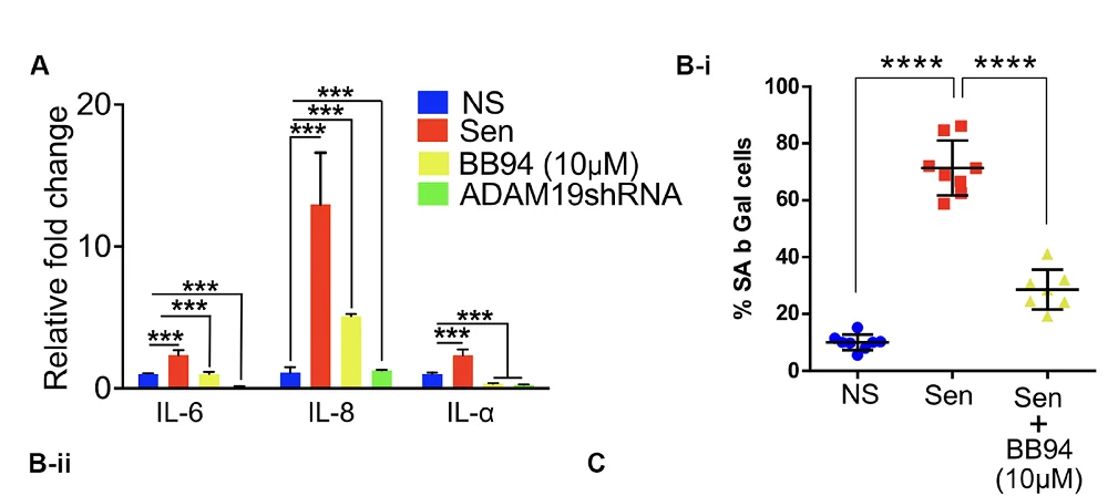
When co-cultured with immune cells from three human blood donors, the cloaked dopaminergic neurons triggered significantly less immune activation, proliferation, and cytokine release. The team then moved to a humanized mouse model with functional human T and B cells.

In this model, uncloaked grafts provoked a strong immune response, while cloaked grafts caused little to no T cell expansion and no rise in pro-inflammatory cytokines. As a result, cloaked grafts grew to about twice the size of uncloaked ones and contained more dopamine-producing neurons.

Functional improvement

To test whether the cloaked neurons could functionally reverse Parkinsonian symptoms, the researchers turned to an immunodeficient rat model of the disease. These rats do not reject either cloaked or uncloaked grafts, so the comparison focused on function, not immune evasion.

Both types of grafts performed comparably: they survived, integrated into the host brain, and reversed motor symptoms by 20 weeks after transplantation. This showed that the cloaking did not compromise the cells’ ability to function.

Finally, the team tested the kill switch in nude mice: animals that lack a functional immune system, making it easier to isolate the effects of the suicide gene. They chose a time point when about half of the NPCs had matured into non-dividing neurons, while the rest were still proliferating. After administering the antiviral drug ganciclovir, the number of proliferating cells dropped significantly while the mature neurons remained unaffected.

“The researchers’ ability to engineer a graftable cell line that can become dopaminergic neurons and evade immune cell targeting fills a gap in the field,” said Dr. Kate Joyce, a researcher at the Lifespan Research Institute. “Not only is the physical process of grafting cells disruptive to the immune system in the brain but in Parkinson’s disease and other neurodegenerative diseases, there is an issue of increased immune surveillance due to a leaky blood brain barrier that further compounds the problem. This method also avoids generating cells from the patients that might harbor harmful mutations and cause more problems later.”

We would like to ask you a small favor. We are a non-profit foundation, and unlike some other organizations, we have no shareholders and no products to sell you. All our news and educational content is free for everyone to read, but it does mean that we rely on the help of people like you. Every contribution, no matter if it’s big or small, supports independent journalism and sustains our future.

Literature

[1] Pavan, C., Davidson, K. C., Payne, N., Frausin, S., Hunt, C. P., Moriarty, N., Berrocal Rubio, M. Á., Elahi, Z., Quattrocchi, A. T., Abu-Bonsrah, K. D., Wang, L., Clow, W., Yang, H., Pellegrini, M., Wells, C. A., Thompson, L. H., Nagy, A., & Parish, C. L. (2025). A cloaked human stem-cell-derived neural graft capable of functional integration and immune evasion in rodent models. Cell Stem Cell.

[2] Simpson, D. J., Olova, N. N., & Chandra, T. (2021). Cellular reprogramming and epigenetic rejuvenation. Clinical Epigenetics, 13, 1-10.

[3] Lanza, R., Russell, D. W., & Nagy, A. (2019). Engineering universal cells that evade immune detection. Nature Reviews Immunology, 19(12), 723-733.

Parkinson's symptom

How Exercise May Fight Parkinson’s Disease

Experimenting on rodents, researchers have found a pathway through which exercise could fight Parkinson’s disease and a molecule that might recapitulate this effect.

Fighting inflammation is crucial in Parkinson’s

Parkinson’s disease is characterized by Lewy bodies and the loss of dopaminergic neurons [1]. Recent work has found that this degeneration is related to neuroinflammation: the increase of microglial activity and inflammatory compounds within the brain [2]. The accumulated α-synuclein in the hippocampus has been found to be a key driver of this inflammation, and reducing neuroinflammation slows the rate of deterioriation [3], suggesting that treatments that suppress this process may be at least somewhat effective in treating the symptoms of Parkinson’s.

Previous work has found that exercise naturally reduces inflammation, including in the brain, and that it may be effective against Parkinson’s disease symptoms [4]. Deriving plasma from exercising rodents, and injecting it into rodents suffering from Parkinson’s symptoms, has also been previously found to have beneficial effects [5].

However, as these researchers note, that previous work did not fully elucidate the biochemical mechanisms involved. They specifically focus on irisin, an exercise-related compound that has been found to have benefits against neuroinflammation [6]. However, the connections between irisin, inflammation, and Parkinson’s have never been fully explored; these researchers, therefore, sought to close that gap.

Exercise affects more than just inflammation

The first experiment involved treating mice with MPTP, a compound that causes Parkinson’s-like symptoms. As expected, this caused both an increase in Parkinson’s pathology and a decrease in the hippocampus’ ability to create new neurons (neurogenesis). Similarly, the MPTP-treated mice performed worse on the Morris water maze test.

However, exercise seemed to partially ameliorate most of these effects. Following 10 weeks of regularly running on treadmills, neurogenesis was largely restored, and performance on the Morris water maze test was improved compared to unexercised MPTP-treated mice. Most interestingly, and most encouragingly, exercise was found to decrease α-synuclein itself: one of the core pathologies of Parkinson’s disease.

In these mice, exercise also decreased cellular death by apoptosis in the hippocampus. MPTP treatment causes an increase in a marker of apoptosis and a decrease in a marker of anti-apoptosis; both of these markers were brought closer to their normal levels by the exercise program. Key, well-known markers of inflammation, including TNF-α, NFκB, and IL-1β, were affected in the same way. NLRP3, an inflammatory protein that increases in Parkinson’s disease, leading to neuroinflammation and mitochondrial dysfunction [7], was also decreased by exercise, while irisin was increased.

A closer look at irisin

The researchers began their irisin experiments in cellular culture. First, they took microglia and cultured them in the presence of α-synuclein. As expected, these cells began to show signs of inflammatory activity, which could be largely reduced by exposing them to MCC950, a compound that suppresses NLRP3. Crucially, irisin was found to accomplish similar results, reducing NLRP3 expression and its downstream effects.

The researchers then subjected rats to four weeks of treadmill running and harvested their serum. When microglia were exposed to both α-synuclein and runner serum, they had reduced NLRP3 and inflammatory markers along with an increase in irisin, as compared to microglia exposed to α-synuclein alone. Further work involving NLRP3 agonists and the irisin blocker cRGDyk confirmed that these beneficial effects were indeed related to irisin’s suppression of NLRP3. Similarly, administering cRGDyk to mice prevented exercise from having beneficial effects.

Most importantly, administering irisin directly to mice recapitulated many of the benefits of exercise. Inflammatory and apoptosis markers were reduced, and signs of neurogenesis were increased. Performance on the Morris water maze test was better as well. Overall, irisin was found to recapitulate many of the benefits of exercise.

These encouraging findings provide hope to people suffering from Parkinson’s disease. Given its significantly deleterious effects on motor function, this particular disease often precludes exercise as a treatment. Therefore, an exercise mimetic, which irisin appears to be, may be a key part of future treatments. However, these findings were in model mice, not people, and a clinical trial would have to be conducted to determine if irisin works in human beings as it does in these rodents.

We would like to ask you a small favor. We are a non-profit foundation, and unlike some other organizations, we have no shareholders and no products to sell you. All our news and educational content is free for everyone to read, but it does mean that we rely on the help of people like you. Every contribution, no matter if it’s big or small, supports independent journalism and sustains our future.

Literature

[1] Zaman, V., Shields, D. C., Shams, R., Drasites, K. P., Matzelle, D., Haque, A., & Banik, N. L. (2021). Cellular and molecular pathophysiology in the progression of Parkinson’s disease. Metabolic brain disease, 36, 815-827.

[2] Han, Q. Q., & Le, W. (2023). NLRP3 inflammasome-mediated neuroinflammation and related mitochondrial impairment in Parkinson’s disease. Neuroscience Bulletin, 39(5), 832-844.

[3] Kouli, A., Camacho, M., Allinson, K., & Williams-Gray, C. H. (2020). Neuroinflammation and protein pathology in Parkinson’s disease dementia. Acta neuropathologica communications, 8, 1-19.

[4] Wang, R., Ren, H., Kaznacheyeva, E., Lu, X., & Wang, G. (2023). Association of glial activation and α-synuclein pathology in Parkinson’s disease. Neuroscience bulletin, 39(3), 479-490.

[5] De Miguel, Z., Khoury, N., Betley, M. J., Lehallier, B., Willoughby, D., Olsson, N., … & Wyss-Coray, T. (2021). Exercise plasma boosts memory and dampens brain inflammation via clusterin. Nature, 600(7889), 494-499.

[6] Zhao, R. (2022). Irisin at the crossroads of inter-organ communications: Challenge and implications. Frontiers in Endocrinology, 13, 989135.

[7] Khot, M., Sood, A., Tryphena, K. P., Khan, S., Srivastava, S., Singh, S. B., & Khatri, D. K. (2022). NLRP3 inflammasomes: A potential target to improve mitochondrial biogenesis in Parkinson’s disease. European Journal of Pharmacology, 934, 175300.

Elderly couple eating food

Dietary Patterns Associated With Healthy Aging

A recent study investigated the impact of eight different mid-life dietary patterns on the odds of healthy aging, including cognitive, mental, and physical health [1].

You are what you eat

Diet is an easily modifiable intervention in aging, as what we eat has a tremendous impact on our health. There is a wealth of evidence that good diets can be beneficial in preventing cardiovascular diseases [2], type 2 diabetes [3], and premature mortality [4]. Diet also impacts age-related cognitive decline and physical performance [5, 6]. Apart from being used as a tool to prevent diseases, a proper diet can help to achieve healthier aging and a better quality of life for the elderly.

In this study, the researchers aimed to determine which of the many dietary approaches and nutritional recommendations is the best. They compared the associations between various measurements of aging and long-term adherence to eight healthy dietary patterns.

“Studies have previously investigated dietary patterns in the context of specific diseases or how long people live. Ours takes a multifaceted view, asking, how does diet impact people’s ability to live independently and enjoy a good quality of life as they age?” said the study’s co-corresponding author Frank Hu, Fredrick J. Stare Professor of Nutrition and Epidemiology and chair of the Department of Nutrition at Harvard Chan School.

Eight healthy diets

The dietary approaches included in the study were the Alternative Healthy Eating Index (AHEI), te Alternative Mediterranean Index (aMED), Dietary Approaches to Stop Hypertension (DASH), the Mediterranean-DASH Intervention for Neurodegenerative Delay (MIND), a healthful plant-based diet (hPDI), the Planetary Health Diet Index (PHDI), an empirically dietary inflammatory pattern (EDIP), the empirical dietary index for hyperinsulinemia (EDIH), and ultraprocessed food (UPF) consumption.

Those approaches aim to promote good health by prioritizing healthy foods, such as fruits, vegetables, and whole grains, while reducing red and processed meats; however, there are differences between the diets.

For example, the aMED emphasizes olive oil, fish, and nuts. In the MIND, berries are an essential diet component. The hPDI focuses on plant-based foods, the PHDI focuses on foods that generate low levels of greenhouse gases, and DASH focuses on sodium restriction.

To investigate the impact of those diets on healthy aging, the researchers used the data of 105,015 participants: 70,091 women and 34,924 men from the Nurses’ Health Study (1986-2016) and the Health Professionals Follow-Up Study (1986-2016). They noted that since the study participants were health professionals, it limits the generalizability of results, and similar studies should be conducted on a more diverse population.

In 1986, at the beginning of the data acquisition step, the participants filled out the Food Frequency Questionnaire, which was repeated periodically over 14 years. At the end of the study, the participants’ health in aging was assessed with self-reported questionnaires that suggested that, after up to 30 years of follow-up, 9,771 (9.3%) of the study participants experienced healthy aging.

Healthy diet, healthy aging

The researchers concluded that “higher adherence to all dietary patterns was associated with greater odds of healthy aging.” However, there were differences between dietary patterns.

The strongest association between healthy aging and diet was observed for the AHEI, followed by the reverse EDIH, while the association for the hPDI was the weakest.

When the researchers compared the participants in the lowest 20% of AHEI adherence to those who were in the highest 20%, they reported “86% greater odds of achieving healthy aging using an age cutoff of 70 years and 2.24 times greater odds using an age cutoff of 75 years” that was independent of other lifestyle factors, including physical activity level, smoking, and BMI.

The AHEI was created by the Harvard T.H. Chan School of Public Health researchers. It’s similar to the US Department of Agriculture’s Healthy Eating Index, a measure of adherence to the federal Dietary Guidelines for Americans. The AHEI focuses on foods that help to reduce the risk of chronic disease.

Different diets for different domains of aging

Aging is not uniform; not everyone goes through it the same way, and its various aspects are affected to different degrees in different people. That’s why the researchers analyzed the impact of various dietary patterns on healthy aging in a few different domains. As previously, they noted that higher adherence to all of the tested diets was associated with healthy aging in all tested domains, but there were some differences between the extent of the effects.

Just as in overall healthy aging, the AHEI showed the strongest association with intact physical function and intact mental health. The strongest association for being free from chronic diseases was observed for the reversed EDIH. Intact cognitive health and surviving to 70 are strongly associated with the PHDI.

Diet effects 1

An apple a day keeps the doctor away

Analysis of individual dietary factors’ impact on healthy aging suggested that healthy aging was associated with higher consumption of fruits, whole grains, vegetables, unsaturated fats, nuts, legumes, and low-fat dairy. Meanwhile, consuming ultraprocessed food, trans fats, sodium, total meats, and red and processed meats decreased the odds of healthy aging. Similar results were observed when different aging domains were analyzed.

“Our findings suggest that dietary patterns rich in plant-based foods, with moderate inclusion of healthy animal-based foods, may promote overall healthy aging and help shape future dietary guidelines,” said co-corresponding author Marta Guasch-Ferré, associate professor in the Department of Public Health at the University of Copenhagen and adjunct associate professor of nutrition at Harvard Chan School.

Diet effects 2

Different people, different approaches

The researchers also analyzed different subgroups. The authors found that for both men and women, there was an association between all dietary patterns and healthy aging; however, that association was stronger for women for most diets.

For most diets, the associations were also stronger for smokers, participants with a BMI above 25, and those whose physical activity was below the median.

The researchers also reported significant interactions between socioeconomic status and two dietary patterns, reversed EDIH and reversed EDIP. They did not observe any statistically significant interactions between these diets and having European or non-European heritage.

Diet effects 3

One size doesn’t fit all

While all diets showed benefits, this research has found that different diets have different effects between the sexes and in people with different health concerns. Future studies could address more individualized approaches in detail.

“Our findings also show that there is no one-size-fits-all diet. Healthy diets can be adapted to fit individual needs and preferences,” summarized lead author Anne-Julie Tessier, assistant professor in the Department of Nutrition at the University of Montreal, researcher at the Montreal Heart Institute, and visiting scientist at Harvard Chan School.

We would like to ask you a small favor. We are a non-profit foundation, and unlike some other organizations, we have no shareholders and no products to sell you. All our news and educational content is free for everyone to read, but it does mean that we rely on the help of people like you. Every contribution, no matter if it’s big or small, supports independent journalism and sustains our future.

Literature

[1] Tessier, A. J., Wang, F., Korat, A. A., Eliassen, A. H., Chavarro, J., Grodstein, F., Li, J., Liang, L., Willett, W. C., Sun, Q., Stampfer, M. J., Hu, F. B., & Guasch-Ferré, M. (2025). Optimal dietary patterns for healthy aging. Nature medicine, 10.1038/s41591-025-03570-5. Advance online publication.

[2] Shan, Z., Li, Y., Baden, M. Y., Bhupathiraju, S. N., Wang, D. D., Sun, Q., Rexrode, K. M., Rimm, E. B., Qi, L., Willett, W. C., Manson, J. E., Qi, Q., & Hu, F. B. (2020). Association Between Healthy Eating Patterns and Risk of Cardiovascular Disease. JAMA internal medicine, 180(8), 1090–1100.

[3] Forouhi, N. G., Misra, A., Mohan, V., Taylor, R., & Yancy, W. (2018). Dietary and nutritional approaches for prevention and management of type 2 diabetes. BMJ (Clinical research ed.), 361, k2234.

[4] English, L. K., Ard, J. D., Bailey, R. L., Bates, M., Bazzano, L. A., Boushey, C. J., Brown, C., Butera, G., Callahan, E. H., de Jesus, J., Mattes, R. D., Mayer-Davis, E. J., Novotny, R., Obbagy, J. E., Rahavi, E. B., Sabate, J., Snetselaar, L. G., Stoody, E. E., Van Horn, L. V., Venkatramanan, S., … Heymsfield, S. B. (2021). Evaluation of Dietary Patterns and All-Cause Mortality: A Systematic Review. JAMA network open, 4(8), e2122277.

[5] Scarmeas, N., Anastasiou, C. A., & Yannakoulia, M. (2018). Nutrition and prevention of cognitive impairment. The Lancet. Neurology, 17(11), 1006–1015.

[6] Tessier, A. J., & Chevalier, S. (2018). An Update on Protein, Leucine, Omega-3 Fatty Acids, and Vitamin D in the Prevention and Treatment of Sarcopenia and Functional Decline. Nutrients, 10(8), 1099.

Brain and food

Fasting Affects the Immune System via the Brain

A new study has found that the immune remodeling associated with fasting can be recapitulated by activating a subset of neurons in the hypothalamus. The findings could be important in the context of fasting mimicking, metabolic disorders, and cancer [1].

Fasting and the immune system

Decades after the modern field of geroscience was born, caloric restriction still holds the crown as the very first and arguably still the most effective intervention. While fasting, a form of caloric restriction, offers numerous benefits [2], there are also many caveats and the practice should be undertaken with caution. For instance, several recent studies tie intermittent fasting to a higher risk of cardiovascular mortality [3].

Fasting can have a profound effect on the immune system. It is known to lower inflammation and reduce the number of circulating pro-inflammatory monocytes – marrow-derived cells that can infiltrate tissue and become macrophages or dendritic cells, part of the innate immune system. However, the mechanisms that drive this immune reorganization are not entirely understood.

In this new study published in Science Immunology, researchers from the University of Manchester posed an intriguing question: does this regulation happen via direct sensing of nutrient levels by the immune system, or does it require signaling from the brain? If the latter is the case, can these effects be induced without fasting, solely by neuronal stimulation?

Mind over body

Hunger and satiety are known to be regulated by two distinct subsets of hypothalamic neurons. The first subpopulation, called Agouti-related peptide (AgRP) neurons, promotes the feeling of hunger, while the second one, pro-opiomelanocortin (POMC) neurons, signals satiety.

The researchers genetically engineered mice in order to allow transient activation of these two groups of neurons. When AgRP neurons were activated in mice that had not fasted, the levels of circulating pro-inflammatory monocytes decreased, mirroring the effect of a 20-hour fast (a 4-hour fast did not have that effect). However, unlike in fasting, no drop in blood glucose was detected.

When fasted mice were re-fed, concurrent artificial activation of AgRP neurons prevented the expected monocyte increase, even though feeding restored normal blood glucose levels. This suggests that the brain’s perceived hunger state, driven by AgRP neurons’ activity, can override the systemic nutrient signal in regulating monocyte abundance.

In another experiment, the researchers activated satiety-promoting POMC neurons in fasted mice. This effectively reversed the fasting effect, increasing the number of monocytes despite the continuing absence of food.

“Our perceptions can shape our bodies in ways we don’t always notice. It’s easy to see how thoughts guide our actions, but this study reminds us that even our internal body adjustments that are not under conscious control respond to the brain’s signals,” said the lead senior researcher and coordinator of the study, Dr. Giuseppe D’Agostino. “This study underlines how important the brain is in regulating the immune system. But if internal or external factors alter the brain’s perception, these processes can go awry, reminding us how deeply the mind and body are – and should remain – connected.”

Collaborator and Manchester immunologist Professor Matt Hepworth added, “This work challenges the long-standing view that fasting’s immunological impact is driven purely by nutrient levels. It highlights the nervous system’s profound influence on how the immune system adapts during fasting.”

The mTOR connection

Activating AgRP neurons mimicked fasting in yet another aspect: by reducing the levels of CCL2, a cytokine known to recruit monocytes from the bone marrow into circulation. Adding exogenous CCL2 partially counteracted this effect.

Since the liver is known to regulate CCL2 levels, the researchers investigated this organ’s potential role. Interestingly, key genes that are usually upregulated in the liver during fasting were not upregulated by activating AgRP neurons. However, the researchers noticed reduced levels of hepatic mTOR activity in what resembled natural fasting. This protein, the mammalian target of rapamycin, is recognized as a potent regulator of longevity in various animal models [4].

Preventing the inhibition of mTOR in the liver blunted the effect of AgRP neuron activation on CCL2 levels and monocyte numbers. “Together, these findings suggest that hepatic mTOR activity is under direct neuronal control via hypothalamic AgRP and POMC neurons and that this control is not exclusively dependent on local nutrient availability,” the paper says.

The mechanism the researchers discovered resembles “fly-by-wire,” as opposed to a direct mechanical connection: a signal goes into the nervous system, which sends outgoing signals that eventually affect the immune system. The authors suggest that their findings might be important in the context of infections, malnutrition, and muscle wasting in cancer (cachexia).

The study’s lead author, Dr. Cavalcanti de Albuquerque, said, “By showing how the brain exerts top-down control over immune cells, we can further explore when and how fasting might deliver health benefits. It also opens up potential ways to treat infectious, inflammatory, metabolic, and psychiatric conditions.”

We would like to ask you a small favor. We are a non-profit foundation, and unlike some other organizations, we have no shareholders and no products to sell you. All our news and educational content is free for everyone to read, but it does mean that we rely on the help of people like you. Every contribution, no matter if it’s big or small, supports independent journalism and sustains our future.

Literature

[1] Hunter, J., Domingues, R. G., Harno, E., Worth, A. A., Liguori, F. M., Aviello, G., Bechtold, D., White, A., Luckman, S. M., & Hepworth, M. R. (2025). Brain sensing of metabolic state regulates circulating monocytes. Science Immunology.

[2] Waziry, R., Ryan, C. P., Corcoran, D. L., Huffman, K. M., Kobor, M. S., Kothari, M., … & Belsky, D. W. (2023). Effect of long-term caloric restriction on DNA methylation measures of biological aging in healthy adults from the CALERIE trial. Nature Aging, 3(3), 248-257.

[3] Sebastian, S. A., Shah, Y., & Arsene, C. (2024). Intermittent fasting and cardiovascular disease: A scoping review of the evidence. Disease-a-Month, 101778.

[4] Miller, R. A., Harrison, D. E., Astle, C. M., Fernandez, E., Flurkey, K., Han, M., … & Strong, R. (2014). Rapamycin‐mediated lifespan increase in mice is dose and sex dependent and metabolically distinct from dietary restriction. Aging cell, 13(3), 468-477.

Ora Rapamycin Project

The mTOR Inhibitors Lifespan Project Enters Next Phase

Ora Biomedical, in partnership with Rapamycin Longevity Lab, announces the successful funding of the first subproject under its ambitious initiative to conduct a rapid lifespan analysis of 601 mTOR inhibitors in roundworms.

With $50,000 secured, Ora Biomedical will now commence the next phase of the first subproject. This will be a high-throughput screening of 301 mTOR inhibitors using its cutting-edge WormBot-AI technology. This milestone marks an important step toward identifying next-generation compounds that could be more effective than rapamycin, which is currently seen as the golden standard because of its good longevity effects in multiple species.

Mitchell Lee, CEO of Ora Biomedical, emphasized the importance of this research by stating: “The potential of targeting aging to broadly improve healthy lifespan is clear from decades of studies with compounds like rapamycin. However, even for well-validated molecular targets like mTOR, we still don’t know the best interventions. We at Ora Biomedical are proud to partner with Rapamycin Longevity Lab to advance our understanding around targeting mTOR and related kinases for maximizing healthy lifespan. None of this work is possible without support from visionary donors and organizations like the Lifespan Research Institute, the nonprofit behind lifespan.io, with whom we have partnered to create pathways for donations to advance longevity science. To all those involved, thank you again, and we are excited to get to work!”

“At Lifespan Research Institute, we believe that rigorous, scalable science is essential to advancing interventions that can combat age-related decline and extend healthspan. We are proud to help catalyze Ora Biomedical’s high-throughput screening of mTOR inhibitors, and deeply appreciate the donors making it possible,” said Keith Comito, Board President of Lifespan Research Institute.

Fundraising for the second subproject

While the first subproject moves forward, fundraising continues for the remaining 300 inhibitors, which requires an additional $40,000. “We’re thrilled to begin screening, but completing the full dataset is crucial. One of the big problems for really advancing the longevity field is that we do not have a large and publicly available dataset of essential basic longevity data. We as a field need to get basic data in place so that we can start to take things to the next level.” said Krister Kauppi, the project leader at Rapamycin Longevity Lab. “We invite organizations and individuals passionate about longevity to join us in this effort to enable research to progress from simple and basic studies to much more advanced and impactful longevity research.”

Those interested in learning more or contributing can reach out to Krister Kauppi at Rapamycin Longevity Lab via email: krister@masteronething.com

Rapamycin Longevity Lab is a research organization focused on advancing the use of mTOR inhibitors as the core component in longevity combinational therapies. Through partnerships with companies like Ora Biomedical, Rapamycin Longevity Lab efficiently screens and tests new combinational therapies, with a long-term goal of translating this research into human clinical trials.

Ora Biomedical, Inc. is a longevity biotechnology company developing therapeutics that target the biological mechanisms of aging to improve health, resilience, and performance. Using proprietary AI-driven robotic screening platforms, Ora identifies and advances small molecule interventions that extend lifespan, fight age-associated disease, and protect against stress-induced physiological decline. The company’s lead programs focus on dual-use applications in defense, aerospace, and preventive health. Ora also leads the Million Molecule Challenge—a groundbreaking citizen science initiative to create the world’s largest open-access longevity interventions database through broad public participation.

We would like to ask you a small favor. We are a non-profit foundation, and unlike some other organizations, we have no shareholders and no products to sell you. All our news and educational content is free for everyone to read, but it does mean that we rely on the help of people like you. Every contribution, no matter if it’s big or small, supports independent journalism and sustains our future.
Spinal pain

Reprogramming Epigenetics to Fight Back Pain

A new paper published in the Nature journal Bone Research has found that reprogramming the epigenetics of spinal disc cells reduces senescence and alleviates pain in a rat model.

The soft tissue degrades

Nucleus pulposus cells, which maintain the discs in the spine [1], are prone to the same age-related deterioration that other cells are. They become senescent, losing the ability to renew themselves [2], and they fail to perform their fundamental functions of extracellular matrix maintenance, thus leading to disc shrinkage and many of the back problems that people experience as they age.

We have reported on studies in this space, including a recent study that focused on targeting a specific pathway to fight senescence and restore function. These researchers, however, took an entirely different approach, using epigenetic reprogramming in an attempt to restore these cells to a youthful state.

These researchers chose a combination of the first three factors, OSK, for this experiment; c-Myc, the M in OSKM, is often omitted because it has been linked to cancer.

One of the key difficulties that they noted in these reprogramming experiments is in simply delivering the necessary factors to cells; using virus-related vectors has been found to be more difficult than expected at the clinical level [3]. Therefore, these researchers chose exosomes, intercellular signaling molecules that researchers frequently derive from youthful cells in order to benefit older tissues [4]. Instead of simply containing the common signals sent by youthful cells; however, they contained a plasmid that codes for OSK.

An engineered exosome seems to bear fruit

These exosomes went through a carefully designed process. They originated from bone marrow stem cells that had their exosomes modified with Cavin2, a protein that enhances the ability of these vesicles to be taken up into other cells. These exosomes were then filled with the OSK plasmid.

In the first experiment, the researchers verified that their OSK plasmid would benefit nucleus pulposus cells as it does other cells. Directly giving the cells plasmids did indeed encourage the production of OSK within the cells, and this led to downregulation of the senescence-related p53 tumor suppressor pathway along with a decrease in DNA damage as measured by the marker γH2AX. With aging, the epigenetic methylation marker H4K20me3 increases while the related marker H3K9me3 decreases; with the OSK plasmid, this was reversed in these cells. There were also benefits for the nuclear envelope, which maintains the cellular nucleus’ structure.

OSK also restored these cells’ fundamental abilities. Compared to a control group of untreated aging cells, markers of proliferation were increased, and critically, it also decreased markers of tissue destruction (anabolism) while increasing markers of tissue creation (catabolism). This is particularly promising, as it suggests that these cells are better able to maintain vertebral discs. There was also a reduction in inflammatory biomarkers.

The researchers then tested the effectiveness of their Cavin2 exosome modification. This change didn’t benefit its ability to enter into young cells; instead, it helped it enter older cells, which, as these researchers found, accept fewer unmodified exosomes than younger cells do. This is because Cavin2 encourages phagocytosis: the ability of cells to consume material near to them.

These modified exosomes were found to have even stronger effects than the initial way in which they had delivered OSK plasmids into the cells. Senescence biomarkers were even further reduced, DNA damage was also reduced, and there was more H3K9me3 than in the original OSK group as well. Inflammatory biomarkers were even more strongly reduced, and there were positive effects on the maintenance-related anabolism/catabolism balance.

Complete restoration in a rat model

Encouraged by these results, the researchers then turned to a rat model. Puncturing rats’ vertebral discs with a needle leads to intervertebral disc degeneration and many of the same problems seen in older people, including both pain and cellular senescence. Two weeks after this injury, the rats were given one of a variety of injections into the nucleus pulposus, including unmodified exosomes, OKS plasmids, and their modified plasmids, along with a sham-injured group and an untreated control group.

Neither unmodified exosomes nor OSK plasmids reached statistical significance on key metrics, including visual analysis of disk degeneration. It appeare that these two treatments may have helped somewhat, particularly eight weeks after treatment, when the severity of their disc degeneration was judged to be lower.

On the other hand, rats treated with Cavin2-modified, OSK-containing exosomes had immediate benefits only four weeks after injection. Looking at disc morphology, there were practically no differences between this group and the sham-injured group at all; it was if they had never been injured in the first place.

RNA sequencing suggested an anti-aging effect. In an examination of genes whose expressions change with aging, rats injected with these exosomes were found to have many of the modifications found in the cellular studies, including a decrease in the p53 pathway and in other pathways related to cellular senescence, compared to the untreated control group.

While this research is promising, it is still in its early stages and needs verification. These were not naturally aged rats, and there may be unforeseen problems with delivering these sorts of exosomes to humans. Still, this work warrants further investigation to determine if these modified exosomes can indeed be a vector for effective epigenetic reprogramming therapies in nucleus pulposus cells or other tissues.

We would like to ask you a small favor. We are a non-profit foundation, and unlike some other organizations, we have no shareholders and no products to sell you. All our news and educational content is free for everyone to read, but it does mean that we rely on the help of people like you. Every contribution, no matter if it’s big or small, supports independent journalism and sustains our future.

Literature

[1] Fine, N., Lively, S., Séguin, C. A., Perruccio, A. V., Kapoor, M., & Rampersaud, R. (2023). Intervertebral disc degeneration and osteoarthritis: a common molecular disease spectrum. Nature Reviews Rheumatology, 19(3), 136-152.

[2] Roberts, S., Evans, E. H., Kletsas, D., Jaffray, D. C., & Eisenstein, S. M. (2006). Senescence in human intervertebral discs. European Spine Journal, 15, 312-316.

[3] Pan, X., Veroniaina, H., Su, N., Sha, K., Jiang, F., Wu, Z., & Qi, X. (2021). Applications and developments of gene therapy drug delivery systems for genetic diseases. Asian journal of pharmaceutical sciences, 16(6), 687-703.

[4] Möller, A., & Lobb, R. J. (2020). The evolving translational potential of small extracellular vesicles in cancer. Nature Reviews Cancer, 20(12), 697-709.

Happy pig

World’s First Pig-to-Human Liver Transplant

Chinese scientists announced the world’s first successful transplantation of a genetically modified pig liver into a brain-dead patient. This represents an important step towards routinely using pig organs to save human lives [1].

Where do we get spare parts?

The shortage of organs for transplantation is a major cause of mortality, but transplantation also plays a role in the broader context of longevity, as it is one of the proposed strategies for radically addressing aging without fully understanding its complexities. Currently, a new organ is transplanted into a patient only when the original organ is failing, but in the future, we might want to proactively “service” our bodies to remain young and fit. However, we must first solve the supply problem.

While growing organs in a lab sounds exciting and might be possible someday, a more practical (albeit less humane) approach involves harvesting organs from one of the most humanlike animals: pigs. However, early attempts to do so had hit the wall of graft rejection. After all, even between humans, finding a good fit is complicated, and transplantations require immunosuppression.

Thankfully, recent advances in genetic engineering have allowed scientists to grow pigs that lack the handful of problematic genes responsible for acute rejection. These pigs also usually have certain human genes inserted to facilitate transplantation. Researchers have transplanted pig hearts and kidneys with relative success before and connected a pig liver to a brain-dead patient for several days. However, until now, no successful pig-to-human liver transplantation had been reported.

A “bridge transplantation”

A paper published in Nature several days ago by scientists from the Fourth Military Medical University in Xi’an, China, describes the first experiment of this kind. The researchers used a liver from a genetically modified Bama miniature pig for heterotopic auxiliary liver transplantation, meaning that the organ was implanted in addition to, and in a different location from, the recipient’s native liver. Specifically, the researchers knocked out the genes GGTA1, CMAH, and B4GALNT2, key mediators of hyperacute rejection, and inserted human transgenes coding for the proteins thrombomodulin, CD46, and CD55.

Although seemingly unusual, this approach mainly serves as a “bridge transplantation,” a temporary measure for patients whose livers have failed while waiting for a suitable human donor. However, this proof of concept lays the groundwork for full-scale transplantations in the future.

After the procedure, the pig’s liver began producing bile and porcine albumin, demonstrating a quick return to function. Liver enzymes were largely normal, aside from a transient spike in AST levels immediately after surgery, potentially originating from tissues other than the liver. The liver “functioned really well” and “smoothly secreted bile,” said study co-author Lin Wang, touting the transplantation as “a great achievement.”

The liver exhibited normal blood flow (hemodynamics) and, importantly, avoided the major coagulation problems encountered in earlier experiments, including the first human cardiac xenotransplantation. The chosen immunosuppression protocol mostly worked well. However, there were also unexpected signs of B-cell activation, which subsided after the immunosuppressant rituximab was used. This suggests that further optimization of immunosuppressive protocols might be needed.

The transplanted liver’s function remained stable for ten days. The experiment’s short duration was a limitation imposed by the patient’s family.

Kidney transplant by the same team

Peter Friend, Professor of Transplantation at the University of Oxford, who was not involved in the study, said: “This is an important study because it advances the field of xenotransplantation from non-human primates to humans, enabling assessment of transgenic xenografts in the context of human immunological and physiological systems.”

While stating that “a very elegant surgical technique” was used, Friend also urged some caution: “The presence of the brain-dead donor’s native liver means that we cannot extrapolate the extent to which this xenograft would have supported a patient in liver failure. However, this study does demonstrate that these genetic modifications allow the liver to avoid hyperacute rejection.”

Another researcher, Iván Fernández Vega, Professor of Pathological Anatomy at the University of Oviedo, Spain, who was also not involved in the study, said: “I found the work very relevant, but we have to be cautious. The quality of the work is very high, both in terms of scientific rigor and the exhaustive clinical, immunological, histological, and hemodynamic characterization of the procedure. It is the first study to demonstrate that a genetically modified porcine liver can survive and exert basic metabolic functions in the human body.”

The team at Xi’an has been working on xenotransplantations for over a decade. Around the same time as the liver transplantation, the researchers also reported one of the world’s first successful pig kidney transplantations. At the time of reporting, the 69-year-old female patient was nearly three weeks post-surgery and reportedly doing well. Pig kidney transplants have so far yielded mixed results: three out of five patients (including the latest one) remain alive, but two others died shortly after their procedures last year.

We would like to ask you a small favor. We are a non-profit foundation, and unlike some other organizations, we have no shareholders and no products to sell you. All our news and educational content is free for everyone to read, but it does mean that we rely on the help of people like you. Every contribution, no matter if it’s big or small, supports independent journalism and sustains our future.

Literature

[1] Tao, K. S., Yang, Z. X., Zhang, X., Zhang, H. T., Yue, S. Q., Yang, Y. L., … & Dou, K. F. (2025). Gene-modified pig-to-human liver xenotransplantation. Nature, 1-8.

Heartbeat

Extracellular Vesicles Restore Some Heart Function to Mice

Publishing in Stem Cell Research & Therapy, researchers have found that small extracellular vesicles (sEVs) derived from young mice alleviate heart dysfunction in older mice.

Reaffirming an approach

These researchers note that there are a few ways to classify extracellular vesicles. Many of the earlier classifications were based on their origin, with microvesicles coming from the membrane and exosomes being made from encapsulated endosomes [1]. While informative, these classifications are seldom useful in practice, as current methods of separating EVs are simply based on their size. The border between small and large EVs is the 200-nanometer mark; small ones can be as low as 30 nanometers, while large ones go up to a micrometer in length.

This is far from the first laboratory success for extracellular vesicles, particularly sEVs. We have previously reported on research demonstrating that taking sEVs from young mice extends life in older mice. These researchers, however, chose to focus on one particular organ that is very often directly related to lifespan: the heart.

Partially reversing an enlarged heart

These researchers derived sEVs from adipose-derived stem cells (ADSCs): mesenchymal stem cells (MSCs) taken from the fat of mice aged 3 to 6 months. The recipient mice were almost exactly 22 months old. The mice received two doses of sEVs, with a week between the doses.

Labeling found that, after injection, sEVs migrate throughout the bodies of mice, with a notable concentration in liver tissue. However, the heart received plenty of sEVs, and they were found to migrate into the muscle tissue that provides the basic functionality of the heart.

The researchers used an ultrasound device to conduct transthoracic echocardiography, an examination process that is commonly used to diagnose heart conditions in people. Heart rate didn’t change as these mice aged, and the treatment had no effect on heart rate either. In line with previous research [2], the ability of the left ventricle to contract (systolic function) was unaffected by aging or sEVs, and in mice, the valves of the aorta tend to become calcified, resulting in higher peak velocities of blood flow [3]; unfortunately, this treatment had no effect on this aging process.

However, the treatment had a substantial effect on diastolic function: the ability of the heart to decompress between beats, which is accompanied by enlargement of the left ventricle, in both mice and people [4]. Compared to a control group, old mice treated with sEVs had significantly reduced wall thickness. The echocardiogram showed a related restoration towards youthful function in the heart, with the left ventricle becoming better at properly expanding to receive more blood. While the mice were not fully restored in this respect, the effect was statistically significant.

sEVs wall thickness

Tissue and metabolic changes

Aging involves significant changes to the tissue of the heart. As mice age, they grow significantly larger overall, so the researchers found no statistically significant difference between total weight and heart weight between young and old mice. The old mice treated with sEVs, however, had hearts that were significantly smaller.

A closer look found less intense fibrosis in the treated mice compared to the control group, although young mice had practically no fibrosis at all. Angiogenesis, the ability to form new blood vessels, was almost completely absent in untreated old mice but somewhat restored in the treatment group.

Other markers were even more promising. Markers of oxidative damage to both fats and proteins increase with age, but with sEV treatment, these markers were significantly reduced. Inflammatory factors related to the senescence-associated secretory phenotype (SASP) were notably decreased, as were markers of cellular senescence itself. Tissue inflammation, as measured by T cell infiltration and the DNA damage marker γH2AX were also beneficially affected by sEV treatment.

There were also positive changes to metabolic markers. Older mice accumulate metabolites that younger mice do not, such as acetyl-CoA; this accumulatedhas been associated with a failure of cellular maintenance [5]. Treatment with sEVs partially reversed this trend as well, putting the metabolome of the treated mice closer to that of the young mice.

While this study did not fully reverse aging of the heart and this is only a mouse study, it builds upon a useful body of research suggesting that sEVs may be useful in lengthening lifespan and treating age-related conditions. Cardiovascular failure is the world’s leading cause of death; if such a treatment can be brought to market, it may save many, many lives.

We would like to ask you a small favor. We are a non-profit foundation, and unlike some other organizations, we have no shareholders and no products to sell you. All our news and educational content is free for everyone to read, but it does mean that we rely on the help of people like you. Every contribution, no matter if it’s big or small, supports independent journalism and sustains our future.

Literature

[1] Raposo, G., & Stoorvogel, W. (2013). Extracellular vesicles: exosomes, microvesicles, and friends. Journal of Cell Biology, 200(4), 373-383.

[2] Zhang, T. Y., Zhao, B. J., Wang, T., & Wang, J. (2021). Effect of aging and sex on cardiovascular structure and function in wildtype mice assessed with echocardiography. Scientific Reports, 11(1), 22800.

[3] De Moudt, S., Hendrickx, J. O., Neutel, C., De Munck, D., Leloup, A., De Meyer, G. R., … & Fransen, P. (2022). Progressive aortic stiffness in aging C57Bl/6 mice displays altered contractile behaviour and extracellular matrix changes. Communications biology, 5(1), 605.

[4] Paulus, W. J., & Tschöpe, C. (2013). A novel paradigm for heart failure with preserved ejection fraction: comorbidities drive myocardial dysfunction and remodeling through coronary microvascular endothelial inflammation. Journal of the American college of cardiology, 62(4), 263-271.

[5] Eisenberg, T., Schroeder, S., Andryushkova, A., Pendl, T., Küttner, V., Bhukel, A., … & Madeo, F. (2014). Nucleocytosolic depletion of the energy metabolite acetyl-coenzyme a stimulates autophagy and prolongs lifespan. Cell metabolism, 19(3), 431-444.

X chromosome

Activation of Silent X Chromosome Might Improve Cognition

Using a mouse model, researchers from UCSF have found that the genes that become activated on the silent X chromosome might explain some sex-dependent differences in cognitive abilities during aging [1].

XX and XY

It is widely known that women live longer than men [2]. Women also show differences in cognitive aging [3]. “In typical aging, women have a brain that looks younger, with fewer cognitive deficits compared to men,” said the senior author of this study, Dena Dubal, MD, PhD, a professor of neurology and the David A. Coulter Endowed Chair in Aging and Neurodegenerative Disease at UCSF.

This study’s authors link the genetic differences between sexes, specifically the X chromosome, of which women have two and men have one, to cognitive aging differences. “Cognition is one of our biggest biomedical problems, but things are changeable in the aging brain, and the X chromosome clearly can teach us what’s possible,” Dubal said.

Controlled inactivation

Even though women have two X chromosomes, they don’t simply express twice as many X-linked genes; instead, one X chromosome in each female cell is kept inactive or silent. However, some genes escape the inactivation of the second X chromosome. Such escapees from the silent X chromosome might be a part of sex-dependent differences, possibly those affecting cognition.

The inactivation of the X chromosome is a random process, and some cells in the same body might have an inactive maternal chromosome, while in others, the paternal X chromosome is inactive.

In experiments, it is challenging to distinguish whether a gene was expressed from the maternal or paternal chromosome. Therefore, the researchers crossed two mouse strains– Mus musculus and Mus castaneus. M. musculus is genetically modified to make the X chromosome from this strain always active, while the M. castaneus-derived X chromosome is inactive in every cell. Any gene expression that comes from M. castaneus, which can be assessed based on genetic differences, must be from the silent chromosome.

Changes on the X chromosome

The researchers used single-nucleus RNA sequencing to analyze the gene expression in 40,000 nuclei derived from different cell types in hippocampi taken from four young and four old female mice. The hippocampus is the the brain structure responsible for learning and memory.

An analysis of X chromosome-linked aging-impacted gene expression across hippocampal cell types uncovered that aging remodeled this expression from both X chromosomes in a cell-type-specific manner, suggesting differential responses to aging in different cells. Among the notable changes were several genes whose expression was activated from the silent X chromosome only in aged animals. “These results show that the silent X in females actually reawakens late in life, probably helping to slow cognitive decline,” said Dubal.

Many of these activated genes from the silent X chromosomes had neural-related functions. Additionally, nearly half of those genes are related to human X-linked conditions of intellectual disability, typically in males, who do not have the second X. In females, the second X can compensate for the mutation in a single X chromosome, suggesting the importance of those genes in cognitive functioning.

Female biology helping everyone

Among the identified genes, the researchers focused on the gene Plp1, which is activated on the silent X chromosome, and its expression increases with aging in a few cell types. Its protein, PLP1, is a component of myelin, the neuronal protective sheath essential for transmitting signals, and it is linked to Pelizaeus-Merzbacher disease, which results in intellectual disability.

A comparison of young and old mice’s hippocampi showed that Plp1 levels increased in aged female mice but not in the male parahippocampus, a region that surrounds the hippocampus and is involved in spatial memory, information, and context. These results were confirmed by measuring Plp1 expression in the aging human parahippocampus. Specifically, PLP1 levels were higher in older women than men in the parahippocampus but not in other tested brain regions.

To further test Plp1‘s effect on cognition in aging, the researchers created a genetically engineered virus that overexpressed Plp1 in oligodendrocytes. The focus on oligodendrocytes, cells that produce myelin, stems from the observation that “the highest overall expression and most robust aging-induced increase in Plp1” was in oligodendrocytes.

The researchers injected the dentate gyrus, one of the hippocampus regions, of mice of both sexes with the engineered virus expressing Plp1 or a control virus, which was identical except for the lack of Plp1 expression. The researchers chose the dentate gyrus because this brain region is essential for cognitive functions like spatial memory, and it exhibited the most differentially expressed genes in their analysis, suggesting sensitivity to aging.

Overexpression of Plp1 in oligodendrocytes of the hippocampi of aging mice didn’t change anxiety-like behaviors and total activity, but it improved learning and memory in both sexes.

These positive changes were observed in mice that received the treatment in relatively old age, showing that long-term treatment is not necessary. This is promising for developing future therapies for neurodegenerative diseases that occur later in life.

Understanding the biology

The authors discuss that the aging-induced activation of the silent X chromosome increases the dose of genes activated in the female hippocampus. They speculate that since the X chromosome is enriched in cognition-related genes, the increased dose of those genes might benefit cognitive abilities. “We immediately thought this might explain how women’s brains remain resilient in typical aging, because men wouldn’t have this extra X,” said Margaret Gadek, a graduate student in the MD PhD program at UCSF and the first author of the paper.

The authors suggest that aging-related epigenetic alterations, specifically methylation, might be responsible for making the chromatin more accessible, thus allowing some genes to be active on the silent X chromosome, but this still needs to be tested.

This research adds to a better understanding of sex-dependent differences in aging and what pathways and molecular processes are responsible for those differences. Understanding those differences might help find targets for interventions to increase both sexes’ healthspan and lifespan.

We would like to ask you a small favor. We are a non-profit foundation, and unlike some other organizations, we have no shareholders and no products to sell you. All our news and educational content is free for everyone to read, but it does mean that we rely on the help of people like you. Every contribution, no matter if it’s big or small, supports independent journalism and sustains our future.

Literature

[1] Gadek, M., Shaw, C. K., Abdulai-Saiku, S., Saloner, R., Marino, F., Wang, D., Bonham, L. W., Yokoyama, J. S., Panning, B., Benayoun, B. A., Casaletto, K. B., Ramani, V., & Dubal, D. B. (2025). Aging activates escape of the silent X chromosome in the female mouse hippocampus. Science advances, 11(10), eads8169.

[2] Zarulli, V., Barthold Jones, J. A., Oksuzyan, A., Lindahl-Jacobsen, R., Christensen, K., & Vaupel, J. W. (2018). Women live longer than men even during severe famines and epidemics. Proceedings of the National Academy of Sciences of the United States of America, 115(4), E832–E840.

[3] McCarrey, A. C., An, Y., Kitner-Triolo, M. H., Ferrucci, L., & Resnick, S. M. (2016). Sex differences in cognitive trajectories in clinically normal older adults. Psychology and aging, 31(2), 166–175.

Exosome inside

Scientists Discover a New Mitigator of Senescence

Working with flies, mice, and human cells, scientists have demonstrated that a highly conserved protein can modulate cellular senescence, potentially opening a new avenue for future therapies [1].

DNA damage and senescence

Imagine what would happen if the computer code in the software we use daily began accumulating errors with time. A somewhat similar process is going on in any living organism: despite robust repair mechanisms, DNA in our cells accumulates damage, rendering the genetic code incorrect. Sometimes, the damage is so extensive that it leads to the cell becoming senescent, which is when it stops dividing and performing many of its functions and starts emitting a mix of mostly pro-inflammatory factors: the senescence-associated secretory phenotype (SASP).

In a new study published in the journal Aging, a group of scientists from the Buck Institute for Research on Aging and the Lifespan Research Institute investigated this process in Drosophila flies, further validating their findings in mice and in human cells.

Less SASP and inflammation

Adult female flies were exposed to radiation to induce DNA damage and ultimately senescence in their guts. The researchers confirmed an increase in markers showing that DNA repair mechanisms were working overtime to cope with the damage. However, there was also a notable increase in the senescence marker β-galactosidase, showing that many cells became senescent nonetheless. A similar picture was observed in aged flies who were not subjected to radiation, as aging causes many of the same types of damage, just more slowly.

Previous research identified several Drosophila genes associated with increased gut permeability following irradiation, including meltrin. In this study, this gene’s expression was significantly lower following exposure to radiation.

To confirm the relationship, the researchers knocked down meltrin in adult flies using RNA interference and then ran a Smurf assay to assess gut permeability. This study gets its name from feeding flies blue dye, and if the intestine is damaged, the dye leaks outside, and the entire fly soon becomes blue. Knocking down meltrin in control flies caused little change, but in the irradiated group, half as many “Smurf flies” appeared. DNA repair markers and senescence markers were reduced as well.

Meltrin is a highly conserved gene, and it has a mammalian homolog: ADAM19. The latter has been tied to gut function by previous research: for instance, its expression is elevated in intestinal cells in inflammatory bowel disease (IBD) [2]. To investigate further, the researchers created a mouse model by treating mice with doxorubicin, a chemotherapy drug that causes gut permeability, and blocking ADAM19 with a different molecule, batimastat (BB-94).

As expected, doxorubicin treatment increased markers of intestinal permeability (fecal albumin), inflammation (IL-6), and cellular senescence (p21) – but not in treated mice. BB-94 essentially blocked this effect, preventing increases in all three markers.

BB-94 1

To look more closely into the effects of ADAM19 on cellular senescence, the researchers experimented with human fetal lung fibroblasts. Irradiation caused a large proportion of these cells to enter senescence and start churning out SASP-associated factors such as β-galactosidase and the pro-inflammatory cytokines IL-6, IL-8, and IL-1α. The researchers then tried either inhibiting ADAM19 with BB-94 or blocking it with an interfering RNA molecule. Both methods were effective in lowering those senescence markers.

BB-94 2

A Disintegrin and Metalloproteases (ADAMs) are a family of enzymes that are involved primarily in ectodomain shedding: the cleavage of transmembrane proteins, which releases their extracellular domain and leaves a “stub” inside the cellular membrane [3]. Ectodomain shedding is an important element of both extracellular and intracellular signaling. Although senescent cells are suspected of tightly regulating ectodomain shedding, the exact mechanism that ties ADAM19 to a reduction in SASP factors is a subject for further studies. Still, the authors suggest that “ADAM19 inhibitors might be a novel class of senomorphics” – drugs that modify senescent cells’ behavior, making it less destructive.

Similarity between flies and mice

“It is interesting to see how a common regulatory mechanism between flies, humans, and mice is involved in senescence programming,” said Dr. Amit Sharma, head of senescence-related programs at the Lifespan Research Institute and a co-author of this study. “To me, one of the most interesting and consequential effects of cellular senescence is in the SASP expression, and now, recognizing how ADAM19, along with other ADAMs, can manipulate the secretion of SASP offers another avenue to explore senescence heterogeneity and novel therapeutic possibilities. However, these data must be tested in other senescence and aging models in the future.”

Indeed, another notable element of this study is the use of fly models with further validation in mice and human cells. The paper notes that this is fast and cost-effective. “It’s surprising to see a senescence phenotype in adult Drosophila intestine, although it has been shown in the larval developing tissue,” said Dr. Kazutaka Akagi, Assistant Professor at the Institute of Natural Medicine, University of Toyama, who was not involved in this study. “The authors’ approach that uses multiple model systems to identify a conserved function of ADAM19 proved the power of Drosophila model as a screening system for aging research.”

“It’s remarkable to see conservation of senescence-related pathways from flies to humans, the significance of which I don’t think we understand well yet,” said Dr. Pankaj Kapahi of the Buck Institute, a co-author of the study. “This discovery of senescence in flies will open new areas of investigation.”

We would like to ask you a small favor. We are a non-profit foundation, and unlike some other organizations, we have no shareholders and no products to sell you. All our news and educational content is free for everyone to read, but it does mean that we rely on the help of people like you. Every contribution, no matter if it’s big or small, supports independent journalism and sustains our future.

Literature

[1] Bar, S., Hilsabeck, T. A., Pattavina, B., López-Domínguez, J. A., Basisty, N., Bons, J., Watson, M., Schilling, B., Campisi, J., Kapahi, P., & Sharma, A. (2025). Inhibition of the metalloprotease ADAM19 as a novel senomorphic strategy to ameliorate gut permeability and senescence markers by modulating senescence-associated secretory phenotype (SASP). Aging

[2] Franzè, E., Caruso, R., Stolfi, C., Sarra, M., Cupi, M. L., Ascolani, M., … & Monteleone, G. (2013). High expression of the “A Disintegrin And Metalloprotease” 19 (ADAM19), a sheddase for TNF-α in the mucosa of patients with inflammatory bowel diseases. Inflammatory bowel diseases, 19(3), 501-511.

[3] Lichtenthaler, S. F., Lemberg, M. K., & Fluhrer, R. (2018). Proteolytic ectodomain shedding of membrane proteins in mammals—hardware, concepts, and recent developments. The EMBO journal, 37(15), e99456.

Rejuvenation Roundup March 2025

Rejuvenation Roundup March 2025

It’s April Fool’s Day, but until we prevent our bodies from gradually deteriorating, the joke will always be on us. Here’s a no-fooling list of what’s been done to fight aging last month.

Interviews

Joshua McClure InterviewJoshua “Scotch” McClure: “Infectious Disease Drives Aging”: According to McClure, his company’s synthetic version of the ubiquitous but naturally unstable anti-microbial peptide LL-37 can fend off almost any infection, potentially leading to a considerable increase in human healthspan and lifespan.

Research Roundup

Genes Affect Aging and Mortality Less Than Extrinsic Factors: A massive study from the University of Oxford has evaluated the relative impact of genetic and non-genetic factors on aging, mortality, and disease prevalence.

Ginkgo bilobaGinkgolide B Improves Healthspan and Lifespan in Female Mice: The authors of a recent study describe Ginkgolide B, a compound with senotherapeutic potential that improved muscle health, metabolism, frailty, inflammation, and senescence metrics and increased lifespan in female mice.

A Klotho Gene Therapy Extends Life in Male Mice: In Molecular Therapy, a team of researchers has described how increasing the expression of a form of Klotho, a protein that has been frequently found to have rejuvenative effects, leads to longer lifespans in male mice.

Histone methylationAn Existing Diabetes Drug May Treat Aspects of Aging: In the Nature publication Signal Transduction and Targeted Therapy, researchers have described how glibenclamide, a drug used to treat type 2 diabetes, partially reverses epigenetic alterations and fights cellular senescence in mice.

A New Study Claims to Challenge Peto’s Paradox: Applying statistical methods to a large dataset spanning almost 300 species, scientists found a positive correlation between body size and cancer prevalence.

Europe from spaceHow Life Expectancy Has Changed in Europe: A recent country-level analysis of life expectancy among several European nations shows changes in life expectancy trends and how well-designed national policies can reduce or minimize exposure to risk factors, thus improving life expectancy.

Human Exposome Project Explores Environmental Disease Causes: Recent research confirms the relatively minor role that genetics plays in our health, with the ‘exposome’, defined as the totality of exposures individuals experience over their lives affecting their health, responsible for 10 times more variation in mortality risk than genetic predisposition.

Lab miceCellular Senescence Prevents Unlearning in Some Male Mice: In Aging Cell, researchers have established a link between cellular senescence and cognitive decline in unmodified male mice.

Using a Surface Biomarker to Target Senescent Cells: Scientists have identified a senescence-associated surface protein that can be targeted using antibodies and published their work as a pre-print.

Older woman lifting weightsElamipretide, a Potential New Drug, Reduces Frailty in Mice: In Aging Cell, Dr. Vadim Gladyshev and a team of researchers have described how elamipretide beneficially affects mitochondrial pathways and reverses frailty in mice.

Short-Term Overeating Alters Brain Insulin Sensitivity: A new study published in Nature Metabolism suggests that even a short period of eating loads of sweet and fatty snacks can cause brain changes reminiscent of those seen in obesity and type 2 diabetes.

HypothalamusA Hypothalamus Neuropeptide Reduces Aging in a Mouse Model: Researchers have improved multiple health metrics in prematurely aged mice by re-establishing the production of neuropeptide Y in the hypothalamus.

A Core Senescence Biomarker Fights Inflammation: Researchers publishing in Nature Communications have found that p53, a biomarker and inducer of senescence, suppresses both inflammation and DNA damage in senescent cells.

RNAScientists Create Cytoskeleton-Like Structures From RNA: In a new study, researchers report producing self-assembling nanotubes and rings made from RNA molecules inside artificial cell-like lipid vesicles. In the future, this technology could facilitate the creation of synthetic cells for various research, diagnostic, and therapeutic applications.

Anti-Amyloid Drug Reduces Alzheimer’s Risk in Small Subgroup: According to an open-label study from Washington University in St. Louis, the anti-amyloid drug gantenerumab reduced the risk of developing familial Alzheimer’s disease in a subgroup of participants.

Hot dayHeat May Speed Up Epigenetic Aging in Older Adults: A recent study reported significant associations between increased heat days and accelerated epigenetic aging.

Researchers Find New Target for Spinal Disc Degeneration: Researchers have found a new avenue for approaching spinal disc degeneration and published their work in Aging Cell.

Brain activityNeed for Cholesterol May Explain Alzheimer’s Brain Patterns: Some brain regions are more vulnerable to Alzheimer’s disease than others. A new study suggests that this might be due to how they uptake cholesterol.

Changes in Aging Adrenal Glands Disturb Hormonal Balance: In the Journal of Endocrinological Investigation, reviewers have described how aging affects the adrenal glands, which has downstream effects on the rest of the human body.

Optimal dietary patterns for healthy aging: These findings suggest that dietary patterns rich in plant-based foods, with moderate inclusion of healthy animal-based foods, may enhance overall healthy aging, guiding future dietary guidelines.

Butter and Plant-Based Oils Intake and Mortality: Substituting butter with plant-based oils, particularly olive, soybean, and canola oils, may confer substantial benefits for preventing premature deaths.

Effectiveness of exercise for improving cognition, memory and executive function: a systematic umbrella review and meta-meta-analysis: These findings provide strong evidence that exercise, even light intensity, benefits general cognition, memory and executive function across all populations.

Home-based heat therapy lowers blood pressure and improves endothelial function in older adults: This could be an alternative nonpharmacological intervention to reduce blood pressure and improve vascular function.

Effects of a natural ingredients-based intervention targeting the hallmarks of aging on epigenetic clocks, physical function, and body composition: These findings suggest that the Cel System supplement range may effectively reduce biological age and improve health metrics

Citrulline regulates macrophage metabolism and inflammation to counter aging in mice: These findings underscore the significance of citrulline deficiency as a driver of aging, highlighting citrulline supplementation as a promising therapeutic intervention.

Metformin and physical performance in older people with probable sarcopenia and physical prefrailty or frailty in England (MET-PREVENT): Metformin did not improve 4-m walk speed and was poorly tolerated in this population.

Neuroprotective effects of SGLT2 inhibitors empagliflozin and dapagliflozin on Aβ1–42-induced neurotoxicity and neuroinflammation in cellular models of Alzheimer’s disease: SGLT2i significantly reduced Aβ1–42-induced reactive oxygen species generation, downregulated NLRP3-inflammasome, and diminished tau pathology.

Sildenafil and risk of Alzheimer disease: a systematic review and meta-analysis: This meta-analysis showed that the use of Sildenafil is associated with a reduced risk of developing AD by two-fold.

Genetically supported targets and drug repurposing for brain aging: A systematic study in the UK Biobank: This study provides insights into the genetic basis of brain aging, potentially facilitating drug development for brain aging to extend healthspan.

Drug-Based Lifespan Extension in Mice Strongly Affects Lipids Across Six Organs: This finding implies that lifespan-extending treatments tend to reverse metabolic phenotypes to a biologically younger stage.

A torpor-like state in mice slows blood epigenetic aging and prolongs healthspan: These findings provide novel mechanistic insight into the decelerating effects of torpor and hibernation on aging

Small extracellular vesicles from young adipose-derived stem cells ameliorate age-related changes in the heart of old mice: These results denote the potential of ADSC-sEVs as a novel, noninvasive therapeutic strategy for mitigating cardiac aging-associated functional decline.

Senolytic treatment for low back pain: Together, these data suggest RG-7112 and o-vanillin as potential disease-modifying drugs for LBP and other painful disorders linked to cell senescence.

Low-intensity pulsed ultrasound inhibits chondrocyte senescence by inhibiting PI3K/AKT/mTOR signaling: findings expanded the clinical application of LIPUS and provide a new, non-invasive, and safe treatment approach to prevent and treat age-related degenerative joint disorders.

Rejuvenation of Senescent Cells, In Vitro and In Vivo, by Low-Frequency Ultrasound: These results indicate that mechanically induced pressure waves alone can reverse senescence and aging effects at the cellular and organismal level.

Reprogramming to restore youthful epigenetics of senescent nucleus pulposus cells for mitigating intervertebral disc degeneration and alleviating low back pain: Collectively, reprogramming by the OKS@M-Exo to restore youthful epigenetics of senescent NPCs may hold promise as a therapeutic platform to treat IVDD.

News Nuggets

Longevity Science SummitEvent Announcement: Longevity Science Summit: The Longevity Science Foundation (LSF) has organized the Longevity Science Summit in Miami – the future hub of longevity sciences in the United States.

LongX Launches 2nd Edition of the Xplore Program: LongX, an initiative dedicated to providing avenues into the longevity space, announced a call for applications for the 2025 Xplore Program.

Rejuve.ai logoRejuve.AI Launches App for Longevity Advice and Treatments: Longevity research network Rejuve.AI has launched Rejuve Longevity, an app that combines AI, cutting-edge research and blockchain technology to widen access to longevity treatments and advice.

We would like to ask you a small favor. We are a non-profit foundation, and unlike some other organizations, we have no shareholders and no products to sell you. All our news and educational content is free for everyone to read, but it does mean that we rely on the help of people like you. Every contribution, no matter if it’s big or small, supports independent journalism and sustains our future.

Adrenal glands

Changes in Aging Adrenal Glands Disturb Hormonal Balance

In the Journal of Endocrinological Investigation, reviewers have described how aging affects the adrenal glands, which has downstream effects on the rest of the human body.

Small but vital

This paper begins with an in-depth description of the anatomy of the adrenal glands, which sit atop the kidneys, and the various hormones that each of their four zones produces; only the inner portion of these glands is responsible for adrenaline itself. The levels of these hormones vary with aging; some are elevated and others are reduced.

This work focuses on one compound in particular. Many tissues, including the adrenal glands, produce the biologically active DHEA, an androgenic steroid that is a precursor to testosterone, but only the zona reticularis of the adrenal glands sulfates it into DHEAS [1]. Production of this hormone is surprisingly rapid before birth, but it falls dramatically after birth until the age of five [2]. Its levels are highest in teenagers and young adults, with men having more than women; after the age of sixty, a man might have a tenth to a fifth of his younger level [3]. Under normal circumstances, levels of DHEA fluctuate during the day, but with aging, that rhythm is disrupted [2].

DHEA fulfills multiple roles within the human body: it has a natural antidepressant function and improves brain performance [2] and it has effects on the immune system, although they are complex and are modulated by other hormones [4]. Low DHEAS levels are associated with increased severity of arthritis and breast cancer along with increased mortality [2] and brain pathologies, including Alzheimer’s [5].

However, these are associations rather than causative proofs, and a lot of work involving DHEA supplements has reached negative or inconclusive results. Four separate studies focusing on daily living found that DHEA supplements provide no tangible benefit. Older women, who are at risk of osteoporosis, may have benefits in bone health, but there is no benefit in this area for men [6]. It also may benefit arterial health [7], although the extent is unclear.

These reviewers suggest that short time periods, a focus on healthy participants, and small sizes prevented most DHEA research from providing useful information.

Other hormonal and physical changes

In non-human primates, previous work has found that all the zones of the adrenal glands are impacted by senescence, as evidenced by increasing amounts of the biomarker p21 along with an increase in amyloid-β peptides, which are seldom discussed outside the context of Alzheimer’s. Signs of inflammaging, such as increased numbers of T cells and macrophages, are present as well [8]. These reviewers note that few examinations of human adrenal glands have been carried out, and they urge more work in this area.

The adrenal glands also produce corticosteroids, most notably the stress hormone cortisol. Under normal circumstances, this hormone spikes in the morning and is low in the evening, and it serves a very wide variety of bodily functions, including dealing with fats and reducing inflammation [9].

Unlike DHEA, however, cortisol levels somewhat increase about 20 to 50 percent between the ages of 20 and 80. While there is still a diurnal rhythm, it is less pronounced and shifted forward with age [10]. However, this may be due to inflammaging and other stressors [11], and while the brain’s receptiveness to cortisol has been found to diminish with aging in animal models [12], the increased levels are correlated with a great many problems, including failures of memory and reduced brain volume [13].

The researchers also point to associations between cortisol and other issues, such as diabetes, although they acknowledge that this work is largely inconclusive; for example, there appears to be no link between cortisol and body mass index. However, confirming common wisdom, elevated cortisol appears to be connected with an increase in blood pressure [14].

It is unclear if another hormone, aldosterone, increases or decreases with age. Some work has found a decrease [15], while other work has found an increase that is explainable by a migration of function from one part of the adrenal glands to another; a system that fluctuates in response to stimuli may be replaced by a constant flow with aging [16].

This is a review article, and it did not provide any novel research on its own. However, the collected information paints a picture of a fairly well-known but poorly explored target. The reviewers believe that adrenal aging should be considered an age-related pathology with its own diagnosis and disease stages, which would provide a framework for future interventions. As the adrenal glands are responsible for hormones throughout the body, their proper function may be key in preventing imbalances and damage in seemingly unrelated organs.

We would like to ask you a small favor. We are a non-profit foundation, and unlike some other organizations, we have no shareholders and no products to sell you. All our news and educational content is free for everyone to read, but it does mean that we rely on the help of people like you. Every contribution, no matter if it’s big or small, supports independent journalism and sustains our future.

Literature

[1] Samaras, N., Samaras, D., Frangos, E., Forster, A., & Philippe, J. (2013). A review of age-related dehydroepiandrosterone decline and its association with well-known geriatric syndromes: is treatment beneficial?. Rejuvenation research, 16(4), 285-294.

[2] Arlt, W. (2004). Dehydroepiandrosterone and ageing. Best Practice & Research Clinical Endocrinology & Metabolism, 18(3), 363-380.

[3] Orentreich, N. O. R. M. A. N., Brind, J. L., Vogelman, J. H., Andres, R., & Baldwin, H. O. W. A. R. D. (1992). Long-term longitudinal measurements of plasma dehydroepiandrosterone sulfate in normal men. The Journal of Clinical Endocrinology & Metabolism, 75(4), 1002-1004.

[4] Prall, S. P., & Muehlenbein, M. P. (2018). DHEA modulates immune function: a review of evidence. Vitamins and hormones, 108, 125-144.

[5] Quinn, T., Greaves, R., Badoer, E., & Walker, D. (2018). DHEA in prenatal and postnatal life: implications for brain and behavior. Vitamins and hormones, 108, 145-174.

[6] Lin, H., Li, L., Wang, Q., Wang, Y., Wang, J., & Long, X. (2019). A systematic review and meta-analysis of randomized placebo-controlled trials of DHEA supplementation of bone mineral density in healthy adults. Gynecological Endocrinology.

[7] Weiss, E. P., Villareal, D. T., Ehsani, A. A., Fontana, L., & Holloszy, J. O. (2012). Dehydroepiandrosterone replacement therapy in older adults improves indices of arterial stiffness. Aging cell, 11(5), 876-884.

[8] Wang, Q., Wang, X., Liu, B., Ma, S., Zhang, F., Sun, S., … & Zhang, W. (2024). Aging induces region-specific dysregulation of hormone synthesis in the primate adrenal gland. Nature Aging, 4(3), 396-413.

[9] Yiallouris, A., Filippou, C., Themistocleous, S. C., Menelaou, K., Kalodimou, V., Michaeloudes, C., & Johnson, E. O. (2024). Aging of the adrenal gland and its impact on the stress response. In Vitamins and Hormones (Vol. 124, pp. 341-366). Academic Press.

[10] Van Cauter, E., Leproult, R., & Kupfer, D. J. (1996). Effects of gender and age on the levels and circadian rhythmicity of plasma cortisol. The Journal of Clinical Endocrinology & Metabolism, 81(7), 2468-2473.

[11] Moffat, S. D., An, Y., Resnick, S. M., Diamond, M. P., & Ferrucci, L. (2020). Longitudinal change in cortisol levels across the adult life span. The Journals of Gerontology: Series A, 75(2), 394-400.

[12] Lee, S. Y., Hwang, Y. K., Yun, H. S., & Han, J. S. (2012). Decreased levels of nuclear glucocorticoid receptor protein in the hippocampus of aged Long-Evans rats with cognitive impairment. Brain research, 1478, 48-54.

[13] Ferrari, E., Cravello, L., Falvo, F., Barili, L., Solerte, S. B., Fioravanti, M., & Magri, F. (2008). Neuroendocrine features in extreme longevity. Experimental Gerontology, 43(2), 88-94.

[14] Di Dalmazi, G., Fanelli, F., Zavatta, G., Ricci Bitti, S., Mezzullo, M., Repaci, A., … & Pagotto, U. (2019). The steroid profile of adrenal incidentalomas: subtyping subjects with high cardiovascular risk. The Journal of Clinical Endocrinology & Metabolism, 104(11), 5519-5528.

[15] Hegstad, R., Brown, R. D., Jiang, N. S., Kao, P., Weinshilboum, R. M., Strong, C., & Wisgerhof, M. (1983). Aging and aldosterone. The American journal of medicine, 74(3), 442-448.

[16] Nanba, K., Vaidya, A., & Rainey, W. E. (2018). Aging and adrenal aldosterone production. Hypertension, 71(2), 218-223.

Rejuve.ai logo

Rejuve.AI Launches App for Longevity Advice and Treatments

Longevity research network, Rejuve.AI, today launches Rejuve Longevity, an app that combines AI, cutting-edge research and blockchain technology to widen access to longevity treatments and advice. Rejuve Longevity analyzes users’ demographic, medical, and lifestyle data to give them a biological age estimate, alongside other personalized health insights. At its core, Rejuve Longevity is designed to help its users understand how to extend their healthy lifespans while putting power back into the hands of its users by ensuring they receive a fair share of proceeds gained from the use of their data.

Health Data

The data on which insight is built is gathered using demographic, medical, and lifestyle data from wearables or self-inputted by the user. Users then have the option to opt-in to studies and databases, being paid for participation using Rejuve.AI’s RJV crypto token, which can be used in-app to purchase products such as supplements and DNA tests. The app is designed to not only help users maximise their health, but also provide a deeper dive into data concerning lifespan. The app enables users to plunge into cutting-edge rejuvenation therapies as they come onboard, alongside a range of AI insights.

Rejuve Longevity calculates longevity recommendations using over 370 biomarkers, with over 300 present in the AI, one of the largest sets available in today’s market. Alongside this, the app’s systems are built on Bayes Expert, Rejuve.AI’s revolutionary approach to integrating diverse scientific studies into a holistic understanding of health risks and intervention. Rejuve.AI’s cutting-edge attitude to the intersection between technology and health makes today’s app launch an exciting development for the health technology community.

Jasmine Smith, Chief Executive Officer of Rejuve.AI, said: “The Longevity app is one-of-a-kind. While other wellbeing apps monitor one or a few aspects of a person’s health, Rejuve Longevity allows users to earn, discover trusted products and providers, and eventually combine various data types such as genetics, epigenetics and telomeres to get a truly holistic view of their health and longevity progress. It also, crucially, allows individuals to take back control of, and benefit from their personal data.

“The level and speed of progress being made in the longevity space is astounding. But that progress shouldn’t be gatekept. Anyone who wants to live a healthier, longer life should have the tools and insight to do so. This is the mission on which we founded Rejuve, and it’s so exciting to see this come to life two years after we penned the initial whitepaper.”

Dr Ben Goertzel, Chief AI Scientist at Rejuve.AI and CEO of SingularityNET, is just one of the AI leaders who has contributed to the app’s development, as well as Chief Technology Officer Dr Deborah Duong, an industry leader in AGI research. This expertise, paired with the app’s presence within the SingularityNET ecosystem (part of the ASI Alliance), means that the app is built on first-class technology.

Goertzel commented: “AI is advancing with remarkable speed, as is the accumulation of valuable biomedical data. There seems little doubt that the application of advanced AI to all this data has tremendous potential to move forward the science of longevity toward deeper understanding and toward powerful therapies for extending human healthspan. The pharmaceutical establishment is being distressingly slow at actualizing this potential. Their business models and ways of thinking are stuck in a previous era, focused on siloing data and insights in proprietary vaults and attacking one disease at a time in isolation and in a generic way, rather than approaching health in a holistic and personal fashion. Rejuve.AI has the modest mission of solving all this, by rolling out the world’s most advanced AI and systems biology modeling technology on decentralized networks leveraging crowdsourced biomedical data, and applying these tools to the reduction and eventual elimination of involuntary human death. It’s a complex and intensive job but someone’s got to do it, our lives are very literally at stake!”

Alongside developing Rejuve Longevity, Rejuve.AI has been building a network of best-in-class partners, spanning supplements, wearables, testing, DNA sequencing, and epigenetics. This includes companies like Garmin, Travala, TruDiagnostics, Glycanage, Lifelength and AVEA. This all happened while carrying out a 4,000-person beta test of the app to ensure the app was ready for launch.

The app is launching both on Android and iOS. The app is free to download, with premium services available by subscription, which is set to arrive later this year. Users can sign up here for Android and here for IOS devices.

About Rejuve.AI

Rejuve AI People

Rejuve.AI, the world’s first decentralized AI longevity research network, brings together blockchain, artificial intelligence, and cutting-edge longevity research. With a firm belief that an enhanced, healthy lifespan shouldn’t be an elite privilege, Rejuve.AI promotes equitable access to longevity benefits.

Users contribute health data via the Longevity app on iOS and Android, earning RJV tokens in return. These tokens unlock a wealth of wellness products and personalized longevity insights.

Central to Rejuve.AI is its unique tokenomic model, encompassing both the RJV utility token and innovative non-fungible tokens (NFTs) – the Data NFT (dNFT) and the Product NFT (pNFT). This structure guarantees a fair reward system for all contributors.

Beyond its platform, Rejuve.AI is carving out strategic partnerships across the longevity ecosystem, from supplement providers to biopharma companies, amplifying its impact.

In essence, Rejuve.AI isn’t just a platform—it’s a movement. Merging the technological promise of Web3 with the age-old quest for longevity, Rejuve.AI envisions a world where healthy aging is democratically accessible to all.

We would like to ask you a small favor. We are a non-profit foundation, and unlike some other organizations, we have no shareholders and no products to sell you. All our news and educational content is free for everyone to read, but it does mean that we rely on the help of people like you. Every contribution, no matter if it’s big or small, supports independent journalism and sustains our future.
Brain activity

Need for Cholesterol May Explain Alzheimer’s Brain Patterns

Some brain regions are more vulnerable to Alzheimer’s disease than others. A new study suggests that this might be due to how they uptake cholesterol [1].

Why do some parts of the brain succumb earlier?

Despite decades of research and vast amounts of funding, scientists still have limited understanding of how Alzheimer’s disease develops and progresses. The field urgently needs new approaches, and a recent study from the University of California, San Francisco, published in Alzheimer’s and Dementia, is an example of one.

The researchers capitalized on the fact that some brain regions appear to be much more vulnerable to Alzheimer’s than others. To understand why, the authors analyzed 22 post-mortem brain samples from patients at various stages of the disease, focusing on two regions: the locus coeruleus (LC) and the substantia nigra (SN). The former is among the first regions to exhibit Alzheimer’s-related damage, while the latter remains relatively resilient.

“These two regions are remarkably similar despite their markedly different vulnerabilities to Alzheimer’s disease,” said study first author Alexander Ehrenberg, Ph.D., an investigator at the UCSF Memory and Aging Center and translational health fellow at the Innovative Genomics Institute.

Both are anatomically and neurochemically similar, and both are also similarly vulnerable to other neurodegenerative diseases like Parkinson’s. Given this, we reasoned that the differences between the brain regions at the beginning of the study would offer clues into the baseline selective vulnerability of the LC to Alzheimer’s disease.

Someone’s hungry for cholesterol!

The researchers analyzed gene expression in these two regions and found that some genes and pathways were expressed markedly differently in the LC versus the SN. These included inflammation-related pathways, which is expected given the known connection between neuroinflammation and Alzheimer’s; estrogen pathways; and, notably, significant differences in cholesterol-related genes.

Cholesterol has previously been linked to Alzheimer’s disease [2]. Importantly, the gene most strongly associated with sporadic Alzheimer’s, APOE, is involved in cholesterol transport. The brain contains about one-fifth of the body’s total cholesterol, mostly within neuronal membranes and the myelin sheaths that cover axons, the long, slender projections of neurons transmitting electrical impulses. Brain cholesterol is primarily produced by glial cells, especially astrocytes, and delivered to neurons through specialized transport mechanisms.

“One key difference between the brain regions had to do with cholesterol metabolism and homeostasis,” said Ehrenberg. “The LC neurons exhibit signatures suggesting that they are super cholesterol-hungry—these neurons are doing both their best to produce their own cholesterol and take in as much as possible. The SN, on the other hand, doesn’t have the same level of demands.”

The paper makes an interesting suggestion as to why the LC has a higher cholesterol demand: “The LC projects widely throughout the neocortex to regions with high metabolic demand, while the SN, comparatively, projects less diffusely.” Essentially, longer and more extensive projections might increase cholesterol requirements for maintaining neuronal membranes, supporting synaptic function, and, where present, sustaining myelin sheaths, although this is currently hypothetical.

Same receptors take up amyloid beta

The researchers identified significantly increased expression of the LDLR gene in the LC compared to the SN. This gene codes for part of the transmembrane receptor complex Sigma-2, which facilitates the uptake of extracellular lipoproteins, including the notorious LDL (low-density lipoprotein, or “bad cholesterol”) and apoE, produced by its namesake gene. However, Sigma-2 also takes in soluble oligomers (small clumps) of amyloid beta, the misfolded peptide central to Alzheimer’s pathology.

While amyloid beta is commonly associated with extracellular plaques, research suggests that its soluble oligomers that enter cells might also be harmful. It should be noted that in 2022, fraud was found in several influential studies dealing with amyloid beta oligomers, but these primarily involved a specific oligomer type (Aβ*56). Nonetheless, substantial unrelated evidence supports a role for amyloid beta oligomers in Alzheimer’s [3].

The study was limited by its small sample size and exploratory nature. Further experimental research is required to confirm this hypothesis. However, the authors’ comparative approach provides valuable insights into some of the mechanisms underlying Alzheimer’s. While focusing on cholesterol metabolism, the study also highlights other gene expression differences between these two brain regions, which are also potentially relevant to Alzheimer’s mechanisms.

“The study highlights how cholesterol regulation not only explains differences between people’s vulnerability to Alzheimer’s but also differences in vulnerability between brain regions at early disease stages,” said senior author Lea Grinberg, MD, Ph.D., the John Douglas French Alzheimer’s Foundation Endowed Professor at the UCSF Memory and Aging Center.

A deeper understanding of the causal factors underlying LC degeneration—and the development of strategies to mitigate its vulnerability—could have a profound impact on the treatment of Alzheimer’s. LC dysregulation impairs critical functions such as sleep regulation and neuroinflammatory control, both of which are recognized as key risk factors that can accelerate Alzheimer’s disease progression.

We would like to ask you a small favor. We are a non-profit foundation, and unlike some other organizations, we have no shareholders and no products to sell you. All our news and educational content is free for everyone to read, but it does mean that we rely on the help of people like you. Every contribution, no matter if it’s big or small, supports independent journalism and sustains our future.

Literature

[1] Ehrenberg, A. J., Sant, C., Pereira, F. L., Li, S., Buxton, J., Langlois, S., Trinidad, M., Oh, I., Paraizo Leite, R. E., Diehl Rodriguez, R., Ribeiro Paes, V., Pasqualucci, C. A., Seeley, W. W., Spina, S., Suemoto, C. K., Temple, S., Kaufer, D., & Grinberg, L. T. (2025). Pathways underlying selective neuronal vulnerability in Alzheimer’s disease: Contrasting the vulnerable locus coeruleus to the resilient substantia nigra. Alzheimer’s & Dementia, 21, Article e70087.

[2] Feringa, F. M., & Van der Kant, R. (2021). Cholesterol and Alzheimer’s disease; from risk genes to pathological effects. Frontiers in Aging Neuroscience, 13, 690372.

[3] Jongbloed, W., Bruggink, K. A., Kester, M. I., Visser, P. J., Scheltens, P., Blankenstein, M. A., … & Veerhuis, R. (2015). Amyloid-β oligomers relate to cognitive decline in Alzheimer’s disease. Journal of Alzheimer’s disease, 45(1), 35-43.

Older man with back pain

Researchers Find New Target for Spinal Disc Degeneration

Researchers have found a new avenue for approaching spinal disc degeneration and published their work in Aging Cell.

A very common problem

Back problems in the elderly are widely known to be caused by a deteriorating spine. Some of these problems are due to the vertebrae themselves losing cohesion, such as in osteoporosis, but the soft discs between the vertebrae also wear out. This condition, intervertebral disc degeneration (IDD), makes discs less elastic, decreasing their ability to bear loads and hold the spine together [1].

Previous work has found that cellular senescence is a major part of disc degeneration, as the SASP has been linked to the degradation of the nucleus pulposus (NP) cells responsible for maintaining spinal discs [2]. Some recent work has directly contradicted other recent work in the mechanisms behind IDD; one paper concluded that the senescence-related STING pathway is responsible for it [3], while another concluded that it is not [4].

This paper, however, does not focus on STING. Instead, it focuses on BRD4, a compound that regulates transcription and gene expression. Previous work has found that BRD4 is directly related to the degradation of NP cells and thus IDD in diabetic patients [5], and these researchers have previously found that inhibiting BRD4 suppresses IDD in rats [6].

However, that prior study did not go into precisely why this is the case. Therefore, the researchers took a very close look at the biochemistry involved, discovering a mechanistic pathway that had not been previously described.

Confirming the target

In their first experiment, the researchers examined cells derived from IDD patients, including pathway analysis. They found that BRD4 indeed spurs senescence in NP cells, including upregulation of the well-known senescence inducer NF-κB. Senescence and severity are strongly correlated in IDD; patients with merely Grade II IDD had far less than patients with Grade V IDD, whose discs had become yellowed and solidified.

The researchers then turned to their rat population. Wild-type Sprague-Dawley rats naturally develop IDD over time in much the same way as humans; 2-month-old rats had no signs of it, while 9-month-old rats began to develop it and 20-month-old rats had notable degeneration. As expected, the SASP and other senescence biomarkers increased over time in these animals’ NP cells. Here, too, BRD4 was directly linked to this increase.

The researchers confirmed this link by cultivating NP cells and driving them senescent by either TNF-α or repeated replication. In both cases, the cells’ BRD4 increased alongside their SASP production. Overexpressing BRD4 caused these maintenance-focused cells to secrete compounds that break down the extracellular matrix (ECM); inhibiting BRD4 caused them to build the ECM instead.

Fundamental mechanisms

An examination of biochemical pathways found that the gene MAP2K7 is expressed alongside BRD4. Previous work has found this gene to be related to the well-known MAPK signaling pathway [7]. Knocking down BRD4 also knocked down MAP2K7, confirming that MAP2K7 is downstream of BRD4 and suggesting that it plays a role in cellular senescence.

Directly upregulating and downregulating MAP2K7 had the same effects as in BRD4, with overexpression leading to cellular senescence and ECM deterioration; similarly, inhibition led to a decrease in senescence and an increase in ECM construction.

Similar work on MAP2K7 and BRD4 found yet another downstream target, PGF. In cells derived from IDD patients, the expression of all three of these genes increased alongside disease severity and cellular senescence. Rat tissues were found to have similar results. Once more, direct upregulation and downregulation of PGF had the same effects as in BRD4 and in MAP2K7. These effects were confirmed in rats; in an IDD model induced by needle puncture, rats with BRD4 knocked down through a silencing lentivirus had reduced levels of both MAP2K7 and PGF, along with reduced senescence and better disc and ECM healing, compared to a control group.

Regulating these three components differently had interesting and sometimes contradictory effects. Downregulating BRD4 while upregulating MAP2K7 decreased senescence but also decreased ECM construction. Doing the reverse seemed to be beneficial, decreasing senescence and increasing ECM construction. Overexpressing MAP2K7 while inhibiting PGF also led to benefits in fighting senescence and improving ECM construction, while doing the reverse of this also reduced senesence but harmed construction.

Overall, the researchers concluded that BRD4, MAP2K7, and PGF form a signaling axis that modulates senescence and ECM maintenance in NP cells. They suggest that the components of this axis are potentially druggable targets, and focusing on this area may lead to effective therapies for age-related back pain and disc deterioration.

We would like to ask you a small favor. We are a non-profit foundation, and unlike some other organizations, we have no shareholders and no products to sell you. All our news and educational content is free for everyone to read, but it does mean that we rely on the help of people like you. Every contribution, no matter if it’s big or small, supports independent journalism and sustains our future.

Literature

[1] Azril, Huang, K. Y., Hobley, J., Rouhani, M., Liu, W. L., & Jeng, Y. R. (2023). Correlation of the degenerative stage of a disc with magnetic resonance imaging, chemical content, and biomechanical properties of the nucleus pulposus. Journal of Biomedical Materials Research Part A, 111(7), 1054-1066.

[2] Gao, J. W., Shi, H., Gao, F. P., Zhou, Z. M., Peng, X., Sun, R., … & Wu, X. T. (2025). Inhibition of OLR1 reduces SASP of nucleus pulposus cells by targeting autophagy-GATA4 axis. The Journals of Gerontology, Series A: Biological Sciences and Medical Sciences, 80(2), glae204.

[3] Wang, P., Zhang, S., Liu, W., Lv, X., Wang, B., Hu, B., & Shao, Z. (2024). Bardoxolone methyl breaks the vicious cycle between M1 macrophages and senescent nucleus pulposus cells through the Nrf2/STING/NF-κB pathway. International Immunopharmacology, 127, 111262.

[4] Ottone, O. K., Kim, C. J., Collins, J. A., & Risbud, M. V. (2022). The cGAS-STING pathway affects vertebral bone but does not promote intervertebral disc cell senescence or degeneration. Frontiers in immunology, 13, 882407.

[5] Wang, J., Hu, J., Chen, X., Huang, C., Lin, J., Shao, Z., … & Zhang, X. (2019). BRD4 inhibition regulates MAPK, NF‐κB signals, and autophagy to suppress MMP‐13 expression in diabetic intervertebral disc degeneration. The FASEB Journal, 33(10), 11555-11566.

[6] Zhang, G. Z., Chen, H. W., Deng, Y. J., Liu, M. Q., Wu, Z. L., Ma, Z. J., … & Kang, X. W. (2022). BRD4 inhibition suppresses senescence and apoptosis of nucleus pulposus cells by inducing autophagy during intervertebral disc degeneration: an in vitro and in vivo study. Oxidative Medicine and Cellular Longevity, 2022(1), 9181412.

[7] Zhang, H., Shao, Y., Yao, Z., Liu, L., Zhang, H., Yin, J., … & Bai, X. (2022). Mechanical overloading promotes chondrocyte senescence and osteoarthritis development through downregulating FBXW7. Annals of the rheumatic diseases, 81(5), 676-686.