The Blog

Building a Future Free of Age-Related Disease

Longevity Med Summit Full

The 3rd Longevity Med Summit Heads to Lisbon in May 2025

Lisbon, Portugal – The Global 3rd Longevity Med Summit, the premier global event in longevity medicine, wellness, and healthcare innovation, is set to take place in Lisbon from May 6 to 8, 2025. This year’s summit promises an expanded agenda featuring groundbreaking topics, world-renowned speakers, and an exclusive Pre-Summit Day focused on the Future of Wellness Spaces: Longevity, Hospitality, and Clinics on May 6.

With over 60+ exhibitors and 70+ speakers, the Longevity Med Summit 2025 is a must-attend event for professionals in healthcare, wellness, and longevity. Attendees will gain access to cutting-edge innovations, insights from leading experts, and the opportunity to connect with global leaders shaping the future of preventive medicine.

Key Highlights of the Global 3rd Longevity Med Summit 2025

  • Cutting-Edge Topics: Explore advancements in regenerative medicine, AI-driven diagnostics, longevity clinics, and wellness innovation.

World-Class Speakers: Featuring notable leaders such as:

  • Dr. Robert Hariri, Founder and CEO of Celularity, on cellular therapies for human performance and longevity.
  • Anna Bjurstam, Senior Strategic Advisor at Six Senses, on integrating clinical practice with wellness services.
  • Simone Gibertoni, CEO of Clinique La Prairie, moderating a panel on investing in longevity startups.
  • Gordan Lauc, Founder of GlycanAge, exploring the use of glycans in personalized health.
  • Elizabeth Yurth, MD, Co-Founder of Boulder Longevity Institute, on optimizing NAD therapies.
  • Ernst Kuipers, Former Minister of Health, Netherlands, on rethinking healthcare systems for longevity.
  • Pre-Summit Day Focus: Discussions on integrating longevity, wellness, and hospitality into real estate projects, creating environments that support health and well-being.
  • LMS App for Networking: The Summit introduces the Longevity Med Summit App, a dedicated platform for seamless 1:1 Matchmaking and networking.

Attendees can connect with industry leaders, schedule meetings, and engage with peers through this innovative digital tool.

  • Global Expertise: Featuring 70+ speakers, including top-tier researchers, clinicians, and thought leaders driving innovation in longevity and wellness.
  • Exhibitor Showcase: With 60+ exhibitors, the Summit’s exhibition hall will highlight cutting-edge solutions, products, and technologies transforming the healthcare and wellness sectors.
  • Networking & Collaboration: A vibrant platform to connect with clinicians, innovators, investors, and thought leaders from around the world.

Why in Lisbon?

Lisbon, with its rich history, vibrant culture, and status as a growing hub for innovation, provides the perfect backdrop for exploring the future of health, wellness, and longevity.

The 3rd Longevity Med Summit is not just an event; it’s a call to action for professionals and organizations dedicated to improving healthspan and advancing longevity science. With its comprehensive program, focus on collaboration, and the innovative LMS App, the Summit is the must-attend event of 2025 for those at the forefront of healthcare and longevity medicine.

For more information and registration, visit the summit’s website www.longevitymedsummit.com or contact us at register@longevitymedsummit.com.

About Longevity Med Summit

The 3rd Longevity Med Summit brings together global leaders, researchers, and innovators to explore and advance the science of longevity and preventive medicine. With a mission to transform the future of healthcare, the Summit offers a platform for collaboration, innovation, and actionable insights.

Media Contact:

press@longevitymedsummit.com

Gut bacteria inside

Maintaining Muscle by Restoring Gut Bacteria

In Aging Cell, researchers have described how different combinations of gut bacteria impact muscle strength in mice.

Expanding upon a known link

The link between gut bacteria and health is well-documented, and multiple biomarkers have confirmed that a healthy gut leads to health elsewhere [1]. This is not just due to inflammation caused by pathogenic bacteria: previous work in mice without existing gut bacterial populations has found that introducing beneficial bacteria leads to better muscle health [2]. The biochemical links have also also been found; for example, beneficial bacteria create short-chain fatty acids (SCFAs) that were demonstrated to benefit muscle health in mice [3].

Research in this area is ongoing; for example, we reported on a paper on a probiotic derived from breast milk earlier this month. These researchers took a different approach to the subject: using bacteria derived from older people with and without sarcopenia, they sought to push towards an effective clinical therapy that uses gut bacterial populations to alleviate frailty.

People with sarcopenia have different gut bacteria

This experiment recruited 51 people with an average age of 74.5 years, and roughly three-fourths were women. 28 of the participants had sarcopenia, and 23 did not.

Sarcopenia was associated with lower levels of acetic acid and butyric acid; this is unsurprising, as butyrate has been documented to have physical benefits. They also trended towards having less SCFAs, although this finding did not meet statistical significance. A total of 37 metabolites were found to be different between the two groups, particularly purine.

People with sarcopenia also had less of Clostridiales and Lachnospira species while having more Butyricimonas virosa, a species that, despite producing butyric acid, has been found to be pathogenic [4]. An evaluation of 16 known probiotics found that one was related to muscle mass and two more were related to physical performance.

The effects of these bacteria were analyzed in mice. There were four groups used in this experiment: mice that were given gut bacteria from people with sarcopenia, mice given gut bacteria from people without it, mice that had their gut bacteria removed through antibiotics, and a pure control group of unaffected mice.

Two weeks later, the antibiotic-treated mice, as expected, had poorer physical metrics than the control group. Their grip strength, interestingly, was on par with the mice given human non-sarcopenic bacteria; the mice given sarcopenic bacteria fared even worse. However, the mice given non-sarcopenic bacteria had greater twitch force than any of the other groups. There were no significant differences in body weight between the four groups. Force induced by repeated (tetanic) contractions was significantly lower in the mice given sarcopenic bacteria, which, unsurprisingly, had the lowest muscle mass.

The gut health of the mice was also affected. The gut mucus of the mice given sarcopenic bacteria was significantly thinner than that of the ones given non-sarcopenic bacteria. They also had more of the inflammatory biomarker Il-1β.

A probiotic solution

In the next part of their study, the researchers looked into probiotics, specifically Lacticaseibacillus rhamnosus (LR), which is correlated with muscle function, and Faecalibacterium prausnitzii (FP), which is correlated with muscle mass. The researchers also tested a combination of the two (LF). Beginning at 20-21 months of age, mice were given one of these treatments alongside a control group for three months.

Only some muscle sizes were improved by the treatments; the quadriceps and gastrocnemius muscle sizes were improved in all treatment groups, and other muscle sizes were only improved in the FP and LF groups. All of the treatments improved grip strength and both twitch and tetanic forces compared to the control group; however, there were no improvements over the baseline, meaning that these probiotic treatments were found to delay but not reverse sarcopenia. Muscle fiber cross-sections were improved by all three treatments.

Metabolism was also found to be positively affected: multiple proteins related to mitochondrial fusion and fission were upregulated, and biochemical cycles that occur in the mitochondrial matrix were upregulated as well. The LF and FP groups had more NRF1, a protein that encourages the creation of mitochondria. Interestingly, either of the bacteria alone improved the NAD+/NADH ratio, but the combination of the two did not. Additionally, a few gene expressions related to muscle atrophy were by from the treatments, although most were unaffected.

There were also improvements in gut health. The gut barrier was improved by all three treatments, but only the LR and LF groups enjoyed increased immunological biomarkers. LR was found to improve amino acid and lipid metabolism, LR and FP separately were found to improve vitamin metabolism, and FP and LF had fewer metabolic diseases than the control group.

While this study was performed with bacteria taken from humans, it was not performed on humans. While substantial work has been done in this overall area, these particular probiotics need clinical verification to determine if they are in fact valuable for fighting sarcopenia.

We would like to ask you a small favor. We are a non-profit foundation, and unlike some other organizations, we have no shareholders and no products to sell you. All our news and educational content is free for everyone to read, but it does mean that we rely on the help of people like you. Every contribution, no matter if it’s big or small, supports independent journalism and sustains our future.

Literature

[1] Hou, K., Wu, Z. X., Chen, X. Y., Wang, J. Q., Zhang, D., Xiao, C., … & Li, J. Microbiota in health and diseases., 2022, 7.

[2] Lahiri, S., Kim, H., Garcia-Perez, I., Reza, M. M., Martin, K. A., Kundu, P., … & Pettersson, S. (2019). The gut microbiota influences skeletal muscle mass and function in mice. Science translational medicine, 11(502), eaan5662.

[3] Liu, C., Wong, P. Y., Wang, Q., Wong, H. Y., Huang, T., Cui, C., … & Wong, R. M. Y. (2024). Short‐chain fatty acids enhance muscle mass and function through the activation of mTOR signalling pathways in sarcopenic mice. Journal of Cachexia, Sarcopenia and Muscle, 15(6), 2387-2401.

[4] García-Agudo, L., & Nilsen, E. (2018). Butyricimonas virosa: a rare cause of bacteremia. Anaerobe, 54, 121-123.

Matthew O'Connor

Cyclarity Launches Human Trial to Cure Atherosclerosis

Recently, Cyclarity Therapeutics announced the launch of a Phase 1 human clinical trial for a drug that aims to remove the arterial plaques that lead to heart attacks and strokes.

Tackling the leading cause of death worldwide

Cyclarity Therapeutics is working on a cost-effective small molecule drug aimed at eliminating arterial plaque, thus potentially addressing the globally leading cause of death: cardiovascular disease.

Its primary cyclodextrin drug candidate, UDP-003, focuses on 7-ketocholesterol, a type of oxidized cholesterol that increases in cells and tissues as people age. Atherosclerosis occurs when plaque accumulates in the arteries, primarily due to this oxidized cholesterol buildup.

This clinical trial will take place at CMAX, a leading clinical research center in Australia, in partnership with Monash University. As the clinical trial is set to start this year, we feel that this is the right moment to connect with Dr. Matthew O’Connor from Cyclarity to learn more about this important advancement.

Dr. Matthew O’Connor formerly worked for the SENS Research Foundation, which merged with lifespan.io last year to establish the Lifespan Research Institute, where the initial concepts for Cyclarity originated. He now holds the position of CEO of Scientific Affairs at Cyclarity.

Hi Matthew, and thanks for joining us for this interview. So, first off, congratulations on reaching this important milestone, it has been quite a while coming. How do you feel about Cyclarity finally achieving this goal?

It’s really a dream come true. For me, or any researcher really, to get something that we developed and worked on over the course of years to this point is great. First, it’s just an idea, then you do some proof-of-concept research, then you put a lot of blood, sweat and tears into it over the course of years. And then to have something that you worked on from the beginning coming all the way to people, and you get to find out if it can actually help people, it’s a dream come true. It’s an amazing feeling.

I can imagine, it’s really quite an achievement, and we’re all very excited about it. Before we dive into things, could you briefly explain how UDP-003 works to remove plaques?

So, what we’re trying to do is convince your own immune cells, in particular a type of immune cell called the macrophage, to go and eat up the plaque. In fact, that’s what the macrophage already wants and tries to do.

But, when it comes to the buildup of plaques, when the macrophage tries to eat them, it absorbs too much oxidized cholesterol (7-ketocholesterol) from inside them. It cannot metabolize the oxidized cholesterol, so it shuts down and balloons in size, becoming a nasty kind of cell called a foam cell.

Foam cells are just big bloated cells that sit there and contribute to the problem of the plaque rather than resolving it like the macrophage is supposed to do. So, what our treatment does is turns the foam cell back into a healthy macrophage and allows it to go back to doing its job. Our drug specifically binds this oxidized cholesterol form, pulls it out of the plaques and macrophages, and just floats away with it to be excreted. That allows the plaque to shrink and the artery wall to heal.

Also, there is a myth about our drug that it works like Drano and is just going to wash away your plaque. It’s not quite that simplistic, and that might be dangerous. What our drug targets is the small amount of oxidized cholesterol that’s in your plaque and then allows it to resolve and heal on its own over the course of a few months after the treatment.

Well, that would be amazing. And we’ve talked about this in our last interview, that it has the potential to help 70 to 80 percent of people who are at risk of heart attacks and or strokes. It would be wonderful to have a real solution to the number one cause of death in the world. I would think that with the Phase 1 that you’re about to start, you’re probably already fully enrolled with participants?

No, not yet.

Well then, how might people go about enrolling if they’re interested?

The enrollment will officially open on or around January 22nd. We’re not managing the trial directly; there’s a professional trial site and company that’s managing it, but we will put up the volunteer information on the Cyclarity website and the Cyclarity Facebook page as soon as that information is available.

That sounds fantastic, and needless to say, I think you’re going to be inundated. Now onto a question I asked you when we did the previous Cyclarity interview last February. It’s been almost a year since we last talked, and based on your experiences since then, what has been the greatest challenge getting UDP-003 into the clinic?

Overall, it’s about creating a product that you’ve tested is safe in every way you can before moving it to people: that you have the drug product manufactured correctly, properly, and well documented. The amount of work and documentation that goes into that process is simply harrowing.

I won’t bore you or your audience with all of the nitty-gritty details, but it’s an enormous amount of work, and we’ve gone through every step of the process and the review committee in Australia.

We’ve also talked to regulators in the UK and in the US to get international feedback to make sure that what we’re doing in Australia is going to be transferable to an international realm. It’s a lot of work to get from something that’s working in a test tube in the lab to being ready to start dosing people.

Yeah, it is a tremendously long journey. You’ve got the in vitro and the animal testing stages before something can even get anywhere near people. So it’s an amazing achievement that you have reached this point.

But anyway, as most conversations eventually turn to, let’s talk about money. Cyclarity has just closed the first tranche of a Series A funding round, which was somewhere around about 6.4 million US dollars. Could you tell us a little bit more about how these funds will be used?

Yes, of course, and I’ll just clarify one thing, which was that it was actually the first part of a series A funding round, and the second part is still open. We’re hoping to close on that in the next month or two. We actually still need to raise the rest of the round in order to complete the entirety of the Phase 1 trial that has been approved by the regulators.

What this 6.4 million will pay for, in terms of the clinical trial, is the safety-only part in healthy volunteers. So, there’ll be 72 healthy volunteers, half of whom will get a single dose of our drug and half of whom will get six doses.

The second part, the multiple dosing program, is what the eventual design of the dosing regimen is planned to be, and that’s what will get used in the second part of Phase 1.. We still need to raise at least another $2.6 million to be able to pay for it.

Raising money is hard to do, and we are always raising money, and there’s not any end to that in sight.

Yes, there’s few things as expensive as research, especially medical research. I definitely sympathize, so hopefully there are some people reading this who have the means and might be interested in putting funds into it.

So let’s talk a little bit about the personalities driving this trial. You’ve opted to work with Dr. Stephen Nicholls, the director of the Monash Victorian Heart Institute in Melbourne, and he’s also a professor of cardiology at Monash University. What made you decide to work with him, and how has your experience been with him so far?

We’re really lucky to get to work with Professor Nichols on this project and to get his advice and feedback on our clinical strategy and on the trial to have him leading the effort. It was really thanks to him that we ended up deciding to do our initial clinical trials, and hopefully later phases of the clinical trials, in Australia.

Really, he’s just an amazing guy to work with. He has a phenomenal history, he came out of cardiology at the Cleveland Clinic in the U. S., and he’s the type of person that the big pharmaceutical companies call to oversee their end-stage clinical trials for cardiovascular drugs that are out there now.

He’s been associated with some of the statins and now the other lipid-lowering drugs that he’s working on. To have somebody like him be excited about our drug and the potential that it has to reverse plaque rather than just slowing down the accumulation, which is what the lipid-lowering drugs do, is just amazing.

He’s been really generous with his advice and his time, and he has an amazing team of people to help make all these things happen. That’s really a lot of the value that he bought, introducing us to people who work at Monash University, who can advise us at the Victorian Heart Institute, and at CMAX where we are running the trial. There’s so many people that he has around him who help make magic happen in the cardiovascular field. It’s a real privilege to work with him.

Yes, he is quite a well-known personality in the medical world. He sounds like the perfect choice. Circling back, you mentioned big pharma just now, has there been much interest from Big Pharma in UDP-003? Or are they just watching from the sidelines waiting to see what happens?

We’ve had a number of productive discussions with pharmaceutical companies, and we anticipate wanting to partner with a large pharmaceutical company later on, especially in a large disease indication space like cardiovascular disease.

Atherosclerosis, broadly speaking, is not a tiny niche group of rare disease patients where you might be able to run that with a small biotech company on your own. It’s really something that requires a lot of international infrastructure that the big partners can bring to the table.

So, we’ve had a lot of good discussions. I’d say the summary of the situation is that they want to see the results. They’re excited about the fact that we’ve gotten into the clinic and they want to see the results of Phase 1. They want to see the safety, the blood work, the urine test results and see if the drug is doing, at least at the molecular level, what we claim it’s supposed to do.

Yeah, so they are hedging their bets at the moment, but they’re definitely interested, which is good because we’re going to need big pharma with any of these interventions our field is working on. Of course, we talked about the scalability of the technology last time we spoke, and it does have potential to be produced at scale, which is where Big Pharma comes in.

This is a question that relates to the road that you’ve traveled to get to this point, because you were originally looking at working in doing it in the US via the FDA. Then, you were exploring working with the NHS in the UK but that didn’t pan out, and you finally moved to start up in Australia. So, how does the Australian healthcare system differ from the U.S. and the U. K. in the context of launching trials?

The biggest difference is that it’s somewhat more decentralized than in most countries. In our case, we are working with a Human Research Ethics Committee (HREC) that has national jurisdiction but is one of a number of HRECs in the Australian system. The effect of doing things like this is that it makes for a more decentralized system that has a lower level of bureaucracy.

The other thing, at least with the FDA, is that you’re expected to file your clinical trial application and get it right the first time, and so you put an enormous amount of work into guessing what they could possibly want to see. They will still come back with questions, but there’s a very tight timeline. If you fail to meet it, you get put on clinical hold, which makes your company look very bad, and you have to start over with the application process.

But, in Australia, the way it works is you submit an application, and it’s not expected to be perfect on the first try. You just submit something that is a little bit briefer than what you would do in some other countries and a little less detailed. Then, you wait for their feedback and their questions and see what they want more information about, what concerns they have, and what feedback they have about your clinical trial protocol.

Then, you respond to their questions, and they look at your responses. They can then respond with more questions, and that back and forth can happen however many times it takes. So, it’s a bit more of an iterative process, which I think makes it just as thorough as the other countries, but it’s a bit more nimble and efficient and designed to get therapies into the testing phase as efficiently as possible.

So, speaking a bit more about the Australian healthcare system. The approach that our field embraces is directly targeting the biology of aging, and UDP-003 is very much targeting one of those processes to prevent disease. In the past, there has been significant pushback against this idea from the medical community. Does the one disease at a time approach, what I call whack-a-mole, still pervade?

From that perspective, the Australian policies and the agencies in Australia are pretty similar to other countries.

They’re focused on disease indications and outcomes measures in that way, so we have described our drug in those terms rather than as an anti-aging drug or something like that. Fortunately, since science is logical, if you’re targeting a fundamental aspect of aging, it should be possible to connect the dots and explain why it’s going to help this disease or that disease.

So, in that respect, it hasn’t been any different than you would expect to see in any other country, and we haven’t tried to sell the regulators on the idea that our drug is an anti-aging drug. But, there’s certainly wide recognition that with chronic diseases like atherosclerosis, the main risk factor is a person’s age, and that’s the target of our drug.

They’re happy to acknowledge a new target for a specific disease indication, and as long as you can explain the logic of it and show some evidence, then they can give you a shot. So in that respect it’s not really any different from any other country.

Yes, that makes sense, and at the end of the day if it’s demonstrated to work through the established systems, then it works. Next, I’d like to take you back in time. You worked at the SENS Research Foundation, which merged last year with lifespan.io to form the Lifespan Research Institute (LRI), for nine years. Can you tell me about your early research experience there and how did it help you move towards launching Cyclarity?

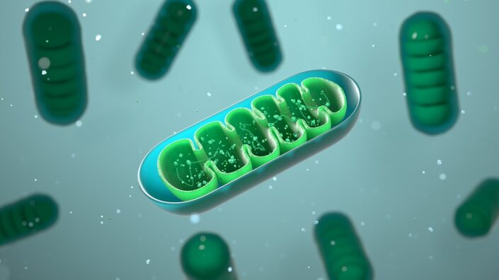
When I first joined the SENS Research Foundation in 2010 I was initially hired to work on mitochondrial aging and to develop a mitochondrial gene therapy to help resolve the mutations in mitochondrial DNA that accumulate with age. That was a great project, and we did a lot of good research. It was challenging but also fruitful and we built a good team and published some nice papers, and that work is ongoing by Dr. Amutha Boominathan and her team at the LRI.

But not too long after being at the institute, I got involved in helping to manage all of the research that we were doing at our research center in Mountain View, California, and research that we were funding around the world focused on damage repair. These were types of projects either characterizing the damage or looking at ways to resolve the kinds of damage that we had identified.

So it was that step of getting exposure to all the different kinds of research that we were doing that led to me taking a project that was being done at a university externally and bringing the project into our research center. Next, we completely changed it into a different approach from what had been done previously by the academics.

This led to us making the discoveries that we made, to being able to write patents, and eventually form a company and spin it out of the foundation bringing some of the team with us. That’s how that all came together through the SENS Research Foundation.

It’s great to see that nonprofits have an important role in getting these things off the ground, and obviously in your case, it’s definitely succeeded, and here we are at this very important moment. I think it’s fair to say that if you guys do succeed, it would be pretty strong validation for the idea that the damage repair approach towards aging and age related diseases is viable.

So, now for the big question and what most readers will be wanting to know. With the caveats that clinical trials have at least three phases and this takes time and assuming everything goes to plan and the data is good, when might this therapy become available?

With the traditional system of doing Phase 1, 2, 3, and then being approved for market, the fastest route would be by 2030. That would be about the quickest time that I could imagine full approval under the current system.

That said, we’re really interested in some of the accelerated and adaptive approaches that are being developed. For example, we received the Innovative Licensing and Access Pathway award (ILAP), in the UK, which allows for partial reimbursement by the UK healthcare system in late-stage clinical trials.

If it looks like the drug is working as promised, if it seems likely that it’s working, if it appears that it’s helping people, then we may be able to start gradually releasing it in places like the UK that may start allowing this. Rather than this sort of all-or-nothing system that most of the world uses now, where it either hasn’t been completely proven to be safe and effective, or it’s been completely proven to be safe and effective.

Right now, the overwhelming practice is that you have to prove beyond any shadow of a doubt that it’s safe and effective. Then, it gets fully released and anyone with the approved diagnosis can receive the treatment. That’s pretty black and white, and I think there’s more gradual approaches that hopefully will be available to us soon in the UK and other places. And who knows, maybe in the US, they’ll start experimenting with systems like that as well.

There’s a lot of innovation in clinical trials that are being experimented with in different places right now that we’re paying close attention to and looking for opportunities to try to bring our therapy to people as soon as it’s ready.

It strikes me that systems like RMAT in the USA may be a possible avenue as well. That’s a framework that’s attempting to get with the times and accommodate approaches like gene therapies, regenerative medicine and the new technology that’s coming. As you say, there could be other systems in development that may allow us to accelerate its distribution globally a little bit faster. Also, existing data in one area may also mean you’re not having to start a Phase 1 in every single country.

The last question I’ve got for you is: what’s next for the Cyclarity team?

Of course, we’re mostly focused on developing our lead drug for atherosclerosis, but there’s other related indications that we’re also exploring. So, atherosclerosis and the associated dyslipidemias could be caused by oxidized cholesterol accumulation, where we’re closely investigating the impact on brain aging, and oxidized cholesterol is strongly implicated. We have also got a grant to study our drug in relation to Alzheimer’s disease. While we’re not emphasizing that disease indication, it’s certainly something we’re looking at in the lab.

Liver disease, particularly for things like non-alcoholic fatty liver, is something that we think has a lot of potential as well. So, we’re working on all aspects of the impact of oxidized cholesterol.

We also have this basic technology that is built to sequester things that are small and toxic that accumulate in biological systems. We could potentially use that as a detoxifier of things that accumulate with age. There’s other things that we’re interested in, like bisretinoids that accumulate in the eye in macular degeneration that we’re looking closely at.

We are also working on finding solutions to nanoplastics, things like BPA and PFAS that we think our technology could potentially address. Firstly, at a therapeutic level, in people who’ve accumulated unhealthy levels of nanoplastics that we could clean out of the blood, cells and tissues where they’re building up.

Secondly, environmental remediation is another possible area of interest. You could create something with our technology that’s very specific and potent for removing it from the environment. So, you might use it to remove nanoplastics and other pollution from the soil or water. Obviously, environmental remediation isn’t our focus. We’re a biomedical company, but we’re looking at collaborating with organizations that want to use our technology for other things unrelated to what we’re working on such as environmental remediation.

Well, if this succeeds, I think you folks deserve the Nobel Prize.

Thank you, Steve, but we’re not in it for the prizes or accolades, we’re in it to have a massive impact on humanity for the better. What I’d like to say to my team is that we could make a billion dollars and it’s not just a billion-dollar industry, it’s a trillion-dollar industry. But it’s not about making a billion dollars or a trillion dollars. It’s about saving a billion lives. And that’s when we’ll be true billionaires. The first true billionaire will be the person or the team who has saved a billion lives. And that’s the kind of billionaire that I want to become.

Well, people do often say that health is the greatest wealth. and I’m going to say I do agree with them. Thank you for taking the time to talk with us today, and we wish you the best of luck with your endeavors.

We would like to ask you a small favor. We are a non-profit foundation, and unlike some other organizations, we have no shareholders and no products to sell you. All our news and educational content is free for everyone to read, but it does mean that we rely on the help of people like you. Every contribution, no matter if it’s big or small, supports independent journalism and sustains our future.
Gerophysics Conference

The Global Conference on Gerophysics

Chaired by Prof Brian Kennedy, Assoc Prof Jan Gruber and Dr Maximilian Unfried, this pioneering conference will bring together leading theoretical physicists and eminent researchers in ageing and rejuvenation biology to explore a transformative new field: ‘Gerophysics’. Organised by the Healthy Longevity Translational Research Programme at the Yong Loo Lin School of Medicine, National University of Singapore (NUS Medicine), this interdisciplinary meeting will investigate the application of rigorous physical principles to unravel the complexities of the ageing process.

In the 1980s, physicists revolutionised financial modelling, introducing quantitative methods that transformed Wall Street and reshaped global markets. More recently, key concepts from physics have profoundly influenced the field of deep learning, culminating in a Nobel Prize awarded for discoveries rooted in theoretical physics. In the same manner, this conference seeks to unlock the secrets of longevity by applying similar rigorous frameworks to the study of ageing.

By using powerful tools of theoretical physics such as statistical mechanics, complex systems theory, and dynamical modelling, researchers can comprehend the collective behaviors of cellular components, model ageing as a complex system of interconnected processes, and predict the trajectory of ageing and identify potential points of intervention. Participants at the conference will explore predictive models that can effectively address ageing challenges and foster collaborations to accelerate progress in the field of gerophysics.

The conference will feature a distinguished faculty, including Prof Uri Alon, Prof Vadim Gladyshev, Prof Marija Cvijovic, Prof Andrew Teschendorff, and Dr Peter Fedichev, who will bring their expertise to this exciting new frontier in ageing research. This inaugural gathering will lay the intellectual and collaborative groundwork for a lasting synergy, harnessing the deep insights of theoretical physics to rewrite our understanding of ageing and rejuvenation. Together, researchers can identify parallels and common frameworks, cultivate a shared language, and forge long-term collaborations that lead to breakthroughs in human healthspan.

The Global Conference on Gerophysics will take place on 5 and 6 March 2025 at the Paradox Singapore Merchant Court.

For more information and sign up, please visit our event page.

We would like to ask you a small favor. We are a non-profit foundation, and unlike some other organizations, we have no shareholders and no products to sell you. All our news and educational content is free for everyone to read, but it does mean that we rely on the help of people like you. Every contribution, no matter if it’s big or small, supports independent journalism and sustains our future.

Telomeres

Maintaining Telomeres Extends Lifespan in Mice

A recent study has found that the overexpression of telomerase reverse transcriptase (TERT), which is a subunit of telomerase, an enzyme essential for telomere maintenance, leads to lifespan extension in mice without significant side effects [1].

Protecting DNA

Telomere shortening is a well-known hallmark of aging. Telomeres are protective DNA sequences at the ends of the chromosomes. In most human cells, they become shorter with each division.

Telomerase and TERT have been found to be essential in maintaining telomere length [2]. Since telomeres become shorter with aging, reversing this process and extending telomere length may have the potential to extend longevity and health [3].

Creating genetically modified mice

The authors of a recent study started their research into TERT’s impact on lifespan and healthspan by creating mice that express the TERT gene. They decided to use a safer, more efficient, and more controllable approach since, as they discuss, such techniques as viruses or exogenous TERT introduction to overexpress it can “potentially lead to unintended effects or immune response,” which raises safety concerns.

The researchers genetically modified embryonic stem cells by inserting the TERT gene under the control of the human EF1α promoter. This promoter was selected to ensure stable inheritance and strong TERT expression. They referred to the genetically modified mice as TertKI.

Mating with wild-type Black 6 mice confirmed that the transgene was correctly inherited and didn’t have a negative impact on the mice’s development, growth, or survival. The researchers confirmed the transgene to be inherited for at least five generations with no negative impact on litter size.

A comparison of TertKI and wild-type animals didn’t show any significant differences in visible features, such as coat color, locomotor activities, or social behaviors, including sniffing, grooming, and play behavior.

However, the researchers noted TERT’s impact on postnatal growth and development, as the TertKI group exhibited quicker weight gain from the fifth to twenty-third day postnatal, compared to wild-type mice.

Analysis of organs revealed that organ-to-body weight ratios of the examined organs and organ cellular and tissue morphology didn’t differ between genetically modified and wild-type mice. However, analysis of organs during the autopsy revealed five cases of enlarged liver and six cases of enlarged spleen but no evidence of tumor growth.

The researchers ran tests to confirm increased TERT expression, telomerase activity, and telomere length in TertKI mice compared to wild-type mice. The results confirmed their expectations, but expression was at different levels in different organs. The authors suggested that organ-specific regulation of the EF1α promoter, TERT transcription, and/or the stability of TERT mRNA all played a role in the observed differences.

The researchers also noted that the increase in telomere length and telomerase activity in various organs was not proportional to the increase in the mRNA levels of TERT in a given organ. They suggest that this may be due to tissue-specific gene regulation.

TertKI 1

Safety first

The researchers addressed some safety considerations regarding their research, especially since TERT gene therapy was previously debated to be either the “natural ally” or the “molecular instigator” of cancer [4]. This debate comes from the observation of telomerase activation in many human cancers.

The researchers did not observe any signs of tumors in the TertKI mice they created. Additionally, they didn’t find differences between TertKI and wild-type mice in the levels of the cancer biomarker CA72-4.

However, when the researchers exposed the mice to a mutagen to establish lung cancer, they observed more rapid cancer development in the TertKI mice compared to control animals, suggesting that the overexpression of TERT “can increase the likelihood of carcinogenesis under chronic harmful stimulation.”

Testing whether the genetic modification and TERT overexpression would cause any DNA damage or disturb fetal growth or development revealed no differences between genetically modified and wild-type mice. Blood test results either didn’t show differences or suggested that the genetically modified mice had better health.

Increased lifespan

Lifespan analysis of generations of genetically modified mice revealed an increase in the maximal lifespan of the TertKI mice by 27.48% and a 16.57% increase in median lifespan compared to WT mice.

TertKI 2

Previous research suggested that TERT might contribute to lifespan extension through oxidative stress modulation and protection from oxidative damage, which is known to contribute to aging [5]. The researchers measured antioxidant molecules, namely glutathione (GSH) and superoxide dismutase (SOD), in mouse livers, since TERT expression was significantly increased in this organ. Both GSH and SOD were increased in the liver, suggesting improved antioxidant capacity.

However, these results might also suggest an increase in oxidative stress in TertKI mice, resulting in an increase in GSH and SOD levels. Future studies would need to address those possibilities.

Tissue repair and regenerative potential

Significant lifespan extension doesn’t seem to be the only characteristic of TertKI mice. The researchers also observed improved hair growth, faster skin wound healing with reduced infiltration of inflammatory cells, and improved collagen fiber remodeling. In vitro experiments also demonstrated that mouse TertKI skin fibroblasts had more migration ability than wild-type fibroblasts. All of these results suggest improvements in tissue repair and an increase in regenerative capacity.

An assessment of inflammatory factors during wound healing suggested a quick inflammatory response followed by a quick resolution of this inflammation. The researchers suggested that this allows for a rapid response to injury while preventing the adverse effects of an sustained inflammatory state.

The increase in the wound healing capacity of TertKI mice was also supported by the upregulation of growth factor expression and protein levels.

TERT was also found to have benefits when the researchers induced colon inflammation (colitis) in these mice. Their results indicated that their TertKI animals “display less colon deformation, functional disruption, and reduced molecular markers of injury compared to WT animals.”

Limitations

Since this study focused on the common Black 6 strain of mice, more studies are needed to test if these results are strain-specific or can be more generalizable to different strains, animal models, and environments. It is also unclear whether these findings can be applied to future human therapies in the future, especially ones that would start in older age and don’t involve TERT overexpression over the entire human lifespan.

Additionally, existng methods of overexpressing genes can be challenging to perform, time- and labor-intensive, expensive, and/or limited to mouse models. The development of easier, human therapy-compatible, and safe methods is essential.

We would like to ask you a small favor. We are a non-profit foundation, and unlike some other organizations, we have no shareholders and no products to sell you. All our news and educational content is free for everyone to read, but it does mean that we rely on the help of people like you. Every contribution, no matter if it’s big or small, supports independent journalism and sustains our future.

Literature

[1] Zhu, T. Y., Hu, P., Mi, Y. H., Zhang, J. L., Xu, A. N., Gao, M. T., Zhang, Y. Y., Shen, S. B., Yang, G. M., & Pan, Y. (2024). Telomerase reverse transcriptase gene knock-in unleashes enhanced longevity and accelerated damage repair in mice. Aging cell, e14445. Advance online publication.

[2] Bodnar, A. G., Ouellette, M., Frolkis, M., Holt, S. E., Chiu, C. P., Morin, G. B., Harley, C. B., Shay, J. W., Lichtsteiner, S., & Wright, W. E. (1998). Extension of life-span by introduction of telomerase into normal human cells. Science (New York, N.Y.), 279(5349), 349–352.

[3] Muñoz-Lorente, M. A., Cano-Martin, A. C., & Blasco, M. A. (2019). Mice with hyper-long telomeres show less metabolic aging and longer lifespans. Nature communications, 10(1), 4723.

[4] Shay J. W. (2016). Role of Telomeres and Telomerase in Aging and Cancer. Cancer discovery, 6(6), 584–593.

[5] Sahin, E., & Depinho, R. A. (2010). Linking functional decline of telomeres, mitochondria and stem cells during ageing. Nature, 464(7288), 520–528.

Amyloid plaques

Fighting Alzheimer’s by Helping Neurons Consume Proteins

Researchers have found that kinesin family member 9 (KIF9), a protein that diminishes with aging, is instrumental in allowing cells to consume harmful proteins and fights Alzheimer’s in a mouse model.

Consuming amyloids before they become a problem

Alzheimer’s is well-known as a proteostasis disease: it is characterized by amyloid beta plaques outside the cells and tau tangles inside them [1]. These protein accumulations have been reported to occur alongside the failure of autophagy, and prior work has found that improving autophagy may be effective in preventing Alzheimer’s [2].

However, autophagy is a complicated biochemical process with many moving parts. Within neurons, the kinesin family is responsible for transporting lysosomes, core components of autophagy, along the microtubules inside the cell, and their decline is associated with brain degeneration [3]. While overexpressing kinesins have been found to improve autophagy [4], there has been little work in their connection to Alzheimer’s, and the researchers noted that KIF9 in particular has gone mostly unevaluated.

Transport is crucial

In their first experiment, the researchers examined a well-known mouse model of Alzheimer’s and compared it to wild-type mice. They found that after six months, compared to the wild-type mice, the Alzheimer’s model mice began to suffer significant reductions in KIF9 and significant increases in the proteins p62 and LCIII, which is evidence of degraded autophagy. These differences became even more stark after 12 months of life.

Next, the researchers turned to human cells. Beginning with HEK293, a commonly used cell line, the researchers used a variant, 2EB2, that produces amyloid precursor proteins. That variant, as expected, experienced diminished KIF9 and substantially decreased autophagy. This effect was found to be directional: forcing the 2EB2 cells to express more KIF9 reduced the presence of amyloid precursors and restored the autophagic components, autophagosomes, that were normally reduced in these cells.

Further research that specifically targeted individual parts of the autophagic process found that KIF9 had no special ability in directly restoring the structures themselves; rather, it was simply doing its job as a transporter, bringing these organelles to where they need to be.

Effective in mice

The researchers then used an adeno-associated virus (AAV) to determine whether an increased expression of KIF9 could ameliorate Alzheimer’s in model mice, performing behavioral tests at 5 months and brain tissue examination at 6 months.

The KIF9 AAV did nothing to the behavior of wild-type mice, as measured by an open-field test, the Barnes maze test, and the Morris water maze test. However, there were significant dfferences in all three tests when the AAV was applied to the Alzheimer’s model mice, restoring their abilities almost exactly to the levels of the wild-type mice.

Anxious mice do not want to spend time in an open space, but once they become acclimated to an area, they become more willing to explore it. Alzheimer’s model mice, at this age, do not become acclimated; the KIF9 AAV allowed this to occur significantly more.

The Barnes maze is a memory test that measures a mouse’s ability to discover and return to the correct hole. Alzheimer’s model mice, as expected, have significant impairments in memory, which the KIF9 AAV ameliorated. The Morris water maze is similar, except that it uses a hidden platform; the KIF9 AAV restored the ability of the Alzheimer’s mice to remember where it was.

However, this treatment was not perfect, as the brain examination revealed. Even after the KIF9 AAV, Alzheimer’s mice still had amyloid plaques and increases in amyloid-related proteins compared to the wild-type mice. However, the extra KIF9 did significantly reduce the amounts of these proteins and plaques.

Like many others of its kind, this is only a mouse study that uses a lab-created model, as mice do not naturally get Alzheimer’s. It is also unclear if this approach, causing neurons to express KIF9 through an AAV, could be successfully implemented in the clinic. However, it provides a crucial starting point for allowing our neurons to fight Alzheimer’s at its protein-accumulation root.

We would like to ask you a small favor. We are a non-profit foundation, and unlike some other organizations, we have no shareholders and no products to sell you. All our news and educational content is free for everyone to read, but it does mean that we rely on the help of people like you. Every contribution, no matter if it’s big or small, supports independent journalism and sustains our future.

Literature

[1] Liu, Y., Tan, Y., Zhang, Z., Yi, M., Zhu, L., & Peng, W. (2024). The interaction between ageing and Alzheimer’s disease: insights from the hallmarks of ageing. Translational Neurodegeneration, 13(1), 7.

[2] Long, Z., Ge, C., Zhao, Y., Liu, Y., Zeng, Q., Tang, Q., … & He, G. (2025). Enhanced autophagic clearance of amyloid-β via histone deacetylase 6-mediated V-ATPase assembly and lysosomal acidification protects against Alzheimer’s disease in vitro and in vivo. Neural Regeneration Research, 20(9), 2633-2644.

[3] Hayashi, K., & Sasaki, K. (2023). Number of kinesins engaged in axonal cargo transport: A novel biomarker for neurological disorders. Neuroscience Research.

[4] Liu, M., Pi, H., Xi, Y., Wang, L., Tian, L., Chen, M., … & Zhou, Z. (2021). KIF5A-dependent axonal transport deficiency disrupts autophagic flux in trimethyltin chloride-induced neurotoxicity. Autophagy, 17(4), 903-924.

Genetic examination

New Study Links Epigenetic Changes to Genetic Mutations

A new paper published in Nature Aging suggests that somatic mutations cause significant remodeling of the epigenetic landscape. The findings might be relevant to future anti-aging interventions [1].

The genome and the epigenome

Genomic instability and epigenetic alterations are two of the hallmarks of aging [2]. The former occurs in somatic cells due to replication errors and stressors such as radiation and reactive oxygen species. DNA mutations can be relatively benign, but they can also impair cellular function, which might contribute to age-related disorders in various ways. The ultimate bad outcome of mutations in a single cell is, of course, cancer.

Epigenetic alterations are different. One type of them, methylation, involves a methyl group being added to or removed from a nucleotide in the DNA molecule, most often a cytosine that is followed by a guanine in the DNA sequence, with the two linked by a phosphate bond (which is why such sites are called CpG). CpG methylation is an important regulator of gene expression.

While the exact role of somatic mutations in aging is not entirely clear [3], CpG methylation is so strongly correlated with aging that it has formed the basis for epigenetic aging clocks, which have become increasingly popular over the last decade. However, what if mutations and epimutations are causally connected? A new study coming from the University of California suggests that this might be the case.

Building a mutation clock

The scientists note that at least one mechanism linking methylation and mutations has been known for a while: when a CpG site is methylated, the cytosine becomes more prone to spontaneous deamination, leading to its conversion into thymine. Since cellular DNA repair machinery does not always correct this change, CpG sites are common mutation hotspots. Conversely, if a mutation alters or eliminates a CpG site, it can prevent future methylation at that location.

Using tissue samples that had both mutation and methylation data available, the researchers identified several types of interaction between somatic mutations and DNA methylation. While they mostly used cancerous tissues, they also made an effort to validate their findings in healthy tissues.

First, the researchers confirmed that mutated CpG sites were methylated less often than non-mutated sites, which concurs with the known data. However, they also found that such mutations created atypical methylation patterns in the sections of the genome surrounding the mutation site, sometimes for tens of thousands of base pairs. This was observed in all tested tissue types.

The effect size in non-cancerous tissues, however, was substantially lower than in cancerous ones. In the latter, abnormal methylation patters were found around 15.5% of mutated sites, while in the former, the number was 8%, and the disturbances’ extent was about 1,000 base pairs from the mutation site.

Having established this correlation, the researchers wanted to see whether mutation patterns can predict biological age, just like methylation clocks do. They constructed a proprietary clock based on the profile of somatic mutations, including the counts of mutations in the vicinity of the CpG sites on which the methylation clock was based.

The methylation clock won the day, showing higher accuracy in predicting chronological age, but the mutation clock was predictive as well (Pearson correlations of r=0.83 and r=0.67, respectively). Predictions from the two clocks were also correlated across individuals. This correlation held for three previously published clocks: Horvath, PhenoAge, and Hannum.

The researchers validated their findings in a smaller number of samples from non-cancerous tissues. Here, both clocks were more predictive of chronological age (which is to be expected, since cancer introduces genomic instability and disrupts normal epigenetic patterns), but the mutation clock was still substantially behind the methylation clock. The researchers concluded that somatic mutations explain more than 50% of variation in methylation age across individuals.

What does it mean for fighting aging?

Dr. Trey Ideker of UCSD, the leading author of the study, gave us a comment:

What our paper shows is that epigenetic clocks can be largely explained by underlying DNA mutations. We think this is a pretty important finding since so much investment is currently being placed in epigenetic clocks – not only as a quantitative measurement of age, but as a means of reversing it. Our study suggests that current efforts to reverse or stabilize epigenetic changes will need to seriously contend with the underlying accumulation of DNA mutations, an area that has received comparatively less attention. On the other hand, perhaps it is worth ‘doubling down’ on treatments that slow the accumulation of DNA mutations in the first place, such as caloric restriction/dieting and certain anti-aging drugs.

The results might be especially relevant to cellular reprogramming, in which cells are being either fully de-differentiated to a pluripotent state or rejuvenated using certain reprogramming factors. Cellular reprogramming is accompanied by a considerable remodeling of the epigenetic landscape. One possibly relevant question is what if, following reprogramming, the underlying mutations cause this landscape to once again become aberrant?

“Yes, this would be one concern,” Ideker noted. “Another is that the epigenetic changes are largely not causal for aging at all, and that aging is related more directly to the mutations themselves and how they disrupt protein expression, structure and function. Essentially, what our paper has done is to open up all of these new questions.”

João Pedro de Magalhães, professor at the University of Birmingham, who was not involved in this study, said, “It’s a very interesting paper, suggesting that mutations may contribute or to some degree explain epigenetic changes, including in the context of epigenetic clocks. They show that somatic mutations with age correlate with methylation changes, which is an important new observation.”

However, he also had some reservations: “The obvious limitation of the study is that it employs data from cancer patients, including mostly from tumor samples – though some noncancerous tissues were also used. Therefore, validating these findings in normal tissues is imperative to assess the relevance of somatic mutations to epigenetic aging changes.”

One company that chose to go after the particularly hard target of fixing somatic mutations is Matter Bio. Its co-founder and CSO, Dr. Sam Sharifi, who was not involved in this study, commented:

While epigenetic clocks have attracted considerable attention as markers of biological aging, they may only reflect downstream changes triggered by a deeper, more permanent force – cumulative DNA damage. This article sheds a fascinating light on the interplay between genetic and epigenetic changes and opens the door to a purely mutation-based clock. It is still early, but once this technology matures, it could provide a more robust measure for age, given the permanent nature of DNA mutations and their steady accumulation with age.

The findings of this study are also potentially relevant to the information theory of aging promoted by Dr. David Sinclair of Harvard. It postulates that epigenetic changes are an upstream cause of aging due to loss of information on how the cell should function; therefore, aging can largely be reversed by restoring this information via cellular reprogramming or other, yet to be discovered, techniques.

“This study provides compelling evidence that epigenetic changes could not only be connected to but actually be downstream of somatic mutations,” Sharifi said. “This means that the changes in epigenetic information could be consequences of genetic information loss. Unlike methylation marks, which are relatively malleable and can be experimentally reset, DNA mutations are permanent. A big question is: are both epigenetic and genetic loss of information due to upstream processes such as DNA damage, which accumulates during aging? Critically, the article’s findings raise the important notion that targeting epigenetic states alone might not suffice to reverse aging if the underlying mutational burden is driving those epigenetic shifts in the first place.”

We would like to ask you a small favor. We are a non-profit foundation, and unlike some other organizations, we have no shareholders and no products to sell you. All our news and educational content is free for everyone to read, but it does mean that we rely on the help of people like you. Every contribution, no matter if it’s big or small, supports independent journalism and sustains our future.

Literature

[1] Koch, Z., Li, A., Evans, D. S., Cummings, S., & Ideker, T. (2025). Somatic mutation as an explanation for epigenetic aging. Nature Aging, 1-11.

[2] López-Otín, C., Blasco, M. A., Partridge, L., Serrano, M., & Kroemer, G. (2023). Hallmarks of aging: An expanding universe. Cell, 186(2), 243-278.

[3] Chatsirisupachai, K., & de Magalhães, J. P. (2024). Somatic mutations in human ageing: New insights from DNA sequencing and inherited mutations. Ageing Research Reviews, 102268.

S. Jay Olshansky Op-Ed

The Battle for Long Life Has Been Accomplished: What’s Next?

How long can people live? This is not just a foundational question in science. The answer has important public policy implications and is of interest to us all. Recent scientific evidence has revealed the answer, so what’s next in humanity’s never-ending battle against disease and the persistent ravages of aging?

Larger increases in lifespan not likely if aging itself remains the same

In our first effort to answer this longevity question more than three decades ago, my colleagues and I estimated how much death rates would need to decline to live decades longer than we do now. The findings were unexpected. Our analysis revealed that a life expectancy of 100, for example, appeared implausible because it would require reductions in death rates greater than those achieved by curing diseases of aging.

To illustrate why this seemed improbable, we calculated the effect on life expectancy if cancer, cardiovascular diseases, and diabetes were cured individually, or collectively all at the same time. Surprising to many, curing cancer increases life expectancy by only about three years; curing heart disease only adds about 4.5 years to life expectancy; and curing all fatal diseases together produces a life expectancy at birth of about 90 years.

This naturally raised the question: if curing everything does not render us immortal or something close to it, what’s holding us back?

The answer eventually hit us like a lightning bolt. There is an immutable force at work in humans and other species that becomes activated and visible only when enough people survive long enough to experience it. It is the aging of our bodies – the natural degradation of parts of the body that wear out over time and with use, that was barely visible throughout history because, so few people lived that long.

This phenomenon not only prevents large increases in life expectancy from occurring in the future as long as aging remains unchanged, it also meant that the rise in life expectancy must slow down in long-lived populations except under extenuating circumstances that have never occurred before. It is the biological aging of our bodies that explains why accelerating gains in life expectancy, and immortality, aren’t in the cards.

As long as aging remains unchanged, large increases in life expectancy, like those experienced in the 20th century, are unattainable.

In other words, a slowdown in the rate of improvement in life expectancy is not a sign of a failing health care system or a byproduct of harmful risk factors (like smoking or obesity) or some new toxins in our food and air. Instead, it is a signal that humanity’s battle for a long life had largely been accomplished. We estimated back in 1990 that once 95% of a birth cohort survives beyond age 65, and 80% of deaths occur between 65 and 95, life expectancy would plateau at around 85 years (88 for women and 82 for men).

A decade later, in an article in Science, we provided a 10-year check on this hypothesis using life expectancy data from Japan, France, and the U.S., and came to the same conclusions: this phenomenon of slowing increases in life expectancy had already begun.

We’ve now waited a full 34 years to look back on what actually happened following our 1990 prediction and 2001 confirmation. The results appear in a paper recently published in Nature Aging. Using data from ten long-lived populations that were first in line to benefit from accelerating advances in life-extending technologies, we’ve now demonstrated definitively that the period of rapid increases in life expectancy is over.

The rise in life expectancy slowed precipitously in the very countries where it should have accelerated higher. Accompanying evidence also demonstrated that mortality is compressing into the time window between ages 65 and 95, and that it is far more difficult to raise life expectancy today than it was just three decades ago.

So, what do these observations mean exactly? What’s next?

First, declare victory in the pursuit of life extension

The miracle of extended life has been given to us by public health, modern medicine, and improved behavioral risk factors. If all medical interventions of every kind were removed from a population, even if everyone lived a healthy lifestyle, life expectancy would be somewhere in the 30-60-year range – which is consistent with the historical record. This means that most people now alive past age 60 are living on “manufactured time” – survival time humanity has created for itself.

In practical terms, this means that the natural limit to life expectancy for long-lived human populations is well behind us – not in front of us as longevity protagonists suggest. Like stretching a rubber band, the further life expectancy extends beyond its natural limits, the more difficult it becomes to live longer.

This means the battle to achieve longer lives has been won. The time has arrived to declare victory in the pursuit of life extension. It’s now time to extend the period of healthy life.

The failures of success

The rising prevalence of heart disease, cancer, stroke, dementia, sensory impairments, etc., is a product of success – not failure. In the early 1990s, we described this as the “expansion of morbidity hypothesis”, which is a cautionary note indicating we should be aware of the diseases and disorders we’re trading for in exchange for our longer lives. We weren’t the first to make this argument – the “Failures of Success” arguments from Ernest Gruenberg, Jim Fries, and Ken Manton, led to related conclusions.

Treating one disease at a time without modulating the rate at which we age, which is a whack-a-mole approach to disease management now in place, will ultimately lead to rising levels of frailty and disability among the survivors. This is not a suggestion that humanity stop battling diseases or improving behavioral risk factors that can accelerate both disease and aging – it is a realization of the population-level health consequences of successful life extension in the absence of modifications to aging itself.

Would healthier lifestyles lead to radical life extension?

The short answer is no, but healthier lifestyles as primary prevention should always be a top priority in public health. Keep in mind that certainly in the United States, but elsewhere, harmful behavioral risk factors, such as obesity and sedentary lifestyles, have been working against the observed gains in life expectancy shown to be slowing down.

However, modern medicine moved swiftly to accommodate these health challenges through medical procedures and pharmaceuticals that have proven successful in restoring length of life close to average. Stents, statins, and blood pressure medications, among many others, are effective in saving and extending lives.

But would we be better off modifying risk factors, so these drugs aren’t needed to begin with? Of course, and that’s the preferred path to a healthier life, but the life-extending benefits of healthier lifestyles at the population level cannot exert that strong of an influence on life expectancy for national populations that is much greater than the medical and pharmaceutical interventions already in place. However, at the individual level, their influence on health and length of life can be profound.

Does the presence of “vanguard groups” of longer-lived people, such as those described as living in Blue Zones, provide optimism for the future of life expectancy for national populations?

At one level, yes, because these population subgroups provide us with clues on what is theoretically possible in human bodies. Furthermore, studying vanguard groups of longer-lived people allows scientists to discover some of the genetic and behavioral risk factors that favor exceptional longevity. However, heterogeneity in survival prospects across genetically diverse populations is a natural part of human biology, so just because some people can live exceptionally long lives, does not mean everyone in a population has the opportunity to do so.

By way of illustration, some people are capable of running a mile in under four minutes, but this does not mean the rest of us can. The same holds true for vanguard longevity and its link to life expectancy. Just because the world record for human longevity is a validated 122 years by the French woman Jeanne Calment, this does not mean the life expectancies of national populations are capable of rising to the maximum lifespan for the species.

The rationale for geroscience

If there is one thing aging research has proven in the last few decades, it’s that the biological process of aging is inherently modifiable. Science and medicine can shape and mold this process just as we have done for major fatal diseases, so one obvious path forward is to change the rules of the whack-a-mole game by bringing in a new hammer with multiple heads that hit all the moles at once.

There is no shortage of pathways to a successful gerotherapeutic because the door is wide open – natural selection could not have given rise to brick walls for longevity or aging time bombs that are set off beyond a certain age. Combine this with empirical evidence suggesting that the economic value of extending healthspan is, at a minimum, $38 trillion for just one year of healthy life, and the conditions are ripe for investments in aging interventions and the healthspan they will manufacture. Geroscience has the potential to redefine what it means to grow old.

We would like to ask you a small favor. We are a non-profit foundation, and unlike some other organizations, we have no shareholders and no products to sell you. All our news and educational content is free for everyone to read, but it does mean that we rely on the help of people like you. Every contribution, no matter if it’s big or small, supports independent journalism and sustains our future.
Roswell Park

Cutting-Edge Facility Expands to Support Cancer Therapy

New York Gov. Kathy Hochul and leaders from The Roswell Park Comprehensive Cancer Center came together on Monday to celebrate the opening of the newly expanded Roswell Park Good Manufacturing Engineering and Cell Manufacturing Facility (GMP). This significant undertaking expands Roswell Park’s GMP facility to 11,000 square feet, including 20 clean rooms across two buildings and an entire floor within the Roswell Park Cancer Cell Center. Now the largest academic GMP facility in the state of New York, this milestone marks a transformative step for cancer research and treatment.

The expansion provides cutting-edge cell therapy equipment, manufacturing capabilities and comprehensive quality control, providing a complete spectrum of resources spanning pre-clinical research, clinical manufacturing, clinical trials and implementation. Top specialists in all aspects of cell and gene therapy will lead the work in and around this innovation hub — including Roswell Park team members who helped develop three of the six CAR T-cell therapies now approved by the FDA for treatment of cancer.

“This state-of-the-art facility gives Roswell Park the unique ability to manufacture customized therapies using patients’ own cells right here on site,” said Candace S. Johnson, PhD, President and CEO of the Roswell Park Comprehensive Cancer Center. “Not only will this give our own world-class physicians and scientists the tools and resources needed to advance cancer care, but it also allows us to partner with research organizations and pharmaceutical companies to foster new products and developments.”

The design of the multi-purpose facility will help grow groundbreaking CAR T-cell therapies and also support smaller biotech companies, helping advance therapies that may otherwise be stalled by funding or production constraints. This will ensure the most promising treatments can reach cancer patients faster.

“Our experts are committed to guiding these groundbreaking developments through every stage of the process, ensuring quality, efficiency, and compliance, while focusing on the ultimate goal – patient care and treatment,” said Yeong “Christopher” Choi, PhD, MBA, Technical Director of the Roswell Park’s GMP Facility. “The meticulously planned infrastructure is designed to expedite access to the most promising immunotherapy treatments and is poised to become the largest academic GMP facility in New York State, with insights from the globally acclaimed cell therapy experts at Roswell Park.”

Roswell Park’s Renier Brentjens, MD, PhD, is one of the pioneers of CAR T-cell therapy and has built a team of leading scientists, engineers and oncologists dedicated to advancing these treatments, improving their safety and efficacy and making them more widely available. Five CAR T clinical trials are slated to begin in 2025, utilizing the facility expansion to create these customized treatments.

“These therapies hold remarkable potential to save lives,” said Brentjens, Deputy Director and Chair of Medicine for the Roswell Park Comprehensive Cancer Center. “The GMP Facility allows us to truly blaze the trail to take these from bench to bedside and get them to the patients who need them as quickly as possible.”

To learn more about the unique resources available at the only National Cancer Institute-designated comprehensive cancer center in Upstate New York, as well as the new technology within the Roswell Park GMP Engineering & Cell Manufacturing Facility (GEM), visit roswellpark.org/gmp.

From the world’s first chemotherapy research to the PSA prostate cancer biomarker, Roswell Park Comprehensive Cancer Center generates innovations that shape how cancer is detected, treated and prevented worldwide. Driven to eliminate cancer’s grip on humanity, the Roswell Park team of 4,000 makes compassionate, patient-centered cancer care and services accessible across New York State and beyond. Founded in 1898, Roswell Park was among the first three cancer centers nationwide to become a National Cancer Institute-designated comprehensive cancer center and is the only one to hold this designation in Upstate New York. To learn more about Roswell Park Comprehensive Cancer Center and the Roswell Park Care Network, visit www.roswellpark.org, call 1-800-ROSWELL (1-800-767-9355) or email ASKRoswell@RoswellPark.org.

Media Contact

Annie Deck-Miller, Director of Public Relations 716-845-8593; annie.deck-miller@roswellpark.org

We would like to ask you a small favor. We are a non-profit foundation, and unlike some other organizations, we have no shareholders and no products to sell you. All our news and educational content is free for everyone to read, but it does mean that we rely on the help of people like you. Every contribution, no matter if it’s big or small, supports independent journalism and sustains our future.
Green mitochondria

Enhancing NAD+ Efficiency by Energizing Sirtuins

Researchers publishing in Physical Review X have discovered compounds that can double the efficiency of the sirtuin SIRT3 in processing NAD+.

Looking for a new way to boost enzymes

The researchers begin their paper by noting that most drugs administered to people are geared towards inhibition of particular enzymes in order to treat a disease. In this case, however, the goal is the opposite: to boost the function of an enzyme, thereby boosting a healthy phenotype rather than battling back a diseased one.

Sirtuins are enzymes that have been heavily investigated in the context of aging. They rely on NAD+ to function, and these researchers describe them as being critical regulators of cellular pathways relating to aging [1]. Upregulating sirtuins has been found in considerable previous work to extend lifespan in mammals [2]. However, most methods of using drugs to boost sirtuins has relied on allosteric activation, a chemical process that relies on an existing substrate that might be limited in quantity [3].

Of course, as sirtuins rely on NAD+, there has been much work on directly influencing that instead. These researchers note two problems with that approach: as it is a common aspect of metabolism, boosting NAD+ across the board may result in broad side effects [4] and converting it into NADH relies on delivering it into cells that have functioning internal machinery [5], which, in the context of aging, is far from guaranteed.

Therefore, these researchers seek to allow sirtuins to do more with less: to continue to function adequately even when NAD+ is diminished. This, the researchers describe, is a trickier thing to do; while allosteric activators fundamentally rely on existing, evolved mechanisms, attempting to modulate these enzymes is similar to designing new enzymes outright.

Also, they needed a compound that works all the time: a steady-state activator. Previous work has created compounds that inhibit, rather than activate, sirtuins most of the time [6], only performing their desired function under specific conditions.

SIRT3 was chosen as the target for two reasons. The first is that it is known to have beneficial effects on mitochondria [7], and previous work has found that the benefits of NAD+ against mitochondrial dysfunction are due to SIRT3 [8]. The second is that natural mutations in the SIRT3 gene are connected to longevity [9].

Needle in a haystack

Using an advanced algorithm, the researchers searched a library of 1.2 million compounds by beginning with Honokiol, a compound that only activates SIRT3 under certain conditions. The researchers were able to find compounds that do steady state and non-steady state activation, with which they refined their experiments further with a close and detailed examination of the specific biochemistry involved, looking for compounds that have strong bonds to certain amino acids on the SIRT3 protein.

This initial work, however, was all done on computers. To verify their findings in the real world, the authors administered their compounds to real SIRT3 in a substrate. While a lot of this type of work uses fluorescent labeling, the authors eschewed that approach as it may have affected the results. One particularly strong compound, number 5689785, was identified as being a plausible drug after this screening process.

The researchers tested their new candidate against a control group, honokiol, and the well-known NAD+ precursor NMN. In nearly all cases, 5689785 performed favorably against these alternatives. Administering nicotinamide (NAM) to cells inhibits NAD+ enzymatic activity, but 5689785 was able to restore it in a way that honokiol could not.

Next steps

This is not a drug yet; it has not been formulated in a way that is consumable by living organisms, and so there were no animal studies done. What the researchers have is an initial compound with which to continue the process of drug development. Their goal was to prove that it is indeed possible to directly enhance the activity of sirtuins without relying on substrate-based methods. If this approach sees success in animal models, it could pave the way for drugs that, due to SIRT3’s mitochondrial effects, fight multiple aspects of aging.

We would like to ask you a small favor. We are a non-profit foundation, and unlike some other organizations, we have no shareholders and no products to sell you. All our news and educational content is free for everyone to read, but it does mean that we rely on the help of people like you. Every contribution, no matter if it’s big or small, supports independent journalism and sustains our future.

Literature

[1] Kaeberlein, M., McVey, M., & Guarente, L. (1999). The SIR2/3/4 complex and SIR2 alone promote longevity in Saccharomyces cerevisiae by two different mechanisms. Genes & development, 13(19), 2570-2580.

[2] Roichman, A., Elhanati, S., Aon, M. A., Abramovich, I., Di Francesco, A., Shahar, Y., … & Cohen, H. Y. (2021). Restoration of energy homeostasis by SIRT6 extends healthy lifespan. Nature communications, 12(1), 3208.

[3] Sinclair, D. A., & Guarente, L. (2014). Small-molecule allosteric activators of sirtuins. Annual review of pharmacology and toxicology, 54(1), 363-380.

[4] Yang, T., & Sauve, A. A. (2006). NAD metabolism and sirtuins: metabolic regulation of protein deacetylation in stress and toxicity. The AAPS journal, 8, E632-E643.

[5] Hu, Q., Wu, D., Walker, M., Wang, P., Tian, R., & Wang, W. (2021). Genetically encoded biosensors for evaluating NAD+/NADH ratio in cytosolic and mitochondrial compartments. Cell reports methods, 1(7).

[6] Reverdy, C., Gitton, G., Guan, X., Adhya, I., Dumpati, R. K., Roy, S., … & Chakrabarti, R. (2022). Discovery of novel compounds as potent activators of Sirt3. Bioorganic & medicinal chemistry, 73, 116999.

[7] Van de Ven, R. A., Santos, D., & Haigis, M. C. (2017). Mitochondrial sirtuins and molecular mechanisms of aging. Trends in molecular medicine, 23(4), 320-331.

[8] Cantó, C., Houtkooper, R. H., Pirinen, E., Youn, D. Y., Oosterveer, M. H., Cen, Y., … & Auwerx, J. (2012). The NAD+ precursor nicotinamide riboside enhances oxidative metabolism and protects against high-fat diet-induced obesity. Cell metabolism, 15(6), 838-847.

[9] Bellizzi, D., Rose, G., Cavalcante, P., Covello, G., Dato, S., De Rango, F., … & De Benedictis, G. (2005). A novel VNTR enhancer within the SIRT3 gene, a human homologue of SIR2, is associated with survival at oldest ages. Genomics, 85(2), 258-263.

Founders Longevity Forum

Founders Longevity Forum and NUS Announce Event

Founders Longevity Forum Singapore, hosted in collaboration with the National University of Singapore (NUS) Academy for Healthy Longevity, Yong Loo Lin School of Medicine, and Longevity.Technology is set to host a pivotal two-day event on 27-28 February 2025, in Singapore. This forum aims to advance knowledge and foster growth in the rapidly evolving field of longevity, with a special emphasis on the Asia-Pacific (APAC) region.

Building upon the immense success of the Unlock Healthy Longevity Conference of the NUS Academy for Healthy Longevity and the inaugural Founders Longevity Forum held in London in 2024, the Singapore event will convene global leaders, clinicians, academics, and investors to explore advancements in extending health span and addressing the challenges of ageing. The forum will feature a dynamic roster of speakers, including experts in epigenetics, AI, cryomedicine, and preventative healthcare.

The event is structured to allow attendees to engage with content relevant to their specific interests through two distinct tracks: Precision Geromedicine and Longevity Investment. Sessions will cover a broad spectrum of topics, from biomarkers of ageing and consumer diagnostics to investment opportunities in the wellness and gym sectors embracing longevity.

Prof Andrea Maier, Oon Chiew Seng Professor in Medicine, NUS highlights the importance of the multidisciplinary approach of the conference, saying: “Precision Geromedicine is an emerging field and increasingly implemented into clinical practice to optimise the health of ageing individuals. Gerodiagnostics to measure the biological age and gerotherapeutics to lower the biological age are needed to build clinically meaningful and cost-effective services. This conference is stimulating the interaction of stakeholders to build this hugely important ecosystem.”

Carolyn Dawson, CEO of Founders Forum Group, said: “We are thrilled to bring Founders Longevity Forum to Singapore in collaboration with the National University of Singapore (NUS) Academy for Healthy Longevity, Yong Loo Lin School of Medicine and Longevity.Technology. This event is a testament to the rapid advancements and investment opportunities emerging in the Asia-Pacific longevity sector. By uniting global experts, investors, and innovators, we aim to catalyse breakthroughs in healthspan and ageing science, empowering the next generation of founders and leaders to shape a healthier and more sustainable future for all.”

Phil Newman, Founder and CEO of Longevity.Technology, emphasised the significance of the event, stating: “Founders Longevity Forum Singapore represents a unique convergence of scientific innovation, investment potential, and consumer engagement in the longevity sector. With the APAC region experiencing rapid growth in longevity marketing, this forum offers unparalleled networking opportunities and insights into the future of healthy ageing.”

Ticketing for the event is live, and attendees are encouraged to register promptly to secure their participation.

About Founders Longevity Forum

Founded by Founders Forum Group and Longevity.Technology, Founders Longevity Forum is a premier event series dedicated to advancing the field of longevity science and technology. In collaboration with leading academic institutions and industry partners, the forum provides a platform for thought leaders, innovators, and investors to drive progress in extending healthspan and addressing the challenges of ageing.

About Founders Forum Group

Founders Forum Group is a global community and group of businesses supporting entrepreneurs at every stage of their journeys.

Its forums unite the world’s most influential founders, investors, corporate and government leaders to tackle era-defining questions in iconic locations across the globe.

Since 2019, Founders Forum has partnered with Informa Tech to celebrate the strength and diversity of UK tech through London Tech Week.

Fuelled by the connections and ideas forged at the group’s flagship events, FF Group businesses support the needs of today’s founders via services (Founders Keepers, Founders Law, Founders Makers, Miroma Founders Network, Founders HR, Founders Comms, Founders Health), education (01 Founders), investment (Founders Factory, firstminute capital), networking (Grip, INDI), philanthropy (Founders Pledge, The Centre for Entrepreneurs), and content (Founders Insights). In 2021, the group sold its innovation strategy consulting firm, Founders Intelligence, to global consultancy, Accenture.

In 2023, FF Group acquired Tech Nation, the UK’s leading growth platform for tech scaleups. Founders Forum Group continues the previously government-funded Tech Nation programmes centred around early-stage and diverse founders, as well as data-driven research into the UK’s tech ecosystem.

For more information, contact:

Sean Lau

Head of Asia, Founders Forum Group

Gut bacteria

The Impact of a Human Breast Milk Probiotic on Sarcopenia

A recent study linked probiotic-induced gut microbiome and metabolite changes to improved muscle functioning in older sarcopenia patients [1].

Sarcopenia and the gut

Sarcopenia is an age-related condition. People with sarcopenia suffer from a reduction in muscle mass, strength, and function, leading to a decreased quality of life and increased morbidity and mortality [2].

The authors of this study, citing evidence of a link between the gut microbiome, muscle health, and sarcopenia, investigated the effect of the consumption of a probiotic on the muscle health of sarcopenia patients. They used Bifidobacterium animalis subsp. Lactis Probio-M8 (Probio-M8), a probiotic strain present in human breast milk [3]. Probio-M8 has already been shown to have a positive impact on bone metabolism [4] and in the treatment of Parkinson’s disease in older adults [5].

Anti-aging effects in mice

The researchers administered Probio-M8 to 19-month-old mice for 28 days. They observed improved muscle function and a significant reduction in senescence in the mice that received probiotics, suggesting an anti-aging effect.

The researchers also investigated inflammatory markers but didn’t see significant differences between the treatment and control groups. They suggest that this might be due to existing low inflammation in the control group, which does not allow probiotic treatment to lower it further.

Impact on microbes and metabolites

The impact of probiotic treatment on the structure and diversity of the gut microbiome in old mice was limited. A deeper look into the 10 most populous bacterial species in these groups’ fecal matter revealed a pathogen (Mucispirillum schaedleri) that was far more abundant in the control group. Previous reports suggested that this microbe may cause ulcerative colitis [6]. This is in contrast to probiotic-treated mice, in which the researchers observed abundant beneficial microbes.

There were also beneficial changes in the metabolites in the fecal and serum samples of old mice treated with the probiotic. The treatment led to a significant increase in anti-inflammatory, anticancer, and antioxidant metabolites, metabolites that have been reported to have benefits against aging, and metabolites that may somewhat alleviate neurological disorders, such as Alzheimer’s disease and Parkinson’s disease.

The chair test

The promising results in mice led to a test of this probiotic on 43 older sarcopenia patients. Following 60 days of supplementation with Probio-M8, the researchers observed a roughly 16% reduction in the five-time chair stand (FTCS) test time among the treated patients. This test requires patients to sit and stand up five times and measures lower limb strength. This significant result suggests an improvement in overall physical performance.

However, other sarcopenia-related measurements didn’t support the optimistic results obtained in the FTCS test. Skeletal muscle mass, grip strength, calf circumference, and BMI didn’t significantly change following the probiotic treatment.

Additionally, an evaluation of multiple physiological sarcopenia-related measurements showed mostly no changes compared to controls, except for reduced total cholesterol.

The role of microbial metabolites

Similarly to the results obtained in mice, human samples also showed modest changes in the richness and structure of the gut microbiome after probiotic treatment. Some of the most significant changes included increased numbers of beneficial gut bacteria, and reduced numbers of pathogenic gut bacteria, in patients with sarcopenia.

Despite modest changes in microbial composition, the researchers observed significant metabolite changes: the probiotic treatment enriched the microbial pathways involved in vitamin C biosynthesis and nucleotide metabolism. The researchers suggest that higher activity of those pathways might play a role in microbes’ support for host antioxidant defenses and nucleotide availability.

Other metabolites that were increased in feces and serum are involved in anti-inflammatory effects and processes essential for vital physiological functions, or they are associated with skeletal muscle, such as metabolites promoting the proliferation of skeletal muscle cells. There was also an increase in a compound that is considered a source of muscle energy and important for promoting muscle protein synthesis: creatine.

The researchers noted that the impact of the probiotic treatment on the composition of the gut microbiome was modest. However, the impact on the metabolite changes was significant, leading them to further investigations into how probiotic-driven metabolite changes influence host physical performance.

A series of bioinformatic analyses and models were employed to identify key players in the connection between Probio-M8 and sarcopenia. The authors summarized that their analysis “suggests that Probio-M8 may positively influence muscle metabolism, potentially through its effects on the gut microbiome and subsequent modulation of creatine synthesis or utilization.”

A computational analysis of metabolites also pointed the researchers toward a hypothesis that one of the harmful molecules known as n-dodecyl-l-homoserine lactone (HSL) “could reduce the absorption of creatine from the gut.” To test this, they created a cell culture monolayer of enterocytes. Enterocytes are intestinal absorptive cells that are located on the inner surface of the small and large intestines. An experiment confirmed that HSL interfered with creatine transport by affecting the level of its transporter (CRT).

Molecular understanding

In the discussion section, the researchers gathered the molecular evidence that they and others presented to assemble a possible mechanism of action.

One of the key players appears to be creatine, and these researchers have found that this probiotic encourages creatine to be delivered into the bloodstream from the gut. Creatine is a compound essential for muscles and, when combined with resistance training, can increase lean mass and muscle strength in older adults. Previous research suggested that creatine supplementation has benefits in older adults with sarcopenia [7]. The authors suggest that creatine might “act as a buffer to inhibit the production of reactive oxygen species (ROS) by serving as a neutralizing agent.” The inhibition of ROS is important, since accumulation of ROS has been linked to muscle function and muscle loss [8].

Probio-M8 can inhibit the enrichment of HSL in patients with sarcopenia, thereby promoting the accumulation of creatine in the serum and improving the host’s overall physical performance.

HSL Creatine
We would like to ask you a small favor. We are a non-profit foundation, and unlike some other organizations, we have no shareholders and no products to sell you. All our news and educational content is free for everyone to read, but it does mean that we rely on the help of people like you. Every contribution, no matter if it’s big or small, supports independent journalism and sustains our future.

Literature

[1] Zhang, Z., Fang, Y., He, Y., Farag, M. A., Zeng, M., Sun, Y., Peng, S., Jiang, S., Zhang, X., Chen, K., Xu, M., Han, Z., & Zhang, J. (2024). Bifidobacterium animalis Probio-M8 improves sarcopenia physical performance by mitigating creatine restrictions imposed by microbial metabolites. NPJ biofilms and microbiomes, 10(1), 144.

[2] Cohen, S., Nathan, J. A., & Goldberg, A. L. (2015). Muscle wasting in disease: molecular mechanisms and promising therapies. Nature reviews. Drug discovery, 14(1), 58–74.

[3] Zhong, Z., Tang, H., Shen, T., Ma, X., Zhao, F., Kwok, L. Y., Sun, Z., Bilige, M., & Zhang, H. (2022). Bifidobacterium animalis subsp. lactis Probio-M8 undergoes host adaptive evolution by glcU mutation and translocates to the infant’s gut via oral-/entero-mammary routes through lactation. Microbiome, 10(1), 197.

[4] Zhao, F., Guo, Z., Kwok, L. Y., Zhao, Z., Wang, K., Li, Y., Sun, Z., Zhao, J., & Zhang, H. (2023). Bifidobacterium lactis Probio-M8 improves bone metabolism in patients with postmenopausal osteoporosis, possibly by modulating the gut microbiota. European journal of nutrition, 62(2), 965–976.

[5] Sun, H., Zhao, F., Liu, Y., Ma, T., Jin, H., Quan, K., Leng, B., Zhao, J., Yuan, X., Li, Z., Li, F., Kwok, L. Y., Zhang, S., Sun, Z., Zhang, J., & Zhang, H. (2022). Probiotics synergized with conventional regimen in managing Parkinson’s disease. NPJ Parkinson’s disease, 8(1), 62.

[6] Kuffa, P., Pickard, J. M., Campbell, A., Yamashita, M., Schaus, S. R., Martens, E. C., Schmidt, T. M., Inohara, N., Núñez, G., & Caruso, R. (2023). Fiber-deficient diet inhibits colitis through the regulation of the niche and metabolism of a gut pathobiont. Cell host & microbe, 31(12), 2007–2022.e12.

[7] Casciola, R., Leoni, L., Cuffari, B., Pecchini, M., Menozzi, R., Colecchia, A., & Ravaioli, F. (2023). Creatine Supplementation to Improve Sarcopenia in Chronic Liver Disease: Facts and Perspectives. Nutrients, 15(4), 863.

[8] Watson, M. D., Cross, B. L., & Grosicki, G. J. (2021). Evidence for the Contribution of Gut Microbiota to Age-Related Anabolic Resistance. Nutrients, 13(2), 706.

Intestinal inflammation

A Gut Metabolite Reduces Senescence and Inflammation

In a preprint study, scientists from Lifespan Research Institute and the Buck Institute for Research on Aging have published their findings that Urolithin A, a molecule that has garnered a lot of attention in the longevity field, potently reduces senescence-related markers in human fibroblasts [1].

Senolytic versus senomorphic

Cellular senescence is a well-documented aspect of aging [2], but the best strategy to counter it remains a question. Senescent cells play an important role in development, wound healing, and anti-cancer defenses, but with age, as their numbers grow, they start doing more harm than good.

While even in aged tissue, the relative prevalence of senescent cells is usually small, they wreak havoc by emitting damage-associated molecular patterns (DAMPs) as well as the SASP. These trigger excessive immune response and are documented to contribute to the systemic, age-related inflammation known as inflammaging.

Clearing out senescent cells, something that the immune system should naturally do, forms the basis of the senolytic approach, which dominates today’s research in academia and biotech. The task is tricky, however, particularly because senescent cells are highly heterogeneous and are also full of harmful molecules that are released when the cell dies. One enticing alternative approach is the senomorphic one, which involves altering senescent cells in a way that would leave them in place but make them less harmful.

Urolithin A might help heal senescent cells

Urolithin A is a molecule that has gained popularity in the longevity community after demonstrating several healthspan and lifespan benefits in animal models and humans. This metabolite is produced by our gut bacteria from precursors found in foods like nuts, berries, and pomegranates, and it has been shown to decrease inflammation and improve muscle function in humans. In animal models, it led to significant increases in lifespan. For instance, in a small study from the Buck Institute for Research on Aging, Urolithin A produced a 19% increase in lifespan in mice, which is among the best results for any intervention [3].

In this study, the researchers induced two types of senescence in human fetal lung fibroblasts: one triggered by the chemotherapy drug doxorubicin and another by cellular division (replicative senescence). While the treatment had little effect on the two popular senescence markers p16 and p21, it did significantly reduce the secretion of the major pro-inflammatory SASP factors interleukin 6 (IL-6) and interleukin 8 (IL-8) as well as the expression of the corresponding genes in both models of senescence.

This translated to lower levels of paracrine senescence, which occurs when SASP from senescent cells induces senescence in neighboring cells. The researchers cultured healthy fibroblasts in the presence of media collected from either control senescent cells or those treated with Urolithin A and found that the latter scenario caused less paracrine senescence.

“Urolithin A has generated a lot of excitement in the last several years based on its potential use as an anti-aging therapeutic,” said Dr. Julie Andersen of the Buck Institute for Research on Aging, a co-author of this study and the earlier one showing that Urolithin A increases lifespan in mice. “This includes clinical data demonstrating its ability to slow loss of muscle function in older individuals. Our studies demonstrate a novel mechanism of action for the compound – suppression of chronic inflammation associated with cellular senescence, a major contributor to multiple age-related diseases. This offers a novel approach for treating a wide range of chronic diseases which could improve overall quality of life in later years.”

The mechanism

The researchers went one step further and tried to elucidate the potential mechanisms behind these effects. Cellular senescence has been linked to cytosolic DNA, which are fragments of DNA floating around in the cytosol instead of staying where they belong: in the nucleus and in mitochondria. Since it can be of viral or bacterial origins, cytosolic DNA is recognized by the cell as a sign of invasion, and the alarm is raised via the cGAS-STING pathway, which drives inflammation.

Urolithin A significantly decreased the abundance of cytosolic DNA in the treated cells. The researchers suggest that this might have something to do with Urolithin A’s ability to induce mitophagy [4], the process of eliminating unhealthy mitochondria. Damaged mitochondria leak DNA, and lowering their burden would be consistent with the observed reduction in cytosolic DNA.

“We discovered that Urolithin A, a remarkable gut-derived metabolite significantly suppresses the expression and release of pro-inflammatory SASP and DAMP factors,” said Dr. Amit Sharma of the Lifespan Research Institute, the study’s lead author. “This effect is driven, at least in part, by reducing cytosolic DNA release and dampening the cGAS-STING signaling pathway – a central player in chronic inflammation.”

Is the hype real?

Urolithin A has been making a lot of buzz lately, given its availability as a supplement, which makes this pre-print particularly timely. “This is an exciting study as it opens up the possibility of thinking how gut metabolites can influence inflammation by modulating the SASP,” said another Buck researcher, Dr. Pankaj Kapahi, who was not involved in this study.

“The growing excitement around Urolithin A as a potent anti-aging molecule is well-founded, and our findings take this a step further, unveiling the precise mechanisms behind its anti-inflammatory power,” Sharma noted. “This breakthrough provides a deeper understanding of how Urolithin A combats the hallmarks of aging.”

Importantly, genetics dictate how we metabolize Urolithin A. According to one study, only 40% of people are able to produce it from natural precursors in meaningful quantities [5]. However, there are still not a lot of human studies that involve Urolithin A.

“Our results open new doors for exploring Urolithin A as a targeted and selective intervention against inflammaging and its associated diseases,” said Sharma. “Its exceptional ability to reduce inflammation and tackle the root causes of inflammaging left us astonished. This molecule could redefine the fight against age-related inflammation and its devastating consequences.”

We would like to ask you a small favor. We are a non-profit foundation, and unlike some other organizations, we have no shareholders and no products to sell you. All our news and educational content is free for everyone to read, but it does mean that we rely on the help of people like you. Every contribution, no matter if it’s big or small, supports independent journalism and sustains our future.

Literature

[1] Barkovskaya, A., Brauning, A., Chamoli, M., Rane, A., Andersen, J. K., & Sharma, A. (2025). Mitigating Proinflammatory SASP and DAMP with Urolithin A: A Novel Senomorphic Strategy. bioRxiv, 2025-01.

[2] Di Micco, R., Krizhanovsky, V., Baker, D., & d’Adda di Fagagna, F. (2021). Cellular senescence in ageing: from mechanisms to therapeutic opportunities. Nature reviews Molecular cell biology, 22(2), 75-95.

[3] Ballesteros-Alvarez, J., Nguyen, W., Sivapatham, R., Rane, A., & Andersen, J. K. (2023). Urolithin A reduces amyloid-beta load and improves cognitive deficits uncorrelated with plaque burden in a mouse model of Alzheimer’s disease. Geroscience, 45(2), 1095-1113.

[4] Zhao, H., Song, G., Zhu, H., Qian, H., Pan, X., Song, X., … & Liu, C. (2023). Pharmacological Effects of Urolithin A and Its Role in Muscle Health and Performance: Current Knowledge and Prospects. Nutrients, 15(20), 4441.

[5] D’Amico, D., Andreux, P. A., Valdés, P., Singh, A., Rinsch, C., & Auwerx, J. (2021). Impact of the natural compound urolithin A on health, disease, and aging. Trends in molecular medicine, 27(7), 687-699.

Atherosclerosis

Cyclarity Launches Human Trial for Atherosclerosis

Cyclarity Therapeutics, a biotechnology company based at the Buck Institute in California, has launched its first human clinical trial.

Its primary candidate cyclodextrin drug, UDP-003, focuses on 7-ketocholesterol, an oxidized cholesterol variant that builds up in cells as we age. Atherosclerosis involves the accumulation of plaque within arteries, and it primarily results from this oxidized form of cholesterol.

Heart disease is the leading cause of death worldwide. If successful, this drug could potentially help 70 to 80 percent of people that have heart disease and are at risk of having heart attacks.

Current treatment of heart disease includes lifestyle and dietary interventions, statins, and surgery. However, these are not that effective, and there is currently no effective way to reverse the condition. If UDP-003 is a success in the coming years, it will be a game changer.

Not only will it transform how we treat heart disease, it will be a clear demonstration of how tackling the root causes of aging can lead to proper solutions to age-related diseases.

Trials and tribulations

The original plan had been to launch the trials in Cambridge, UK working with the MHRA (similar to the FDA in the USA), but, unfortunately, there were setbacks.

Regular readers may recall our last interview with Dr. Matthew O’ Connor from Cyclarity, CEO of Scientific Affairs, where he explained the delay:

The bad thing is that post Brexit, it seems that the MHRA has gotten a bit backlogged and isn’t able to keep up with our current demands on their time. It takes too long to get meetings and responses to applications currently. We’ve had to take our first human clinical trial to Australia, where it’s a faster, more streamlined, and cheaper process.

While this has led to a delay in starting trials for this potentially transformative therapy, it is great to see it finally moving forward.

Dr. Matthew O’ Connor was previously the Vice President of Research at the SENS Research Foundation for nine years (now the LRI, of which lifespan.io is part), where initial research for what was to become UDP-003 was conducted. This trial is a proud moment for both Cyclarity and our organization.

Australia is leading the charge

This research will now take place at CMAX, a leading clinical research center in Australia, in partnership with Monash University. The trials will proceed under the guidance of Dr. Stephen Nicholls, the Director of the Monash Victorian Heart Institute in Melbourne and a Professor of Cardiology at Monash University.

In the recent Cyclarity press release, Dr. Matthew O’Connor said: “We are excited to be working with Dr. Nicholls on a groundbreaking advancement in cardiovascular care. As we advance into being a clinical stage company, Cyclarity is focused on bringing truly disease-modifying treatments for the world’s deadliest disease into reality.”

The Phase 1 clinical study will include a section featuring single ascending dose and multiple ascending dose methodologies as well as a unique segment involving 12 patients suffering from acute coronary syndrome.

This trial is intended to assess the safety of UDP-003 in patients with pre-existing plaque and to collect initial insights on its efficacy.

Cyclarity has already finished the manufacturing process for the human-quality drug material in what’s known as the Current Good Manufacturing Practice (the CGMP). They have human-grade material packaged and in sterile, single-use vials ready for patients to receive.

Thorough studies necessary for investigational new drug approval have been completed, showing no expected toxicity issues and ensuring a safe route for clinical advancement. All essential documents for trial authorization have been submitted and accepted, which means that the clinical trial should commence in the very near future.

We will be interviewing Dr. Matthew O’ Conner from Cyclarity and finding out more about this exciting development, so stay tuned for that in the next week. Finally, we wish to congratulate the Cyclarity team on this important milestone for our field. Perhaps this will lead to more acceptance of the idea that to tackle age-related diseases, we need to tackle the underlying reasons we age.

We would like to ask you a small favor. We are a non-profit foundation, and unlike some other organizations, we have no shareholders and no products to sell you. All our news and educational content is free for everyone to read, but it does mean that we rely on the help of people like you. Every contribution, no matter if it’s big or small, supports independent journalism and sustains our future.
Time-restricted eating

Intermittent Fasting Improves Coordination in Mice

Researchers have discovered that intermittent fasting increases myelin in aged mice, leading to better neural function and coordination.

Crucial proteins and a well-known intervention

Normally, neuronal axons are coated in a protein sheath made of myelin, which is necessary for their proper function [1]. Myelination is most known to be impeded by multiple sclerosis, but it also decreases with aging [2]. It is predominantly formed from two key proteins, myelin basic protein (MBP) and myelin-associated glycoprotein (MAG) [3], and previous work has found that upregulating the expression of these proteins has a beneficial effect on myelination [4].

Other work has found that myelination can be affected by diet and nutrition [5]. However, that work did not focus on these researchers’ chosen intervention: intermittent fasting, which has been found in substantial previous research to have metabolic and anti-inflammatory benefits, particularly in the context of aging [6].

Fasting for 18 hours a day

For their experiments, the researchers used three groups of mice: ten young mice, ten older mice, and eight older mice that had undergone intermittent fasting fof ten weeks, in which they were only allowed to eat for six hours a day. The researchers first began by testing overall markers of physical function: on the wire hanging test, the fasting mice were able to hold on for longer than the old control group, and they trended towards being able to run faster and longer than this group as well.

In a balance beam test, the fasting proved exceptionally potent: the fasting group was able to perform just as well as the young mice, far outpacing their same-aged counterparts. However, cognitive function was found to be unaffected: there was no benefit according to a Y maze test.

Stronger motor signals

A closer look at the mice’s muscles may have revealed why. While the maximum electrical signal strength going from the nerves to the muscles was not significantly affected, the treatment group had higher average signal strength. Looking at the frequency ranges involved revealed that the treatment group could exert more force and could react more quickly than similarly aged mice that were fed freely.

The brain was affected as well. Measuring whole-brain connectivity, the researchers found that the brains of the treated mice were less connected in ten areas but more connected in seven, particularly in places related to motor function and sensory input. Comparing these connection differences to the physical tests, the researchers concluded that these changes may also be responsible for the improvements they found.

Myelin was directly improved

Finally, the researchers looked directly at the myelin in the brain. Interestingly, and possibly of concern, the fasting group had reduced axonal diameters compared to the aged control group, suggesting an increase in degeneration. However, they had substantially more myelin, particularly on their smaller axons. These findings were true for both motor and non-motor portions of the brain, and the researchers note that this has been documented to occur in other animals, including people, who are recovering from demyelinating diseases [5].

Both MBP and MAG were positively affected. The treated mice had significantly more of both proteins in both of the tested areas, although there was no significant increase in MAG in the motor cortex. Myelinated fibers were found to trend towards being more common and longer in the fasting group. Overall, these results suggest that fasting somewhat changes the brain, and the researchers hold that these changes are beneficial.

While this is only a mouse study, it is in line with previous research showing that such dietary interventions may have beneficial effects on the brain. Furthermore, while it may not be appropriate for everyone, intermittent fasting is a freely available intervention. More studies may reveal whether or not it has beneficial effects on the myelin, and muscle coordination, of older people.

We would like to ask you a small favor. We are a non-profit foundation, and unlike some other organizations, we have no shareholders and no products to sell you. All our news and educational content is free for everyone to read, but it does mean that we rely on the help of people like you. Every contribution, no matter if it’s big or small, supports independent journalism and sustains our future.

Literature

[1] Almeida, R. G., & Lyons, D. A. (2017). On myelinated axon plasticity and neuronal circuit formation and function. Journal of Neuroscience, 37(42), 10023-10034.

[2] Nickel, M., & Gu, C. (2018). Regulation of central nervous system myelination in higher brain functions. Neural plasticity, 2018(1), 6436453.

[3] Deng, S., Shu, S., Zhai, L., Xia, S., Cao, X., Li, H., … & Xu, Y. (2023). Optogenetic stimulation of mPFC alleviates white matter injury‐related cognitive decline after chronic ischemia through adaptive myelination. Advanced science, 10(5), 2202976.

[4] Zhang, Q., Zhu, W., Xu, F., Dai, X., Shi, L., Cai, W., … & Hu, X. (2019). The interleukin-4/PPARγ signaling axis promotes oligodendrocyte differentiation and remyelination after brain injury. PLoS biology, 17(6), e3000330.

[5] Langley, M. R., Triplet, E. M., & Scarisbrick, I. A. (2020). Dietary influence on central nervous system myelin production, injury, and regeneration. Biochimica et Biophysica Acta (BBA)-Molecular Basis of Disease, 1866(7), 165779.

[6] De Cabo, R., & Mattson, M. P. (2019). Effects of intermittent fasting on health, aging, and disease. New England Journal of Medicine, 381(26), 2541-2551.