The Blog

Building a Future Free of Age-Related Disease

Human heart

A Protein That Exacerbates Heart Disease With Age

Researchers publishing in Aging Cell have found that Hevin, a protein found in the extracellular matrix that increases with age, leads to heart problems in older male mice.

Inflammaging contributes to heart disease

The researchers begin by outlining the mechanisms involved in inflammaging, noting how it is closely connected to various progressive problems in the heart, leading to its gradual loss of function [1, 2]. Unsurprisingly, suppressing inflammatory cytokines led to better cardiac outcomes in mice [3], and prolonging lifespan by preventing inflammaging-related heart disease has been a biotechnology goal for some time.

Macrophage behavior is a key part of this relationship. Pro-inflammatory M1-polarized macrophages are good at killing off pathogens, but they cause damage to surrounding tissues; M2-polarized macrophages reverse this process and repair tissues. Aging drives a shift from M2 to M1 behavior [4], and one of the main chemical culprits is CCL5, a ligand that impedes the polarization of macrophages towards M2 [5].

Hevin, which is found in the extracellular matrix, plays a variety of roles in disease. We have previously reported that Hevin has significant benefits against brain aging in mice, but other work has found entirely different functions, including activity against cancer by recruiting macrophages [6]; however, the recruitment is of M1 macrophages, potentially worsening diseases such as pneumonia [7] and non-alcoholic fatty liver disease [8].

Hevin is hell for older animals

Unsurprisingly, Hevin in the plasma increases with age in people, particularly in people over 60 [9]. In line with previous work finding it to be a biomarker of heart failure [10], these researchers found that it is associated with a decrease in ejection fraction.

In mice, Hevin expression does not increase in the heart; rather, it increases in fatty tissues and is then distributed throughout the body, and this increase in Hevin distribution is accompanied by a decrease in the heart’s ability to effectively pump blood. Directly introducing Hevin to 20-month-old mice did not change blood pressure nor heart rate, but it promoted cellular senescence and shortened telomeres in the heart, increased macrophage infiltration along with inflammatory cytokines in cardiac tissue, and increased fibrosis and hypertrophy while contributing to the heart weakening already seen in older mice. However, these negative effects were only seen in older animals; injecting 8-month-old mice with Hevin caused none of these problems.

Hevin effects

The researchers then used an adeno-associated virus (AAV) to knock down Hevin. Just like with introducing Hevin itself, this treatment had no effects on younger animals; however, in the 20-month-old mice, many age-related diseases were ameliorated by this treatment. Senescent cell biomarkers decreased, telomeres were lengthened, hypertrophy was diminished, and both hypertrophy and fibrosis were significantly decreased.

Inflammation plays a key role

These effects were found to be largely due to Hevin’s effects on CCL5 in particular, as it significantly promotes the expression of this ligand and CCL5 was indeed found to promote M1 macrophage polarization in the heart. Older mice that were also treated with an anti-CCL5 antibody were partially spared from the inflammatory effects of Hevin injection, and this antibody also partially reversed the associated hypertrophy and fibrosis. Blocking toll-like receptor 4 (TLR4) and its associated p65 pathway had similar effects.

This study had certain limitations. While Hevin expression has sex-related differences in people, only male mice were used in this study, and further work will have to discover its effects on females. While the researchers surmise that Hevin has an amplifying effect on existing age-related processes, precisely why it had no effects on younger animals was not thoroughly investigated.

The connection between Hevin and fat was noted, and the authors suggest that reducing adipose tissue, such as through semaglutide, may be effective against its age-related increase. This, however, remains an open question as well. As Hevin is known to have beneficial effects in other contexts, whether or not it is wise to target it directly or indirectly is still a topic that requires more detailed investigation.

We would like to ask you a small favor. We are a non-profit foundation, and unlike some other organizations, we have no shareholders and no products to sell you. All our news and educational content is free for everyone to read, but it does mean that we rely on the help of people like you. Every contribution, no matter if it’s big or small, supports independent journalism and sustains our future.

Literature

[1] Liberale, L., Badimon, L., Montecucco, F., Lüscher, T. F., Libby, P., & Camici, G. G. (2022). Inflammation, aging, and cardiovascular disease: JACC review topic of the week. Journal of the American College of Cardiology, 79(8), 837-847.

[2] Liberale, L., Montecucco, F., Tardif, J. C., Libby, P., & Camici, G. G. (2020). Inflamm-ageing: the role of inflammation in age-dependent cardiovascular disease. European heart journal, 41(31), 2974-2982.

[3] Hu, C., Zhang, X., Hu, M., Teng, T., Yuan, Y. P., Song, P., … & Tang, Q. Z. (2022). Fibronectin type III domain‐containing 5 improves aging‐related cardiac dysfunction in mice. Aging Cell, 21(3), e13556.

[4] Becker, L., Nguyen, L., Gill, J., Kulkarni, S., Pasricha, P. J., & Habtezion, A. (2018). Age-dependent shift in macrophage polarisation causes inflammation-mediated degeneration of enteric nervous system. Gut, 67(5), 827-836.

[5] Li, M., Sun, X., Zhao, J., Xia, L., Li, J., Xu, M., … & Xia, Q. (2020). CCL5 deficiency promotes liver repair by improving inflammation resolution and liver regeneration through M2 macrophage polarization. Cellular & molecular immunology, 17(7), 753-764.

[6 Zhao, S. J., Jiang, Y. Q., Xu, N. W., Li, Q., Zhang, Q., Wang, S. Y., … & Zhang, Z. G. (2018). SPARCL1 suppresses osteosarcoma metastasis and recruits macrophages by activation of canonical WNT/β-catenin signaling through stabilization of the WNT–receptor complex. Oncogene, 37(8), 1049-1061.

[7] Zhao, G., Gentile, M. E., Xue, L., Cosgriff, C. V., Weiner, A. I., Adams-Tzivelekidis, S., … & Vaughan, A. E. (2024). Vascular endothelial-derived SPARCL1 exacerbates viral pneumonia through pro-inflammatory macrophage activation. Nature Communications, 15(1), 4235.

[8] Liu, B., Xiang, L., Ji, J., Liu, W., Chen, Y., Xia, M., … & Lu, Y. (2021). Sparcl1 promotes nonalcoholic steatohepatitis progression in mice through upregulation of CCL2. The Journal of clinical investigation, 131(20).

[9] Lehallier, B., Gate, D., Schaum, N., Nanasi, T., Lee, S. E., Yousef, H., … & Wyss-Coray, T. (2019). Undulating changes in human plasma proteome profiles across the lifespan. Nature medicine, 25(12), 1843-1850.

[10] Di Salvo, T. G., Yang, K. C., Brittain, E., Absi, T., Maltais, S., & Hemnes, A. (2015). Right ventricular myocardial biomarkers in human heart failure. Journal of cardiac failure, 21(5), 398-411.

Arthritis

A Small Molecule Regenerates Cartilage in Aged Mice

By inhibiting the aging-related enzyme 15-PGDH, scientists have shifted cartilage cells towards a healthier phenotype, leading to a significant improvement in a mouse model of osteoarthritis [1].

The hard-to-repair part

Articular cartilage (the smooth, load-bearing cartilage on the ends of bones) doesn’t repair well with age or after injury [2], which is why osteoarthritis is hard to treat. This disease affects 1 in 5 adults, leading to reduced quality of life for 33 million patients in the US alone. Current treatments primarily focus on pain relief and joint replacement, with no approved therapies targeting the cartilage loss that causes osteoarthritis.

Previous research has shown that 15-hydroxyprostaglandin dehydrogenase (15-PGDH) increases with age in multiple tissues and can blunt regeneration by degrading key prostaglandins, lipid signaling molecules that influence inflammation and tissue repair. In those earlier models, which studied muscle, nerve, bone, and blood, inhibition of 15-PGDH boosted endogenous prostaglandin signaling and improved tissue repair [3].

Since osteoarthritis is fundamentally a problem of failed repair in articular cartilage, and cartilage regeneration strategies based on endogenous repair have been limited, a team led by researchers from Stanford Medicine decided to investigate the role of 15-PGDH in aged and injured cartilage. Their study was published in the journal Science.

More healthy cartilage

Using immunohistochemistry on knee joints from young (4 months) and aged (24 months) mice, the team discovered that cells expressing 15-PGDH were present in multiple joint tissues. In cartilage specifically, 15-PGDH abundance was about twice as high in aged mice. Aged knee joints had much thinner cartilage and multiple breaks in the cartilage surface.

A cohort of aged male mice was treated daily intraperitoneally with a small molecule 15-PGDH inhibitor (PGDHi) for one month. As a result, the knee joints of PGDHi-treated aged mice showed increased cartilage thickness and uniformness, almost on par with young mice.

The “extra” cartilage in treated aged mice was not fibrous and rough, as often happens after an injury heals, but bore many signs of normal cartilage, with increased expression of type II collagen (COL-2) and aggrecan (ACAN), the main structural building blocks of healthy cartilage, and of lubricin (PRG4), a surface lubricant that helps cartilage glide with low friction.

Cartilage condition PGDHi

Safranin O staining: red/orange marks proteoglycan-rich cartilage matrix (healthier cartilage); fading indicates cartilage matrix depletion.

The researchers then wanted to know whether local joint delivery is sufficient in an injury-driven osteoarthritis model. Three-month-old male mice were treated with a series of intra-articular injections of PGDHi starting one week after injury, twice a week for two weeks.

The response was similar to what the team had seen with systemic administration: improved cartilage quality, higher COL-2, and increased aggrecan/lubricin. Pain responses were also better in treated mice: PGDHi-injected mice looked closer to uninjured controls across gait and mechanical pain measures. This particular experiment is relevant to young people as well: even after a successful repair, half of the people who suffer an ACL tear develop osteoarthritis in the injured joint within about 15 years.

No stem cells involved

Tissue regeneration often involves proliferation and differentiation of stem cells, but such cells in cartilage have rarely been seen, which might be a reason why cartilage regenerates poorly. The team made an exciting discovery: the regeneration they had witnessed was mostly due to gene expression changes in existing differentiated cartilage cells rather than a result of stem cell expansion.

The researchers identified multiple chondrocyte clusters in aged cartilage and described three that shift with PGDHi. Hypertrophic chondrocytes, the type that drives cartilage ossification, showed high expression of 15-PGDH. The treatment lowered the abundance of this subtype from 8% to 3%. Another largely harmful subset, fibro-chondrocytes, shifted from 16% down to 8% in the presence of PGDHi.

Conversely, the healthy subtype that actively maintains the extracellular matrix increased in prevalence from 22% to 42%. There was no evidence of drastically increased cellular division, supporting the idea that the positive effect mostly came from the existing cells shifting their behavior.

To make their findings more relevant to humans, the researchers studied samples from 11 osteoarthritis patients undergoing knee replacement and found signs of increased 15-PGDH expression and lower prostaglandin levels. Finally, they treated human cartilage with PGDHi in vitro and saw results similar to those in mice, with increased stiffness pointing to healthy load-bearing behavior.

“This is a new way of regenerating adult tissue, and it has significant clinical promise for treating arthritis due to aging or injury,” said Helen Blau, PhD, professor of microbiology and immunology and a senior author on the study. “We were looking for stem cells, but they are clearly not involved. We are very excited about this potential breakthrough. Imagine regrowing existing cartilage and avoiding joint replacement.”

“Millions of people suffer from joint pain and swelling as they age,” added Nidhi Bhutani, PhD, associate professor of orthopedic surgery, and another senior author. “It is a huge unmet medical need. Until now, there has been no drug that directly treats the cause of cartilage loss. But this [PGDH] inhibitor causes a dramatic regeneration of cartilage beyond that reported in response to any other drug or intervention. Cartilage regeneration to such an extent in aged mice took us by surprise. The effect was remarkable.”

We would like to ask you a small favor. We are a non-profit foundation, and unlike some other organizations, we have no shareholders and no products to sell you. All our news and educational content is free for everyone to read, but it does mean that we rely on the help of people like you. Every contribution, no matter if it’s big or small, supports independent journalism and sustains our future.

Literature

[1] Singla, M., Wang, Y. X., Monti, E., Bedi, Y., Agarwal, P., Su, S., … & Bhutani, N. (2025). Inhibition of 15-hydroxy prostaglandin dehydrogenase promotes cartilage regeneration. Science, eadx6649.

[2] Hu H, et al. “Endogenous Repair and Regeneration of Injured Articular Cartilage: A Challenging Balance.” Cells. 2021.

[3] Palla, A. R., Ravichandran, M., Wang, Y. X., Alexandrova, L., Yang, A. V., Kraft, P., … & Blau, H. M. (2021). Inhibition of prostaglandin-degrading enzyme 15-PGDH rejuvenates aged muscle mass and strength. Science, 371(6528), eabc8059.

RNA

How Harmful Extracellular Vesicles Cause Brain Inflammation

In a paper published in Aging Cell, researchers have described how older cells send long interspersed nuclear element-1 (LINE-1) RNA to other cells in extracellular vesicles (EVs), spurring inflammation.

Evil EVs

In the literature, EVs are often discussed in a therapeutic context, as they can be used to send beneficial signals. However, EVs are the natural communications method of cells; their contents can consist of nearly any molecular information, not all of which is beneficial.

EVs can even pass through the blood-brain barrier (BBB), the protective layer between the brain’s vasculature and its cells, and previous work has found that this can indeed spur inflammation [1] and even Parkinson’s disease [2]. However, what these harmful EVs contain and where they come from are questions that have not been thoroughly answered.

These researchers focus on LINE-1, a series of mobile genetic elements that comprise a full sixth of the human genome and whose activation has been associated with mutation [3] and aging [4]. LINE-1 RNA is known to be reverse transcribed into DNA [5], and previous work had found that this RNA can be transmitted through EVs [6]. These researchers, therefore, sought to determine the effects of these EVs on microglial inflammation and brain aging.

LINE-1 dramatically increases with aging

First, the researchers purified and quantified the EVs derived from 185 people between 20 and 95 years old. As expected, EVs containing distinctly identifiable LINE-1 elements were far lower in younger people; 20- to 45-year-olds had roughly a third of the LINE-1 elements of 46- to 65-year-olds, who themselves had roughly a quarter of the LINE-1 elements in people over 65. There was a significant correlation between LINE-1 elements in EVs and markers of brain aging, including amyloid beta, even when adjustments were made for age and other possibly confounding factors such as heart disease and diabetes.

To find out where these harmful EVs were coming from, the researchers studied 3-month-old and 21-month-old mice. The older animals had many more EVs coming from the brain and heart, but there was no significant upregulation in the kidney and liver. In the older animals, LINE-1 elements were significantly upregulated in the brain and lung but not in other tissues.

Harmful in younger animals

The researchers then confirmed that these older EVs were harmful by administering them to 10-month-old mice. Compared to a control group, the mice that had received these EVs suffered significant cognitive impairment, including depressive behaviors, a lack of interest in novel objects, and poorer results on the Y-maze test. However, if the mice were injected with EVs derived from older animals that had been previously treated with a LINE-1 inhibitor, the harmful effects were considerably blunted.

These findings were recapitulated in an examination of the mice’s brains. Older EVs without LINE-1 had very limited harmful effects that did not always rise to the level of statistical significance, but LINE-1 EVs significantly increased markers of both microglial activation and cellular senescence, and they decreased the number of healthy neurons in the hippocampus as well.

These effects were found to be due to the well-known cGAS/STING pathway, which promotes inflammation. Its downstream effects, such as an increase in the pro-inflammatory cytokine TNF-α, were readily apparent. Alongside the LINE-1 EVs, the researchers injected the treated mice with inhibitors of either LINE-1 retrotranscription or STING. Both approaches were found to be effective; these treated animals had microglial effects and inflammation levels that were much more like the control group, compared to animals that had received LINE-1 EVs alone.

As it is extremely difficult to intercept EVs in transit, the researchers suggest investigating both LINE-1 and STING inhibition as possible treatments. However, this was only a mouse study, and much more work needs to be done to determine whether such approaches can be developed into safe therapies for human beings.

We would like to ask you a small favor. We are a non-profit foundation, and unlike some other organizations, we have no shareholders and no products to sell you. All our news and educational content is free for everyone to read, but it does mean that we rely on the help of people like you. Every contribution, no matter if it’s big or small, supports independent journalism and sustains our future.

Literature

[1] Ramos-Zaldívar, H. M., Polakovicova, I., Salas-Huenuleo, E., Corvalán, A. H., Kogan, M. J., Yefi, C. P., & Andia, M. E. (2022). Extracellular vesicles through the blood–brain barrier: a review. Fluids and Barriers of the CNS, 19(1), 60.

[2] Guo, M., Wang, J., Zhao, Y., Feng, Y., Han, S., Dong, Q., … & Tieu, K. (2020). Microglial exosomes facilitate α-synuclein transmission in Parkinson’s disease. Brain, 143(5), 1476-1497.

[3] Cordaux, R., & Batzer, M. A. (2009). The impact of retrotransposons on human genome evolution. Nature reviews genetics, 10(10), 691-703.

[4] Miller, K. N., Victorelli, S. G., Salmonowicz, H., Dasgupta, N., Liu, T., Passos, J. F., & Adams, P. D. (2021). Cytoplasmic DNA: sources, sensing, and role in aging and disease. Cell, 184(22), 5506-5526.

[5] Thawani, A., Florez Ariza, A. J., Nogales, E., & Collins, K. (2024). Template and target-site recognition by human LINE-1 in retrotransposition. Nature, 626(7997), 186-193.

[6] Kawamura, Y., Sanchez Calle, A., Yamamoto, Y., Sato, T. A., & Ochiya, T. (2019). Extracellular vesicles mediate the horizontal transfer of an active LINE-1 retrotransposon. Journal of Extracellular Vesicles, 8(1), 1643214.

Connecting neurons

Recombinant Human Protein Stops Neuronal Loss in Alzheimer’s

A recent study investigated biomarkers that can help monitor trajectories of Alzheimer’s disease-related molecular processes, such as neuronal cell death, and how patients respond to treatments. The authors reported that using biomarkers enabled them to gain insights into the molecular processes that contribute to improved cognition following human recombinant granulocyte macrophage colony-stimulating factor (GM-CSF, sargramostim) treatment [1].

Measuring the damage

Alzheimer’s disease is accompanied by neuronal loss and increased inflammation [2, 3]. However, to monitor those processes, whether for diagnostic purposes or to aid in the development of Alzheimer’s disease therapies, easy-to-measure blood biomarkers are indispensable.

The authors of this study investigated such biomarkers. They focused on three proteins: ubiquitin C-terminal hydrolase-L1 (UCH-L1) for neuronal cell loss, neurofilament light (NfL) for neuron and axon damage, and glial fibrillary acidic protein (GFAP) for astrogliosis (an abnormally high number of astrocytes in response to neuronal destruction) and inflammation.

Changes in the healthy population

The researchers assessed the levels of UCH-L1, NfL, and GFAB in 317 healthy participants aged 2 to 85. Plasma UCH-L1 and NfL concentrations grew exponentially from ages 2 to 85. However, there were some differences between the two markers. For UCH-L1, the rate of exponential increase in females is faster than in males. For NfL, the estimated change per year is greater than that of UCH-L1, and the rate of exponential increase in females is slower than in males (opposite to that of UCH-L1).

Such an exponential age-dependent increase in neuronal damage markers suggests that brain aging is a lifelong process; however, its effects are evident in older age, “as the accumulated neuronal damage overcomes neurogenesis, functional redundancy, and resiliency in some individuals but not all.”

GFAP showed a different type of relationship with age. GFAP levels remain relatively constant up to 25, and around 40, they start to rise exponentially. As with previous markers, there are sex-specific differences, with females showing higher GFAP plasma levels across all ages.

It appears that plasma biomarkers of neuronal damage, UCH-L1 and NfL, increase earlier in life than the astrogliosis marker (GFAP), suggesting that astrogliosis and inflammation are responses to age-related neuronal damage.

Alzheimer’s disease trajectories

Once the trajectories of plasma markers of neurodegeneration (UCH-L1 and NfL) and of astrogliosis and inflammation (GFAP) in healthy individuals throughout their lifespan were established, the researchers compared them with those observed in 36 people with Alzheimer’s disease and 32 patients with mild cognitive impairment.

Patients diagnosed with mild cognitive impairment or mild-to-moderate Alzheimer’s disease had higher levels of NfL and GFAP compared to healthy controls of the same age. The same was true for UCH-L1 levels in plasma from participants with mild cognitive impairment, but in patients with mild-to-moderate Alzheimer’s disease, UCH-L1 levels in plasma were comparable to those of healthy participants of the same age.

Efficacy

The biomarkers investigated in this study can provide a better understanding of the mechanisms and trajectories of brain aging and help assess the efficacy of interventions aimed at slowing brain aging and neurodegenerative diseases.

The researchers used their previous study to evaluate the usefulness of those biomarkers in testing the efficacy of Alzheimer’s disease treatment, specifically using the immune-system-modulating cytokine GM-CSF, a long-approved drug that has also been investigated in the treatment of many other neurological injuries and diseases, including age-related cognitive decline, Down syndrome, stroke, traumatic brain injury, and Parkinson’s disease [4-8].

Treatment of a mouse model of Alzheimer’s disease with GM-CSF “reverses cognitive decline and the rate of neuron death after just a few weeks of treatment,” said the study’s senior author, Professor Huntington Potter, PhD, director of the University of Colorado Alzheimer’s and Cognition Center at CU Anschutz. The benefits of GM-CSF extend beyond Alzheimer’s disease and also benefit normal brain aging, as it improves impaired cognition and reduces neuronal function in aged wild-type mice [9].

The benefits are not limited to mice. These researchers conducted a phase 2, double-blind, randomized, placebo-controlled trial of human recombinant GM-CSF (sargramostim) in people suffering from mild-to-moderate Alzheimer’s disease [10]. “This drug improved one measure of cognition and reduced a blood measure of neuron death in people with AD in a relatively short period of time in its first clinical trial,” Potter said.

However, one of the most significant improvements was observed in the plasma UCH-L1 concentrations. “When people with AD were given sargramostim in the clinical trial, their blood levels of the UCH-L1 measure of neuronal cell death dropped by 40% – in our study, this was similar to levels seen in early life,” Potter said. “We were very surprised.”

Given the high sensitivity of UCH-L1 to sargramostim/GM-CSF, this biomarker might be a good candidate for assessing the efficacy of many Alzheimer’s disease treatments, including lifestyle changes.

NfL and GFAP were not reduced. The most likely reason is the very short treatment period (only 3 weeks) and the short plasma half-life of UCH-L1 relative to NfL and GFAP.

GM-CSF Effectiveness

Understanding the mechanism

An aged rat model of Alzheimer’s disease helped to understand the mechanism behind GM-CSF treatment’s effects on caspase-3, a marker of cellular death by apoptosis. The number of caspase-3-positive cells is increased in humans and animal models with Alzheimer’s disease, and this was also evident in the aged Alzheimer’s disease rats used in this experiment, specifically in hippocampal neurons. GM-CSF treatment significantly reduced those numbers to levels comparable to those of wild-type untreated animals, suggesting that GM-CSF reduces neuronal cell death. Assessment of GFAP staining showed that GM-CSF treatment also reverses astrogliosis in some hippocampal regions of the Alzheimer’s disease rat model.

Those results suggest that the beneficial effects of GM-CSF treatment on cognition and various biomarkers are “likely due to a reduction in the number of apoptotic neurons in the brain.” The authors also add that since the GM-CSF can stimulate some of the immune cells, it can also contribute to “removal of damaged, apoptotic, and senescent neurons, thus allowing the remaining neurons to function more effectively.”

Informative biomarkers

Potter summarized, “These findings suggest that the exponentially higher levels of these markers with age, likely accelerated by neuroinflammation, may underlie the contribution of aging to cognitive decline and AD and that sargramostim treatment may halt this trajectory.”

Additionally, using those markers, the researchers can identify people with mild cognitive impairment, suggesting they may be used to predict future Alzheimer’s disease. However, variability in marker levels indicates that other players also influence the risk of developing the disease, and factors such as genetics or lifestyle can shape the trajectory of cognitive decline.

We would like to ask you a small favor. We are a non-profit foundation, and unlike some other organizations, we have no shareholders and no products to sell you. All our news and educational content is free for everyone to read, but it does mean that we rely on the help of people like you. Every contribution, no matter if it’s big or small, supports independent journalism and sustains our future.

Literature

[1] Sillau, S. H., Coughlan, C., Ahmed, M. M., Nair, K., Araya, P., Galbraith, M. D., Ritchie, A., Ching-Jung Wang, A., Elos, M. T., Bettcher, B. M., Espinosa, J. M., Chial, H. J., Epperson, N., Boyd, T. D., & Potter, H. (2025). Blood measure of neuronal death is exponentially higher with age, especially in females, and halted in Alzheimer’s disease by GM-CSF treatment. Cell reports. Medicine, 102525. Advance online publication.

[2] Risacher, S. L., Anderson, W. H., Charil, A., Castelluccio, P. F., Shcherbinin, S., Saykin, A. J., Schwarz, A. J., & Alzheimer’s Disease Neuroimaging Initiative (2017). Alzheimer disease brain atrophy subtypes are associated with cognition and rate of decline. Neurology, 89(21), 2176–2186.

[3] Hyman, B. T., Van Hoesen, G. W., Damasio, A. R., & Barnes, C. L. (1984). Alzheimer’s disease: cell-specific pathology isolates the hippocampal formation. Science (New York, N.Y.), 225(4667), 1168–1170.

[4] Kim, N. K., Choi, B. H., Huang, X., Snyder, B. J., Bukhari, S., Kong, T. H., Park, H., Park, H. C., Park, S. R., & Ha, Y. (2009). Granulocyte-macrophage colony-stimulating factor promotes survival of dopaminergic neurons in the 1-methyl-4-phenyl-1,2,3,6-tetrahydropyridine-induced murine Parkinson’s disease model. The European journal of neuroscience, 29(5), 891–900.

[5] Kelso, M. L., Elliott, B. R., Haverland, N. A., Mosley, R. L., & Gendelman, H. E. (2015). Granulocyte-macrophage colony stimulating factor exerts protective and immunomodulatory effects in cortical trauma. Journal of neuroimmunology, 278, 162–173.

[6] Kong, T., Choi, J. K., Park, H., Choi, B. H., Snyder, B. J., Bukhari, S., Kim, N. K., Huang, X., Park, S. R., Park, H. C., & Ha, Y. (2009). Reduction in programmed cell death and improvement in functional outcome of transient focal cerebral ischemia after administration of granulocyte-macrophage colony-stimulating factor in rats. Laboratory investigation. Journal of neurosurgery, 111(1), 155–163.

[7] Schneider, U. C., Schilling, L., Schroeck, H., Nebe, C. T., Vajkoczy, P., & Woitzik, J. (2007). Granulocyte-macrophage colony-stimulating factor-induced vessel growth restores cerebral blood supply after bilateral carotid artery occlusion. Stroke, 38(4), 1320–1328.

[8] Olson, K. E., Abdelmoaty, M. M., Namminga, K. L., Lu, Y., Obaro, H., Santamaria, P., Mosley, R. L., & Gendelman, H. E. (2023). An open-label multiyear study of sargramostim-treated Parkinson’s disease patients examining drug safety, tolerability, and immune biomarkers from limited case numbers. Translational neurodegeneration, 12(1), 26.

[9] Boyd, T. D., Bennett, S. P., Mori, T., Governatori, N., Runfeldt, M., Norden, M., Padmanabhan, J., Neame, P., Wefes, I., Sanchez-Ramos, J., Arendash, G. W., & Potter, H. (2010). GM-CSF upregulated in rheumatoid arthritis reverses cognitive impairment and amyloidosis in Alzheimer mice. Journal of Alzheimer’s disease : JAD, 21(2), 507–518.

[10] Potter, H., Woodcock, J. H., Boyd, T. D., Coughlan, C. M., O’Shaughnessy, J. R., Borges, M. T., Thaker, A. A., Raj, B. A., Adamszuk, K., Scott, D., Adame, V., Anton, P., Chial, H. J., Gray, H., Daniels, J., Stocker, M. E., & Sillau, S. H. (2021). Safety and efficacy of sargramostim (GM-CSF) in the treatment of Alzheimer’s disease. Alzheimer’s & dementia (New York, N. Y.), 7(1), e12158.

Year 2025

Longevity Biotech in 2025: The Expert Roundup

How did the year 2025 turn out for longevity biotech? Was it surprising or more of the same? Exciting or disappointing? Was the progress fast-moving or excruciatingly slow? What should we expect in 2026? We asked five leading experts to weigh in.

Where did longevity biotech exceed your expectations in 2025, and where did it underperform or stall? Has this changed your long-term outlook for how fast the field will translate into approved therapies?

Kristen Fortney, CEO, BioAge

Longevity biotech exceeded my expectations in 2025 in how quickly it went mainstream within big pharma, largely because the GLP‑1 era made it obvious that metabolism, inflammation, and aging biology are deeply connected. Where it stalled is still clinical translation: we have many compelling preclinical mechanisms, but a comparatively thin set of rigorous human trials and hard endpoints. Approvals take time and real capital, so speed will come from anchoring aging mechanisms in tractable indications and using modern biomarkers to de‑risk early.

Mehmood Khan, CEO, Hevolution Foundation

2025 marked a real inflection point for healthspan science. We saw growing recognition from investors, policymakers, and the public that extending healthy years of life is not a niche ambition but a macroeconomic and societal imperative. At the same time, it became clear that human judgment remains essential. AI and digital tools are powerful, but they must be deployed within strong ethical, governance, and regulatory frameworks. These technologies are only as effective as the data, intent, and oversight behind them, making human wisdom and accountability non-negotiable.

Translation from discovery to clinical impact continues to be the bottleneck. Despite exciting scientific advances, moving from laboratory insights to scalable interventions is still slow. A major constraint is the absence of universally accepted biomarkers of biological aging: a shared language that researchers, industry, and regulators can trust, much like LDL cholesterol in cardiovascular disease. Overall, I remain optimistic but realistic. Progress will come from disciplined science, robust standards, and sustained collaboration rather than shortcuts.

Jamie Justice, Executive Director, XPRIZE Healthspan

Insilico Medicine brought my biggest breakthrough of the year: their AI-driven drug discovery pipeline focused on longevity targets delivered a novel target, a small molecule design for an inhibitor of Traf2- and Nck-interacting kinase (TNIK), and an accelerated testing pipeline that culminated in a successful Phase 2a clinical trial in the age-related disease idiopathic pulmonary fibrosis. This is without a doubt a huge win for biotech in general, but seeing this breakthrough launched by a biotech rooted in longevity is beyond expectation. The potential of generative AI-discovered pathways and accelerated translational testing for aging-related conditions and longevity is difficult to overstate.

[Full disclosure: Alex Zhavoronkov is a scientific advisor to XPRIZE Healthspan, but I assure you that this in no way influenced my opinion. Insilico is not a team in our competition, and I am not an advisor to Insilico; I just get to fan-girl from the arena like everyone else!]

Nathan Cheng, General Partner at Healthspan Capital, Co-Founder of Vitalism and Longevity Biotech Fellowship

Venture funding in longevity still hasn’t recovered, but that’s mostly macroeconomic; biotech broadly went through a downturn and only started bottoming out mid-year. Progress is taking longer than I expected. For investors with dry powder, it’s an opportunity for good deals and higher impact.

What exceeded expectations was Washington. I didn’t anticipate the new administration being so explicitly pro-longevity. Jim O’Neill appointed as HHS Deputy Secretary, Dr. Oz at CMS, the new NIH and FDA leadership – each is knowledgeable on longevity and/or an outright advocate. The recent MAHA summit in DC had a panel on age reversal in the brain. On the state level, multiple states recently advanced right-to-try legislation (New Hampshire, Montana). The leverage of policy is seriously underrated in this space; more attention needs to be paid here.

Karl Pfleger, Investor, Founder of AgingBiotech.info

Nothing that happened in 2025 significantly changed my long-term outlook. Things move slowly in biotech. Most of what happened in terms of clinical progress, especially late-stage clinical, was in view long before 2025. Clinical trial stages advance slowly. A highlight was Stealth Bio finally getting approval (accelerated approval in this case) for elamipretide (aka SS-31) for a rare disease (Barth) after a long road of ups and downs. This approval is notable for being the first – likely of many – significant approval of a mitochondria therapeutic. Approaches targeting mitochondria will likely have wide applicability to many aging diseases.

An unexpected stall was that the manufacturing plant producing Scholar Rock’s anti-myostatin antibody had problems with the FDA, delaying its approval despite positive Phase 3 trial data, but this should hopefully just be a temporary setback. On the funding side, ARPA-H has been great, but NIH funding cuts and attacks on universities have been unexpectedly severe. Venture funding for biotechs also remains very tight.

Which event in longevity biotech from 2025 do you consider the most influential, and why? You’re welcome to name a runner-up.

Kristen Fortney

My pick is the “FGF21 moment”: within months, GSK (Boston Pharma), Roche (89bio), and Novo Nordisk (Akero) basically bought the class, putting ~$10B of validation behind a key longevity pathway that hits metabolism, inflammation, and fibrosis. It looks very real for MASH, with potential upside beyond that based on what we know from aging biology.

Mehmood Khan

One of the most influential developments in 2025 was the growing number of aging biology programs entering mid-stage clinical trials. Several senolytic, immune modulation, and epigenetic reprogramming approaches are now moving beyond early safety studies into trials that assess functional outcomes, reflecting the progression of the field. At the same time, strategic partnerships between longevity biotech startups and major pharmaceutical and technology companies signaled that healthspan is moving from the fringe into mainstream research and development.

For me, a defining moment was the Global Healthspan Summit in Riyadh. Bringing together global leaders across science, policy, investment, and industry demonstrated that healthspan has become a top-tier global priority and showed what is possible when the world aligns around a shared ambition for healthier, longer lives.

As a runner-up, the momentum of the XPRIZE Healthspan was notable. Seeing teams advance toward measurable restoration of function, not just lifespan extension, reinforced the field’s shift toward outcomes that truly matter to people such as mobility, cognition, and independence. Hevolution’s early anchoring support reflects our belief in bold, results-driven science.

Jamie Justice

As Executive Director of XPRIZE Healthspan, I am of course most influenced by the incredible progress within our own competition. We now have over 700 teams of academic scientists, biotechs, clinics, and students from 71 countries engaged in our competition. Our judges vetted more than 200 complete applications to name the Top 100 teams and awarded the Top 40 teams. Instituting rigorous processes to vet teams and setting audacious milestones to accelerate the translation of therapeutics to trials is incredibly rewarding professionally, and influential for our global teams, investors, funders, and partnership ecosystem.

Beyond our competition, other ecosystem drivers and announcements have been influential. Of note: 1) in the US, the ARPA-H Proactive Solutions for Prolonging Resilience (PROSPR) program received well-deserved attention as a driver of progress in healthy aging and longevity; 2) large pharmaceutical companies are investing and signaling strong interest in aging and longevity. This includes new XPRIZE Healthspan sponsors GSK – announced in May 2025 – and Eli Lilly and Novo Nordisk, who were both present and vocal about interests in longevity and age-related disease pathways at the Aging Research and Drug Discovery (ARDD) event hosted in Copenhagen in August. Collectively, these announcements and investments signal a watershed moment and promise for commercial development and scientific discovery in aging and longevity. The convergence of regulatory validation (see below), clinical trial initiation, and massive capital influx suggests we’re transitioning from pure research to translational medicine.

Nathan Cheng

Jean Hébert’s FRONT program launching at ARPA-H, easily. On the surface, it’s a US government program using stem cells and tissue engineering to repair brain damage. Few understand that this is actually an opening shot on goal to solve aging in the brain, not just slow aging or target one hallmark. FRONT is a significantly funded moonshot, and it will catalyze substantial matching private investment in replacement therapies.

The replacement paradigm is gaining traction more broadly. ARDD 2025 had its first replacement workshop. YZi/Binance Labs invested in Renewal Bio (stembroids). Vadim Gladyshev is doing serious work on heterochronic organ transplantation at Harvard and spoke optimistically about this strategy for longevity. Watch this space.

Karl Pfleger

I’d say that the collective, nearly simultaneous financing rounds of the big epigenetic partial reprogramming companies Life Bio, NewLimit, and Retro Bio, together with the involvement from big pharma’s biggest company, Lilly, in at least NewLimit’s round, is the collective “biggest thing.” The size of these rounds and their valuations may not be officially public but are on the whole quotable as numbers of, or fractions of, billions; a $1.6B valuation for NewLimit is reported on some websites. The whisper numbers for Life Bio’s valuation are lower but only by a fewfold, and multiple reports suggest Retro is trying to raise at a $5B valuation, though this round does not seem to be done unlike the others. In aggregate, these are gigantic numbers and will likely influence other valuations.

Thinking back to what you believed about longevity biotech on January 1, 2025, did anything this year genuinely surprise you or make you update your priors?

Kristen Fortney

I was surprised in a good way by how quickly big pharma and the government started treating longevity biology as a core R&D strategy. Lilly and Novo pushed their GLP‑1 ambitions well beyond weight loss and embraced the “longevity” label; Novartis is building out DARe around diseases of aging; and multiple biotechs that frame their work around extending healthspan are landing large partnerships from top pharma – not despite that framing, but because of it. On the government side, ARPA‑H is making big swings in healthspan and regeneration with PROSPR and FRONT.

Mehmood Khan

What stood out most this year was the strength of public demand. Citizens are increasingly calling for prevention and healthy aging to be central responsibilities of health systems. Our Global Healthspan Report reflects this growing expectation that governments invest earlier rather than simply treating later.

I was also reminded that capital alone cannot unlock this field. The real constraint is infrastructure, shared data, agreed biomarkers, and regulatory pathways. Without these foundations, innovation cannot scale, no matter how much money is deployed. This has updated my view on the next phase of progress. It will be driven less by isolated breakthroughs and more by global collaboration, aligned standards, and shared priorities across science, industry, and policy.

Jamie Justice

Our XPRIZE Healthspan registration closed in January 2025, leaving me the daunting task of directing the review of global submissions to award $10M at Milestone 1. I was uncertain about field quality and therapeutic originality. Our competition’s moonshot depends entirely on exceptional teams; snake oil won’t deliver results. To my relief, teams arrived with both practical approaches and novel therapies testable through rigorous, transparent methods.

We created an Innovations Landscape Report documenting team development, global scope, and paths to clinical trials for our teams – some entering trials for the first time, others seasoned. The report identifies potential and critical gaps requiring attention fo field progress. The response to our competition has been both overwhelming and surprising in the best possible way.

Nathan Cheng

Big Pharma’s interest in longevity surprised me. I had the standard model in my head: pharma is conservative, waits for de-risked assets, and acquires rather than innovates. Then at ARDD 2025, representatives from Eli Lilly and Novo Nordisk stood up and suggested GLP-1s (like semaglutide) are longevity drugs. Whether that’s true is debatable. What matters is the vibe shift.

GLP-1 agonists made Eli Lilly the first pharma ever to hit $1 trillion USD in market cap. That success rewired how pharma thinks about markets. The old model was to find people with a disease and treat it. The new model is to find the largest possible patient population and prevent disease. Listen to Dave Ricks on the Stripe podcast. Eli Lilly’s CEO sounds like a tech founder – talking direct-to-consumer models, AI partnerships with NVIDIA. The most conservative industry in healthcare is suddenly hungry for what’s next, and after obesity, what’s next is aging.

Karl Pfleger

Big pharma companies talking about and explicitly devoting resources to aging programs became much clearer in 2025. Their presence and explicitness in talking about aging at ARDD, such as specifically addressing it with respect to GLP-1 drugs, was much more direct than in prior years. It’s become clear that several big pharma companies are supporting the subsector.

Age1’s pharma report card is another source that reflects some of this shift. The partnerships data highlighted there was largely in the AgingBiotech.info/companies table already, but the report gathered that plus other indicators, and a few of the partnerships, such as Juvena’s, were only announced in 2025. A lowlight within this overall category, however, is Abbvie pulling out of their Calico partnership in 2025. But, 2025 showed me that big pharma is turning towards the aging sector a bit faster than I expected, which is great.

What are your expectations for 2026 in longevity biotech?

Kristen Fortney

In 2026, I’m focused on the clinical pipeline. Multiple clinical milestones are on the horizon, including readouts from the FGF21 class, NewAmsterdam’s CETP inhibitor, NLRP3 inhibitors (including BioAge’s), Insilico’s TNIK inhibitor, as well as Phase 1 data from exciting mechanisms at Retro, Rubedo, Juvena, Life, Cyclarity, and more. I’m also hopeful for sharper biomarker strategies and tighter trial designs in the space as a whole, so we can learn faster per dollar.

Mehmood Khan

Equity must be front and center. My greatest hope is that healthspan science does not become the privilege of a few. At Hevolution, our mission is to help ensure these advances are affordable, accessible, and globally relevant, so healthier aging becomes a universal opportunity rather than a luxury. I would like to see more multinational collaboration, including shared data, joint research, and co-funded initiatives. From a business perspective, public-private partnerships with structured risk-sharing between sectors will be critical to accelerate development and adoption. Ultimately, can this field help accelerate the shift from sickcare to preventive healthcare?

Jamie Justice

2026 marks a pivotal transition from preclinical promise to clinical reality in longevity biotech. XPRIZE Healthspan will award our Top 10 finalists $10M each to support one-year clinical trials restoring functional capacity. Beyond the competition, mega-funded programs like Retro Biosciences, and possibly Altos Labs, will advance toward human study, while Life Biosciences launches its epigenetic reprogramming therapeutic in trials.

Senolytic trials for Alzheimer’s continue progressing, mitochondrial therapeutics advance, and inflammation- or immune-targeting companies pursue new indications and fundraising rounds. Critically, some companies, trials, and therapeutics will inevitably fail to meet registered endpoints. This is not evidence of overhype or field-wide failure; it’s essential to testing. As we develop therapeutics, we must simultaneously develop testing frameworks: building trial ecosystems, establishing appropriate biomarkers and lead indicators, and identifying suitable populations and contexts of use. The field’s maturation depends on conducting this work openly, transparently, and with appropriate risk tolerance. Success requires embracing both breakthrough and setback as equally valuable data points informing our path toward interventions that genuinely extend human healthspan.

Nathan Cheng

Three things. First, replacement goes mainstream. The FRONT program will accelerate the space. Retro Biosciences is also advancing cellular replacement approaches: microglia replacement for the brain, blood stem cell replacement for the immune system. Investors will be forced to pay attention.

Second, the U.S.-China biotech race forces regulatory innovation. China surpassed the U.S. in clinical trial volume in 2024 – over 7,100 trials versus about 6,000. First-in-human trials launch faster and cheaper there. That data has compounding value, especially as AI gets better at learning from clinical outcomes. The U.S. will have to respond.

Third, AI gets serious about biology. Almost every major AI lab head – Dario, Demis, Sam – has longevity as a core motivation for their work. We’ve already seen AI solve protein folding, so what’s next? Perhaps virtual cells? Lots of hype in 2025, but still early. Gen-AI drugs? The first bets there are advancing in the clinic. The rate of progress of AI in jagged domains is likely to continue, and I’m curious to see what miracle the silicon gods will perform next.

Karl Pfleger

This is a broad question. Obviously, there will be more science, some clinical progress along with probably some setbacks, and more funding rounds along with some companies failing. The most important thing that I expect is continued progress at the late stage of the clinical pipeline. I expect Scholar Rock’s anti-myostatin antibody that succeeded in its Phase 3 trial to get FDA approval once the hiccups at the manufacturing plant are worked out or the company establishes a new location for production. While I haven’t analyzed the expected end dates of all interventions currently in Phase 3 that truly target core aging mechanisms, hopefully a couple of the 14 or so will announce some positive data.

We would like to ask you a small favor. We are a non-profit foundation, and unlike some other organizations, we have no shareholders and no products to sell you. All our news and educational content is free for everyone to read, but it does mean that we rely on the help of people like you. Every contribution, no matter if it’s big or small, supports independent journalism and sustains our future.

A Single Gut Microbe Suppresses Weight Gain in Mice

Scientists have found that a single microbial species can blunt the negative effects of a high-fat diet due to the unique mix of lipids it produces [1]. They intend to identify its specific lipids in future work.

Good neighbors

The billions of gut microbes that we share our bodies with can profoundly influence our health. For instance, microbiome diversity is generally reduced in obese people [2]. Transferring microbiota from obese to lean mice causes the latter to put on weight [3], while entirely germ-free mice stay lean under a high-fat diet (HFD) [4], suggesting that some bacteria promote weight gain while others restrict it.

Scientists have known for a while that spore-forming (SF) bacteria support healthy metabolism and leanness. The team at the University of Utah that originally discovered this connection recently looked for particular bacterial strains that can produce an oversized effect, and they published their result in Cell Metabolism.

The lone hero

The researchers found that among all the SF species, the bacterium Turicibacter single-handedly improved the metabolic health of mice on HFD when supplied continuously, including lowering triglyceride levels more robustly than the entire SF community. Turicibacter also reduced weight gain and shrank white adipose tissue (WAT). It pushed down sphingolipid metabolism in the small intestine and lowered circulating ceramides. Ceramides, a subclass of sphingolipids, tend to rise on HFD and are often linked to insulin resistance and lipid overload.

“I didn’t think one microbe would have such a dramatic effect; I thought it would be a mix of three or four,” said June Round, PhD, professor of microbiology and immunology at U of U Health and senior author on the paper. “So when [we did] the first experiment with Turicibacter and the mice were staying really lean, I was like, ‘This is so amazing.’ It’s pretty exciting when you see those types of results.”

The team then used a human metagenomic database to compare Turicibacter levels across people categorized by obesity status. In that dataset, Turicibacter was markedly lower in individuals with obesity, which matches several prior studies.

As HFD in mice is also associated with reduced microbial diversity, the researchers hypothesized that diet might directly suppress Turicibacter, rather than the latter simply being a passive marker of obesity. To prove this, they used Turicibacter-monocolonized mice (germ-free mice colonized with Turicibacter alone), feeding them either HFD or normal chow. HFD almost eliminated Turicibacter from the small intestine and significantly reduced it in the lower GI tract, despite it being the only organism in the gut. Interestingly, palmitate, a major saturated fat in HFD, reduced Turicibacter growth in vitro.

These results suggest that HFD may promote weight gain in part by suppressing the bacteria that normally counteract it. Since HFD is hostile to stable colonization, in the in vivo experiments, the mice had to be not only monocolonized with Turicibacter but also constantly fed it to keep them exposed to the bacteria.

The secret is in the mix

Using bacterial lipidomics, the researchers showed that Turicibacter produces a highly specific mix of lipids dominated by galactolipids with relatively low phosphatidylcholine. They note, however, that 95% of the Turicibacter lipidome is unannotated, so it could be making sphingolipid-like molecules that current databases miss.

Importantly, experiments showed that lipids from Turicibacter can get into intestinal epithelial cells. Once there, they downregulate genes that support ceramide synthesis. The team suggests that this slowing down of ceramide production, which leads to reduced fatty acid uptake by epithelial cells, is likely a major contributor to the bacterium’s effect on weight gain. Apparently, HFD does not just reduce Turicibacter abundance, it also shifts its lipid composition, blunting its effect on ceramide synthesis.

Treating epithelial cells with Turicibacter-derived lipids in vitro recapitulated the bacterium’s effect on lipid uptake. When this lipid extract was fed to mice, the animals showed reduced weight gain, lower fasting glucose, lower WAT, and blunted sphingolipid-related gene expression.Bacterium lipid effects

Next, the researchers hope to identify the particular lipids responsible. “Identifying what lipid is having this effect is going to be one of the most important future directions,” Round said, “both from a scientific perspective because we want to understand how it works, and from a therapeutic standpoint. Perhaps we could use this bacterial lipid, which we know really doesn’t have a lot of side effects because people have it in their guts, as a way to keep a healthy weight.”

“With further investigation of individual microbes, we will be able to make microbes into medicine and find bacteria that are safe to create a consortium of different bugs that people with different diseases might be lacking,” said Kendra Klag, PhD, MD candidate at the Spencer Fox Eccles School of Medicine at the University of Utah and first author of the paper. “Microbes are the ultimate wealth of drug discovery. We just know the very tip of the iceberg of what all these different bacterial products can do.”

We would like to ask you a small favor. We are a non-profit foundation, and unlike some other organizations, we have no shareholders and no products to sell you. All our news and educational content is free for everyone to read, but it does mean that we rely on the help of people like you. Every contribution, no matter if it’s big or small, supports independent journalism and sustains our future.

Literature

[1] Klag, K., Ott, D., Tippetts, T. S., Nicolson, R. J., Tatum, S. M., Bauer, K. M., … & Round, J. L. (2025). Dietary fat disrupts a commensal-host lipid network that promotes metabolic health. Cell Metabolism.

[2] Davis, C. D. (2016). The gut microbiome and its role in obesity. Nutrition today, 51(4), 167-174.

[3] Ridaura, V. K., Faith, J. J., Rey, F. E., Cheng, J., Duncan, A. E., Kau, A. L., … & Gordon, J. I. (2013). Gut microbiota from twins discordant for obesity modulate metabolism in mice. Science, 341(6150), 1241214.

[4] Bäckhed, F., Manchester, J. K., Semenkovich, C. F., & Gordon, J. I. (2007). Mechanisms underlying the resistance to diet-induced obesity in germ-free mice. Proceedings of the National Academy of Sciences, 104(3), 979-984.

Rejuvenation Roundup December 2025

Rejuvenation Roundup December 2025

We’re closer to 2050 than to 2000, and technology is advancing apace; this includes technologies that help us stop aging in all its forms. Here’s the highlights of what’s been accomplished last month.

Team and activities

Christmas-Editorial.pngA Year of Rejuvenation Research and Journalism: Winter is in full swing for those of us in the Northern Hemisphere. It is a time for cozy nights by the fire and a great time to catch up on what has been happening at Lifespan News and LRI.

Interviews

Maximina Yun on the Wonders of the Axolotl: Dr. Maximina Yun, principal investigator at Chinese Institutes for Medical Research in Beijing (CIMR), studies some of the most amazing animals in the world: salamanders, a group of amphibians that includes newts and species such as the universally loved axolotl.

Advocacy and Analysis

Close-look-at-documents.jpgAnalyzing the Quality of Preclinical Anti-Aging Research: Researchers have investigated the reporting quality of preclinical studies’ outcomes in anti-aging research. They analyzed how study quality changed over time, shortcomings in research, and the improvements that can be made in the future in order to yield as many valuable insights as possible.

Research Roundup

Arginine Reduces Signs of Alzheimer’s in Mice: The amino acid arginine shows promise in animal models of amyloid aggregation due to its ability to promote protein folding. This study’s researchers suggest that it could be useful for early prevention and treatment of Alzheimer’s.

Used cigarettesSecond-Hand Smoke Alters Protein Expression: A recent study investigated plasma proteins in people exposed to secondhand smoke and found that exposure affected multiple molecular processes, including immune, inflammatory, and tissue repair pathways.

Engineering Immune Cells to Fight Gut Senescence: In Nature Aging, researchers have published their finding that targeting urokinase plasminogen activator receptor (uPAR), a senescence-associated protein, restores gut function in mice.

Microglial cell on neuronMicroglia Replacement Already Working in Humans: A new review highlights the promise of microglia replacement, a strategy that made the leap from mouse studies to the first successful human trial in just five years.

Nasal Tissue Extracellular Vesicles Improve Health in Mice: Researchers have discovered that extracellular vesicles (EVs) derived from the nasal mucosa provide significant regenerative benefits to tissues throughout the body, including the brain.

Rhesus macaqueStem Cell-Derived Vesicles Improve Cognition in Aged Monkeys: In a new study, extracellular vesicles (EVs) derived from mesenchymal stem cells (MSCs) improved spatial working memory in rhesus macaques, suggesting a possible reversal of age-related cognitive decline.

A Key Molecular Link Between Aging and Osteoporosis: In Aging Cell, researchers have described how an age-related deficiency in another compound leads the antioxidant FoxO1 to contribute to bone deterioration in osteoporosis by siphoning from a bone-building pathway.

Suspended stem cellsHuman Umbilical Cord Blood Metabolites Lengthen Worms’ Lives: A comparison of human umbilical cord blood with adult plasma revealed hundreds of metabolites whose abundances were age-dependent. Two different formulas, each a mix of a few metabolites, demonstrated anti-senescence properties in cell cultures and model organisms.

Molecule From Chocolate Linked to Slower Epigenetic Aging: A new observational study spanning two human cohorts has found a link between theobromine, a phytochemical abundant in cocoa, and slower epigenetic aging.

Obese mouseTargeting a Metabolic Regulator Reduces Fat in Mice: In Aging, researchers have described how they removed visceral fat from older male mice by targeting the metabolic regulatory protein CD47.

Senescence Exacerbated by Mitochondrial RNA Leakage: Scientists have investigated a little-known mechanism that fuels cellular senescence: mitochondrial RNA leaking into the cytoplasm. Targeting this mechanism showed promise in a mouse model of fatty liver.

GeneticsA Gene That Changes Function With Aging in Mice: In Nature Aging, researchers have discovered how growth differentiation factor 3 (GDF3), a cytokine that increases with aging, is related to more inflammatory macrophages in older animals.

Producing Thymic Factors in the Liver Rejuvenates Immunity: A new study proposes a novel approach to fighting immune system decline caused by thymic involution: making the liver produce proteins that support T cell development and function.

Auditory pathwayResearchers Find a Potential Target for Hearing Loss: In Aging Cell, researchers have identified bone marrow stromal antigen 2 (BST2) as a key protein in age-related hearing loss in a mouse model.

Bacterium From Frogs Completely Destroys Colon Cancer: Scientists in Japan have discovered that multiple strains of bacteria taken from frogs, newts, and lizards can be effective against human colon cancer in a mouse model. One particular microbe achieved a perfect response rate and survival.

SupplementsHow Multivitamins and Minerals Impact Health and Longevity: A recent review evaluated studies that included over 5.5 million participants to assess the impact of multivitamin and mineral supplements on different aspects of health.

Blood measure of neuronal death is exponentially higher with age, especially in females, and halted in Alzheimer’s disease by GM-CSF treatment: An exponential increase in neurodegeneration with age, accelerated by astrogliosis/inflammation, may underlie the contribution of aging to cognitive decline.

Pharmacologic reversal of advanced Alzheimer’s disease in mice and identification of potential therapeutic nodes in human brain: Forty-six proteins aberrantly expressed in advanced 5xFAD mouse brain and normalized by P7C3-A20 show similar alterations in human Alzheimer’s brain, revealing targets with potential for optimizing translation to patient care.

High- and Low-Fat Dairy Consumption and Long-Term Risk of Dementia: Evidence From a 25-Year Prospective Cohort Study: Higher intake of high-fat cheese and high-fat cream was associated with a lower risk of all-cause dementia, whereas low-fat cheese, low-fat cream, and other dairy products showed no significant association. APOE ε4 status modified the association between high-fat cheese and Alzheimer’s.

Long-Term High-Protein Diet Intake Accelerates Adipocyte Senescence Through Macrophage CD38-Mediated NAD+ Depletion: These findings establish macrophage-adipocyte NAD+ crosstalk as a central axis linking dietary protein excess to white adipose tissue aging, providing actionable targets for the prevention and treatment of age-related metabolic disorders.

Dietary fatty acids and epigenetic aging in US adults: results from the National Health and Nutrition Examination Survey: The researchers found consistent positive associations for saturated fat subtypes and negative associations for polyunsaturated fat subtypes with epigenetic aging; associations of monounsaturated fat subtypes varied.

Relationships among dietary patterns and heterogeneous biological aging at system and organ-specific levels and mortality risks: Weighted Cox regression models revealed that aging-related diet scores were more strongly associated with mortality risk than their respective diet scores alone.

Accelerometer-measured weekend catch-up sleep and incident dementia: A prospective cohort study: Moderate weekend catch-up sleep was linked to lower dementia risk, especially among individuals with less weekday sleep.

Oral sodium hyaluronate improves skin hydration, barrier function and signs of aging: a randomized, double-blind, placebo-controlled trial in 150 healthy adults: This approach improved multiple aspects of skin physiology, supporting its use as a functional food ingredient with measurable benefits for skin health and healthy aging.

An unbiased comparison of 14 epigenetic clocks in relation to 174 incident disease outcomes: Second- and third-generation epigenetic clocks show promise for disease risk prediction, particularly in relation to respiratory and liver-based conditions.

Small Extracellular Vesicles From Human Amniotic Membrane Mesenchymal Stem Cells Rejuvenate Senescent β Cells and Cure Age-Related Diabetes in Mice: According to the researchers, this establishes a framework for targeting cellular senescence in metabolic disorders.

Platelet-bioengineered hiPSC-sEVs achieve targeted repair of fibrotic sinoatrial node in preclinical SND models: This study establishes a targeted, cell-free nanotherapeutic platform for resolving fibrosis and electrophysiological dysfunction in sinus node disease.

Abrogation of aberrant glycolytic interactions eliminates senescent cells and alleviates aging-related dysfunctions: A new senolytic molecule that targets the PGAM-Chk1 interaction creates a specific vulnerability of senescent cells to potentially fight age-related diseases.

Enhanced non-enzymatic H2S generation extends lifespan and healthspan in male mice: These findings uncover the potential of enhanced hydrogen sulfide generation to promote healthy aging.

Long-term consequences of soft political repression on psychological well-being, systemic inflammation and cellular ageing: This study investigates the long-term sequelae of soft political repression in the former East Germany, which was linked to higher levels of interleukin-6, indicating increased systemic inflammation.

From sick care to healthspan: educating the longevity physician for health maintenance and health promotion: Education may represent one enabling factor in efforts to shift, where feasible, from predominantly reactive care toward more proactive approaches to health maintenance.

Why we age: The geroscience hypothesis, which states that intervention on the rate of aging should also modulate the incidence of age-related diseases, is likely to be correct.

We would like to ask you a small favor. We are a non-profit foundation, and unlike some other organizations, we have no shareholders and no products to sell you. All our news and educational content is free for everyone to read, but it does mean that we rely on the help of people like you. Every contribution, no matter if it’s big or small, supports independent journalism and sustains our future.

Longevity investors

Longevity Investors Lunch 2026

Longevity Investors announces the upcoming Longevity Investors Lunch 2026, an exclusive, application-only gathering designed specifically for investors seeking exposure to the most compelling opportunities emerging at the intersection of longevity science, technology, and capital. Held during the World Economic Forum in Davos, the event convenes a highly curated group of global investors alongside select longevity scientists, researchers, and industry leaders shaping the future of health and aging.

Since its launch in 2022, the Longevity Investors Lunch has established itself as a trusted platform – one that bridges capital allocators with frontier science, applied longevity technologies, and academically-driven innovation moving toward commercialization. The event is purpose-built to translate scientific progress into investable insight, enabling informed capital deployment.

LIL2026 1

Inside the Longevity Investors Lunch in Davos — curated discussions bringing together investors, scientists, and industry leaders in an intimate setting.

The 2026 program will feature expert-led panels, focused insights, and curated networking sessions designed to foster meaningful dialogue and long-term collaboration between investors and the scientific community. Discussions will focus on areas including translational longevity science, computational biology, precision medicine, AI-enabled healthcare, and scalable interventions targeting aging and age-related disease. All discussions are shaped through an investor lens rather than purely academic exploration.

Announced Speakers for LIL 2026

LIL2026 2

Prof. Evelyne Yehudit Bischof, MD, PhD, MPH

Director, Sheba Longevity Center

Internationally recognized physician-scientist advancing translational longevity medicine, clinical innovation, and evidence-based health optimization.

Jordan Shlain, MD

Founder & Chairman, Private Medical

Physician, entrepreneur, and healthcare innovator known for advancing prevention-first medical care and long-term health strategy.

Dr. Liv Kraemer, MD, PhD

Founder, Dr. Liv Kraemer Skin Longevity Clinic

Dermatologist and longevity researcher specializing in skin biology, preventive dermatology, and personalized aesthetic-health integration.

Peter Fedichev

CEO, GERO.ai

Computational biophysicist and AI pioneer focused on aging biomarkers, systemic resilience, and data-driven longevity therapeutics.

Dr. Andrea Gartenbach, MD

Longevity Physician, Axmann/Gartenbach

Internal medicine specialist and longevity expert focusing on cardiometabolic prevention, functional and hormone-based therapies, and personalized performance-driven health optimization.

Dr. Neven Pičuljan, PhD

Co-Founder, Aion Longevity

AI engineer and technology entrepreneur applying machine learning to integrate wearable data, lab results, and longitudinal wellbeing signals—advancing personalized, data-driven longevity analytics and precision health insights.

“We are proud to host the fifth edition of our Longevity Investors Lunch in January 2026 in Davos as a side-event during the World Economic Forum. Our vision and ambition is still the same: to bring more money into the emerging longevity industry by attracting investors in Davos who support research and startups in the industry.“ says Marc P. Bernegger, Co-Founder and Host of Longevity Investors. “Longevity is developing at an extraordinary pace, yet many still wonder where the meaningful inflection points lie. LIL 2026 in Davos gives us the opportunity to filter out the noise, examine what the science really tells us, and connect investors with the innovators building solutions that can genuinely enhance healthspan.” says Dr. Tobias Reichmuth, Co-Founder and Host, Longevity Investors

LIL2026 3

Longevity Investors Co-Founders Marc P. Bernegger and Dr. Tobias Reichmuth, CEO Lucy Kupcova, and LIL 2025 speaker Dr. Deepak Chopra at the Longevity Investors Lunch in Davos.

LIL 2026 builds on a longstanding tradition of convening leaders who shape the future of healthspan, medicine, and scientific progress. As longevity science accelerates, the gathering offers an essential platform for investors and innovators to align on the breakthroughs, trends, and foundational work driving the next wave of global health transformation.

That dialogue continues with the seventh edition of the Longevity Investors Conference, taking place September 14–17, 2026, in Gstaad, Switzerland. Hosted in a private, invitation-only setting at a 5-star luxury hotel, the Longevity Investors Conference is the world’s most private investor conference dedicated to longevity, bringing together key opinion leaders, institutional and private investors, family offices, and funds to explore the scientific and investment foundations of human healthspan extension.

Together, the Longevity Investors Lunch and the Longevity Investors Conference reflect a shared conviction: as advances in biotechnology, medicine, and data-driven science accelerate, longevity is emerging as one of the defining investment opportunities of the decades ahead—and requires informed capital, rigorous dialogue, and long-term perspective.

Press Contact

Longevity Investors

info@longevityinvestors.ch

www.longevityinvestors.ch

https://www.longevityinvestorsnews.ch/

Follow us on social media: X: @Longevity_Inv IG: LongevityInvestors LinkedIn: Longevity Investors

Supplements

How Multivitamins and Minerals Impact Health and Longevity

A recent review evaluated studies that included over 5.5 million participants to assess the impact of multivitamin and mineral supplements on different aspects of health [1].

A big market

Multivitamin and mineral (MVM) supplements are widely used worldwide, and in the United States, around 40% of adults report taking MVMs regularly [2]. While there is scientific evidence regarding the beneficial effects of individual vitamins or micronutrients on health, the role of MVMs in lifespan and longevity remains to be evaluated.

Andrea Maier, whom we recently interviewed, and her team conducted a review to assess findings from 19 meta-analyses published in the last 25 years on the impact of MVMs on different aspects of health, with each study analyzing data from a few thousand to almost a million participants. While the analysis showed no effect of MVM supplementation on all-cause mortality, it did affect the risks and progression of various health conditions.

Impacting disease risk

The researchers began by examining the impact of MVM supplements on cancer. They noted that seven prospective cohort studies suggest a protective effect of MVM supplementation against colorectal cancer, with an 8% lower incidence among supplement users. However, no association was observed between MVM supplementation and breast cancer incidence, prostate cancer incidence, advanced/metastatic prostate cancer, or prostate cancer mortality.

Similarly, the pooled analysis of 16 cohort studies found no significant association between MVM use and cardiovascular disease mortality, coronary heart disease mortality, stroke mortality, or stroke incidence. This analysis, however, showed a lower risk of coronary heart disease incidence, but two randomized controlled trials (RCTs) didn’t support this association. Additional evidence for cardiovascular disease came from a set of 12 RCTs, which suggested “MVM intake lowers blood pressure in individuals with hypertension or chronic disease but has little effect in normotensive individuals and does not prevent hypertension.”

Differences by subgroup were also observed regarding infections. While no significant effect of MVM supplementation on infection rates was found in healthy people 65 and older, among people under 65, MVM supplementation was associated with fewer infections. Similarly, MVM supplementation for at least 6 months in older, undernourished people reduced the number of infections. Two RCTs that focused explicitly on COVID-19 infection found no effect of MVM supplementation on COVID-19 outcomes in hospitalized patients.

Regarding psychological health, the researchers reported positive effects of MVM supplementation, with studies showing that MVM supplementation reduces stress, mild psychiatric symptoms, anxiety, and fatigue in healthy adults, but not depression. The effectiveness was robust for supplements with high doses of B vitamins.

An additional positive impact of MVMs was observed on cognition and musculoskeletal health. Studies reported that MVM supplementation benefits global cognition and measures of cognitive performance, such as immediate free recall and episodic memory, and is associated with a lower risk of fragility-related hip fractures.

There was also one negative observation. When 13 RCTs of peopple aged 42-89 years old with varying stages of age-related macular degeneration were analyzed, the researchers reported that “MVM supplementation does not improve visual acuity and increases the risk of progression to late age-related macular degeneration.” Data from different vision-related studies were more positive, suggesting that MVM supplementation is associated with a lower risk of cataracts (according to observational findings). RCTs also suggest a protective effect, but only for one cataract subtype, nuclear cataracts, in older adults.

Pregnancy

While many studies exclude pregnant women, studies on MVM supplementation analyzed the MVM supplementation during pregnancy and its outcomes. They observed that there was no association between MVM supplementation containing folic acid and the risk of gestational hypertension or preeclampsia in observational studies. However, RCTs suggest a protective effect.

Studies suggest no significant effect of MVM supplementation on preterm birth, low birth weight, and stillbirth. Observational studies differed from RCTs regarding small-for-gestational-age births, in which observational studies, but not RCTs, suggest a protective effect of MVM supplementation. Observational studies also found a significant association between MVM supplementation and a lower risk of neural tube defects, congenital cardiovascular defects, urinary tract defects, and limb deficiencies, but not of Trisomy 21.

Additionally, children of mothers who took MVMs before or during pregnancy suggest that prenatal MVM supplementation is positively associated with reduced risk of specific pediatric cancers (leukemia, brain tumors, and neuroblastoma), and there is a possible protective effect of prenatal MVM use against autism spectrum disorder in offspring according to some, but not all, studies.

Supplement effects

The need for a standardized definition

The overall results indicate that MVM supplementation can be beneficial in specific populations or for particular conditions, suggesting that multiple variables, such as age, sex, health status, dietary intake, and food frequency, should be taken into account when recommending MVMs. The authors point out that nutritional deficiencies should receive special attention, as deficiencies in specific nutrients can lead to health problems and accelerated aging [3].

Other studies also suggest that “nutritional status may modify the response to supplementation,” and the benefits of supplementation can be more pronounced in individuals with lower dietary quality [4]. Stratifying study participants into subgroups based on different factors might help identify those most likely to benefit from supplementation. However, as the reviewers conclude, “the value of MVM supplementation in optimizing the health and healthspan of relatively healthy individuals remains uncertain.”

Analysis of multiple studies also highlighted general limitations and improvements needed when conducting MVM studies. One of the most significant problems in this field is the lack of standardized definitions of what qualifies for MVM supplements, which vitamins and minerals should be included, and in what quantities. Lack of definition can result in inconsistencies in study outcomes and makes comparing studies more challenging, since MVMs in various studies can have different compositions. Additionally, some studies use self-reported questionnaires to gather information, which often lack information about the type of supplement used.

There is also a possibility that some studies on MVMs, especially observational cohorts that recruit self-selected supplement users, suffer from “healthy user effect.” The cohorts in those studies might contain more health-conscious individuals, who are interested in taking supplements and engaging in other health-oriented behaviors, making the samples of MVM supplement users not representative of the general population. Such cohorts might be lacking participants who might benefit the most from MVM supplementation: less health-conscious people who might have nutritional deficiencies.

We would like to ask you a small favor. We are a non-profit foundation, and unlike some other organizations, we have no shareholders and no products to sell you. All our news and educational content is free for everyone to read, but it does mean that we rely on the help of people like you. Every contribution, no matter if it’s big or small, supports independent journalism and sustains our future.

Literature

[1] Lou, C., Wang, G., Xiong, Z., Jiang, Y. C., Li, Y., Zhu, M., Yang, H., Wang, L., He, L., Chang, H. M., Wang, J., Zhu, W., Dong, X., Li, T. Y., Yuan, S., Zhao, Y., & Mu, L. (2025). Association of female reproductive traits with altered aging trajectories: Insights from genetic and observational analyses. Cell reports. Medicine, 6(12), 102481.

[2] Knippen, K. L., Mahas, R., & Van Wasshenova, E. (2020). Outcome Expectancies, Health Information Seeking, and Cancer Beliefs Associated with Multivitamin/Mineral Use in a National Sample, HINTS-FDA 2015. Journal of the Academy of Nutrition and Dietetics, 120(8), 1368–1376.

[3] Mahadzir, M. D. A., Tan, S., Elena, S., Chin, E. M., Garg, V., Mantantzis, K., Péter, S., & Maier, A. B. (2025). Towards defining optimal concentrations of micronutrients in adults to optimize health. Mechanisms of ageing and development, 225, 112062.

[4] Rautiainen, S., Gaziano, J. M., Christen, W. G., Bubes, V., Kotler, G., Glynn, R. J., Manson, J. E., Buring, J. E., & Sesso, H. D. (2017). Effect of Baseline Nutritional Status on Long-term Multivitamin Use and Cardiovascular Disease Risk: A Secondary Analysis of the Physicians’ Health Study II Randomized Clinical Trial. JAMA cardiology, 2(6), 617–625.

Japanese tree frog

Bacterium From Frogs Completely Destroys Colon Cancer

Scientists in Japan have discovered that multiple strains of bacteria taken from frogs, newts, and lizards can be effective against human colon cancer in a mouse model. One particular microbe achieved a perfect response rate and survival [1].

Bacteria against cancer

Recent research has illuminated the enormous impact of the microbiome on overall health. Microbes that live inside us can influence metabolism, modulate inflammation, tune hormone and neurotransmitter signaling, and alter drug breakdown and absorption [2]. Via their immunomodulatory action, bacteria can influence susceptibility not just to infections and autoimmunity but also cancer, affecting its initiation, progression, and responses to therapy [3].

In a new study published in Gut Microbes, the research team of Prof. Eijiro Miyako at the Japan Advanced Institute of Science and Technology (JAIST) ventured one step further from the human microbiome. They gathered and analyzed bacteria from three vertebrate species – two amphibians (Dryophytes japonicus and Cynops pyrrhogaster) and a reptile (Takydromus tachydromoides) – to see if they could find strains that actively suppress cancer.

A 100% kill rate

After an initial assessment, the team chose nine strains for further evaluation in a mouse colon cancer model. Immunocompetent mice were inoculated with human colon cancer cells subcutaneously, and after the tumors grew to 200 mm3, the study group received a single intravenous injection of a bacterial suspension. Tumor growth was then monitored for 40 days.

One strain produced no antitumor activity, which is still an important result, “indicating that not all gut bacteria possess intrinsic anticancer properties,” the paper says. The rest showed at least some slowing of tumor progression and survival improvement.

One particular bug stood out: Ewingella americana, taken from the guts of Dryophytes japonicus, the Japanese tree frog. Rarely do cancer studies produce such convincing results: E. americana achieved fast and complete tumor eradication in all mice. The entire study group survived until the end of the follow-up, while the entire control group was dead before day 30. When the control group was subjected to tumor rechallenge, none developed cancer, suggesting long-lasting anti-cancer immunity.multi

The researchers then compared E. americana’s efficacy to that of two established anti-cancer treatments: PD-1L checkpoint blockade and the chemotherapy drug doxorubicin. While the two treatments showed some efficacy, none of them came close to E. americana’s results.

The therapy was well-tolerated and safe. “Comprehensive analysis of hematological and biochemical parameters revealed no significant differences between E. americana-treated mice and PBS-treated control groups across all measured parameters,” the paper notes. Histology confirmed no detectable organ toxicity or structural damage.

Essentially, after the injection, E. americana colonized the tumor, quickly destroyed it, and then was itself cleared out by the immune system, leaving no trace except for the prolonged immunity. In any case, E. americana is known to respond well to antibiotics, so any lingering infection can probably be easily treated.

Direct cytotoxicity combined with immune recruitment

The authors then delve deeper into possible mechanisms of action. Interestingly, two more bacteria achieved initial tumor regression, but then cancer rebounded. Like E. americana, these two are facultative anaerobic bacteria, meaning that they can thrive in both oxygen-rich and hypoxic environments. The researchers note thhis finding is “consistent with established principles of bacterial cancer therapy, as anaerobic bacteria possess the unique capability to selectively accumulate and colonize within solid tumors due to the characteristically hypoxic and immunosuppressive tumor microenvironment.”

Working with an in vitro tumor model, the team found that E. americana killed lumps of cancer cells (cancer spheroids) directly by secreting cytolysins, toxins that create pores in cellular membranes. However, in vivo, the bacteria also recruited B cells, T cells, and neutrophils. This massive immune cell mobilization and accompanying increase in inflammatory signals, including interferon-gamma (IFN-γ) and tumor necrosis factor-alpha (TNF-α), probably contributed a lot to the treatment’s effectiveness. The researchers confirmed tumor destruction using techniques for detecting cellular death by apoptosis.

“Collectively, these mechanistic investigations demonstrate that E. americana employs a multifaceted approach to achieve tumor elimination, combining direct bacterial-mediated cytotoxicity with robust activation of host immune responses,” the paper concludes. “The bacterium efficiently infiltrates and proliferates within tumors, where it exerts direct cytotoxic effects while simultaneously activating immune cells (particularly T cells, B cells, and neutrophils) to effectively eliminate cancer cells through complementary mechanisms.”

Like any study, this one had certain limitations. For instance, the colon cancer model was subcutaneous instead of gastrointestinal. However, as the authors note, such a model can be especially relevant for metastatic cancers, the deadliest variety. It also remains to be seen whether E. americana is effective against other cancers and how it fares in combination with other bacteria or treatments.

We would like to ask you a small favor. We are a non-profit foundation, and unlike some other organizations, we have no shareholders and no products to sell you. All our news and educational content is free for everyone to read, but it does mean that we rely on the help of people like you. Every contribution, no matter if it’s big or small, supports independent journalism and sustains our future.

Literature

[1] Iwata, S., Yamasita, N., Asukabe, K., Sakari, M., & Miyako, E. (2025). Discovery and characterization of antitumor gut microbiota from amphibians and reptiles: Ewingella americana as a novel therapeutic agent with dual cytotoxic and immunomodulatory properties. Gut Microbes, 17(1).

[2] Hou, K., Wu, Z.-X., Chen, X.-Y., et al. (2022). Microbiota in health and diseases. Signal Transduction and Targeted Therapy, 7, Article 135

[3] Dutta, D., & Lim, S. H. (2020). Bidirectional interaction between intestinal microbiome and cancer: opportunities for therapeutic interventions. Biomarker Research, 8(1), 31.

Auditory pathway

Researchers Find a Potential Target for Hearing Loss

In Aging Cell, researchers have identified bone marrow stromal antigen 2 (BST2) as a key protein in age-related hearing loss in a mouse model.

Neural myelination is necessary for function

Demyelination, the loss of neurons’ protective sheaths, is known to be fundamental in multiple serious neurological disorders, including both amyotrophic lateral sclerosis (ALS) and multiple sclerosis (MS). The proliferation and migration of Schwann cells (SCs) is critical in maintaining neuron myelination [1]; this includes the spiral ganglion neurons (SGNs) that govern hearing, and impairment of these cells leads to motor and sensory problems in mice [2].

Both ALS and MS are accompanied by an increase in BST2, and in a mouse model of experimental autoimmune encephalomyelitis (EAE), another demyelinating disease, suppressing BST2 in SCs halts its progression [3]. However, the precise relationship between BST2, SCs, and aging was “unclear in peer-reviewed literature” according to these researchers, and therefore, they performed this study to investigate it more closely.

Hearing loss is correlated with higher BST2 expression

In their first experiment, the researchers took a look at the SGNs of mice from 3 to 18 months old, with groups being 3 months apart. They noticed that even at only 6 months of age, the mice had very slightly less myelination; at 12 months, they had considerably less, and at 18 months, there was practically no myelin left. Similarly, the mice began to lose their hearing at 12 months, with many tones being far less audible.

Unsurprisingly, this was linked to an increase in BST2 in the SCs, with a direct correlation between hearing thresholds and BST2 expression. This was also accompanied by a decrease in N-cadherin and an increase in E-cadherin, which demonstrates that the SCs had a reduced ability to migrate.

The researchers then administered a BST2-suppressing adeno-associated virus (AAV) into the ears of group of 8-month-old mice. Compared to the untreated and empty-virus control groups, the treated mice enjoyed significantly improved hearing, although not to the level of 4-month-old mice. The myelin of their auditory nerves had been significantly restored, and further experimentation found that the treated mice had significantly fewer senescent Schwann cells.

A correlation with inflammation

SCs with elevated BST2 were also found to have elevations in the senescence- and inflammation-related NF-κB pathway, which led to a decrease in the crucial factor POU6F1. POU6F1 was found to be crucial to SC function, improving healing and cellular migration of SCs while increasing the myelin-promoting factor MPZ, and knocking out POU6F1 blunted the effects of knocking out BST2. Downregulating POU6F1 in 6-month-old mice impaired their hearing, and overexpressing it improved their hearing. This finding corroborates previous research demonstrating that POU6F1 promotes the growth of neurons [4].

The researchers noted that this experiment was performed on a very specific subset of Black 6 mice, C57BL/6J, and that C57BL/6N mice do not lose their hearing in the same way. While this paper reports that “the translational potential of targeting the BST2/POU6F1 axis is substantial,” it also recognizes that related experiments will have to be performed on other animal models and on human tissues before this line of research could proceed to clinical trials.

We would like to ask you a small favor. We are a non-profit foundation, and unlike some other organizations, we have no shareholders and no products to sell you. All our news and educational content is free for everyone to read, but it does mean that we rely on the help of people like you. Every contribution, no matter if it’s big or small, supports independent journalism and sustains our future.

Literature

[1] Alhamdi, A. A., Mackie, S., Trueman, R. P., & Rayner, M. L. (2025). Pharmacologically targeting Schwann cells to improve regeneration following nerve damage. Frontiers in Cell and Developmental Biology, 13, 1603752.

[2] Gambarotto, L., Russo, L., Bresolin, S., Persano, L., D’Amore, R., Ronchi, G., … & Cescon, M. (2025). Schwann Cell‐Specific Ablation of Beclin 1 Impairs Myelination and Leads to Motor and Sensory Neuropathy in Mice. Advanced Science, 12(5), 2308965.

[3] Manouchehri, Navid, et al. “CD11c+ CD88+ CD317+ myeloid cells are critical mediators of persistent CNS autoimmunity.” Proceedings of the National Academy of Sciences 118.14 (2021): e2014492118.

[4] McClard, C. K., Kochukov, M. Y., Herman, I., Liu, Z., Eblimit, A., Moayedi, Y., … & Arenkiel, B. R. (2018). POU6f1 mediates neuropeptide-dependent plasticity in the adult brain. Journal of Neuroscience, 38(6), 1443-1461.

Thymus

Producing Thymic Factors in the Liver Rejuvenates Immunity

A new study proposes a novel approach to fighting immune system decline caused by thymic involution: making the liver produce proteins that support T cell development and function [1].

Bringing back the Ts

Immunosenescence, the gradual deterioration of the immune system, is a central aspect of aging. Research has tied it to increased cancer incidence, vulnerability to infections, weak vaccine responses, and so on [2]. Learning how to keep the immune system active and functional would be a huge leap towards meaningful life extension, although some recent studies suggest this would require simultaneously curbing autoimmunity, which also increases with age.

T cells, an important part of the immune system, mature in the thymus, a small organ near the heart. With age, the thymus experiences involution: the functional tissue gets replaced by fat, and the output falls dramatically. This leads to a loss of naive T cells, which are ready to be primed against a specific new pathoges, and a rise in memory and exhaustion-like T cell states, reducing immune resilience [3].

A new study by MIT researchers, published in Nature, suggests a novel approach to solving this problem. “Efforts to counter immune ageing have primarily focused on reversing thymic involution through hormones, cytokines, small molecules and heterochronic parabiosis, or by directly modulating haematopoiesis,” the paper says. “Although these strategies have provided valuable insights into immune ageing, they have been limited by effect size, toxicity or clinical feasibility.”

Instead of trying to rebuild the thymus, the team used the liver to produce factors that are usually made in the thymus and are central to T cell development.

“If we can restore something essential like the immune system, hopefully we can help people stay free of disease for a longer span of their life,” said Feng Zhang, the James and Patricia Poitras Professor of Neuroscience at MIT, who has joint appointments in the departments of Brain and Cognitive Sciences and Biological Engineering.

Increased thymic output

The authors first tried to pin down which thymic support signals actually fade with age. They profiled thymus tissue across many ages and used spatial assays to study cell–cell communication between thymocytes (immune cells maturing in the thymus) and thymic epithelial cells (TECs). The analysis pointed to age-linked weakening of Notch1/3 and IL-7 signaling programs, alongside reduced interstitial FLT3-L in aged thymus and classic involution metrics, such as a decrease in thymus weight.

Based on these findings, the researchers decided to put back the Notch ligand DLL1 in aged hosts along with FLT3-L and IL-7 to create a combined DFI treatment. They focused on the liver, because its protein-synthesis capacity is preserved at advanced ages and blood circulation, including T cells, passes through it.

The team used mRNA encapsulated in lipid nanoparticles (LNPs) to deliver “production instructions” to the liver, rather than simply flooding the blood with recombinant proteins. This was done for several reasons, including the rapid clearance of recombinant cytokines, toxicity issues associated with frequent dosing, and the fact that Notch ligands are transmembrane and normally require cell-cell contact. This means that simply putting them in the blood would not do the trick, as they must be expressed by cells on their surface, which hepatocytes can do.

The four-week DFI treatment of aged mice (~18 months) increased naive T cell counts and improved the naive-to-memory T cell ratio. Crucially, the team showed that these new cells were not just clones of a few old ones (“peripheral expansion”). Analysis of T cell receptor sequences instead supported the idea that more new T cells are being produced, signifying increased thymic output. None of the three factors alone showed the same effect.

Testing the concept

To determine if this actually makes the immune system work better, the researchers used a vaccination model based on ovalbumin, a harmless protein that the immune system can be primed against as if it were a pathogen. Aged mice normally generate fewer antigen-specific CD8+ T cells and show weaker vaccine responses.

Preconditioning with the DFI treatment improved vaccine-induced T cell responses in aged mice, increasing ovalbumin-specific CD8+ T cells significantly. It also preserved a higher naive T cell fraction post-vaccination, supporting more functional immunity.

Then came the big test: cancer. The team challenged aged mice with melanoma (B16-OVA) or colon carcinoma (MC38-OVA) cells and examined how well older animals could control tumors, including in the context of anti-PD-L1 checkpoint blockade, a current state-of-the-art immunotherapy. As expected, aged mice had faster tumor progression and worse survival, and PD-L1 blockade that controlled tumors in adults had little effect in aged cohorts.

In the melanoma model, DFI pre-treatment followed by anti-PD-L1 drove complete rejection in 40% of aged mice, while all controls died within about 3 weeks. In the colon carcinoma model, DFI pre-conditioning (with a short washout) improved endogenous tumor control, increasing spontaneous rejection rates and prolonging survival. In follow-up profiling, DFI was associated with a higher fraction of intratumoral CD8+ T cells and lower expression of exhaustion-associated markers.

Finally, the researchers checked whether DFI might increase autoimmunity. In a mouse model of type 1 diabetes, an autoimmune disease in which the immune system attacks beta cells in the pancreas, DFI didn’t raise blood sugar or make diabetes start sooner, and it didn’t increase self-reactive T cells. The team then ran tests in two additional autoimmunity models, supporting DFI’s immunological safety in those settings.

We would like to ask you a small favor. We are a non-profit foundation, and unlike some other organizations, we have no shareholders and no products to sell you. All our news and educational content is free for everyone to read, but it does mean that we rely on the help of people like you. Every contribution, no matter if it’s big or small, supports independent journalism and sustains our future.

Literature

[1] Friedrich, M. J., Pham, J., Tian, J., Chen, H., Huang, J., Kehl, N., … & Zhang, F. (2025). Transient hepatic reconstitution of trophic factors enhances aged immunity. Nature, 1-9.

[2] Liu, Z., Liang, Q., et al. (2023). Immunosenescence: Molecular mechanisms and diseases. Signal Transduction and Targeted Therapy, 8(1), 200.

[3] Liang, Z., et al. (2022). Age-related thymic involution: Mechanisms and functional impact. Aging Cell, 21(8)

Maximina Yun Interview LRI

Maximina Yun on the Wonders of the Axolotl

Dr. Maximina Yun, principal investigator at Chinese Institutes for Medical Research in Beijing (CIMR), studies some of the most amazing animals in the world: salamanders, a group of amphibians that includes newts and species such as the universally loved axolotl. On top of being cute, salamanders possess unparalleled regeneration abilities for vertebrates, being able to regrow organs and limbs.

Salamanders are also notoriously long-lived for their body sizes, with axolotls hitting around 20 years while demonstrating negligible senescence and cave olms sporting a mind-boggling maximal lifespan of over 100 years. Studying salamanders is not easy, but the potential rewards are enormous. We talked to Maximina about her interest in these animals and the ways that we can utilize their phenomenal adaptations for ourselves.

How did you become a geroscientist, and what does studying human aging mean to you personally?

I arrived in this area because of my interest in DNA repair. I did my PhD on mechanisms of genome stability and maintenance, and I had a strong interest in cellular plasticity. That is what brought me to do a postdoc in Jeremy Brockes’ lab and begin to understand the mechanisms of cellular plasticity commonly used during salamander regeneration.

Eventually, working with this model, I realized that there is a strong potential for using salamanders to understand the links between regeneration and aging. One of the reasons is that salamanders have long been considered organisms of negligible senescence. This is largely based on mortality studies indicating that the rate of death does not increase with age in salamanders, and actually this is seen in all species studied so far.

Moreover, if you go into the very early literature, you will find reports stating that we are unable to determine a salamander’s age. There is no such thing as differentiating a newt that is one year old or two years old versus one that is twenty years old. That presented a lot of potential, and I became interested from the biological side. Obviously, aging is a pressing challenge – probably the biggest challenge of the 21st century. But for me, as a biologist, what really pulled me into the field was the potential link with cellular plasticity and understanding how these two big processes, regeneration and aging, interplay.

I think salamanders are a phenomenal model. Tell me more about them – their regeneration abilities, the negligible senescence, and all the things they can do that we can’t.

Salamanders are a very special type of organism. They are amphibians, very close to Xenopus (frogs). But while Xenopus loses its regenerative ability through adulthood, salamanders keep it regardless of whether they go through metamorphosis or not. All salamander species reported so far are known to regenerate structures. It is thought that the ancestor of salamanders was able to regenerate. Actually, there is an area in Germany called Pfalz which has fossil records of ancient salamanders at different stages of regeneration. My postdoctoral mentor, Jeremy Brockes, used to have a couple of such fossils in his office and they never failed to leave me in awe.

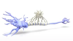
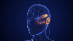
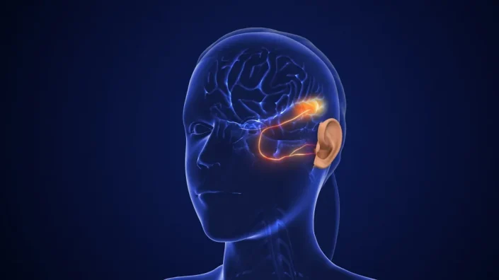
This ability is remarkable among vertebrates. Particularly, they are the only tetrapods able to regenerate their nearly-full limbs as adults. They can regenerate parts of their brains – in experiments with newts, if you remove half the optic tectum, it will grow back. It takes a long time, over six months, but it will grow back.

They are also able to regenerate their ovaries, and work from my talented student Yuliia Haluza looking at thousands of matings over 15 years in the Dresden axolotl colony indicates that the axolotl retains fertility through lifespan, in keeping with their extreme aging resilience.

multi

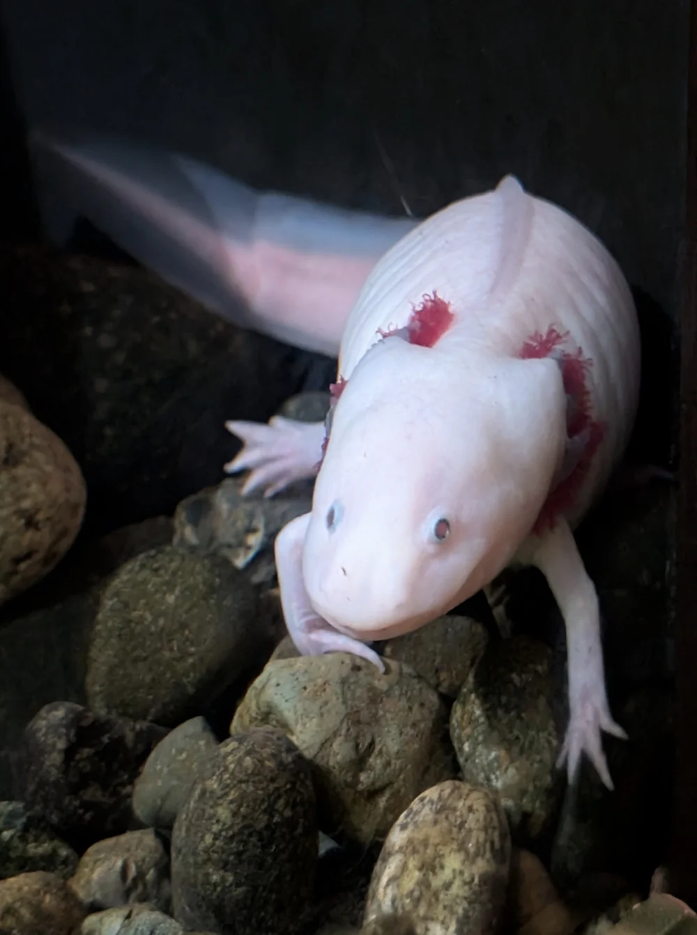
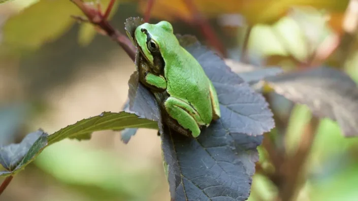
Axolotl at Vancouver Aquarium. Photo: Arkadi Mazin

They can also regenerate up to a third of their heart, their tail including the spinal cord, maxillary bones, gills. This is really remarkable. They even regrow structures they don’t necessarily need. For example, the axolotl never undergoes metamorphosis, but it can regrow its lung even though it’s never going to use the lung for breathing on land. It’s not selected for utility, which is interesting. It’s an example of residual regeneration.

They are powerful at regenerating, but there is a catch: in all known cases of regeneration – except for the newt lens, which is a particular example – you always needed the remnant of the structure in order to regrow it. However, our lab has just discovered that the limits of regeneration can be extended further: axolotls are able to regrow their thymus de novo. This means they can regrow their thymus completely from scratch, and it’s the one example of a complex organ that can be fully regrown among vertebrates! This is truly exciting, as it present a completely different regeneration paradigm and further highlights the power of this model for regeneration research.

With regards to ageing, this raises key questions too: does the thymus involute with time in axolotls, as it does in mammals? Is its age-related homeostasis enhanced due to the regenerative abilities? Largely, how does a super regenerator age? Many exciting avenues ahead of us.

When they regenerate their brains, are memory and learning preserved?

We don’t know yet. Until recently, salamanders were not fully experimentally tractable. For example, the axolotl genome only came out in 2018, and the first chromosome assembly for the Spanish ribbed newt – the most tractable newt model – came out just in 2025, an effort led by the labs of Nick Leigh, Andras Simon and mine. Compared with other model organisms, this is all too recent. However, it means we can now truly exploit the wide range of tools existing for more traditional systems. There are memory paradigms currently being developed by a few research groups, both for axolotls and newts, and we are all looking forward to the results.

I think their genome is about ten times larger than the human genome, right?

It is ten times larger. There were a lot of complicatons because most salamanders exhibit genomic gigantism. Largely due to massive expansions of repetitive elements, not genome duplication (actually, the axolotl and the Iberian ribbed newt are both diploid). As they are extremely large and highly repetitive, standard sequencing approaches did not work well. Recent advances in genome sequencing tech, particularly PacBio long-read sequencing, made this possible.

So, for geroscience, it’s a fairly new model, and they also usually have long lifespans, correct?

That’s true. Although different salamanders have very different lifespans. For axolotls, the average is 10 to 13 years of age while their maximum lifespan is about 20 or 21. Newts are significantly longer lived, and then cave olms, such as Proteus anguinus, live well over 100 years! They are all lifespan outliers based on their body size.

But we don’t know for sure, say, from your colonies?

It is in fact from our colonies! We had a 21-year-old, one of the longest-lived axolotls. When the model is so long-lived, it’s difficult to do the studies. With newts for example, there have been a lot of capture-recapture studies in the wild. Critically, whenever a salamander species has been studied in terms of lifespan, one thing is obvious: there is no increase in mortality rate with age.

So, that’s negligible senescence.

Exactly. We classify them as having negligible senescence because this has been tested in several species and none of them exhibit mortality increase with age. But nothing is known at the molecular level, and this is where our lab jumps in: in one huge project, my lab is characterizing how these animals age, and how this is different from senescent species such as us.

I’m wondering if there is a relationship between regeneration and negligible senescence in these species.

That’s exactly what we are working on right now. The first attempt to address this was through building epigenetic clocks, together with Steve Horvath’s team. We found that we can build an epigenetic clock for up to four years of aging in the axolotl, which is the early life. Past that point, it seems to stabilize epigenetically, a phenomenon we have never seen before and could relate to negligible senescence traits.

With the part of the clock that works after four years of age, we used this to ask what happens to limbs or tails which have undergone several regeneration cycles. The answer is that if you compare a forelimb that has regenerated three times versus the contralateral limb that has never regenerated, the one that has regenerated three times is epigenetically younger by DNA methylation age than the contralateral limb.

This suggests there might be some rejuvenation events associated with regeneration, which is exciting because it’s in a natural setting. This organism does it naturally; it’s not reprogramming with Yamanaka factors, and it raises multiple questions which we’re trying to address. The epigenetic and other tools we are currently developing will help providing important answers with regards to the interplay between regeneration and aging.

If I remember correctly, regeneration in forlimbs is linked to cellular senescence.

Cell senescence is elicited every time you regenerate a limb. It appears at particular stages during regeneration, and it contributes to fueling the process, particularly by secreting factors – for example, Wnt signals – that will promote plasticity (in newts) or proliferation of the neighboring cells (in axolotls).

As regeneration progresses, you cannot have these processes turned on forever. We have seen that the number of senescent cells starts to decline as the limb progresses through regeneration. This depends on clearance by the immune system, in particular the macrophages.

We know that senescent cells play a role in wound healing in mammals. Is this some sort of a remnant of this mechanism?

It’s not clear. If you read Marco Demaria’s work – he and the late Judith Campisi uncovered the roles of senescence in wound healing – they are acting by secreting PDGF factors that promote the more rapid migration of the cells to close the wound. This is a different mechanism from what we see in salamanders, which fuels formation and proliferation of the blastema progenitors.

We don’t know exactly if they are related. Whether there are commonalities between this context of “beneficial” senescence that are different from the “negative,” pro-inflammatory senescence you see in aging is a very interesting question. We would love to explore this more with others in the field.

Salamanders also appear to have a superior clearance mechanism for senescent cells, right?

The clearance we see is very effective. Not only do the dynamics of regeneration lead to regenerative limbs that have no senescent cells – you can try to find them, but you won’t – but they never linger. Not even a couple, which is remarkable. We know that macrophages are required for their clearance, but little beyond this. There is a great opportunity here to probe the basis of efficient clearance mechanism, which I am convinced could inform strategies to improve clearance in mammalian systems.

Senescent cells are proposed to play roles in limb development, is it also the case in salamanders?

This is also different from development. If you see a developing limb bud, you won’t see senescent cells, but if you induce regeneration in that developing limb bud, then you will see senescent cells. I sometimes get asked if regeneration is similar to development. I would say it is not a full recapitulation. There are many differences. Senescent cell intervention and usage is different, and there are many other aspects. Of course, some of the patterning programs used to regrow the structure are conserved between development and regeneration, but there are many differences, especially at the beginning of the process.

There’s also dedifferentiation, maybe transdifferentiation of cells during regeneration. It’s a really interesting phenomenon, something like cell reprogramming in vivo.

What we know is that salamanders are masters at regulating cellular identity and plasticity. The cells know very well what they are, and when they go back, they never forget what they are. When a limb regenerates, connective tissue only gives you connective tissue; muscle only gives you muscle.

For example, in the newt, the muscle is generated by dedifferentiation. Even though the muscle comes from dedifferentiation, the resulting regenerative progenitors never fully lose their muscle marks and they only give you muscle.

Experiments with genetic tracing, single-cell analysis, and transplantations tell you the specific tissues overall maintain their identity. Dedifferentiation is also critical for axolotl limb regrow, whereby the connective tissue cells dedifferentiate to give you all CT-derived tissues. This is one of the most important principles when these animals regenerate.

Transdifferentiation is another mechanism these animals use. For example, in the newt lens: you can remove the lens of the eye completely, and the entire lens will come back by a transdifferentiation of the dorsal pigmented epithelial cells from the iris. When you remove the lens, these pigmented epithelial cells undergo transdifferentiation to form crystalline cells that form the lens. This is one example where pure transdifferentiation is used.

But in the context of many other complex structures, we haven’t seen much of this transdifferentiation. There are recent reports, such as an exciting preprint from Wouter Masselink, Elly Tanaka and Prayag Murawala, which indicates that the tail mostly regenerates via specific stem-like progenitors. These stem cell-like cells can give you different tissues in the tail, but it’s a bit different; it’s like playing with the potency of a stem cell, not necessarily direct transdifferentiation as in the lens case.

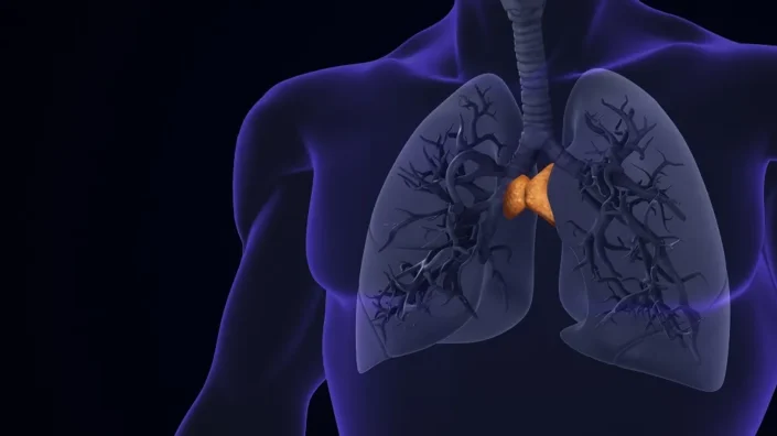
Your group is also working on thymus regeneration in salamanders. This is exciting since thymus involution clearly plays a role in human aging.

It is really amazing because, first, it’s something you can see with your own eyes. The thymus in the axolotl sits at the base of the gills, formed by three bilateral nodules. You can remove the entire nodules including the connective tissue surrounding them, and, in six to eight weeks, the entire organ will be back. It’s a complex organ because it’s formed by thymic epithelial cells, thymic progenitors, hematopoietic progenitors, dendritic cells, endothelium, macrophages, and obviously the different lymphocytes, and everything just reappears.

We found that when these animals regenerate the thymus, they recapitulate the morphology, cell populations, and function. One can do these very cool experiments where you transplant a regenerated structure from one salamander – a thymus nodule from a salamander that is fluorescently labeled – into a [transparently] white host. Then you can see if this regenerated thymus can support the hematopoiesis and lymphopoiesis of the host.

You can see that it does, because after a year or two, this thymus nodule will still have its original thymic epithelial cells fluorescently labeled, but it will be supporting the entire thymopoiesis of the host. It’s wonderful.

How can we translate insights from these species into humans? What are the main hurdles? I must confess that every time I speak with someone about long-lived or regenerating species, I get envious. I immediately want to have those abilities.

The first thing is to do solid basic science that gets deep into cellular and molecular mechanisms. Understand what really underlies these remarkable traits, and only then you can generate a blueprint for guiding interventions.

Regarding hurdles and timescales, these are intertwined. Primarily, you need to consider the physiology and biological differences between species. Sometimes you may have a target that seems sensible, but it’s actually tied to the particular species’ physiology.

In the case of salamanders, they are ectotherms. They live at different temperatures and control their energy differently. If they’re adapted to live at 20 degrees, can we adapt their mechanisms for an organism that lives at 37 degrees? We have to think about all this. Sometimes you can try to replicate these interventions by playing with the organism’s own proteome. That’s great because you are not subject to these differences.

The biggest hurdle is: do we need one hit or ten hits to reproduce this? But that is just trial and error, leveraging datasets and doing preclinical studies. We have started to take some of our thymus findings into other models to see if we can promote thymus rejuvenation or delay involution by giving them these molecular features that the axolotl has. We’re already doing it, and it’s very exciting.

It’s great to know that you are already looking at the translational angle.

Very much so. This goes hand-in-hand with generating a solid fundamental basis.

What about the evolutionary aspect? Do we know why salamanders have preserved regenerative abilities and mammals lost them almost completely?

It is not necessarily clear in the field of regeneration whether regeneration is a conserved trait of all animals or an acquired trait and an example of convergent evolution. A number of scientists, including my postdoctoral mentor Jeremy Brockes, sustained the latter, supported by the fact that there are salamander-specific proteins involved in regeneration. It is quite possible that regeneration has evolved in salamanders in a different way than in other organisms. For example, if you look at Hydra or planarians, they regenerate in very different ways. It’s not that all organisms that regenerate use the same mechanisms. Different organisms have different solutions.

So, it’s not necessarily settled that we lost the ability to regenerate. There are still many views regarding the evolution of regeneration. Among them is the cancer connection – that organisms that regenerate very well would be more prone to cancer, but that’s not the case in salamanders. They’re actually very resistant to cancer.

They seem to have all the bases covered somehow.

Exactly. And if you think of salamanders as an organism, they occupy almost every niche. They are really evolutionarily successful. For example, you can find salamanders in Siberia. You have newts in the tundra that get frozen and stay dormant in a block of ice for long periods, but when the ice melts they just walk away. There’s a lot of adaptability there.

Obviously, humans are a threat to salamanders worldwide. The axolotl is a good example; in Lake Xochimilco, human activity resulted in water contamination from rapid urbanization and the introduction of carps into the system. They started to wipe out axolotl populations in the natural environment to the point of near extinction in their natural environment (though they exist in high numbers in pet shops and colonies!). Right now, there are efforts towards bringing them back, and we are all rooting for this.

Do you have an opinion on Michael Levin’s bioelectricity-focused research into regeneration?

Bioelectricity is very important. There were early studies in the sixties and seventies showing that reverting currents in the water prevents for example, newt limb regeneration. So, we know that bioelectricity plays an important role. It’s a way in which cells communicate. I think Mike’s work is very interesting. For the field, we want to see this expanded into how the different molecular networks impact bioelectricity and how this is coordinated during regenerative processes. It’s a very interesting angle.

You moved to China recently. How do you see China’s place in the modern geroscience landscape, and how is the research and biotech climate different from Europe?

As in many other sectors, China’s influence is growing at speed. The trend is for science to be heavily influenced by China soon. I think the government has made very right steps in investing in science and deciding China should become the next scientific powerhouse. The environment here is certainly very welcoming, increasingly so towards internationals.

The current international context has led to a significant import of foreign scientists, which are in turn strengthening China’s position worldwide. There are significant funding opportunities here, for both basic research and the biotech sphere, combined with the chance to tap into a pool of talented and super hardworking students and staff.

Things happen at a very fast pace, and the regulatory frameworks for experimentation are much more science-friendly than in Europe. There is still perhaps too much focus placed on high-impact publications, but there is a clear effort towards promoting rigorous science. I’ve been here for four months, so we’ll see how things develop, but what I’m experiencing so far is very exciting.

Do you see the agility that is often invoked with regard to China – things being done faster, more efficiently?

Things are being done much faster than anything I have ever experienced. Sometimes if you have too much speed, that can be counterproductive. But institutes like the one I am in, Chinese Institutes for Medical Research (CIMR), are set up with more than 90% of the faculty coming from Europe and the US, so it’s a bit different.

Personally, I was attracted by the idea conceived by Yi Rao and others to create an HHMI-style institute in Beijing. Here, the support system and evaluation processes are based on the success stories that happen in the West, modeling institutes like the Laboratory of Molecular Biology in Cambridge, where I did my PhD, and Janelia Farm.

One of the features of such places is also the possibility to have resources and time to explore great, bold ideas, and I feel this component is important for doing significant science. Overall, there is a view that this will lead to success here.

We would like to ask you a small favor. We are a non-profit foundation, and unlike some other organizations, we have no shareholders and no products to sell you. All our news and educational content is free for everyone to read, but it does mean that we rely on the help of people like you. Every contribution, no matter if it’s big or small, supports independent journalism and sustains our future.

Aging Evolution Book

Aging: Why Does Evolution Kill?

Why do we age at all, and why do different species age at such dramatically different rates? After decades of longevity research, biologists still disagree on the most basic questions: What is aging? Is it possible to stop or reverse it? And what strategies stand a real chance of working?

Most gerontologists explain aging as the gradual accumulation of cellular and molecular damage, much like rust in a car or the decay of a house. But this viewpoint struggles to explain a series of biological paradoxes. Tiny sparrows can live twenty years. Naked mole-rats survive twice as long. Hydra and planaria appear not to age at all. Queens in eusocial species can live fifty times longer than workers with the same genome. Some animals–jellyfish, comb jellies, and eusocial insects–can rejuvenate, yet they do so only under stress, not under ideal conditions. If youth is mechanistically available, why do they “choose” aging and death?

A new book, titled Aging: Why Does Evolution Kill?, written by Hong Kong–based professor Peter Lidsky, and published with the support of Open Longevity, offers a bold, non-orthodox answer. The book argues that classical evolutionary theories of aging are inconsistent with recent empirical and theoretical results–and develops instead a novel pathogen control theory of aging, in which aging is not just damage, but an evolved, adaptive program.

Early evolutionary thinking, dating back to August Weismann in the late 19th century, proposed that aging is a programmed process that removes maimed individuals from a population. This view fell out of favor because it relied on group selection: individuals supposedly sacrifice their own fitness for the good of the species, a mechanism later considered too weak to explain such costly traits. As a result, for much of the 20th century, theories of programmed adaptive aging were largely abandoned, and non-programmed, damage-based explanations took over.

Lidsky’s pathogen control theory revives the idea of programmed aging but grounds it in kin selection, the same well-accepted evolutionary force that explains parental care. As J.B.S. Haldane quipped, he would give his life “for two brothers or eight cousins”— a vivid illustration of kin selection, in which an individual may sacrifice even its life if this helps relatives, who share its genes, to survive and reproduce.

How, then, could death from aging ever help one’s “two brothers or eight cousins”? The book’s central claim is that the missing piece is chronic, sterilizing infections–pathogens such as syphilis or gonorrhea in humans, which do not kill quickly but prevent reproduction. Individuals carrying such infections become evolutionarily “worthless”: they cannot have offspring, and they can transmit these infections to relatives, harming their genetic interests. In this context, the early death of infected, non-reproductive individuals can be favored by kin selection.

Because the probability of acquiring such infections increases with time, evolution, according to the pathogen control theory, can favor mechanisms that remove older individuals as a function of age. In this view, aging is an immune strategy: a program that sacrifices older individuals to protect their kin from the infections they accumulate over long lives.

This perspective leads to a series of striking, testable predictions. If aging evolved to protect relatives from infection, then the population structure–who interacts and infects whom–becomes a major determinant of lifespan and aging patterns. The book argues that many “outlier” species fit this logic: eusocial insects, naked mole-rats, salmon, flying birds, and bats all have atypical population structures that can explain their unusual aging and death schedules.

In the closing chapters, Aging: Why Does Evolution Kill? explores the implications of the pathogen control theory for modern gerontology. It places aging squarely within the context of the immune system and outlines new research directions that could reshape how we think about interventions to slow or reverse aging. The book presents an ongoing research program: many of its hypotheses remain to be rigorously tested, and readers are invited to evaluate the theory critically.

Whether or not one ultimately accepts the pathogen control theory, this book offers a provocative rethinking of one of biology’s most fundamental problems. It will interest researchers, clinicians, and lay readers concerned with aging and longevity.

https://www.amazon.com/dp/B0G4R3DDH6

We would like to ask you a small favor. We are a non-profit foundation, and unlike some other organizations, we have no shareholders and no products to sell you. All our news and educational content is free for everyone to read, but it does mean that we rely on the help of people like you. Every contribution, no matter if it’s big or small, supports independent journalism and sustains our future.
Genetics

A Gene That Changes Function With Aging in Mice

In Nature Aging, researchers have discovered growth differentiation factor 3 (GDF3), a cytokine that increases with aging, is related to more inflammatory macrophages in older animals.

Fat and inflammation

We have recently reported on a potential treatment for visceral fat, as this kind of fat is known to generate health problems, including an increase in inflammation [1]. Macrophages are the cause of some of this inflammation, which also makes fat loss more difficult; this effect has been linked to the GDF3 signaling axis [2].

GDF3 has been heavily studied in multiple contexts, including aging. Unsurprisingly, other research has linked it to fat gain [3], but it has also been found to be effective against blood sepsis [4] and even encourages muscle regeneration in older organisms [5]. As a member of the TGF-β superfamily, it acts on SMAD, a group of molecules that modify gene expression and help to manage chromatin [6], which governs the availability of DNA.

GDF3 changes toxic shock response

In their first experiment, the researchers challenged young and old mice with lipopolysaccharide (LPS), a toxic compound that encourages inflammation. Unsurprisingly, the old mice reacted more strongly than the younger mice, reducing their body temperatures and increasing their numbers of inflammatory macrophages compared to other macrophage types. Gdf3, correspondingly, also substantially increased in the older mice.

The researchers then created a breed of mice that don’t express Gdf3. These mice had few differences from their unmodified counterparts, including in adipocytes, and there were no changes in metabolism. However, among older animals, their numbers of inflammatory macrophages were significantly lower than those of wild-type mice, and they did not have the inflammatory phenotype that makes fat burning difficult. Old Gdf3-knockout mice also appeared to have healthier responses to LPS than old wild-type mice; younger mice saw no benefit.

Animals that had Gdf3 knocked out of only their bone marrow (myeloid) cells had significant reductions in multiple inflammatory factors, including IL-1b and IL-6. In old age, these animals also had significant improvements in glucose metabolism, a better ability to burn fat, and less strong reactions to LPS.

The researchers then attempted to treat GDF3 in older mice by using JQ1 to inhibit BDF4, which binds to GDF3. Older mice treated with JQ1 did not develop hypothermia upon LPS injection the way younger mice did, and they had fewer inflammatory macrophages. These results suggest that GDF3 is treatable.

Changing what genes are accessible

These results were found to be directly related to SMAD. Increasing GDF3 levels also increased the phosphorlyzation of SMAD2/3, which was found to lead to the increased inflammation in macrophages. The researchers confirmed this by directly suppressing SMAD3, which stopped the negative effects of GDF3 in the macrophages of old mice but did not affect younger mice. This suppression also inhibited other gene expressions in young mice, but those effects were not found in the older mice, leading the researchers to conclude that SMAD3, and GDF3, affect different pathways with aging.

Further work found that chromatin remodeling was a significant part of this change. Comparing old murine macrophages with and without Gdf3 revealed significant differences in these cells’ chromatin, which significantly altered which genes were accessible. Not only was there a significant decrease in chromatin-related inflammation in the Gdf3-knockout group, there was significant overlap between genetic pathways that were more accessible in the Gdf3-expressing macrophages and genetic pathways related to aging.

The researchers admit their study’s limitations, most notably that this research only involved mice and murine cells, with no human cells being used. Additionally, GDF3 serves vital biological functions, including in the immune system, and its effects on human beings may be different from those on lab mice kept in a controlled environment. Further work will determine if people have the same age-related changes in GDF3/SMAD function as mice do, along with whether or not this compound can be targeted to fight inflammaging and help people live longer.

We would like to ask you a small favor. We are a non-profit foundation, and unlike some other organizations, we have no shareholders and no products to sell you. All our news and educational content is free for everyone to read, but it does mean that we rely on the help of people like you. Every contribution, no matter if it’s big or small, supports independent journalism and sustains our future.

Literature

[1] Carey, A., Nguyen, K., Kandikonda, P., Kruglov, V., Bradley, C., Dahlquist, K. J., … & Camell, C. D. (2024). Age-associated accumulation of B cells promotes macrophage inflammation and inhibits lipolysis in adipose tissue during sepsis. Cell reports, 43(3).

[2] Camell, C. D., Sander, J., Spadaro, O., Lee, A., Nguyen, K. Y., Wing, A., … & Dixit, V. D. (2017). Inflammasome-driven catecholamine catabolism in macrophages blunts lipolysis during ageing. Nature, 550(7674), 119-123.

[3] Wang, W., Yang, Y., Meng, Y., & Shi, Y. (2004). GDF-3 is an adipogenic cytokine under high fat dietary condition. Biochemical and biophysical research communications, 321(4), 1024-1031.

[4] Wang, P., Mu, X., Zhao, H., Li, Y., Wang, L., Wolfe, V., … & Fan, G. C. (2021). Administration of GDF3 into septic mice improves survival via enhancing LXRα-mediated macrophage phagocytosis. Frontiers in immunology, 12, 647070.

[5] Patsalos, A., Simandi, Z., Hays, T. T., Peloquin, M., Hajian, M., Restrepo, I., … & Nagy, L. (2018). In vivo GDF3 administration abrogates aging related muscle regeneration delay following acute sterile injury. Aging cell, 17(5), e12815.

[6] Bertero, A., Brown, S., Madrigal, P., Osnato, A., Ortmann, D., Yiangou, L., … & Vallier, L. (2018). The SMAD2/3 interactome reveals that TGFβ controls m6A mRNA methylation in pluripotency. Nature, 555(7695), 256-259.