The Blog

Building a Future Free of Age-Related Disease

Close-look-at-documents.jpg

Analyzing the Quality of Preclinical Anti-Aging Research

Researchers have investigated the reporting quality of preclinical studies’ outcomes in anti-aging research. They analyzed how study quality changed over time, shortcomings in research, and the improvements that can be made in the future in order to yield as many valuable insights as possible [1].

The need for quality reporting aging research

Aging research has grown substantially; however, conducting human trials in the aging field is time-consuming and requires substantial resources. Therefore, initial testing is done in preclinical models, such as mice, worms, fruit flies, and other model animals, as many genes, molecular processes, and aging mechanisms are conserved between those animals and humans [2]. To increase the likelihood of translating results from animal models to humans, high-quality studies are essential.

To assess the quality of preclinical studies in the anti-aging field, the researchers analyzed 667 studies published in peer-reviewed journals between 1948 and 2024, which included 720 experiments, from the DrugAge database. This is “a curated database of preclinical experiments investigating the effects of interventions on aging and lifespan in non-human animals.” The analyzed studies varied in the animal species they used, with a small fraction including more than one model organism. The researchers aimed to assess the quality of reporting, methodological rigor, the distribution of observed effect sizes, and the presence of biases in those studies.

Assessing quality

The researchers assessed the studies using the CAMARADES (Collaborative Approach to Meta-Analysis and Review of Animal Data from Experimental Studies) score. This score, which usually involves scoring studies accordingly to a 10-item checklist, allows for assessing methodological quality and risk of bias.

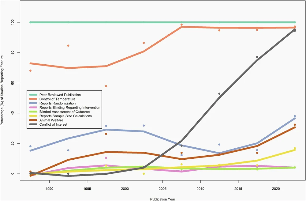
The median CAMARADES score across the analyzed studies was 3, but the researchers observed differences depending on the species used. Only two assessed parameters were consistent across all studies. First, all studies went through peer review. Second, blinding was generally absent. Specifically, blinding to intervention was discussed in only 4% of studies and blinded assessment of outcomes in 3%.

Among the assessed parameters, the researchers noted that almost one-fifth of studies mention randomization. Randomization, along with sample size calculation, was rarely reported when Caenorhabditis worms and Drosophila fruit flies were used, and overall, it was uncommon, with only 6% of all studies reporting it. However, studies using Caenorhabditis and Drosophila almost always gave information regarding the temperature at which animals were undergoing experiments. Temperature information was also common across all experiments, regardless of species used, with over 90% of analyzed studies reporting it. Those who didn’t report it mainly used mice. However, mouse studies did better with other parameters assessed by the researchers.

Other measured parameters included animal welfare, reported in 13.9% of studies, and conflict of interest statements, reported in more than half of the studies.

Since the studies used in the analysis spanned eight decades, the researchers analyzed how reporting changed over time. They noted that reporting of some parameters, especially conflicts of interest, compliance with animal welfare regulations, temperature control, and sample size calculations, increased over time, contributing to an increase in the CAMARADES score. However, there was no significant increase in reporting of randomization and blinding.

multi

The critical parameters

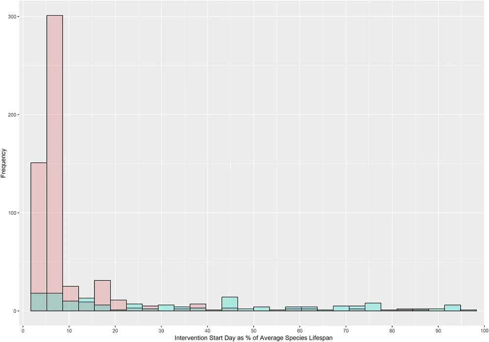
The critical parameter for anti-aging interventions is the timing of initiation, since there is a need for effective interventions that can extend lifespan when taken in mid-life or in the elderly, and the intervention’s effect might differ depending on the start time. However, among the analyzed experiments, the vast majority (over 80%) begin early in life, while only around 8% start at 50% of average lifespan or later, a gap that future studies should address. The researchers also note that, in the pre-clinical studies analyzed, mammal experiments tend to start later in lifespan than non-mammal experiments.

Another critical component in aging research is the animal’s sex. It is well known that there are sex-dependent differences in aging trajectories, and interventions should be assessed in both sexes, as they may respond differently to the same treatment. However, among the experiments analyzed by the authors that included animals that reproduce sexually, fewer than half used both sexes; 35.7% used only males, 12.9% used only females, and some didn’t report the sexes used at all.

multi

Anti-aging compounds

The researchers noted that, among the studies in the DrugAge database, most compounds tested in non-mammalian models increased lifespan.

Additionally, the researchers compared the results between the mammalian and non-mammalian models. They noted that of 35 compounds tested in both mammalian and non-mammalian models, 21 significantly increased lifespan in non-mammalian models, but only one-third of those also significantly increased mammalian lifespan: curcumin, spermidine, epithalamin, D-glucosamine, estradiol, SKQ, and taurine. At the same time, two showed inconsistent results when compared to non-mammalian models, decreasing mammalian lifespan (quercetin and butylated hydroxytoluene). This suggests that in the case of those experiments, “non-mammal results do not seem to reliably predict mammal results, raising further concern for translation.”

The experiments in mammalian and non-mammalian models also differed in other parameters across compounds, including the median percentage increase in lifespan, which was smaller in mammalian models at 7.4% than in non-mammalian models at 17.5%.

Room for improvement

This study suggests that there is room for improvement in the way preclinical antiaging research is performed. The researchers noted that “important design features such as randomization, blinding of intervention, blinded assessment of outcome, compliance with animal welfare regulations, and sample size calculations were infrequently reported, despite evidence that the absence of such features can bias experimental results.” [3,4,5] Some of the most essential experiment design features, such as randomization and blinding, didn’t see substantial improvements over time. They conclude that “generally, most studies did not meet standard reporting guidelines for preclinical experiments.”

While this is not an excuse for failing to meet the standards necessary for high-quality research, those flaws are not limited to anti-aging research, as many studies addressing various diseases exhibit similar reporting and study design problems [6], suggesting a need for improvement.

We would like to ask you a small favor. We are a non-profit foundation, and unlike some other organizations, we have no shareholders and no products to sell you. All our news and educational content is free for everyone to read, but it does mean that we rely on the help of people like you. Every contribution, no matter if it’s big or small, supports independent journalism and sustains our future.

Literature

[1] Parish, A., Ioannidis, J. P. A., Zhang, K., Barardo, D., R Swindell, W., & de Magalhães, J. P. (2025). Reporting quality, effect sizes, and biases for aging interventions: a methodological appraisal of the DrugAge database. npj aging, 11(1), 96.

[2] Kenyon C. (2001). A conserved regulatory system for aging. Cell, 105(2), 165–168.

[3] Schulz, K. F., Chalmers, I., Hayes, R. J., & Altman, D. G. (1995). Empirical evidence of bias. Dimensions of methodological quality associated with estimates of treatment effects in controlled trials. JAMA, 273(5), 408–412.

[4] Schulz, K. F., & Grimes, D. A. (2002). Blinding in randomised trials: hiding who got what. Lancet (London, England), 359(9307), 696–700.

[5] Kringe, L., Sena, E. S., Motschall, E., Bahor, Z., Wang, Q., Herrmann, A. M., Mülling, C., Meckel, S., & Boltze, J. (2020). Quality and validity of large animal experiments in stroke: A systematic review. Journal of cerebral blood flow and metabolism : official journal of the International Society of Cerebral Blood Flow and Metabolism, 40(11), 2152–2164.

[6] Kilkenny, C., Parsons, N., Kadyszewski, E., Festing, M. F., Cuthill, I. C., Fry, D., Hutton, J., & Altman, D. G. (2009). Survey of the quality of experimental design, statistical analysis and reporting of research using animals. PloS one, 4(11), e7824.

Mitochondria folds

Senescence Exacerbated by Mitochondrial RNA Leakage

Scientists have investigated a little-known mechanism that fuels cellular senescence: mitochondrial RNA leaking into the cytoplasm. Targeting this mechanism showed promise in a mouse model of fatty liver [1].

The new target

The increasing abundance of senescent cells with age has been linked to numerous diseases and is considered a hallmark of aging. Understanding why cells become senescent, and how we can either save them from this fate or mitigate the consequences, is an important target for geroscience.

It has been known that mitochondria in senescent cells leak mitochondrial DNA into the cytoplasm. The cells’ defense mechanisms often mistake it for viral DNA and trigger response mechanisms that exacerbate the cell’s senescent or pre-senescent state [2]. In a new study coming from Mayo Clinic and published in Nature Communications, a team of scientists focused on a different, much less-studied mechanism: the leakage of mitochondrial RNA.

Mitochondria have their own small circular genomes that encode several proteins essential for the organelle’s function. While the transcription and translation of mtDNA differ from those of nuclear DNA, the basics are the same: DNA is transcribed into RNA and then translated into proteins by ribosomes.

Mitochondrial RNA (mtRNA) can sometimes form double-stranded RNA (mtdsRNA), such as when complementary “sense” and “antisense” mitochondrial transcripts overlap. Because cytosolic dsRNA is a classic viral-like danger signal, its appearance outside mitochondria can trip the cell’s antiviral RNA sensors and set off an inflammatory “fire alarm”, which is similar in spirit to mtDNA leakage but occurs via a different sensing pathway [3].

The mtRNA-senescence connection

The team found that mtdsRNA levels are higher in the cytosol of senescent cells (specifically, fibroblasts), which sets off the RNA sensors RIG-I and MDA5. This happened across multiple senescence triggers (replicative, doxorubicin/etoposide) and cell lines. The levels of those RNA sensors also increase with age in multiple mouse tissues, along with the senescence markers p16 and p21 in addition to SASP factors.

To determine cause and effect, the researchers injected non-senescent fibroblasts with purified mitochondrial RNA, which likely contained and/or generated mtdsRNA. This boosted common SASP factors and RNA sensors, suggesting that the presence of mtRNA in the cytosol is enough to drive the SASP program.

They then did something roughly opposite, depleting mitochondria from already senescent cells. As a result, the cells stopped producing the SASP while still staying senescent. The researchers then added purified mtRNA to the cells to see what its impact would be in the absence of working mitochondria. In these mitochondria-depleted senescent cells, mtRNA add-back partially restored interferon/NF-κB inflammatory transcriptional programs – a key element of SASP regulation – rather than fully restoring SASP secretion.

The researchers then pharmacologically reduced mtRNA production in senescent cells by inhibiting mitochondrial RNA polymerase (POLRMT). This lowered cytosolic mtdsRNA, reduced RNA sensors and several SASP components, but did not lower p16 and p21, which can be interpreted as dampening SASP without reverting senescence. Interestingly, blocking STING to blunt mtDNA sensing reduced SASP more than blocking MAVS to blunt mtRNA sensing, and doing both didn’t help further, suggesting that these pathways overlap, with cGAS-STING likely being the main driver of SASP here.

The researchers suspected that mtRNA escape was driven by pores formed by the proteins BAX and BAK in a subset of mitochondria. Indeed, deleting both BAX and BAK reduced cytosolic mtRNA, lowered RNA sensors, reduced MAVS aggregation, and suppressed SASP components.

In vivo validation

The team validated their findings in a mouse model of metabolic dysfunction-associated steatohepatitis (MASH), a dangerous and increasingly prevalent subtype of fatty liver that is often triggered by obesity. They found increased RNA-sensing/SASP signals in livers of MASH mice and then showed that either hepatocyte-targeted Bax deletion or hepatocyte-targeted MAVS knockdown dampens inflammatory and fibrotic markers.

“Liver scarring and inflammation are hallmarks of MASH,” said Stella Victorelli, Ph.D., who is the lead author of the study. If left untreated, it can progress to liver cancer. This is why it’s so important to understand the mechanisms driving the disease so that we can prevent it or develop more effective treatments.”

“With age, we accumulate ‘zombie’ cells, which can lead to more disease,” added João Passos, Ph.D., senior author of the study. “Our idea is that if we can quiet these cells earlier, we can prevent runaway inflammation and the development of many age-related conditions, including liver disease. Understanding the mechanisms that drive disease allows us to target and delay those processes – potentially benefiting more than one condition.”

We would like to ask you a small favor. We are a non-profit foundation, and unlike some other organizations, we have no shareholders and no products to sell you. All our news and educational content is free for everyone to read, but it does mean that we rely on the help of people like you. Every contribution, no matter if it’s big or small, supports independent journalism and sustains our future.

Literature

[1] Victorelli, S., Eppard, M., Martini, H., et al. (2025). Mitochondrial RNA cytosolic leakage drives the SASP. Nature Communications, 16, 10992.

[2] Victorelli, S., Salmonowicz, H., Chapman, J., Martini, H., Vizioli, M. G., Riley, J. S., … & Passos, J. F. (2023). Apoptotic stress causes mtDNA release during senescence and drives the SASP. Nature, 622(7983), 627-636.

[3] Dhir, A., Dhir, S., Borowski, L. S., Jimenez, L., Teitell, M., Rötig, A., … & Proudfoot, N. J. (2018). Mitochondrial double-stranded RNA triggers antiviral signalling in humans. Nature, 560(7717), 238-242.

Obese mouse

Targeting a Metabolic Regulator Reduces Fat in Mice

In Aging, researchers have described how they removed visceral fat from older male mice by targeting the metabolic regulatory protein CD47.

A key regulator of metabolism

Visible fat carried around the body is subcutaneous fat. While this kind of fat is obviously not healthy to have in large amounts, it is not as immediately dangerous as visceral fat, which accumulates around organs and drives many metabolic diseases [1]. This metabolic damage is often associated with similar age-related problems [2], including sarcopenia, a loss of muscle mass that leads to frailty [3].

To combat visceral fat, the researchers focus on CD47, a multifunctional membrane protein that increases during aging and is known to drive several age-related disorders, including a loss of the ability to create new blood vessels [4]. Cancers also use it to protect themselves from the immune system [5].

Most relevant to this research, however, is the fact that excessive CD47 has been repeatedly found to lead to age-related metabolic disorders in animal models, including obesity and diabetes [6]. These researchers previously found that a CD47 deficiency leads to the browning of white fat, putting it into a state in which it can be burned for heat, and then encourages that burning [7]. Curiously, further work found that these results only appear to apply to male animals [8].

A potential treatment for fat generation

This work takes that previous research a bit further; while the previous work used modified mice, this uses an actual treatment: an antisense oligonucleotide that specifically targets CD47 (CD47 ASO). A group of 20-month-old wild-type Black 6 mice was injected twice a week, along with a saline-injected group and a control ASO group.

Overall body weight did not significantly change between the groups. Total fat mass, on the other hand, was significantly decreased in the CD47 ASO group compared to either of the controls, and this was accompanied by a significant loss of visceral fat. multi
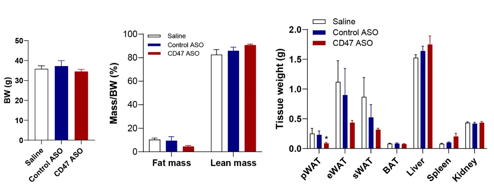
There was also a marked improvement in diabetes-related biomarkers; the treated mice were better able to handle glucose, and HOMA-IR, a biomarker of insulin sensitivity, was improved. The fat cells themselves had become smaller with this treatment as well.

These findings were accompanied by marked changes in gene expression. While many genes related to fat usage were unchanged, genes related to the formation of fat were significantly downregulated in the CD47 ASO group. Additionally, there appeared to be upregulation of genes related to the anti-inflammatory M2 macrophage type, although most genes related to the pro-inflammatory M1 type were unaffected.

The lack of CD47 discouraging fat cells from forming was confirmed in a cellular study. The researchers grew and differentiated fat cells for 15 days, subjecting some of them to CD47 ASO. The cells so treated were not significantly affected for the first week, but by the end of this experiment, at which point the cells had become senescent due to replication, lipogenesis-related genes were significantly downregulated. A closer examination revealed a more nuanced finding: this approach appears to encourage cells to differentiate into fat cells while preventing senescent cells from accumulating fat.

The researchers noted the very specific effects of this treatment: while there was a slight improvement in the liver’s ability to metabolize glucose, CD47 ASO had no apparent effects on skeletal muscle and other tissues. These effects were sex-specific in mice, and it is not yet clear if they apply to human beings; a clinical trial would need to be done to determine if targeting CD47 could be effective in helping elderly men or women to lose weight.

We would like to ask you a small favor. We are a non-profit foundation, and unlike some other organizations, we have no shareholders and no products to sell you. All our news and educational content is free for everyone to read, but it does mean that we rely on the help of people like you. Every contribution, no matter if it’s big or small, supports independent journalism and sustains our future.

Literature

[1] Shuster, A., Patlas, M., Pinthus, J. H., & Mourtzakis, M. (2012). The clinical importance of visceral adiposity: a critical review of methods for visceral adipose tissue analysis. The British journal of radiology, 85(1009), 1-10.

[2] Tam, B. T., Morais, J. A., & Santosa, S. (2020). Obesity and ageing: Two sides of the same coin. Obesity Reviews, 21(4), e12991.

[3] Nishikawa, H., Asai, A., Fukunishi, S., Nishiguchi, S., & Higuchi, K. (2021). Metabolic syndrome and sarcopenia. Nutrients, 13(10), 3519.

[4] Ghimire, K., Li, Y., Chiba, T., Julovi, S. M., Li, J., Ross, M. A., … & Rogers, N. M. (2020). CD47 promotes age-associated deterioration in angiogenesis, blood flow and glucose homeostasis. Cells, 9(7), 1695.

[5] Sun, J., Chen, Y., Lubben, B., Adebayo, O., Muz, B., & Azab, A. K. (2021). CD47-targeting antibodies as a novel therapeutic strategy in hematologic malignancies. Leukemia Research Reports, 16, 100268.

[6] Maimaitiyiming, H., Norman, H., Zhou, Q., & Wang, S. (2015). CD47 deficiency protects mice from diet-induced obesity and improves whole body glucose tolerance and insulin sensitivity. Scientific reports, 5(1), 8846.

[7] Li, D., Gwag, T., & Wang, S. (2021). Absence of CD47 maintains brown fat thermogenic capacity and protects mice from aging-related obesity and metabolic disorder. Biochemical and biophysical research communications, 575, 14-19.

[8] Li, D., Gwag, T., & Wang, S. (2023). Sex differences in the effects of brown adipocyte CD47 deficiency on age-related weight change and glucose homeostasis. Biochemical and biophysical research communications, 676, 78-83.

Chocolate

Molecule From Chocolate Linked to Slower Epigenetic Aging

A new observational study spanning two human cohorts has found a link between theobromine, a phytochemical abundant in cocoa, and slower epigenetic aging [1].

A yummy geroprotector?

Popular wisdom has it that not many things in life are both healthy and delicious, but according to research, one of them is chocolate. Studies have repeatedly linked chocolate and cocoa consumption with improved health outcomes, including beneficial effects on cholesterol, inflammation, and cellular senescence [2].

Cocoa and coffee share a family of alkaloids called methylxanthines, which includes caffeine, theophylline, paraxanthine, theobromine, and 7-methylxanthine. Coffee is caffeine-heavy, while cocoa is theobromine-heavy.

Theobromine has been reported to extend lifespan in certain strains of C. elegans nematode worms [3] and has been linked to cardiovascular and other health benefits in observational human data [4]. A new study conducted by scientists from King’s College London and several German institutions, published in Aging, looks at theobromine in the context of human aging by analyzing the correlation between the chemical’s blood levels and biological age acceleration.

Two clocks confirm slower aging

The team used two metrics: GrimAge, one of the most robust methylation clocks and trained to predict mortality, and DNAmTL, a DNA methylation-based estimator of telomere length. The latter predicts telomere length in leukocytes from methylation at a specific set of CpG sites.

The researchers pulled together two population cohorts in which people had both blood metabolomics and DNA methylation data. TwinsUK, which includes 509 women with a median age of around 60, served as the discovery cohort. The results were then replicated in KORA, a cohort of 1,160 German adults of both sexes.

They started by measuring the association between the difference between the GrimAge reading and the person’s chronological age (clock acceleration) and six coffee/cocoa-related metabolites: five methylxanthines and the amino acid theanine.

Theobromine immediately stood out, showing the strongest negative correlation. In the researchers’ main model, higher theobromine levels were linked to roughly 1.6 years less GrimAge acceleration per standard step up in theobromine. Theobromine levels were also associated with higher DNAmTL readings, suggesting slower telomere attrition.

Testing the results

The researchers then extensively stress-tested these results. To see if this was just a generic “coffee drinker” signal, they added caffeine and its breakdown products to the model and asked whether the theobromine effect would disappear. It did not; the association with slower GrimAge acceleration stayed, becoming only slightly weaker.

Next, they used penalized regression models (LASSO and elastic net), which automatically shrink or drop less informative variables. Even under these harsher conditions, theobromine consistently remained one of the key predictors of GrimAge acceleration.

In TwinsUK, metabolomics and methylation measurements could be taken up to 5 years apart, so the researchers re-ran the analyses within narrower time windows to see how the association would change. The shorter the distance between the two readings (latency), the stronger the effect size became, strengthening their confidence that this was not just an artifact of long gaps between measurements.

In the KORA replication cohort, higher serum theobromine again tracked with younger epigenetic profiles. After adjustment for age, BMI, blood cell composition, technical factors, and the other methylxanthines, each standard step up in theobromine was linked to about one year of reduced GrimAge acceleration and to slightly longer telomeres.

Because the discovery cohort consisted entirely of women, the authors checked whether the pattern held in women from the KORA study and found a similar, slightly weaker association there, reinforcing their original discovery. In the full KORA cohort, which includes both men and women, the overall effect of theobromine on GrimAge was actually stronger than in women alone. This suggests that men also show a negative association between theobromine and epigenetic age that is at least as strong, if not stronger, than in women.

Dr. Ramy Saad, lead researcher at King’s College London, who is also a researcher at University College London and holds a doctorate in clinical genetics, said: “This is a very exciting finding, and the next important questions are what is behind this association and how can we explore the interactions between dietary metabolites and our epigenome further? This approach could lead us to important discoveries towards ageing, and beyond, in common and rare diseases.”

Dr. Ricardo Costeira, a Postdoctoral Research Associate from King’s College London, added: “This study identifies another molecular mechanism through which naturally occurring compounds in cocoa may support health. While more research is needed, the findings from this study highlight the value of population-level analyses in aging and genetics.”

Caveats and limitations

Being observational and cross-sectional, this study cannot prove a causal relationship between theobromine and aging or even the methylation-based aging metrics that the researchers used. While they adjusted for age, BMI, blood cell counts and several related metabolites, residual confounding by factors like overall diet, lifestyle, socioeconomic status, or other cocoa components, especially flavanols, remains a real possibility.

Both cohorts are European and middle-aged/older, and the discovery sample is composed entirely of female twins, which limits generalization to men, younger people, and other ancestries. Finally, epigenetic clocks and DNAm-based telomere estimates are useful but still imperfect proxies for biological aging. Nevertheless, it’s more encouraging news for chocolate lovers.

We would like to ask you a small favor. We are a non-profit foundation, and unlike some other organizations, we have no shareholders and no products to sell you. All our news and educational content is free for everyone to read, but it does mean that we rely on the help of people like you. Every contribution, no matter if it’s big or small, supports independent journalism and sustains our future.

Literature

[1] Bell, J., Saad, R., Costeira, R., Garcia, P. M., Villicaña, S., Gieger, C., … & Waldenberger, M. (2025). Theobromine is Associated with Slower Epigenetic Ageing. Aging.

[2] Arranz, S., Valderas‐Martinez, P., Chiva‐Blanch, G., Casas, R., Urpi‐Sarda, M., Lamuela‐Raventos, R. M., & Estruch, R. (2013). Cardioprotective effects of cocoa: Clinical evidence from randomized clinical intervention trials in humans. Molecular Nutrition & Food Research, 57(6), 936-947.

[3] Li, H., Roxo, M., Cheng, X., Zhang, S., Cheng, H., & Wink, M. (2019). Pro-oxidant and lifespan extension effects of caffeine and related methylxanthines in Caenorhabditis elegans. Food Chemistry: X, 1, 100005.

[4] Sharifi‐Zahabi, E., Hajizadeh‐Sharafabad, F., Nachvak, S. M., Mirzaian, S., Darbandi, S., & Shidfar, F. (2023). A comprehensive insight into the molecular effect of theobromine on cardiovascular‐related risk factors: A systematic review of in vitro and in vivo studies. Phytotherapy Research, 37(9), 3765-3779.

Suspended stem cells

Human Umbilical Cord Blood Metabolites Lengthen Worms’ Lives

Comparison of human umbilical cord blood with adult plasma revealed hundreds of metabolites whose abundances were age-dependent. Two different formulas, each a mix of a few metabolites, demonstrated anti-senescence properties in cell cultures and model organisms [1].

Young blood has benefits

Previous research has provided evidence that surgically connecting the circulatory systems of young and aged mice, known as parabiosis, in which older animals receive blood from young animals, can lead to reversal of age-associated deterioration in the older animal [2].

Going one step further (or rather, one step younger) is to investigate the properties of human umbilical cord blood and their anti-aging effects, something that has been done by the researchers who just published this study in Aging Cell.

The unique metabolites

Previous studies suggested that human umbilical cord blood has potential applications in anti-aging interventions [3, 4], with some studies showing that human umbilical cord plasma transfusion into aged mice improved their cognitive function [5]. However, no study has compared the metabolite composition of umbilical cord blood and adult blood. Those researchers filled that gap with an analysis of all the metabolites (metabolomics) in human umbilical cord blood, which they compared to adult plasma metabolites.

The researchers recruited 60 mother-offspring pairs with healthy pregnancies and 270 healthy adults, who were divided into groups based on age: young adults (18-25 years), middle-aged individuals (40-55 years), and elderly individuals (65-86 years), and they analyzed the metabolites in their blood. They identified 1092 compounds, with 662 showing significantly different abundances between cord plasma and adult samples.

multi

Further analysis of the identified metabolites revealed 43 molecular pathways that differed between cord plasma and adult plasma, suggesting extensive metabolic changes. Ten of the most differing pathways showed an increase in metabolite abundance in cord plasma. Some of those pathways were linked to amino acid metabolism, biotin metabolism, and pantothenate and CoA biosynthesis, which have been previously shown to positively impact health through their antioxidant properties, promoting wound healing and immune modulation [6-8].

There were 56 metabolites that showed at least a fourfold difference between cord and adult plasma, with 42 of them being more abundant in the cord plasma. Some of those 42 metabolites were also previously reported to have anti-aging properties. One example is inosine, whose antioxidant and anti-inflammatory properties were linked to improvements in learning and memory in aged rats [9]. The 56 metabolites were grouped into nine distinct functional groups, with metabolites that increased or decreased in cord blood forming separate clusters.

multi

Newborn plasma’s anti-aging potential

Previous research suggested that cord plasma metabolites might include some anti-aging properties. Given the apparent differences between cord and adult plasma metabolites, the researchers set out to identify such candidates. They searched for metabolites that were “characterized by significantly higher abundance in cord blood and a declining trend with age progression.” Their analysis yielded 211 potential candidates grouped into 34 distinct pathways, with the most notable related to amino acid metabolism. Some of those metabolites were previously reported to have anti-aging properties; however, many others have not been linked to aging, suggesting potential new approaches and research avenues.

Looking at broader cellular processes, the identified metabolites were grouped into six functional categories: inflammation, oxidative stress, energy and nutrition, proteostasis, DNA damage, and others. The five listed categories are well known to be implicated in aging processes.

To narrow down the list of 211 candidates based on their anti-aging potential, the authors ranked them based on two criteria: “(i) the fold change in abundance between cord and adult plasma, and (ii) the inverse correlation between their abundances and age.” Using those, they created a composite metric for antiaging potential. They identified 42 metabolites that ranked highest on both metrics, making them the most promising candidates. On the flip side, they noted 101 metabolites that had the potential to promote aging, as they were less abundant in cord blood than in adult blood and their levels increased with age.

multi

Validating candidates

The researchers experimentally validated the anti-aging potential of a selected group of metabolites that they identified using cell culture experiments and the model organism C. elegans, a nematode frequently used in aging research. First, they focused on carnosine, taurocholic acid, inosine, L-histidine, and N-acetylneuraminic acid, the five metabolites that ranked highest in their analysis (increased levels in umbilical cord plasma and decline with aging), and they created a formula they refer to as Cord-Formula 1 (CF1).

They treated human embryonic lung fibroblasts with CF1 after first inducing senescence using two different approaches: etoposide treatment, which causes DNA damage, and hydrogen peroxide-induced, oxidative stress-associated senescence. CF1 treatment reduced senescence markers and suppressed the upregulation of senescence-related genes, including SASP genes.

Similar anti-aging effects were seen when the same experiments (induction of senescence in lung fibroblasts) were conducted, but this time the cells were treated with a different formula: Cord Formula 2 (CF2), made of five short peptides. Those short peptides were also identified during the analysis, and their levels were increased in umbilical cord plasma and decreased with age. However, the researchers note that “they have no prior reports of antiaging activity.“

Treating C. elegans with either CF1 or CF2 significantly extended the animals’ lifespan. It also led to reduced markers of aging, improved motor ability (which declines with age), and enhanced stress resistance. There was no negative impact on the animal’s body length or fertility, suggesting a lack of toxicity.

multi

Developing future interventions

Those experiments, while they need to be confirmed in different systems and in humans, provide initial proof-of-concept that some metabolites from the umbilical cord plasma can indeed have anti-senescence and anti-aging activity. What’s more, since those are metabolites that naturally occur in plasma, there shouldn’t be concerns regarding their safety, which should make the development of potential interventions easier and faster.

The authors point out that while their analysis provided insightful new information, there is room for improvement. Metabolite levels have not yet been correlated with people’s broader health and aging profiles. Future studies could increase the number of participants with diverse aging trajectories, ranging from people with comorbidities to centenarians. These studies would allow researchers to assess “whether these metabolites are mere markers of aging or active modulators of healthy aging processes,” how they correlate with various health trajectories, and what kinds of interventions would be most beneficial to populations at different levels of health.

We would like to ask you a small favor. We are a non-profit foundation, and unlike some other organizations, we have no shareholders and no products to sell you. All our news and educational content is free for everyone to read, but it does mean that we rely on the help of people like you. Every contribution, no matter if it’s big or small, supports independent journalism and sustains our future.

Literature

[1] Liu, J., Jiang, S., Shen, Y., Wang, R., Jin, Z., Cao, Y., Li, J., Liu, Y., Qi, Q., Guo, Y., Wang, Y., Xie, B., Li, J., Cao, A., Wang, Y., Yan, C., Han, Q., Zhu, Y., Peng, J., Dong, F., … Xia, Q. (2025). Human Umbilical Cord Plasma Metabolomics Uncover Potential Metabolites for Combating Aging. Aging cell, e70295. Advance online publication.

[2] Ma, S., Wang, S., Ye, Y., Ren, J., Chen, R., Li, W., Li, J., Zhao, L., Zhao, Q., Sun, G., Jing, Y., Zuo, Y., Xiong, M., Yang, Y., Wang, Q., Lei, J., Sun, S., Long, X., Song, M., Yu, S., … Liu, G. H. (2022). Heterochronic parabiosis induces stem cell revitalization and systemic rejuvenation across aged tissues. Cell stem cell, 29(6), 990–1005.e10.

[3] Mei, Q., Mou, H., Liu, X., & Xiang, W. (2021). Therapeutic Potential of HUMSCs in Female Reproductive Aging. Frontiers in cell and developmental biology, 9, 650003.

[4] Bae, S. H., Jo, A., Park, J. H., Lim, C. W., Choi, Y., Oh, J., Park, J. M., Kong, T., Weissleder, R., Lee, H., & Moon, J. (2019). Bioassay for monitoring the anti-aging effect of cord blood treatment. Theranostics, 9(1), 1–10.

[5] Castellano, J. M., Mosher, K. I., Abbey, R. J., McBride, A. A., James, M. L., Berdnik, D., Shen, J. C., Zou, B., Xie, X. S., Tingle, M., Hinkson, I. V., Angst, M. S., & Wyss-Coray, T. (2017). Human umbilical cord plasma proteins revitalize hippocampal function in aged mice. Nature, 544(7651), 488–492.

[6] Miallot, R., Millet, V., Galland, F., & Naquet, P. (2023). The vitamin B5/coenzyme A axis: A target for immunomodulation?. European journal of immunology, 53(10), e2350435.

[7] Cararo, J. H., Streck, E. L., Schuck, P. F., & Ferreira, G.daC. (2015). Carnosine and Related Peptides: Therapeutic Potential in Age-Related Disorders. Aging and disease, 6(5), 369–379.

[8] Solana-Manrique, C., Sanz, F. J., Martínez-Carrión, G., & Paricio, N. (2022). Antioxidant and Neuroprotective Effects of Carnosine: Therapeutic Implications in Neurodegenerative Diseases. Antioxidants (Basel, Switzerland), 11(5), 848.

[9] Srinivasan, S., Torres, A. G., & Ribas de Pouplana, L. (2021). Inosine in Biology and Disease. Genes, 12(4), 600.

Christmas-Editorial.png

A Year of Rejuvenation Research and Journalism

Happy holidays! Winter is in full swing for those of us in the Northern Hemisphere. It is a time for cozy nights by the fire and a great time to catch up on what has been happening at Lifespan News and LRI.

A cultural intelligence platform for effective advocacy

We are delighted to announce that the campaign to develop a cultural intelligence platform has been a success!

Thanks to donations from the community and Longevity Alliance members, the project will now start. The goal of the campaign was $100,000, and we managed to raise $104,395, taking us to 104% needed to start this important initiative.

About the project

Public trust is the key that opens all other doors. For a long time, the field of longevity research has needed better tools to gauge public opinion and cultivate that trust.

To achieve this, LRI is supporting the development of a cultural intelligence platform by the Public Longevity Group (PLG). The goal is to create tools for measuring public opinion. It will help us to evaluate media coverage and analyze social media engagement in the field. It will also help us to develop effective ways to communicate with audiences that have been overlooked.

The cultural intelligence platform is an AI-driven platform. It will be able to examine all kinds of information, such as media articles and journal papers. With this information, it will be able to track public sentiment in real time and can conduct A/B testing for advocacy messaging.

This set of tools will allow us to engage with audiences worldwide and in much more effective ways, such as by finding the best messaging for different demographic groups. This is excellent news, and our field urgently needs tools like this to support effective advocacy.

We will be reporting on progress and findings from the cultural intelligence platform in the coming years. Many thanks to everyone who helped to make this a reality.

Top longevity news stories of Autumn 2025

As usual, we have been busy bringing you the best in longevity journalism in these past weeks; here are some of the highlights.

Longevity medicine for everyone

Andrea Maier InterviewDr. Andrea Maier is a well known and respected researcher in the aging research field. She works at the National University of Singapore and runs her own company, Chi Longevity, which focuses on longevity medicine.

Many longevity centers provide what are effectively high-level healthcare services to rich clients. Andrea wants to go beyond that and has been working on ways to bring longevity medicine to the wider public.

With the NUS launching a clinical trial center focused on longevity, Arkadi Mazin caught up with Andrea. In this interview, he talked to her about the goal of longevity for everyone.

Multilingualism Is Associated With Delayed Aging

Multiple languagesAnna Drangowska-Way brought us an interesting article about people who speak multiple languages and longevity. A recent paper suggested that there is a link between spoken languages and delayed aging.

While the exact reasons for this association are not clear, this does touch upon what we know about brain aging: staying mentally active and engaged gives one the motivation to keep going. The quote “The mind is just like a muscle – the more you exercise it, the stronger it gets and the more it can expand.” is apt here.

Anna speaks a number of languages herself, so that’s also good news for her! It isn’t too late to start learning a new language. With many language apps available today, it has never been easier to get started. You never know; it might just help you live a longer, healthier life.

Improving the Cultural Image of Longevity

ALSAEThe Alliance for Longevity Science, Arts & Entertainment (ALSAE) has recently launched. This new nonprofit organization aims to challenge misconceptions about longevity research.

Its goal is to engage with people who create culture in our societies. The organization already has a list of “cultural ambassadors” that includes Oscar- and Grammy-winning artists. Arkadi Mazin spoke with the founders about their goal to improve the cultural image of longevity.

Repairing our DNA

Sam Sharifi InterviewDNA damage is a primary reason we age. Over time, our DNA experiences imperfect repair or mutations. The burden of this damage grows over time and raises the risk of cancer and other age-related diseases.

Matter Bioworks is a new biotech company with a bold vision: to repair our DNA.

We spoke with Sam Sharifi, the Chief Scientific Officer at Matter Bio, who shared how this company plans to fix damaged DNA. Matter Bio plans to achieve this by replacing only the damaged parts of DNA using a transposon-based editor. This technique offers more fidelity than CRISPR gene editing, according to Sam.

The company is also exploring the potential of giving people multiple copies of beneficial genes, which might make it possible to make the genome more efficient and robust.

Indefinite lifespans are being discussed more often

Hourglass sandWe are likely some time from when aging becomes optional, but it is interesting to see the topic being discussed more often.

A recent poll by YouGov explored public sentiment about living indefinitely thanks to science. The fact that these discussions are happening at all shows there is a change from just ten years ago.

Unfortunately, the questions and even the available answers were a mixed bag, so what this survey ended up with was a somewhat confusing and contradictory set of responses. It does really prompt the question “Do the people making these polls consult with experts in the field before they publish them?”

Judge for yourself about what the public had thought of the question “if death were optional, would you still choose it?

Building a scientific superintelligence

George Church LilaProfessor George Church is a well-known name in the aging research field. He has an almost legendary reputation for entrepreneurship due to the number of companies he has co-founded.

Normally, he only plays advisory roles in these companies, so our interest was piqued when it was announced that he would be the Chief Scientist at the new Lila Sciences.

Lila has the goal of reinventing the scientific method. This company wants to use AI to advance science, but on a scale never seen before. Lila’s executives believe that they can create an AI capable of designing and running thousands of experiments at a time. This high throughput, along with AI’s ability to make unique connections, is an interesting idea.

Arkadi Mazin spoke with Professor Church to learn more about how that this company intends to create a scientific superintelligence.

Support the science for longer, healthier lives for all

While the world celebrates another trip around the sun, help us to turn ‘growing old’ into ‘staying young longer.’ Make a US tax-deductible gift so that we can continue to develop therapies to delay, prevent, and reverse age-related diseases.

We really are making a difference here at our research center in Mountain View, California. The Sharma Lab is conducting important research on senescent cells and therapeutic approaches to removing them. The Boominathan Lab is developing mitochondrial repair therapies, with a focus on the repair or replacement of damaged mitochondrial DNA and the maintenance of mitochondrial health.

Your help also supports our advocacy efforts and allows us to keep bringing you the latest rejuvenation biotech news. As a non-profit, we are free from commercial and government influence and are your trusted source for longevity-related news and information.

Help us accelerate the science and systems driving longer, healthier lives for all. Donate today!!

Join the Lifespan Alliance

If you are interested in supporting us as a company, including the benefits of doing so, please consider becoming a member of the Lifespan Alliance.

We sincerely appreciate all the organizations that have partnered with us already. Your contributions are invaluable and enable us to persist in our vital research, advocacy, journalism, and educational efforts. Thank you for your support.

multi

Bone structure

A Key Molecular Link Between Aging and Osteoporosis

In Aging Cell, researchers have described how an age-related deficiency in another compound leads the antioxidant FoxO1 to contribute to bone deterioration in osteoporosis by siphoning from a bone-building pathway.

A harmful antioxidant?

Because they fight against harmful reactive oxygen species (ROS), antioxidants, both external and internal, are normally viewed as having positive effects against aging. This includes bone tissue, as previous work has found that knocking out FoxO1 harms bone formation while inducing its overexpression leads to more bone building [1].

On the other hand, another team of researchers has found that knocking out FoxO1 in osteoblasts, the cells responsible for building bone, can lead to greater bone formation rather than any depletion [2]. Those researchers discovered some of the reasons why, finding that FoxO1 can have a negative effect on the Wnt/β-catenin signaling pathway in older animals [3].

This paper builds upon that research, focusing on MACF1, a protein that diminishes with age and has been pinpointed as playing a key role in osteoporosis. Unsurprisingly, it too plays a crucial role in the Wnt/β-catenin signaling pathway that osteoblasts need to function [4]. These researchers, therefore, decided to investigate the relationship between FoxO1 and MACF1 in this context.

MACF1 knockout leads to oxidative stress

The researchers took populations of mesenchymal stem cells (MSCs), some of which had MACF1 knocked out, and exposed them to hydrogen peroxide, a strong oxidant. Runx2 and Alp, two factors necessary for the differentiation and function of osteoblasts, were significantly reduced by both MACF1 knockout and by oxidative stress. While either MACF1 knockout or oxidative stress had measurable negative effects on mineralization, mineralization loss was especially profound in the MACF1-knockout cells exposed to the peroxide.

Overall, this experiment led the researchers to hold that “the absence of MACF1 in cells results in persistent and high levels of ROS, leading to chronic oxidative stress and inhibiting the differentiation of osteoblastic cells.”

A further experiment on mice found that oxidative stress was indeed a key link in this relationship. Untreated, mice with MACF1 knocked out experience significantly greater frailty and live significantly shorter lives than unmodified mice, having a 50% mortality rate at only 19 months of age. Treatment with the antioxidant NAC increased this survival rate to 100%, the same as wild-type mice, and restored some of the frailty markers.

Too focused on survival to differentiate

The researchers then turned to the main thrust of their paper, linking MACF1, β-catenin, and FoxO1. They found that exposing cells to hydrogen peroxide significantly reduced β-catenin, but supplying them with NAC did not affect it. MACF1-knockout cells always had elevated levels of FoxO1, whether or not they were exposed to hydrogen peroxide. A fluorescence measurement found that treatment with NAC reduced the intensity of FoxO1.

Most crucially, the researchers found that FoxO1 “seizes” β-catenin away from TCF7, a crucial compound in osteoblast differentiation. This altered the cells’ fate; instead of properly differentiating into osteoblasts, the affected cells were focused on fighting their own oxidation. The MACF1 knockdown spurred this transition, causing FoxO1 and β-catenin to co-locate in greater amounts than in unmodified cells. Treatment with NAC partially alleviated this condition.

This research pinpoints MACF1 as a key target in future work, particularly since it dovetails with other research showing that MACF1 plays a key function in the stability of other cells, including neurons [5]. While treatment with antioxidants, which reduces the need for FoxO1, appears to be beneficial in allowing osteoblasts to properly differentiate, restoring the levels of MACF1, a key compound that decreases with age, appears to be necessary. Further work will have to be done to determine how this may be accomplished.

We would like to ask you a small favor. We are a non-profit foundation, and unlike some other organizations, we have no shareholders and no products to sell you. All our news and educational content is free for everyone to read, but it does mean that we rely on the help of people like you. Every contribution, no matter if it’s big or small, supports independent journalism and sustains our future.

Literature

[1] Rached, M. T., Kode, A., Xu, L., Yoshikawa, Y., Paik, J. H., DePinho, R. A., & Kousteni, S. (2010). FoxO1 is a positive regulator of bone formation by favoring protein synthesis and resistance to oxidative stress in osteoblasts. Cell metabolism, 11(2), 147-160.

[2] Xiong, Y., Zhang, Y., Guo, Y., Yuan, Y., Guo, Q., Gong, P., & Wu, Y. (2017). 1α, 25-Dihydroxyvitamin D3 increases implant osseointegration in diabetic mice partly through FoxO1 inactivation in osteoblasts. Biochemical and biophysical research communications, 494(3-4), 626-633.

[3] Xiong, Y., Zhang, Y., Zhou, F., Liu, Y., Yi, Z., Gong, P., & Wu, Y. (2022). FOXO1 differentially regulates bone formation in young and aged mice. Cellular Signalling, 99, 110438.

[4] Yin, C., Tian, Y., Hu, L., Yu, Y., Wu, Z., Zhang, Y., … & Qian, A. (2021). MACF1 alleviates aging‐related osteoporosis via HES1. Journal of cellular and molecular medicine, 25(13), 6242-6257.

[5] Okenve-Ramos, P., Gosling, R., Chojnowska-Monga, M., Gupta, K., Shields, S., Alhadyian, H., … & Sanchez-Soriano, N. (2024). Neuronal ageing is promoted by the decay of the microtubule cytoskeleton. PLoS biology, 22(3), e3002504.

Rhesus macaque

Stem Cell-Derived Vesicles Improve Cognition in Aged Monkeys

In a new study, extracellular vesicles (EVs) derived from mesenchymal stem cells (MSCs) improved spatial working memory in rhesus macaques, suggesting a possible reversal of age-related cognitive decline [1].

The “normal” decline

Even “healthy” aging (not accompanied by obvious age-related diseases such as dementia) leads to cognitive impairments, particularly in working memory, executive function, and recognition memory [2]. This incessant cognitive decline, which starts at midlife, is linked to myelin pathology in the brain, rather than widespread neuronal loss [3].

In this new study published in the journal GeroScience, researchers at Boston University Chobanian & Avedisian School of Medicine tested the hypothesis that extracellular vesicles derived from young MSCs can reverse this age-related decline in rhesus monkeys.

EVs are tiny lipid bubbles secreted by cells. Containing molecules such as proteins and RNA, they are used for intercellular communication. EVs derived from stem cells have been shown to recapitulate some of the benefits of stem cell therapies without the immunogenic risk that comes with them [4].

Thirteen late middle-aged rhesus monkeys (17-24 years old, roughly equivalent to 51-72 human years) were selected for this study. The animals underwent baseline cognitive testing and MRI scans before being randomly assigned to MSC-EV or control groups for treatment. Bone-marrow MSC-derived EVs were taken from a single young monkey (about 6 years old) and administered intravenously bi-weekly for 18 months. The control group got the same schedule of sham injections without EVs.

MRI scans were conducted at baseline and every six months during treatment to monitor changes in white matter integrity and functional connectivity. For cognitive assessments, the team used two tests: the Delayed Non-Matching to Sample (DNMS) task and the Delayed Recognition Span Task – Spatial (DRSTsp). The former evaluates recognition memory, while the latter assesses spatial working memory.

Improvements in working memory and white matter

Over the course of the study, MSC-EV treatment led to significant improvements in spatial working memory. The treatment not only maintained performance but also reversed age-related cognitive decline, with treated monkeys performing at levels comparable to younger monkeys in one of the team’s previous studies.

It was, however, a bit more complicated with recognition memory. While the difference between the two groups at the end of the study was not statistically significant, the control group had better baseline scores. When this and some other variables were accounted for, the results of one part of the DNMS test (the two-minute delay) did cross the significance threshold.

MRI results indicated improvements in white matter structural integrity in regions such as the right middle temporal area and the fornix along with preserved functional connectivity in the MSC-EV group compared to the control group. In correlation analyses, certain connectivity patterns at the end of treatment were associated with better performance, suggesting that structural and functional brain changes are associated with cognitive outcomes.

While this study did not include biochemical or histological readouts, the same group has previously shown that MSC-derived EVs reduce microglia-mediated neuroinflammation and injury-related pathology and support synapse remodeling in aged rhesus monkeys after cortical injury. In related rodent and primate models, similar EV therapies have been shown to also promote remyelination and reduce white-matter damage.

Outline for future studies

“By applying secreted stem cells, specifically EVs, we found that the aging brain retains a remarkable capacity for resilience. Our findings suggest that aging is not set in stone; that brain health can be supported and maintained even in older age,” explained corresponding author Evan Mackie, a Ph.D. student in the school’s department of anatomy and neurobiology.

“Because similar vulnerabilities in brain structure and function also occur in conditions such as Alzheimer’s disease, multiple sclerosis, stroke and brain injury, this approach may one day help protect the brain in both healthy aging and disease,” added senior author Tara L. Moore, Ph.D., professor of anatomy and neurobiology.

This study had several limitations. For instance, the MSC-EV group was not sex-balanced (four females, two males). Given that the females showed better baseline scores in some of the tasks, this might have affected the results.

The researchers recommend larger sample sizes and longer treatment periods for future studies to enhance statistical power and explore treatment effects. If those future studies replicate the results of this one, investigation into molecular changes and histological analysis of brain pathways is still needed to better understand the mechanisms behind the cognitive improvements.

We would like to ask you a small favor. We are a non-profit foundation, and unlike some other organizations, we have no shareholders and no products to sell you. All our news and educational content is free for everyone to read, but it does mean that we rely on the help of people like you. Every contribution, no matter if it’s big or small, supports independent journalism and sustains our future.

Literature

[1] Mackie, E. C., Cheng, C. H., Alibrio, M. N., Rutledge, C., Xin, H., Chopp, M., … & Moore, T. L. (2025). Mesenchymal cell-derived extracellular vesicles ameliorate age-related deficits in working memory and in vivo MRI measures of white matter structure and function in rhesus monkeys. GeroScience, 1-25.

[2] Buckner, R. L. (2004). Memory and executive function in aging and AD: multiple factors that cause decline and reserve factors that compensate. Neuron, 44(1), 195-208.

[3] Gong, Z., Bilgel, M., Kiely, M., Triebswetter, C., Ferrucci, L., Resnick, S. M., … & Bouhrara, M. (2023). Lower myelin content is associated with more rapid cognitive decline among cognitively unimpaired individuals. Alzheimer’s & Dementia, 19(7), 3098-3107.

[4] Tan, F., Li, X., Wang, Z., Li, J., Shahzad, K., & Zheng, J. (2024). Clinical applications of stem cell-derived exosomes. Signal transduction and targeted therapy, 9(1), 17.

Nasal tissue

Nasal Tissue Extracellular Vesicles Improve Health in Mice

Researchers have discovered that extracellular vesicles (EVs) derived from the nasal mucosa provide significant regenerative benefits to tissues throughout the body, including the brain.

A very regenerative tissue

Previous work involving the transection of sizable portions of the nasal mucosa has found that this tissue grows back quickly and without scarring, even in older people [1], signifying that the tissue has intrinsic regenerative properties that persist despite aging. These researchers hypothesized that extracellular vesicles (EVs), which cells use to send proteins, RNA, and DNA to one another [2] and are a common subject of aging research, may be a key part of this ability.

However, the effects of EVs from regenerative tissues on entirely different, far less regenerative, tissues are little explored. Therefore, these researchers decided to take a broad look at mice injected with human-derived nasal mucosa EVs, with a focus on multiple crucial organs including the brain.

Overall benefits

The experimenters began by introducing human nasal mucosa EVs (nmEVs) into the tail veins of 20-month-old mice twice weekly for two months, assessing their physical performance every two weeks. The researchers observed no toxic effects from this treatment; instead, the treated mice had healthier fur, more body weight, stronger grips, and better balance on a rotating rod compared to a similarly aged control group.

Basic biomarker tests also suggested that the treated animals were in better health and more like younger animals: albumin was higher, while uric acid, aspartate aminotransferase, and blood lactate were lower than the control group. There were also improvements in bone health: while mice lose bone volume and density with age, both of these metrics was substantially improved in the treatment group.

There were improvements in memory as well. The treated mice performed much better on the Morris water maze test, with results approximating those of much younger mice, and they expressed significantly less inflammatory and senescence-related compounds along with more compounds related to neuroplasticity and the generation of axons.

Benefits for the brain

The researchers sequenced the RNA of individual nuclei derived from ten different cell types from the hippocampus, including types of glia and neurons. Compared to the control group, the treatment group experienced a marked shift in cell types, with a significant increase in neurons and a related decrease in glial cells. Examining the cells’ phases, the researchers found that the cells of the treated mice were proliferating more rapidly. Overall, the researchers hold that “systemic administration of nmEVs appears to preserve a more balanced neuro–glial composition in the aged hippocampus.”

An even closer look found very particular shifts. While glial cells were generally decreased in the treatment group, one particular type of astrocyte was markedly increased. These cells were found to express genes that relate to the circadian rhythm along with several pathways related to neuronal function and regulation. The researchers hold that this remodeling is likely to be neuroprotective.

Similarly, two types of metabolically active inhibitory neurons suspected to be prone to inflammation were decreased, while another type of inhibitory neuron was increased. The increased type was also related to the circadian rhythm, and other upregulated pathways led the researchers to the idea that this upregulation may improve the integrity of the brain’s synapses.

The most upregulated population, however, was a group of excitatory neurons. These neurons were found to be strongly enriched in both plasticity and metabolism, along with prevention of cellular death by apoptosis and more robust responses to DNA damage and oxidative stress. The researchers describe these neurons as “functionally enhanced” and suggest that this is why the mice’s cognitive abilities were improved.

Benefits for multiple other organs

Across multiple other tissue types, circadian rhythm-related genes were downregulated rather than upregulated, but the researchers suggested that this may be due to a reset of age-related circadian dysfunction. To test this hypothesis, they monitored the animals’ behavior during light and dark periods. In addition to running on a wheel for longer and being less prone to exhaustion, young mice and the treatment group had a distinct preference for running during the dark periods, while the untreated animals did not follow this rhythm.

Cellular senescence was downregulated as well, particularly in the spleen and heart. Certain immune pathways were also upregulated, but the researchers interpret this as being a beneficial immune reconfiguration rather than a sign of chronic inflammation. p53, which is overexpressed in aged tissues and is related to death by apoptosis, was also downregulated; further work established a strong link between p53 and the circadian rhythm at the cellular level, suggesting that this senescence-related factor affects that rhythm as well. The researchers also found that exposure to nmEVs reduces senescence in human bone marrow stem cells.

Overall, a detailed examination of protein expression, including senescence-related and inflammatory proteins, led the researchers to conclude that “nmEV treatment attenuated tissue fibrosis, reduced cellular senescence, and promoted an anti-inflammatory tissue environment.”

Sourcing autologous EVs, which are derived from the patient’s own cells, is usually difficult for older people. However, as the regenerative capacity of the nasal mucosa continues into old age, the researchers note that nmEVs can be repeatedly derived, making this tissue a significantly easier and most likely more potent source of these beneficial signals. Time, and clinical trials, will tell if nmEVs have the same effects in people as they do in mice, and more work needs to be done to determine which components of nmEVs are responsible for these effects.

We would like to ask you a small favor. We are a non-profit foundation, and unlike some other organizations, we have no shareholders and no products to sell you. All our news and educational content is free for everyone to read, but it does mean that we rely on the help of people like you. Every contribution, no matter if it’s big or small, supports independent journalism and sustains our future.

Literature

[1] Agrawal, A. (Ed.). (2024). Skull Base Surgery-Pearls and Nuances: Pearls and Nuances. BoD–Books on Demand.

[2] Lei, Q., Gao, F., Liu, T., Ren, W., Chen, L., Cao, Y., … & Guo, A. Y. (2021). Extracellular vesicles deposit PCNA to rejuvenate aged bone marrow–derived mesenchymal stem cells and slow age-related degeneration. Science translational medicine, 13(578), eaaz8697.

Microglial cell on neuron

Microglia Replacement Already Working in Humans

A new review highlights the promise of microglia replacement, a strategy that made the leap from mouse studies to the first successful human trial in just five years [1].

Repair or replace

Microglia, the resident immune cells of the brain, have been implicated in various diseases, including Alzheimer’s [2]. However, treatments modulating microglial behavior are scarce, partly because they hide behind the blood-brain barrier (BBB), which blocks many potential drugs and makes it hard to target them precisely [3].

Replacing defective microglia is an interesting solution, but until several years ago, it sounded like something out of this world. Surprisingly, the required technology has matured fast, making its way from mouse studies to a successful human trial in five years. Now, the team behind these breakthroughs, from Fudan University in China, has published an enlightening review of the field in the journal Cell Stem Cell.

“Microglial gene mutations can either cause or accelerate the course of CNS disorders. Conceptually, replacing pathogenic microglia with gene-corrected or wild-type counterparts offers a promising therapeutic avenue to restore homeostatic function and mitigate disease progression,” said corresponding author and team leader Bo Peng, professor at Fudan University.

Success at a cost

As is the case with many promising but ambitious directions, the team chose a rare and severe disease, adult-onset leukoencephalopathy with axonal spheroids and pigmented glia (ALSP), as their first target. Like some other primary microgliopathies, ALSP is caused by mutations in microglial genes such as CSF1R. In July of this year, the researchers reported highly encouraging results of microglia replacement in human patients, with a two-year follow-up suggesting that disease progression had slowed or halted.

While done in a small, highly selected cohort, the trial showed that large-scale microglia replacement is possible in humans and can change the trajectory of a devastating microgliopathy. However, it came at a cost.

Microglia replacement is easier said than done. In a healthy brain, microglia form a dense, self-renewing grid. They occupy territories and suppress each other’s proliferation, so newcomers cannot easily move in. Early attempts mostly involved local injections or incomplete depletion and ended up with small patches of donor cells or rapid rebound of the original microglia.

“Even though microglia replacement is recognized for its potential for disease treatment, early approaches in the pre-replacement era lacked an efficient and robust strategy for microglia replacement, which is key for a meaningful and effective therapy,” co-author Junhao Rao said.

Hey, MISTER!

A successful therapy must combine effective clearance of the resident microglia with a strong influx of donor cells. In practice, that means depleting resident microglia simultaneously with myeloablative conditioning similar to what is used before bone marrow transplantation. Conditioning wipes out much of the host’s hematopoietic system and triggers strong chemokine signals in the brain, which invites donor-derived myeloid cells from bone marrow or peripheral blood to enter the CNS and differentiate into microglia-like cells.

This led the researchers to develop Microglia Intervention Strategy for Therapy and Enhancement by Replacement (MISTER), which includes several protocols. In Mr BMT, microglia are replaced using classical bone marrow transplantation. In Mr PB, the donor source is peripheral blood, which is easier on donors and still achieves high levels of replacement (80% compared to 90% for Mr BMT in mice).

Looking into the future

Can this therapy, today or in the future, treat more common diseases? Some genetic mutations have been linked to dementia. Even if they do not cause disease on their own, they can heavily tilt the odds. “TREM2 mutations may not be sufficient to cause Alzheimer’s disease independently, but they can act as pathogenic amplifiers that synergistically drive disease risk,” Peng said, noting that this is just one example; another one would be mutations in APOE, an Alzheimer’s-related gene strongly expressed in glia, including microglia.

However, the authors are careful about the limitations of their approach. Myeloablative conditioning is still a harsh, cancer-level intervention, which currently confines microglia replacement to rare, life-threatening indications. For common neurodegenerative diseases or risk reduction, the risk-benefit balance must be better, especially in frail older patients. The review explicitly points to safer, more targeted conditioning and better control over engineered microglia as key design goals.

If more tolerable protocols are developed, the authors envisage an even broader use for microglia replacement: genetically engineered microglia that essentially work as drug factories, secreting missing lysosomal enzymes, anti-amyloid antibodies, or neurotrophic factors from within the brain. This would turn microglia replacement into a long-lived delivery system behind the blood-brain barrier.

“Overall, microglia replacement is a newly emerging but rapidly progressing field,” Peng said. “Challenges in safety, compatibility, and long-term function remain, yet they represent solvable design targets. With continued mechanistic insight, clinical innovation, and broad collaboration, microglia replacement can mature from early breakthroughs into a generalizable platform across neurological diseases.”

We would like to ask you a small favor. We are a non-profit foundation, and unlike some other organizations, we have no shareholders and no products to sell you. All our news and educational content is free for everyone to read, but it does mean that we rely on the help of people like you. Every contribution, no matter if it’s big or small, supports independent journalism and sustains our future.

Literature

[1] Peng, B., Rao, Y., & Wu, J. (2025). The evolution of microglia replacement: A new paradigm for CNS disease therapy. Cell Stem Cell, 32(12), 1487–1503.

[2] Hansen, D. V., Hanson, J. E., & Sheng, M. (2018). Microglia in Alzheimer’s disease. Journal of Cell Biology, 217(2), 459-472.

[3] Pardridge, W. M. (2005). The blood-brain barrier: bottleneck in brain drug development. NeuroRx, 2(1), 3-14.

Engineering Immune Cells to Fight Gut Senescence

In Nature Aging, researchers have published their finding that targeting urokinase plasminogen activator receptor (uPAR), a senescence-associated protein, restores gut function in mice.

One way the gut lining ages

Of all the tissues in the human body, the intestinal epithelium, which lines the gut, replaces its cells most quickly [1]. This self-renewal diminishes with aging [2], leading to leaky gut and an overall decline in function [3]. While there has been substantial previous research in this area, leading to multiple potential treatments, these researchers note that the safety and efficacy of such approaches remain unproven in human beings.

They point out two main hallmarks of aging that are of interest in this context: the chronic, age-related inflammation known as inflammaging and the increasing numbers of senescent cells. Their previous work has revealed that senescent cells that express uPAR are harmful in excess and that CAR T cells programmed to attack this receptor may be useful in dealing with them [4]. Other researchers have concurred, finding that using CAR T cells against uPAR-expressing cells fights liver fibrosis in a mouse model [5].

That previous work was on other tissues, and this is the first study that specifically uses CAR T cells to target uPAR cells in the intestinal epithelium.

Removing uPAR cells restores gut function

To begin their study, the researchers analyzed cells from the small intestines of 3-month-old and 20-month-old mice. Unsurprisingly, the older mice had more uPAR-expressing cells, and these cells were also very likely to express the senescence biomarker SA-β-gal and have other presentations of senescence, such as a lack of proliferation. A gene expression analysis found that these uPAR cells had upregulated DNA repair and immune response. Of all the cells identified as senescent by SenMayo analysis [6], roughly three-fifths expressed uPAR.

Similar results were found in cells derived from human beings; using samples taken from 25- to 30-year-olds and 65- to 70-year-olds, the researchers found that, like mice, older people have more uPAR-expressing cells and that these cells have similar gene expression profiles and a similar relationship to senescence.

The researchers then introduced their CAR T cells into the bloodstreams of 3-month-old and 18- to 20-month-old mice. In the small intestines of the older animals, this cell population rapidly expanded, dramatically reducing the numbers of cells that expressed uPAR and SA-β-gal, while restoring intestinal integrity as measured by FITC-Dextran.

The numbers of stem cells and proliferating cells were also restored, with the stem cells of treated mice more readily able to form organoids. There was also a decrease in inflammation and dysbiosis, with the treated animals having gut flora that more strongly resembled that of younger animals. Further work found that these results were due to the CAR T cells’ effects on intestinal tissues rather than on immune cells.

The study also took a look at the well-known senolytic combination of dasitnib and quercetin. The results were similar to CAR T cells, with this combination also reducing senescent cell levels and restoring stem cells in the small intestine.

Long-term benefits

Amazingly, one treatment with anti-uPAR CAR T cells in 3-month-old mice persisted throughout the lifespans of these animals, despite having negligible effects during youth. The mice so treated had detectable uPAR-fighting cells 15 months later, with a signfiicant decline in cellular senescence along with improvements in stem cell numbers, intestinal integrity, and gut health.

In total, the researchers hold that “uPAR+ epithelial cells are key drivers of intestinal aging and associated inflammation and dysfunction.” While regeneration-promoting approaches have been previously linked to cancer, the researchers note that none of the mice that received CAR T cells developed intestinal cancer as a result. Clinical trials are needed to determine if this approach is safe and effective in humans.

We would like to ask you a small favor. We are a non-profit foundation, and unlike some other organizations, we have no shareholders and no products to sell you. All our news and educational content is free for everyone to read, but it does mean that we rely on the help of people like you. Every contribution, no matter if it’s big or small, supports independent journalism and sustains our future.

Literature

[1] Barker, N. (2014). Adult intestinal stem cells: critical drivers of epithelial homeostasis and regeneration. Nature reviews Molecular cell biology, 15(1), 19-33.

[2] Brunet, A., Goodell, M. A., & Rando, T. A. (2023). Ageing and rejuvenation of tissue stem cells and their niches. Nature Reviews Molecular Cell Biology, 24(1), 45-62.

[3] Dumic, I., Nordin, T., Jecmenica, M., Stojkovic Lalosevic, M., Milosavljevic, T., & Milovanovic, T. (2019). Gastrointestinal tract disorders in older age. Canadian Journal of Gastroenterology and Hepatology, 2019(1), 6757524.

[4] Amor, C., Feucht, J., Leibold, J., Ho, Y. J., Zhu, C., Alonso-Curbelo, D., … & Lowe, S. W. (2020). Senolytic CAR T cells reverse senescence-associated pathologies. Nature, 583(7814), 127-132.

[5] Dai, H., Zhu, C., Huai, Q., Xu, W., Zhu, J., Zhang, X., … & Wang, H. (2024). Chimeric antigen receptor-modified macrophages ameliorate liver fibrosis in preclinical models. Journal of hepatology, 80(6), 913-927.

[6] Saul, D., Kosinsky, R. L., Atkinson, E. J., Doolittle, M. L., Zhang, X., LeBrasseur, N. K., … & Khosla, S. (2022). A new gene set identifies senescent cells and predicts senescence-associated pathways across tissues. Nature communications, 13(1), 4827.

Used cigarettes

Second-Hand Smoke Alters Protein Expression

A recent study investigated plasma proteins in people exposed to secondhand smoke and found that exposure affected multiple molecular processes, including immune, inflammatory, and tissue repair pathways [1].

Carrying the consequences of something you didn’t do

It is widely known and researched that smoking is bad for health, and there are even similarities between smoking and aging on the molecular level. However, second-hand smoke, despite affecting around one third of the population [2], appears to get less attention, even though it’s also bad for human health and has been linked to 1.2 million deaths per year among non-smokers [3]. In non-smokers, second-hand smoke was also linked to developing coronary artery disease, lung cancer cases, breast cancer, chronic obstructive pulmonary disease (COPD), and diabetes [2, 4].

A broader look

To better understand the molecular mechanism underlying second-hand smoke’s systemic impact on health, the researchers investigated all the plasma proteins (the proteome) of 48 healthy restaurant workers occupationally exposed to second-hand smoke. They conducted an initial study on those same workers previously reporting on the impact of second-hand smoking on nasal epithelial cells, which showed changes in levels of various proteins involved in oxidative stress and cell detoxification. This time, they aimed to investigate “global protein expression changes in plasma” following exposure to second-hand smoke.

The researchers divided the cohort into three main categories: Non-Smokers (NS), Non-Smokers Exposed (NSE), and Current Smokers (S), or into five subgroups: Never Smokers (N), Never Smokers Exposed (NE), Former Smokers (F), Former Smokers Exposed (FE), and Current Smokers (S). The authors analyzed differentially expressed proteins across those groups, with the restriction that proteins had to be detected in at least 80% of each group or subgroup.

Smoking proteomic analysis

The researchers highlighted several proteins and functions that were differentially expressed across subgroups. This data showed that processes linked to immunity, inflammatory responses, gene transcription, autophagy, blood transport, mTORC1 signalling, and protein breakdown, among others, were impacted by smoking and secondhand smoke.

Focusing on single proteins

The researchers also discussed specific proteins identified in their analysis, including those that showed the most significant differences in expression across groups, and suggested the possible impact of those proteins on people exposed to second-hand smoke. They found that one of the proteins whose levels were increased in such people is butyrylcholinesterase (BChE), an enzyme that can neutralize toxic compounds. The authors suggest that increased levels of this protein can be “a compensatory detoxification response.”

Another protein that had reduced levels in the plasma of the Never and Former Exposed subgroups is Vitamin D-binding protein (GC), which is essential for vitamin D transport and clearance of actin. The researchers hypothesized that exposure to second-hand smoke “may lead to epithelial injury and actin release into circulation, increasing the demand for these scavenger proteins.”

Vitamin D-binding protein also plays a role in inflammatory processes; therefore, this result suggests that there is endothelial inflammation in smokers and people exposed to second-hand smoke. This is unsurprising, as previous research also linked second-hand smoke exposure to increased levels of systemic inflammatory markers [5]. This study, besides Vitamin D-binding protein, identified eight more inflammation-related proteins linked to second-hand smoke, strengthening the evidence of its pro-inflammatory effects.

Because of these proteomic results, the researchers proposed that second-hand smoke exposure might disrupt the clearance of cellular debris, modulate the immune system, and reduce the capacity for tissue repair. These disruptions contribute to tissue damage, chronic inflammation, or autoimmune-like responses.

The researchers also note that previous research has linked “mild systemic inflammation coupled with cellular debris from cell death” to cardiovascular disease [6]. Earlier research also showed secondhand smoke to induce acute atherothrombosis, that is, the formation of a blood clot on atherosclerotic plaque that leads to such conditions as heart attack or stroke.

The current study also shows links between those processes and secondhand smoke. For example, in the Never Smokers Exposed, Former Smokers, and Former Smokers Exposed groups, they identified an increased level of the protein alpha-2-antiplasmin (SERPINF2), which plays an essential role in blood clotting, making secondhand-exposed people more prone to blood clotting. Elevated levels of this protein have previously been linked to atherosclerosis and thrombosis risk [7].

Another protein, whose low levels have previously been linked to poor outcomes in cardiovascular disease [8], apoA4, was downregulated in the Non-Smokers Exposed group.

Those are only a few examples, and the researchers briefly discuss a few more, some of which are linked to insulin signaling, thyroid hormones, or immune function, indicating that second-hand smoke dysregulates multiple molecular processes.

Possible biomarkers

The researchers conclude that exposure to second-hand smoke was associated with changes in protein expression that are linked to the “response to toxic elements in the blood, systemic inflammation/autoimmunity, and blood vessel diseases.” Long-term follow-up to detect changes over time would provide more answers about how second-hand smoke affects health in the long term. Still, even this small study provides valuable information on possible changes. It also helps identify protein signatures that can aid in developing biomarkers to assess the health risks associated with second-hand smoke.

We would like to ask you a small favor. We are a non-profit foundation, and unlike some other organizations, we have no shareholders and no products to sell you. All our news and educational content is free for everyone to read, but it does mean that we rely on the help of people like you. Every contribution, no matter if it’s big or small, supports independent journalism and sustains our future.

Literature

[1] Neves, S., Pacheco, S. A., Vaz, F., Valentim-Coelho, C., Saraiva, J., James, P., Simões, T., & Penque, D. (2025). Second-hand smoke exposure modulates plasma proteins linked to detoxification, inflammation and atherothrombosis. Environmental toxicology and pharmacology, 120, 104864. Advance online publication.

[2] Flor, L. S., Anderson, J. A., Ahmad, N., Aravkin, A., Carr, S., Dai, X., Gil, G. F., Hay, S. I., Malloy, M. J., McLaughlin, S. A., Mullany, E. C., Murray, C. J. L., O’Connell, E. M., Okereke, C., Sorensen, R. J. D., Whisnant, J., Zheng, P., & Gakidou, E. (2024). Health effects associated with exposure to secondhand smoke: a Burden of Proof study. Nature medicine, 30(1), 149–167.

[3] GBD 2019 Risk Factors Collaborators (2020). Global burden of 87 risk factors in 204 countries and territories, 1990-2019: a systematic analysis for the Global Burden of Disease Study 2019. Lancet (London, England), 396(10258), 1223–1249.

[4] Daylan, A. E. C., Miao, E., Tang, K., Chiu, G., & Cheng, H. (2023). Lung Cancer in Never Smokers: Delving into Epidemiology, Genomic and Immune Landscape, Prognosis, Treatment, and Screening. Lung, 201(6), 521–529.

[5] DiGiacomo, S. I., Jazayeri, M. A., Barua, R. S., & Ambrose, J. A. (2018). Environmental Tobacco Smoke and Cardiovascular Disease. International journal of environmental research and public health, 16(1), 96.

[6] Ząbczyk, M., Ariëns, R. A. S., & Undas, A. (2023). Fibrin clot properties in cardiovascular disease: from basic mechanisms to clinical practice. Cardiovascular research, 119(1), 94–111.

[7] Humphreys, S. J., Whyte, C. S., & Mutch, N. J. (2023). “Super” SERPINs-A stabilizing force against fibrinolysis in thromboinflammatory conditions. Frontiers in cardiovascular medicine, 10, 1146833.

[8] Peng, J., & Li, X. P. (2018). Apolipoprotein A-IV: A potential therapeutic target for atherosclerosis. Prostaglandins & other lipid mediators, 139, 87–92.

Amyloid beta

Arginine Reduces Signs of Alzheimer’s in Mice

In a new study, the amino acid arginine shows promise in animal models of amyloid aggregation due to its ability to promote protein folding. The researchers suggest that it could be useful for early prevention and treatment of Alzheimer’s [1].

Hold it and fold it

Amino acids, the building blocks of proteins, can be potent bioactive molecules in their own right. Arginine, an amino acid abundant in foods like pumpkin and meat, has been shown to act as a chaperone, a molecule that assists in protein folding, [2] and is already used to treat several diseases. In this study, published in Neurochemistry International, researchers from Kindai University in Japan and partner institutions attempted to use this quality of arginine to tackle Alzheimer’s disease.

While scientists still don’t fully understand the etiology of Alzheimer’s, protein misfolding definitely plays a big role [3]. Misfolded amyloid beta (Aβ) protein forms fibrils and then plaques, which are Alzheimer’s most iconic hallmark, although the role of soluble Aβ may be even greater. Chaperones can sometimes inhibit misfolding of aggregation-prone proteins [4].

Preventing fibril formation

First, the researchers incubated synthetic Aβ42 peptide (the 42-amino acid form of amyloid-beta that is especially prone to aggregation) and monitored aggregation in vitro. As a positive control, they used epigallocatechin gallate (EGCG), a green tea polyphenol known to prevent amyloid aggregation [5].

Adding arginine reduced the fibril formation signal in a concentration-dependent way, up to roughly 80% inhibition at 1 mM arginine. Transmission electron microscopy (TEM) showed shorter, less developed fibrils.

Interestingly, EGCG, a ‘gold-standard’ amyloid inhibitor in vitro, was more potent than arginine. The authors, however, did not take EGCG into their fly or mouse experiments, possibly because its profile is already well explored and less drug-like: EGCG has poor oral bioavailability, binds promiscuously to many proteins, is slow to cross the blood-brain barrier, and has shown liver toxicity at therapeutic doses.

Arginine amyloid beta

Fruit flies with human Aβ

The researchers then experimented with drosophila flies genetically modified to express human Aβ42 in the eye (a standard neurodegeneration model). Arginine reduced the fraction of cells with Aβ aggregates in a dose-dependent manner. The authors reported no change in Aβ transgene expression, meaning that arginine affected aggregation/clearance, not production. Aβ toxicity, which in this model, manifests in eye shrinkage, was reduced as well.

“Our study demonstrates that arginine can suppress Aβ aggregation both in vitro and in vivo,” explaind Prof. Yoshitaka Nagai, a senior author. “What makes this finding exciting is that arginine is already known to be clinically safe and inexpensive, making it a highly promising candidate for repositioning as a therapeutic option for AD.”

Fewer dense plaques in mice

Finally, the researchers moved to a mouse model, which carries three amyloid precursor protein (APP) mutations and is used to mimic Aβ42 plaque deposition starting around 3-4 months. These mice also develop behavioral abnormalities.

Mice received 6% arginine in drinking water starting at 5 weeks of age. This translates to a human equivalent of 940 mg/kg/day, about twice the maximum oral arginine dose currently approved in Japan for urea cycle disorders.

At 6 months (mid-stage), immunohistochemistry for Aβ showed a clear reduction in plaque area and number in the cortex and hippocampus compared to controls. However, at 9 months (near saturation of plaque load), the effect was weaker, with only a nonsignificant trend toward reduced plaque area in the hippocampus, likely because deposition was already near the ceiling. Notably, arginine-treated mice had fewer dense-core plaques than controls at both 6 and 9 months.

Insoluble Aβ42 was significantly reduced by arginine at 6 months, while soluble Aβ42 was unchanged. Like with the flies, App mRNA expression was unchanged, again arguing for an aggregation/clearance effect rather than changes in APP production.

The researchers then tested the mice’s cognitive abilities. In the Y-maze test, which assesses memory and anxiety via spontaneous alternation and locomotor activity, arginine significantly improved results at 9 months. At 6 months, however, only a weak trend toward improvement was observed, which somewhat contradicts the Aβ accumulation results.

Variability between individual mice was high, which the authors note as a possible reason for inconsistent behavioral results. However, it is also possible that the level of dense plaques, which was lower at 9 months than at 6 months, played a decisive role.

Aβ42 accumulation drives neuroinflammation, so the researchers measured mRNA levels of the pro-inflammatory cytokines IL-1β, IL-6, and TNF-α in the cortex. These were all significantly reduced in treated mice compared to controls.

A candidate for early prevention

The authors concluded that arginine behaves as a disease-modifying candidate that targets Aβ aggregation rather than just symptoms, with the benefit of being orally available, relatively cheap, and already clinically used for other indications. Because Aβ pathology begins 15 to 20 years before Alzheimer’s symptoms, they see arginine as particularly suited to long-term, preventive, or early-stage use, in contrast to expensive intravenous antibodies.

“Our findings open up new possibilities for developing arginine-based strategies for neurodegenerative diseases caused by protein misfolding and aggregation,” noted Nagai. “Given its excellent safety profile and low cost, arginine could be rapidly translated to clinical trials for Alzheimer’s and potentially other related disorders.”

We would like to ask you a small favor. We are a non-profit foundation, and unlike some other organizations, we have no shareholders and no products to sell you. All our news and educational content is free for everyone to read, but it does mean that we rely on the help of people like you. Every contribution, no matter if it’s big or small, supports independent journalism and sustains our future.

Literature

[1] Fujii, K., Takeuchi, T., Fujino, Y., Tanaka, N., Fujino, N., Takeda, A., … & Nagai, Y. (2025). Oral administration of arginine suppresses Aβ pathology in animal models of Alzheimer’s disease. Neurochemistry International, 106082.

[2] Tanimoto, S., & Okumura, H. (2024). Why is arginine the only amino acid that inhibits polyglutamine monomers from taking on toxic conformations?. ACS Chemical Neuroscience, 15(15), 2925-2935.

[3] Bloom, G. S. (2014). Amyloid-β and tau: the trigger and bullet in Alzheimer disease pathogenesis. JAMA neurology, 71(4), 505-508.

[4] Liberek, K., Lewandowska, A., & Ziętkiewicz, S. (2008). Chaperones in control of protein disaggregation. The EMBO journal, 27(2), 328-335.

[5] Fernandes, L., Cardim-Pires, T. R., Foguel, D., & Palhano, F. L. (2021). Green tea polyphenol epigallocatechin-gallate in amyloid aggregation and neurodegenerative diseases. Frontiers in neuroscience, 15, 718188.

Rejuvenation Roundup November 2025

Many researchers in the United States have finished eating turkey and begun shopping, but their work continues. Here’s what people around the world have been doing to fight aging in November.

Interviews

George Church LilaGeorge Church on Building “Scientific Superintelligence”: This involves creating an array of AI models and building huge robotic labs to quickly test AI-generated hypotheses and feed the data back into the model.

Advocacy and Analysis

If Death Were Optional, Would You Still Choose It?: The idea of living longer, healthier lives thanks to rejuvenation biotechnology has steadily become more common, but the answers to questions about it depends on how they are presented.

Scientific questionsThe Key Questions of Longevity Research: In GeroScience, a large team of researchers, including João Pedro de Magalhães, has described a hundred currently unsolved problems in the field.

Research Roundup

Skin Aging Underlined by Loss of Capillary Macrophages: A new study ties the disappearance of capillary-associated macrophages to age-related vascular degeneration in the skin.

IVFRapamycin May Delay Age-Related Fertility Decline: In a recent study, researchers identified that an increase in the expression of ribosome-related genes and a loss of protein homeostasis contribute to the age-related decline in female fertility.

EDA2R May Be an Aging Biomarker and Inflammaging Target: A review in Aging Cell has cataloged the harmful effects of EDA2R, a protein that affects three distinct inflammation-related pathways.

AstrocytesMice With Reduced Astrocytic Oxidative Stress Live Longer: Directly reducing the production of reactive oxygen species at their source in astrocytes, mitochondrial complex III, improves neuronal health and significantly increases lifespan in a mouse model of Alzheimer’s.

High-Fiber Foods May Fight T Cell Senescence: Researchers have discovered that butyrate, a short-chain fatty acid with well-documented gut benefits, fights senescence in T cells.

CRISPR EditingNew Gene Therapy Robustly Lowers LDL and Triglycerides: A new Phase 1 trial produced encouraging safety and efficacy results for a CRISPR-based gene therapy that silences a gene important for lipid regulation.

Multilingualism Is Associated With Delayed Aging: The protective effect of speaking one foreign language diminished with age, while the protective effect of speaking two or more foreign languages was more robust with aging

DNA CloseupNAD+ Rescues Mouse Tauopathy by Fixing Alternative Splicing: A new study reveals a surprising mechanism that might be behind the beneficial effects of NAD+ in preclinical models of Alzheimer’s disease.

A Sarcopenia-Related MicroRNA May Help Pinpoint Its Origin: In Aging Cell, researchers have discovered a potential way to use a microRNA to diagnose sarcopenia, the age-related loss of muscle.

Cynomolgus monkeyImproved Stem Cells Rejuvenate the Brains of Monkeys: Scientists have genetically modified human mesenchymal progenitor cells to express a more potent version of the “longevity gene” FOXO3, producing rejuvenative effects in monkeys, mice, and human cells.

The Impact of Plant Polyphenols on Ovarian Aging: A recent review in the Journal of Ovarian Research summarizes current knowledge of the impact of various polyphenols on different aspects of ovarian aging.

MelanomaHow Senescent Cells Encourage Melanoma Growth: Researchers publishing in Aging Cell have documented a key reason why older people are much more likely to get melanoma, finding that it is directly attracted to senescent cells.

Nanoparticles Improve Intercellular Mitochondrial Transfer: Scientists have created “nanoflowers” that nudge donor cells to produce more mitochondria, which can then be transferred to recipient cells to boost their mitochondrial function.

Bone marrowFixing Lysosomes Improves Blood Stem Cell Function: In a recent study, scientists have demonstrated that lysosomal dysfunction actively decreases the potency of hematopoietic stem cells. Calming lysosomes reversed this process, opening avenues for new treatments.

The Roles of Phenylalanine and Tyrosine in Lifespan: Using UK Biobank data, the researchers reported an association between tyrosine and shorter lifespan, with sex-specific differences. The results for phenylalanine were more inconsistent.

Human Robot HandshakeAn AI-Based System Has Found a Potential Longevity Drug: Prof. Vadim Gladyshev and a team of researchers have used an artificial intelligence-based system to discover a wide variety of potential interventions, including a drug that significantly improves biomarkers of frailty in mice.

A Subtype of T Cells Counters Senescence in Mice: Scientists have discovered that a cytotoxic subtype of CD4 T cells, which is enriched in old people, helps control cellular senescence.

A collagen amino acid composition supplementation reduces biological age in humans and increases health and lifespan in vivo: Oral supplementation in humans demonstrated improved skin features within three months and a reduction in biological age by 1.4 years (p = 0.04) within 6 months.

Body-wide multi-omic counteraction of aging with GLP-1R agonism: These findings have broad implications for the mechanisms behind GLP-1RAs’ pleiotropic benefits, guiding clinical trials, and informing the development of anti-aging-based therapeutics.

NAD+ reverses Alzheimer’s neurological deficits via regulating differential alternative RNA splicing of EVA1C: NAD+ reduces Alzheimer’s pathologies, at least partially, via amplification of the NAD+-EVA1C splicing axis, pointing to a potential splice-switching therapy for Alzheimer’s.

Organ-specific proteomic aging clocks predict disease and longevity across diverse populations: The brain aging clock further stratified Alzheimer’s disease risk across APOE haplotypes, and a super-youthful brain appears to confer resilience to APOE4.

Human Umbilical Cord Plasma Metabolomics Uncover Potential Metabolites for Combating Aging: These findings provide novel insights into the distinctive characteristics of the human cord plasma metabolome and identify promising metabolites with therapeutic potential for antiaging and other cord blood-based medical applications.

Nasal Mucosa-Derived Extracellular Vesicles as a Systemic Antiaging Intervention: These findings support the translational potential of nmEVs as a multifaceted therapeutic candidate for systemic aging intervention.

Low-dose ionizing radiation promotes lifespan extension and stress resistance of C. elegans via DAF-16/SKN-1 mediated adaptive response: This work identifies a critical regulatory network that drives lifespan extension and stress resistance in C. elegans, and provides candidate targets and mechanistic insights for preventive interventions.

Reporting quality, effect sizes, and biases for aging interventions: a methodological appraisal of the DrugAge database: Although anti-aging interventions may have different effects depending on when they are started, most studies began giving the intervention under investigation very early in the organism’s lifespan.

Life-extending interventions do not necessarily result in compression of morbidity: a case example offering a robust statistical approach: This framework offers a valuable tool for future studies, and further refining this method will be crucial to determine under which circumstances lifespan extension leads to morbidity compression.

Targeting RhoA nuclear mechanoactivity rejuvenates aged hematopoietic stem cells: Together, these data outline an intrinsic RhoA-dependent mechanosignaling axis, which can be pharmacologically targeted to restore aged stem cell function.

Anti-uPAR CAR T cells reverse and prevent aging-associated defects in intestinal regeneration and fitness: These findings reveal the deleterious role of uPAR-positive cells on intestinal aging in vivo and provide proof of concept for the potential of targeted immune-based cell therapies to enhance tissue regeneration in aging organisms.

Machine learning predicts lifespan and suggests underlying causes of death in aging C. elegans: Different life-extending treatments result in distinct patterns of suppression of senescent pathology.

REVIVE: a computational platform for systematically identifying rejuvenating chemical and genetic perturbations: When applied to a large-scale in silico screen of more than 10000 compounds and genetic perturbations, REVIVE recapitulates known interventions as well as 477 novel compounds that restore a more youthful transcriptional state.

News Nuggets

Insilico MedicineInsilico Unveils Portfolio of Unique Cardiometabolic Assets: Insilico Medicine, a clinical-stage drug discovery and development company, announced the launch of its innovative cardiometabolic disease portfolio of unique highly-differentiated molecules discovered using generative AI.

Vincere Biosciences Awarded $5 Million Grant: Vincere Biosciences announced the receipt of a $5 million grant from The Michael J. Fox Foundation for Parkinson’s Research through its Therapeutics Pipeline Program.

We would like to ask you a small favor. We are a non-profit foundation, and unlike some other organizations, we have no shareholders and no products to sell you. All our news and educational content is free for everyone to read, but it does mean that we rely on the help of people like you. Every contribution, no matter if it’s big or small, supports independent journalism and sustains our future.

T cell

A Subtype of T Cells Counters Senescence in Mice

Scientists have discovered that a cytotoxic subtype of CD4 T cells, which is enriched in old people, helps control cellular senescence. This hints at a new type of anti-senescence strategy but also suggests that an immune system can be “overly youthful.”

When generals become fighters

CD4 T cells usually behave as “generals”, directing immune attacks from behind the front lines. However, previous research suggests that some of these cells can themselves become killers under certain conditions; for instance, cytotoxic CD4 T cells are enriched in very old people, according to a study of Japanese centenarians [1].

Since aging is also associated with an increasing burden of senescent cells, scientists from the Ben-Gurion University of the Negev, in a new study published in Nature Aging, asked whether these “killer” CD4 T cells are induced by senescent cells and if they help to control senescent cells and the damage they cause.

The senescence connection

First, the researchers transferred a mixture of splenic immune cells from young mice to either young or old recipient mice and tracked what happened to the CD4 T cells. The donors and the recipients were slightly different genetically so that the researchers could follow the donor-originated cells.

After a month, the researchers analyzed the fraction of T cells expressing the “killer” phenotype, which is characterized in particular by the expression of Eomesodermin (Eomes), a transcription factor that, in T cells, helps drive a cytotoxic program. In old recipients, the transferred young CD4 T cells acquired a much higher fraction of Eomes-positive cells while other subtypes (naïve, effector, and Treg) were unchanged or reduced. T cells transplanted into old hosts also showed increased markers of exhaustion.

Interestingly, the proportion of CD4-Eomes among transferred young cells in old mice matched that of the endogenous old CD4 pool, implying that this differentiation is dictated by the environment.

Using a dye that gets diluted every time a cell divides, the team found that donor-derived CD4-Eomes were created by extensive cell division. That shows that the old, senescent environment actively drives young CD4 T cells to proliferate and then differentiate into this cytotoxic state.

Treating old recipients with the senolytic drug navitoclax before transfer reduced liver senescence markers and lowered the ratio of transferred/host CD4-Eomes cells without altering other CD4 subsets. This suggests that the senescent cell burden specifically drives the expansion and differentiation of CD4-Eomes cells.

The researchers then created genetically modified mice with tamoxifen-inducible CD4 T cell-specific Eomes deficiency. When Eomes knockout was triggered in 20-month-old mice, CD4-Eomes frequencies dropped, and the animals developed worse grip strength and reduced spontaneous activity. The livers of Eomes-KO mice showed increased accumulation of senescent cells, both immune and non-immune.

Next, the team placed 15-month-old mice on a long-term tamoxifen treatment (40 weeks). As a result, in the Eomes-KO cohort, survival dropped precipitously compared to Eomes-normal animals. However, in Eomes-KO mice treated with navitoclax, survival rates were closer to controls. These results suggest that CD4-Eomes cells in old mice restrain senescent cell accumulation and help preserve function and lifespan.

Is appropriate better than young?

In addition to aging, increased senescent cell accumulation also occurs in various diseases. To investigate the role of CD4 Eomes cells in a disease setting, the researchers used a mouse model of liver cirrhosis.

Eomes-KO livers had more extensive scarring and more severe fibrosis than controls as well as an increased senescent cell burden. Adding navitoclax reduced both fibrosis and senescence markers. It also reduced the frequency of CD4-Eomes in wild type mice, consistent with the idea that less senescence leads to fewer cytotoxic Eomes-positive T cells.

This is not the first recent study suggesting that simply keeping the immune system young and strong might not be an ideal anti-aging strategy. Another one recently uncovered a connection between “overly youthful” immune systems and autoimmune disease, which become more prevalent as we age [2]. In the context of this study, according to its authors, it means that a more “aged” phenotype of increased CD4-Eomes T cells may be important for countering age-related senescence.

“People say that to reverse aging and ‘rejuvenate’, we need to reset their immune system like the immune systems of people in their 20s,” said Prof. Alon Monsonego, a senior author. “However, our research shows that this might not be the case. People don’t need a super-charged immune system; they need one that is working properly and appropriate for their stage in life. So, one of the ‘axioms’ of how to reduce aging may be incorrect.”

“The authors integrate rigorous in vivo and ex vivo approaches to make a compelling case that immune-mediated senescence surveillance is not a peripheral feature of aging biology, but a central regulatory axis,” said Dr. Amit Sharma, a senior researcher from Lifespan Research Institute, who was not involved in this study. “For years, I’ve believed that unraveling this immune–senescence interface is key not only to understanding the biology of aging but also to developing effective therapeutics for age-related diseases. This paper strongly reinforces that view.”

We would like to ask you a small favor. We are a non-profit foundation, and unlike some other organizations, we have no shareholders and no products to sell you. All our news and educational content is free for everyone to read, but it does mean that we rely on the help of people like you. Every contribution, no matter if it’s big or small, supports independent journalism and sustains our future.

Literature

[1] Hashimoto, K., Kouno, T., Ikawa, T., Hayatsu, N., Miyajima, Y., Yabukami, H., … & Carninci, P. (2019). Single-cell transcriptomics reveals expansion of cytotoxic CD4 T cells in supercentenarians. Proceedings of the National Academy of Sciences, 116(48), 24242-24251.

[2] Weyand, C. M., & Goronzy, J. J. (2025). Sustained immune youth risks autoimmune disease in the aging host. Nature Aging, 5(8), 1404-1414.