The Blog

Building a Future Free of Age-Related Disease

Elderly Person Coughing

Teasing Apart the Effects of Aging and COVID-19

Researchers have used a big data approach to disentangle cellular and physiological changes that are specific to COVID-19 from those caused by aging [1]. Identifying these improves our understanding of the disease and may help explain why COVID-19 has a more severe effect on older people.

A tangled skein

The enormous impact of COVID-19 around the world has led to a massive research effort to characterize the disease, including how our immune system responds to the challenge it poses. Many studies have investigated the differences in the immune cell population of COVID-19 patients and healthy individuals, but they did not distinguish between changes caused by COVID-19 and age-related changes.

There is good reason to expect significant age-related immune changes to color these analyses. COVID-19 has disproportionately affected older people. People over 65 have the highest fatality rate and make up nearly 80% of COVID-19 deaths in the U.S., and COVID-19 infections are significantly more likely to become severe in older people. The disproportionate representation of the elderly among COVID-19 patients means that analyses of the changes in patients might capture not only its effect on the immune system but also age-related immune changes.

Separating threads

To distinguish age-related changes from COVID-19-driven changes, an international team of researchers analyzed data from healthy individuals and people with respiratory diseases, including COVID-19. The data included clinical blood tests, immune cell populations, and plasma proteomic profiles. The inclusion of patients with other respiratory conditions enabled the team to identify changes specific to COVID-19 (rather than pulmonary distress more generally), and comparing people of different age groups with and without COVID-19 distinguished between the COVID-driven changes and changes caused by aging.

The team found an increase in the proportion of B cells in patients with COVID-19 and other respiratory illnesses. Changes in certain T cell populations were specific to COVID-19 patients, and a unique population of T cells was seen in patients with moderate COVID-19. A proteomics analysis showed that the pathways that were the most upregulated in COVID-19 patients – matrisome proteins and extracellular matrix proteins – were, in fact, strongly upregulated with age. Without this analysis, the changes in these pathways would be considered a consequence of the disease, clouding our understanding of its effects.

Further analysis identified pathways that are specifically upregulated in COVID-19 patients independently of age. These include interferon response, interleukins, complement, and lysosome proteins. These pathways merit further investigation for potential biomarkers or perhaps even interventions aimed at COVID-19.

We examine the cellular and soluble determinants of coronavirus disease 2019 (COVID-19) relative to aging by performing mass cytometry in parallel with clinical blood testing and plasma proteomic profiling of ~4,700 proteins from 71 individuals with pulmonary disease and 148 healthy donors (25–80 years old). Distinct cell populations were associated with age (GZMK+CD8+ T cells and CD25low CD4+ T cells) and with COVID-19 (TBET-EOMES- CD4+ T cells, HLA-DR+CD38+ CD8+ T cells and CD27+CD38+ B cells). A unique population of TBET+EOMES+ CD4+ T cells was associated with individuals with COVID-19 who experienced moderate, rather than severe or lethal, disease. Disease severity correlated with blood creatinine and urea nitrogen levels. Proteomics revealed a major impact of age on the disease-associated plasma signatures and highlighted the divergent contribution of hepatocyte and muscle secretomes to COVID-19 plasma proteins. Aging plasma was enriched in matrisome proteins and heart/aorta smooth muscle cell-specific proteins. These findings reveal age-specific and disease-specific changes associated with COVID-19, and potential soluble mediators of the physiological impact of COVID-19.

Conclusion

This study isn’t about longevity in any way, but it demonstrates the importance of geroscience in general. Age is not only a risk factor for many conditions but also a confounding factor of many diseases. This is strikingly clear in the case of COVID-19, making it critical to separate how age and COVID-19 affect individuals and to understand how their interaction increases risk.

We would like to ask you a small favor. We are a non-profit foundation, and unlike some other organizations, we have no shareholders and no products to sell you. All our news and educational content is free for everyone to read, but it does mean that we rely on the help of people like you. Every contribution, no matter if it’s big or small, supports independent journalism and sustains our future.

Literature

[1] Arthur, L., et al. Cellular and plasma proteomic determinants of COVID-19 and non-COVID-19 pulmonary diseases relative to healthy aging. Nature Aging (2021), doi: 10.1038/s43587-021-00067-x

Glioblastoma

Senescent Cells, p65, and a Possible Treatment for Cancer

A report in the Journal of Cell Science has outlined different stages of cellular senescence alongside a mechanism that reverses it, along with a potential new treatment for glioblastoma.

Old paradigms in cellular senescence

Cellular senescence contributes to aging and age-related diseases, likely primarily through the inflammatory SASP. Senescence is often discussed in black-and-white terms: a cell is either senescent or it isn’t. It’s also widely accepted that once a cell enters a senescent state, its cell cycle arrest is permanent. However, more research is coming to light that paints a more complicated picture, including a recent study from the Homi Bhabha National Institute that challenges both of these assertions. [1]

On first consideration, the reversal of senescence may seem like a good thing. However, given the anti-cancer effects of senescence, the greater result of its reversal may be tumorigenesis rather than an anti-aging effect. In fact, previous research by the same lab has presented evidence of various cancer cells becoming senescent after treatment with anti-cancer therapies and then later resuming their proliferation.

Ciprofloxacin induces senescence in glioblastoma cells

Ciprofloxacin is an antibiotic that has also shown anti-tumor properties in some old, largely overlooked studies. However, these studies did not investigate a mechanism of action for these effects. Here, the researchers first showed it inhibited growth in four tumor cell lines in vitro primarily by inducing senescence. Ciprofloxacin caused an increase in SA ß-gal staining, SASP factors, reactive oxygen species (ROS), DNA damage, and senescence gene signatures via RNAseq. Many of these measures became positive in upwards of 80% or 90% of cells.

When ciprofloxacin was removed after 15 days, cells did not begin to proliferate, indicating that their senescence was not reversed. However, cells in which the treatment was removed after 5 or 10 days resumed proliferation. These cells were sorted by size (senescent cells typically show an enlarged morphology) to test whether senescent cells were returning to proliferation or if the proliferation observed was simply from the non-senescent cells within this treated population. The larger cells showed extensive SA ß-gal staining and, after a short delay, proliferated to equal numbers as the smaller, SA ß-gal negative cells.

p65 moves into the cell nucleus during senescence reversal

Whole-transcriptome analysis identified the p65 gene network as highly differential in ciprofloxacin-treated cells. Diving deeper, the researchers found that p65 was localized primarily in the cytoplasm of senescent cells and in the nucleus after removal of the treatment. Preliminary experiments suggested that SMAD7 may mediate this process. However, this did not occur in the cells that were treated for 15 days, which did not resume proliferation.

To test whether movement of p65 to the nucleus was involved in reversing senescence in the 5- or 10-day treated cells, the researchers also treated the cells with drugs that prevented this movement of p65. These cells showed increased levels of senescence, especially with respect to ROS-related measures. Rather than return to proliferation after ciprofloxacin removal, these cells began to die. When implanted into mice, cells treated for 10 days with ciprofloxacin (both the senescent and non-senescent subpopulations) formed tumors while cells treated for 15 days or for 10 days plus a p65 nuclear localization inhibitor did not.

Here, we demonstrate that ciprofloxacin-induced senescence in glioma-derived cell lines and primary glioma cultures is defined by SA-ß-gal positivity, a senescence-associated secretory phenotype (SASP), a giant cell (GC) phenotype, increased levels of reactive oxygen species (ROS), ?-H2AX and a senescence-associated gene expression signature, and has three stages of senescence –initiation, pseudo-senescence and permanent senescence. Ciprofloxacin withdrawal during initiation and pseudo-senescence reinitiated proliferation in vitro and tumor formation in vivo. Importantly, prolonged treatment with ciprofloxacin induced permanent senescence that failed to reverse following ciprofloxacin withdrawal. RNA-seq revealed downregulation of the p65 (RELA) transcription network, as well as incremental expression of SMAD pathway genes from initiation to permanent senescence. Ciprofloxacin withdrawal during initiation and pseudo-senescence, but not permanent senescence, increased the nuclear localization of p65 and escape from ciprofloxacin-induced senescence. By contrast, permanently senescent cells showed loss of nuclear p65 and increased apoptosis. Pharmacological inhibition or genetic knockdown of p65 upheld senescence in vitro and inhibited tumor formation in vivo. Our study demonstrates that levels of nuclear p65 define the window of reversibility of therapy-induced senescence and that permanent senescence can be induced in GBM cells when the use of senotherapeutics is coupled with p65 inhibitors.

Conclusion

The framework presented by this study suggests three stages of senescence: initiation, pseudo-senescence, and permanent senescence. During pseudo-senescence, many of the typical characteristics of cellular senescence can be seen, but this state is reversible. Stopping a senescence-inducing, anti-cancer therapy too early may result in partially senescent cells returning to proliferation. However, this study also opens the door to combination therapies that block this reversal in order to prevent cancer recurrence.

In addition to the study’s relevance to the age-related disease of cancer, it also has important implications for the broader longevity field. More and more, scientists are recognizing the heterogeneity of senescent cells based on original cell type, senescence inducer, and other factors. So too, must they now grapple with the added complication of senescence as a spectrum, rather than a binary switch that is turned on or off.

The question remains, what should ultimately become of these pseudo-senescent cells when designing a longevity treatment? Should they be cleared by senolytics or left alone? Is there a safe way to reverse senescence in these cell populations without triggering cancerous proliferation? Furthermore, what do these cells look like in non-cancerous cell lines? Armed with this knowledge, researchers can build more targeted therapeutics to help minimize side effects and maximize benefits.

We would like to ask you a small favor. We are a non-profit foundation, and unlike some other organizations, we have no shareholders and no products to sell you. All our news and educational content is free for everyone to read, but it does mean that we rely on the help of people like you. Every contribution, no matter if it’s big or small, supports independent journalism and sustains our future.

Literature

[1] Salunkhe, S., et al. Nuclear localization of p65 reverses therapy-induced senescence. Journal of Cell Science (2021). https://doi.org/10.1242/jcs.253203

Mice playing

Enriched Environments Youthen the Brains of Mice

A study published in Nature Communications has shown that an environment rich in stimuli changes the epigenetics of the mouse hippocampus, making the brains of aged mice more like those of young mice [1].

What is an enriched environment?

For mice, enriched environments (ENRs) are “large cages equipped with frequently rearranged toys, providing physical, cognitive, sensory, and social stimulation”. In this experiment, the researchers took an enclosure roughly 3/4 of a square meter in size and filled it with tunnels and plastic toys. Ten mice were placed into this enclosure. The control group was put into standard polycarbonate cages, five mice per cage. Food and water were provided ad libitum to both groups, and there was no difference in the food quality.

The researchers cite many previous studies showing the positive effects of environmental enrichment, including but not limited to neurogenesis in the hippocampus [2], behavioral and cognitive improvements [3], resistance to neurodegenerative diseases [4], and improved learning and brain plasticity [5]. The fact that mental stimulation is good for the brains of mammals is backed by substantial amounts of research.

While previous studies have examined the effects of ENRs on methylation [6], this study is the first to examine the relationship between age-related epigenetic alterations and epigenetic alterations caused by an enriched environment.

The relationship between environment and aging

To begin, the researchers first examined the effects of ENRs on the epigenetics of cells in the dentate gyrus, a part of the brain next to the hippocampus that is responsible for forming memories. Compared to the control mice, ENR-exposed mice had altered gene expression related to synaptic plasticity, synaptic organization, axon guidance, receptor use, signaling, and metabolism. The critical neuronal protein Npas4 was upregulated as well.

The researchers compared the methylation of both young and aged mice in both control groups. The results were striking. Overall methylation is drastically decreased with aging in these cells, but ENR mitigated much of this. Approximately 40% of the specific methylation sites affected by aging were countered by ENR. In multiple visualizations, young control group mice, young ENR mice, and older ENR mice look similar in their methylation profiles; the aged control group clearly stands out.

Methylation profiles

The researchers conducted further testing on aged mice, and they showed that late-life ENR provides similar benefits to lifelong ENR after three months. 17-month-old mice were expressing multiple genes critical to neurogenesis in the same way as their three-month-old counterparts; the control group was expressing these genes little if at all.

Finally, the researchers studied how many of the affected genes were found in human beings, and they found that there was a significant overlap between the genes positively affected by ENR in mice and the genes that decline in expression alongside human cognitive decline, including Alzheimer’s disease.

Abstract

The decline of brain function during aging is associated with epigenetic changes, including DNA methylation. Lifestyle interventions can improve brain function during aging, but their influence on age-related epigenetic changes is unknown. Using genome-wide DNA methylation sequencing, we here show that experiencing a stimulus-rich environment counteracts age-related DNA methylation changes in the hippocampal dentate gyrus of mice. Specifically, environmental enrichment prevented the aging-induced CpG hypomethylation at target sites of the methyl-CpG-binding protein Mecp2, which is critical to neuronal function. The genes at which environmental enrichment counteracted aging effects have described roles in neuronal plasticity, neuronal cell communication and adult hippocampal neurogenesis and are dysregulated with age-related cognitive decline in the human brain. Our results highlight the stimulating effects of environmental enrichment on hippocampal plasticity at the level of DNA methylation and give molecular insights into the specific aspects of brain aging that can be counteracted by lifestyle interventions.

Conclusion

This research makes it extremely clear that lifestyle interventions relating to the brain have identifiable biological effects. Mental stimulation and a changing environment, at least in mice, is shown to drastically improve not only mental abilities but the fundamental gene expression that makes these abilities possible, and it has been shown to have positive effects on the human brain as well [7].

Further studies may allow us to identify the exact sorts of stimulation that promote neurogenesis in humans and the genes that they affect. It may be possible to one day develop a treatment that stimulates expression of these genes, boosting human intelligence and directly counteracting epigenetic age-related cognitive decline.

We would like to ask you a small favor. We are a non-profit foundation, and unlike some other organizations, we have no shareholders and no products to sell you. All our news and educational content is free for everyone to read, but it does mean that we rely on the help of people like you. Every contribution, no matter if it’s big or small, supports independent journalism and sustains our future.

Literature

[1] Zocher, S., Overall, R. W., Lesche, M., Dahl, A., & Kempermann, G. (2021). Environmental enrichment preserves a young DNA methylation landscape in the aged mouse hippocampus. Nature Communications,12(1), 1-15.

[2] Kempermann, G., Kuhn, H. G., & Gage, F. H. (1997). More hippocampal neurons in adult mice living in an enriched environment. Nature, 386(6624), 493-495.

[3] Sampedro-Piquero, P., & Begega, A. (2017). Environmental enrichment as a positive behavioral intervention across the lifespan. Current neuropharmacology, 15(4), 459-470.

[4] Fischer, A. (2016). Environmental enrichment as a method to improve cognitive function. What can we learn from animal models?. Neuroimage, 131, 42-47.

[5] Speisman, R. B., Kumar, A., Rani, A., Pastoriza, J. M., Severance, J. E., Foster, T. C., & Ormerod, B. K. (2013). Environmental enrichment restores neurogenesis and rapid acquisition in aged rats. Neurobiology of aging, 34(1), 263-274.

[6] Zhang, T. Y., Keown, C. L., Wen, X., Li, J., Vousden, D. A., Anacker, C., … & Meaney, M. J. (2018). Environmental enrichment increases transcriptional and epigenetic differentiation between mouse dorsal and ventral dentate gyrus. Nature communications, 9(1), 1-11.

[7] Hertzog, C., Kramer, A. F., Wilson, R. S., & Lindenberger, U. (2008). Enrichment effects on adult cognitive development: can the functional capacity of older adults be preserved and enhanced?. Psychological science in the public interest, 9(1), 1-65.

Bad news

HDAC Inhibitors Linked to Cellular Senescence

A study published in Aging has shown that inhibiting histone deacetylases (HDACs), an approach used for treating cancer and other diseases, can lead to cellular senescence.

What is a histone, anyway?

In biology, a histone is a protein that binds to DNA in order to control its access. Histone acetyl-transferases (HATs) add acetyl groups to histones. These extra groups take away the histones’ positive charge, thus reducing their binding to DNA and opening the door to transcription: the accessible DNA can be used to make proteins. On the other hand, histone deacetylases (HDACs) remove these acetyl groups, causing histones to tightly bind to DNA once more and closing off access.

A histone deacetylase inhibitor, therefore, holds the door open; genes that would otherwise be prevented from making proteins are allowed to continue doing so. HDAC inhibitors have seen use in treating cancer, cardiovascular diseases, and inflammation-related disorders [1].

This new research has demonstrated a negative aspect of this approach, showing that HDAC expression is decreased in cellular senescence and that inhibiting HDACs can cause cells to become senescent.

Which HDACs are important?

In order to test the generalized inhibition of HDACs, the researchers first used SAHA, which affects all HDACs, in dermal fibroblasts. The results were starkly negative, as four components of the SASP skyrocketed: the inflammatory cytokines IL-6 and IL-8 along with the matrix metalloproteinases MMP-1 and MMP-3. Lamin B1, a part of the nuclear lamin that protects DNA from damage, was dramatically decreased. The proportion of cells that express SA beta-galactose (SA ß-gal), another well-known marker of cellular senescence, was also increased.

Using targeted silencing RNA, researchers then inhibited the two HDACs most reduced in dermal fibroblasts, HDAC2 and HDAC7. While HDAC2 inhibition caused the cells to become senescent, halting growth, only the HDAC7 silencer RNA caused the cells to consistently and heavily express the SASP proteins. The researchers showed that they were able to partially offset this effect by blocking the NF-kB pathway.

Causing HDAC expression

Given the results of inhibiting HDACs, the researchers then set out to discover what would happen if they were to cause the expression of HDACs in dermal fibroblasts rather than inhibit it. For this purpose, they infected pre-senescent cells with HDAC2 and HDAC7 lentiviruses. While HDAC2 had little effect, cells given the HDAC7 lentivirus were able to resume proliferation, and the number of cells expressing SA ß-gal was decreased.

There was just one problem: the SASP. Far from decreasing SASP expression, stimulating cells to produce HDAC7 also caused the SASP factors IL-6, IL-8, MMP-1, and MMP-3 to increase, just as inhibiting HDAC7 had. Apparently, directly targeting this HDAC, in either direction, triggers dermal fibroblasts to express the SASP.

Abstract

Originally simply reported to be in a stable and irreversible growth arrest in vitro, senescent cells are now clearly associated with normal and pathological ageing in vivo. They are characterized by several biomarkers and changes in gene expression that may depend on epigenetic factors, such as histone acetylation, involving a balance between histone acetyltransferases (HATs) and histone deacetylases (HDACs). In this study, we investigate the expression and the role of HDACs on the senescent phenotype of dermal fibroblasts. We report that during replicative senescence, most canonical HDACs are less expressed. Moreover, treatment with SAHA, a histone deacetylase inhibitor (HDACi) also known as Vorinostat, or the specific downregulation of HDAC2 or HDAC7 by siRNA, induces the appearance of senescence biomarkers of dermal fibroblasts. Conversely, the ectopic re-expression of HDAC7 by lentiviral transduction in pre-senescent dermal fibroblasts extends their proliferative lifespan. These results demonstrate that HDACs expression can modulate the senescent phenotype, highlighting their pharmaceutical interest in the context of healthy ageing.

Conclusion

While illuminating for people interested in the relationship between histones and senescence. this research shows bad news for people who intend to develop HDAC inhibitors as treatments for age-related diseases. While HDAC inhibitors have been used as treatments for some diseases, more research needs to be done into their long-term effects, whether or not other cells act the same way as dermal fibroblasts when exposed to HDAC inhibitors, and a more complete picture of the role that HATs and HDACs have in cellular senescence as a whole.

We would like to ask you a small favor. We are a non-profit foundation, and unlike some other organizations, we have no shareholders and no products to sell you. All our news and educational content is free for everyone to read, but it does mean that we rely on the help of people like you. Every contribution, no matter if it’s big or small, supports independent journalism and sustains our future.

Literature

[1] Yoon, S., & Eom, G. H. (2016). HDAC and HDAC inhibitor: from cancer to cardiovascular diseases. Chonnam medical journal, 52(1), 1-11.

Blockchain looks set to play a key role in funding aging research.

PulseChain Airdrop Raises $20m for SENS

The SENS Research Foundation has apparently already raised four times its annual income thanks to the PulseChain Airdrop.

The PulseChain airdrop supporting aging research

Richard Heart, the founder of HEX, is about to launch a new cryptocurrency called PulseChain. As part of that launch, he has also arranged an airdrop to give away some of the new cryptocurrency in order to support the SENS Research Foundation (SRF).

The SRF is one of the most important players in the context of organizations conducting aging research. Its goal is to combat age-related diseases through the development of therapies that can reverse the damage that aging does, thus keeping us biologically younger and healthier.

At the time of writing, the PulseChain airdrop has raised an amazing 20 million dollars for aging research at SRF. To put this into perspective, this is four times the annual revenue that the SRF normally receives and around half of the Buck Institute’s revenue for a year. If that was not incredible enough, this money was raised in just 2 days!

This kind of money will be transformative for the SRF, and with it, we could likely see an escalation of its key projects, such as MitoSENS to create backup copies of mitochondrial genes in the nucleus. The funds could allow them to increase the scope and pace of research at the foundation and potentially help to bring therapies to the clinical trial stage sooner.

The airdrop works by people making a “sacrifice”, or making a donation to SRF during this sacrifice phase. People who donate during this time will be placed into the airdrop and will get the chance to win some of the new cryptocurrency once it launches. SRF has asked people to read the instructions for donating through the airdrop before doing anything.

Growing support for life extension from the cryptocurrency community 

The PulseChain airdrop is another example of how the worlds of cryptocurrency and healthy life extension technology have been moving closer together in the past year or two. There is a real intersection between cryptocurrency and aging research, and the interest and activity in this area has really started picking up pace recently.

Earlier this year, cryptocurrency mogul Vitalik Buterin donated significant funds to the Methuselah Foundation and has also donated to SRF and other aging research initiatives. Indeed, he is such an avid supporter of healthy life extension that he said that The Best Thing to Donate Money to Is the Fight Against Aging during a 2018 interview with us.

Under the guidance of Tyler Golato, VitaDAO also launched this year. The last few years have seen the rise of decentralized autonomous organizations, called DAOs, which is where the name VitaDAO is derived from. VitaDAO is built on blockchain technology, the same technology that Bitcoin uses. However, Bitcoin’s blockchain serves as a ledger to record transactions of Bitcoin between users. DAOs use other blockchains, most often Ethereum, to record things like voting records and membership, which are usually represented by a token specific to the DAO. DAOs have already enjoyed some success in the cryptocurrency space, with many DAOs currently valued at a billion dollars or more each.

PulseChain may be a great opportunity for the SRF

Cryptocurrency-based funding offers a great alternative to traditional sources of funding such as grants, which come with a host of issues, including risk aversion to fund moonshot research goals like healthy life extension. It isn’t hard to see how progress could be greatly sped up thanks to cryptocurrency, which is free from these restraints and so allows science to explore the directions it truly wishes to instead of having to appease grant givers or pander to conservative views within the establishment.

PulseChain is one such initiative that has the potential to help drive our field forward and make healthy life extension one step closer. If you do decide to donate make sure that you read these instructions for donating first.

Solving brain puzzle

Loss of Peptide Not Amyloid Plaques a New Alzheimer’s Target

A recent study, led by the University of Cincinnati in collaboration with the Karolinska Institute in Sweden, challenges the popular amyloid hypothesis that has dominated Alzheimer’s research for decades.

Amyloid accumulation may be the consequence, not the cause

Over 100 years ago, Alois Alzheimer originally identified amyloid-beta plaques in the brains of people with the disease, and since then, a lot of focus has been on targeting those plaques. This has also meant that the greater share of funding has gone into developing drugs that target the plaques rather than exploring other possible avenues of approach.

Going against the established amyloid hypothesis, the researchers suggest that the formation of amyloid plaques is a consequence but not the cause of Alzheimer’s disease. 

Results from the Alzheimer’s Disease Neuroimaging Initiative study suggests that the successful treatment of Alzheimer’s disease might not be in targeting amyloid plaques [1]. Instead, normalizing the levels of amyloid-beta peptide, a protein present in the brain, may be the key to combating this disease.

Amyloid-beta peptide is an important protein for brain health while in its soluble form. However, it can begin to misfold and form clumps known as amyloid plaques. The accumulation of amyloids is linked to loss of proteostasis and is believed to be one of the reasons we age.

Testing their new hypothesis

The researchers believe that the cognitive impairment caused by Alzheimer’s could be due to a decline of the soluble amyloid-beta peptide and not the buildup of amyloid-beta.

In order to test this hypothesis, the team analyzed brain scans and spinal fluid samples from 600 participants enrolled in their study. The participants all had amyloid plaques present in their brains. 

Using the data from this, they then compared the presence of plaques and levels of soluble amyloid-beta peptide against people with healthy cognitive function. They discovered that regardless of the presence of amyloid plaques, people with high levels of soluble amyloid-beta peptide had normal cognitive function.

In addition, they also reported that there was an association with a high level of soluble amyloid-beta peptide and a larger hippocampus, a part of the brain responsible for memory.

The researchers noted that as we grow older, most people will have some level of amyloid plaques. They suggest that by the age of 85, around 60% of people have some level of amyloid accumulation, and yet only 10% will develop Alzheimer’s.

The next step for the researchers is to test their findings in animal models of Alzheimer’s disease. This will also include testing replenishing soluble amyloid-beta peptide levels in order to restore healthy brain function. 

Another approach that could work in combination is to prevent the peptide from clumping and forming plaques in the first place.

Abstract

Brain amyloidosis does not invariably predict dementia. We hypothesized that high soluble 42-amino acid β amyloid (Aβ42) peptide levels are associated with normal cognition and hippocampal volume despite increasing brain amyloidosis.

This cross-sectional study of 598 amyloid-positive participants in the Alzheimer’s Disease Neuroimaging Initiative cohort examined whether levels of soluble Aβ42 are higher in amyloid-positive normal cognition (NC) individuals compared to mild cognitive impairment (MCI) and Alzheimer’s disease (AD) and whether this relationship applies to neuropsychological assessments and hippocampal volume measured within the same year. All subjects were evaluated between June 2010 and February 2019. Brain amyloid positivity was defined as positron emission tomography-based standard uptake value ratio (SUVR) ≥1.08 for [18] F-florbetaben or 1.11 for [18]F-florbetapir, with higher SUVR indicating more brain amyloidosis. Analyses were adjusted for age, sex, education, APOE4, p-tau, t-tau, and centiloids levels.

Higher soluble Aβ42 levels were observed in NC (864.00 pg/ml) than in MCI (768.60 pg/ml) or AD (617.46 pg/ml), with the relationship between NC, MCI, and AD maintained across all amyloid tertiles. In adjusted analysis, there was a larger absolute effect size of soluble Aβ42 than SUVR for NC (0.82 vs. 0.40) and MCI (0.60 vs. 0.26) versus AD. Each standard deviation increase in Aβ42 was associated with greater odds of NC than AD (adjusted odds ratio, 6.26; p < 0.001) or MCI (1.42; p = 0.006). Higher soluble Aβ42 levels were also associated with better neuropsychological function and larger hippocampal volume.

Conclusion

The data appears to suggest that the development of Alzheimer’s may be more dependent on the decline of soluble amyloid-beta peptide than the accumulation of amyloid plaques. 

As the old saying goes, never put all your eggs in one basket. In biology, there are often multiple possible routes to take to tackle a problem, and we should be open to exploring all of those with scientific merit. 

Let us hope that this direction of research yields positive results and breaks the stalemate that amyloid-targeting drugs appear to have reached. Speaking as someone who has watched a close relative develop Alzheimer’s and have their memories and who they were slowly stolen from them, the day we find a cure could not come soon enough!

We would like to ask you a small favor. We are a non-profit foundation, and unlike some other organizations, we have no shareholders and no products to sell you. All our news and educational content is free for everyone to read, but it does mean that we rely on the help of people like you. Every contribution, no matter if it’s big or small, supports independent journalism and sustains our future.

Literature

[1] Sturchio, A., Dwivedi, A. K., Young, C. B., Malm, T., Marsili, L., Sharma, J. S., … & Espay, A. J. (2021). High cerebrospinal amyloid-β 42 is associated with normal cognition in individuals with brain amyloidosis. EClinicalMedicine, 100988.

Invisibility

Engineering Cells to Avoid Immune Detection in Transplants

Gene editing can make stem cells invisible to the immune system, making it possible to carry out cell therapy transplants without suppressing the patients’ immune response [1].

Help me help you

Cellular therapies involve transplanting cells into a patient to replace the activity of malfunctioning or damaged cells. For example, cardiomycetes could be transplanted to repair or restore heart function, and endothelial cell transplants could be used to treat vascular diseases. While these therapies hold exciting potential, rejection of the transplanted cells is a significant hurdle to their widespread use. Rejection can be overcome by immunosuppression or by growing custom cells using a sample from the patient, but both approaches have drawbacks.

Instead, scientists in the US and Germany are using immune engineering to develop universal cell products that could be used in all patients. The idea is to create stem cells that evade the immune system; these hypoimmune stem cells are then used to generate cells of the desired type that can be transplanted into any patient without the need for immunosuppression, since the cells won’t elicit an immune response.

Hidden helpers

To accomplish this, the researchers used CRISPR-Cas9 to knock out two genes involved in the major histocompatibility complex, which is used for self/non-self discrimination. They also increased the expression of a protein that acts as a “don’t eat me” signal to protect cells from macrophages. Together, these changes made the stem cells look less foreign and avoid clearance by macrophages.

The team then differentiated endothelial cells and cardiomycetes from the engineered stem cells, and they used these to treat three different diseases in mice. Cell therapy treatments using the hypoimmune cells were effective in rescuing hindlimbs from vascular blockage, preventing lung damage in an engineered mouse model, and maintaining heart function following a myocardial infarction. In all three cases, transplanted cells derived from ordinary stem cells, which hadn’t been engineered to evade the immune system, were not effective.

“We showed that immune-engineered hypoimmune cells reliably evade immune rejection in mice with different tissue types, a situation similar to the transplantation between unrelated human individuals,” said Tobias Deuse of the University of California, San Francisco, in a press release.

Abstract

The emerging field of regenerative cell therapy is still limited by the few cell types that can reliably be differentiated from pluripotent stem cells and by the immune hurdle of commercially scalable allogeneic cell therapeutics. Here, we show that gene-edited, immune-evasive cell grafts can survive and successfully treat diseases in immunocompetent, fully allogeneic recipients. Transplanted endothelial cells improved perfusion and increased the likelihood of limb preservation in mice with critical limb ischemia. Endothelial cell grafts transduced to express a transgene for alpha1-antitrypsin (A1AT) successfully restored physiologic A1AT serum levels in mice with genetic A1AT deficiency. This cell therapy prevented both structural and functional changes of emphysematous lung disease. A mixture of endothelial cells and cardiomyocytes was injected into infarcted mouse hearts, and both cell types orthotopically engrafted in the ischemic areas. Cell therapy led to an improvement in invasive hemodynamic heart failure parameters. Our study supports the development of hypoimmune, universal regenerative cell therapeutics for cost-effective treatments of major diseases.

Conclusion

This technology is still far from clinical application, but this is nevertheless an exciting first step. Immunosuppression poses obvious risks to a patient, and generating custom cells for transplant therapy is expensive, often prohibitively so. The development of universal donor cells that can be used as therapeutics could bring the cost down significantly, making cellular therapeutics available to many more patients in a much safer way.

We would like to ask you a small favor. We are a non-profit foundation, and unlike some other organizations, we have no shareholders and no products to sell you. All our news and educational content is free for everyone to read, but it does mean that we rely on the help of people like you. Every contribution, no matter if it’s big or small, supports independent journalism and sustains our future.

Literature

[1] Deuse, T. Hypoimmune induced pluripotent stem cell–derived cell therapeutics treat cardiovascular and pulmonary diseases in immunocompetent allogeneic mice. PNAS (2021), doi: 10.1073/pnas.2022091118

Aging timeline

The Enormous Economic Benefits of Targeting Aging

Using Peter Pan, Wolverine, and a powerful economic model, a trio of prominent scientists argues that slowing aging can bring gargantuan economic benefits – an order of magnitude bigger than previously thought [1].

The marriage of economics and geroscience

The paper, published in Nature Aging, looks rather unusual. It is co-authored by one of the leaders in geroscience, David Sinclair, and two prominent economists, Andrew J. Scott of the London Business School and Martin Ellison of the University of Oxford. It is full of economic jargon, and it deals with the economic value of fighting aging. While this question had been raised before by the proponents of the Longevity Dividend (LD) theory, such as Jay Olshansky [2], the new paper employs a different methodology and arrives at different results, albeit reinforcing LD’s point.

Why should we even consider the economic value of extending human lifespan and healthspan? Isn’t it a moral imperative to invest massive efforts in this enterprise? In an ideal world, maybe. In real life, though, decision makers must take the financial aspects into account. Today, increased longevity is associated with the growing mountain of expenses incurred by fighting the swarm of age-related diseases. An average person’s HLE (healthy life expectancy) is around 10 years shorter than their total lifespan, which means this last decade is spent in bad health and frailty, with the society sponsoring the increasingly expensive healthcare.

While modern society does invest considerable efforts in keeping the elderly alive, the returns on investment quickly diminish with age. According to the theory of competing risks, even if an elderly person has successfully evaded or fought off cancer, other age-related diseases will catch up with that person quite soon. This is the reason why even full eradication of cancer or any other age-related disease would increase average life expectancy only slightly. As Olshanski notes in his commentary on the paper, chronic fatal conditions are not independent of each other because they are underpinned by the same processes of biological aging [3].

Philanthropic and govenmental resources are both limited and distributed among many different social and economic programs. Causes compete for finances and attention, and this important paper aims to place fighting aging high on that list by arguing that it brings along enormous economic benefits.

To get decisionmakers’ attention, the authors base their research on the model called VSL – Value of Statistical Life. The obvious advantage of this approach is that this model is already widely used for making decisions that affect citizens’ lifespan and healthspan. To influence politicians, it is important to speak their language.

Is Peter Pan stronger than Wolverine?

The authors offer four scenarios corresponding to four different hypothetical ways in which life expectancy can be increased. They wittily chose to name those scenarios after famous fictional characters. The first one is named after Struldbruggs, one of the many strange peoples Jonathan Swift’s Gulliver encounters during his travels. Struldbruggs have discovered the secret of eternal life but not of eternal health. Therefore, Struldbruggs’ health diminishes indefinitely as they age, making them miserable and longing for death. This first scenario is about extending the maximal lifespan without a similar effect on healthspan. The authors then analyze the economic value of this particular type of life extension according to VSL.

The second is the “Dorian Grey scenario”, after the Oscar Wilde’s character who retained good health and youthful looks until his timely death, with his portrait soaking up all the damages of aging in his stead. This scenario corresponds to increasing healthspan without noticeably affecting lifespan.

The third scenario is named after Peter Pan – a character who ages, albeit very slowly. In this scenario, both healthspan and lifespan are increased, which corresponds to slowing aging. This scenario, the preferred one in terms of VSL, is precisely what geroscience is all about.

The fourth scenario, called Wolverine, is worth mentioning as well, though the authors admit that it is more hypothetical than the rest. This scenario imagines treatments that reverse aging. Hypothetical, but not outlandish. Some novel approaches currently in development, such as parabiosis, senolytics, and thymic rejuvenation, aim specifically at repairing the damages of aging.

In real world, healthspan and lifespan are, of course, interconnected, and one cannot be extended without affecting the other to some degree. Yet, those simplified scenarios are enough to prove that targeting aging itself is a superior strategy. According to the paper, not only is it superior to other types of life extension but also to eradication of any single age-related disease.

A real-life example

In a forceful move, the authors apply their model to an actual intervention that most probably works according to the Peter Pan scenario: metformin. Long before becoming a promising anti-aging drug, metformin had been prescribed to people with Type 2 diabetes. Yet, according to Nir Barzilai, the leading scientist behind the upcoming TAME (Targeting Aging with Metformin) study, diabetic people who receive metformin tend to live longer than diabetics on other medications and even longer than people without diabetes.

As Barlizai notes in his recent lifespan.io interview, we cannot definitely say whether metformin also increases average human lifespan (it does moderately increase lifespan in model organisms), since such research would require decades to complete, but we can reasonably suggest that metformin works that way, since it seems to successfully slow the advance of numerous age-related diseases at once. When all the competing risks are addressed simultaneously, the result should be an extension of both healthspan and lifespan.

According to the paper, if metformin, a cheap, safe, and readily available drug, lives up to current estimates, its use as an anti-aging drug can bring enormous economic benefits.

Getting to the bottom line

The authors have calculated that just one year of average lifespan extension done the “Peter Pan way” – i.e., by slowing aging – will bring the US economy a benefit of 38 trillion dollars, and a 10-year gain would be a staggering 370 trillion. This is an order of magnitude larger than the previous estimates done by LD theorists. The main reason is that, according to the paper, there is a positive feedback loop at play: when we slow aging, older people stay healthier for longer, which means they value the next extension of their healthy lifespan even more. On top of that, the proportion of older people in the population grows, causing the overall value of life extension (as a sum of individual values) to increase.

The optimistic projection of this is that as soon as society makes a bold step that slows aging (such as approving wide use of metformin), the demand for further gains and their economic impact will both skyrocket.

We asked one of the co-authors, Andrew J. Scott, a few questions about the paper.

Could you explain in a few words the main takeaways and the importance of the paper?

We consider the economic value of various improvements in health and show that whilst longer lives are valuable, most valuable of all is making sure that both healthspan and lifespan increase – i.e., improvements that delay or reverse aging. Given how long people live and the rising incidence of age-related diseases, a focus on delaying aging has to be central to modern health systems. Treatments that delay aging bring forth multi-trillion-dollar benefits, as they reduce the incidence of numerous diseases and thus have a bigger impact on improving the quality of life. Further, the better we age and the more older people there are, the bigger the gains to delaying aging.

Who is the paper aimed at? Is it aimed primarily at decision makers?

We wanted to use sophisticated but standard tools of health economics to evaluate economic gains from treatments focused on aging. Too often, the longevity debate slips into discussions of science fiction and metaphysics, but we set the issue up as a health debate. Converting it into trillions of dollars seems the way to get greater focus and to shift the debate more into the worlds of decision makers sitting in finance ministries etc. That said, as an economist, I would say that there isn’t much study on the impacts of healthy longevity at later ages on the economy, and so I also want this paper to start a research theme, paying greater attention to the importance, opportunities, and problems that it could bring.

What do you expect the effect of the paper to be? What is your preferred outcome?

We would like to raise awareness of the practical value of tackling aging as a source of age-related diseases, increasing funding around biological pathways of aging and potential treatments arising from it, starting an economic debate about how we achieve the economic and health gains that healthy longevity can bring.

The COVID pandemic highlighted the case for adding infectious diseases to the list of age-related diseases. How does that impact your conclusions?

We calibrated our model using US health and mortality data but didn’t have any data which showed the influence of COVID. Allowing for COVID would obviously make the number even larger, although it’s hard to guess how much larger, but it would clearly be sizeable.

How do you see the relationship between the economic argument for life extension and the moral one?

In this paper, we treat anti-aging therapies as a health issue and like any other health intervention that leads to improvements in mortality and health. That means it’s separate from any moral issues. There always are, of course, moral issues involved in health treatments, and in arriving at a calculation of economic value, these are not taken into account. However, that is standard when applying this approach to any health intervention. I would hope that the modelling we do here might be useful to help frame moral debate in more concrete ways.

What were your reasons for illustrating your point with metformin?

Our paper is based on simulations and ‘what ifs’. There was a published study that presented some estimates of the impact of metformin on various age-related diseases, and that helped us calibrate our simulations in a more detailed way than just making assumptions.

Discussing metformin, you mention education as something that impacts health outcomes and, as such, can be considered an anti-aging intervention. Does the same logic apply to physical activity and healthy diet?

Absolutely. The methodology is very general. You evaluate the gains post and pre-treatment, and a broad range of things can be considered as treatments.

Your model does not account for income inequality. You mention this in the paper and add that a wide access to anti-aging therapies is needed in order to maximize the gains. Could you elaborate on this?

Healthy longevity is a function of many factors, including behavior, environment, healthcare systems, and available treatments. Whilst there is great interest in treatments that could target aging, the fact that large health inequalities exist today even in the absence of such treatments points to a range of behavioral and environmental factors that could be addressed. Life expectancy in the bottom 10% of the US income distribution is 14 years less than the top 10%. That speaks to the importance of socioeconomic determinants of health and longevity. When widespread gains in infant health were achieved, it was done through concerted public health efforts. Something similar needs to be done today at older ages.

Literature

[1] Scott, A.J., Ellison, M. & Sinclair, D.A. The economic value of targeting aging. Nat Aging (2021).

[2] Olshansky, S. J., Perry, D., Miller, R. A., & Butler, R. N. (2007). Pursuing the longevity dividend.

[3] Olshansky, S.J. Aging Like Struldbruggs, Dorian Gray or Peter Pan. Nat Aging (2021).

Runner

NMN Enhances Aerobic Capacity in Amateur Runners

A clinical trial of endurance runners recently published in the Journal of the International Society of Sports Nutrition found nicotinamide mononucleotide (NMN) to increase several measures of aerobic capacity.

Could NMN and exercise be synergistic?

NMN is a NAD+ precursor well known to longevity researchers for its role in various aging pathways, particularly ones related to energy usage and metabolism. NMN supplementation has been shown in both rodents and humans to trigger some of the same molecular pathways as exercise. However, there’s also reason to believe that NMN and exercise may work together for even greater effects.

The NAD+ salvage pathway helps replenish NAD+ levels by generating it from NAD+ precursors. These precursors come from both our diets and recycling NAD+ that has been used up and broken down into other molecules. Exercise increases the rate-limiting enzyme in this process (NAMPT), potentially allowing more NMN in the body to be converted to NAD+. Furthermore, studies in both young and aged rodents have shown NMN plus exercise to increase cardiovascular fitness more than by either intervention alone.

Researchers at Guangzhou University recently conducted the first clinical trial studying the combined effects of NMN and exercise in humans. [1]

NMN supplementation routines and exercise regimens

40 men and 8 women between the ages of 27 and 50 were recruited for the study. Each subject was a regular endurance runner from the same running club in China and abstained from caffeine for the duration of the study. For six weeks, participants took either a placebo or 150 mg, 300 mg, or 600 mg of powdered NMN twice per day, once in the morning and once at lunch or in the afternoon prior to a training run.

For training, all subjects exercised 5-6 sessions per week, with 2 sessions of cycling and 3-4 of running. Each session lasted between 40-60 minutes. Target heart rates were given individually for all subjects based on their aerobic fitness at the start of the study.

Did NMN help improve aerobic fitness? 

No statistically significant differences were found with NMN supplementation in measures of weight loss and body composition. The researchers also conducted a multitude of aerobic fitness measurements before and after the interventions. In general, at peak effort, most measures were unchanged with NMN supplementation, such as VO2 max, maximum heart rate, and peak power. However, subjects showed a dose-dependent improvement of fitness at more moderate effort of the first ventilatory threshold (VT1).

For example, at VT1, subjects who took 600 mg NMN twice per day had higher VO2 and lower heart rates than the lower doses and placebo. The optimal dose of NMN in humans is not clear, but several experiments in rodents have found better results with low doses of NMN compared to high doses, including lower doses than the human equivalents used in this study. However, these previous studies were looking at pathological conditions rather than exercise. This suggests that the optimal NMN dose in humans is likely context-specific.

The authors attributed these dose-dependent improvements to an increased utilization of O2 by skeletal muscle. An improvement in cardiac function is another typical cause of improved aerobic fitness. However, this would also have likely led to higher peak performances, which were not seen in this study. NMN is also known to disproportionately impact skeletal muscle relative to other tissues. Therefore, NMN likely acted by improving muscular efficiency, although the researchers did not take any muscle biopsies or conduct any biochemical analyses.

No adverse events were reported during the intervention, consistent with previous studies in rodents and humans.

Here, we administrated three dosages (300, 600, and 1200 mg/d) of NMN supplementation to healthy amateur runners during a 6-week exercise training program. The main finding of this study is that NMN supplementation during exercise improved first ventilatory threshold (VT1) and power@VT2 without changing the VO2 max and that this improvement was dose-dependent.

Conclusion

NMN, a drug known for its effects on longevity, has had mixed results in human clinical trials despite robust data in rodents. A decline in physical fitness is one of the most noticeable effects of aging, and in this study, NMN further improved fitness above and beyond regular exercise.

Although there was no NMN-only group to compare to, it seems likely from previous studies that such a group would have been outperformed by the exercise groups. With this assumption, it can be concluded that the two interventions have a synergistic effect. The double-blind, placebo-controlled study design, as well as the dose-dependent relationship borne out in the results (i.e. placebo < 300 mg < 600 < 1200 mg/day), provide a lot of confidence that NMN truly was responsible for the improvements seen in these subjects.

However, as always, questions remain. Subjects were exclusively Chinese, young or middle-aged, healthy, and regular but amateur runners. NMN has shown sex-dependent effects in previous studies, but there were not enough female participants to do a separate statistical analysis. Additionally, the effects were only investigated at 6 weeks, so more long-term results remain unknown. Only physiological measures were taken from subjects, so little can be concluded as to why these effects were seen. This study hopefully will inspire more investigation regarding the applicability of these results to other populations and the molecular pathways involved.

We would like to ask you a small favor. We are a non-profit foundation, and unlike some other organizations, we have no shareholders and no products to sell you. All our news and educational content is free for everyone to read, but it does mean that we rely on the help of people like you. Every contribution, no matter if it’s big or small, supports independent journalism and sustains our future.

Literature

[1] Liao, B., et al. Nicotinamide mononucleotide supplementation enhances aerobic capacity in amateur runners: a randomized, double-blind study. Journal of the International Society of Sports Nutrition (2021). https://dx.doi.org/10.1186%2Fs12970-021-00442-4

Fibrotic liver

Telomere Loss And The Development Of Fibrosis

A new study published in Aging has shown a link between a reduction in telomerase reverse transcriptase (TERT), the gene that allows for telomere maintenance, and the development of myofibroblasts, cells that are a part of organ fibrosis. It also documents the link between telomere attrition and cellular senescence, two of the hallmarks of aging.

What’s wrong with myofibroblasts?

Like many other human diseases, fibrosis is caused by a normal process going out of control. Under normal conditions, myofibroblasts play a role in wound healing, and their ability to contract makes them crucial in repairing damaged tissues. Unfortunately, they are also strongly linked to organ fibrosis, as they accumulate scar tissue [1]. As these cells are found in nearly every organ in the body, preventing their overabundance is key in preventing a host of fibrotic diseases.

A straightforward link between telomeres and senescence

Telomerase, which is produced through the TERT gene, is required for stem cells to continue to divide. The expression of telomerase is one of the main differences between stem cells and the somatic (ordinary) cells into which they differentiate.

This cellular research shows that the expression of TERT is directly linked to the repression of two separate genes. The first is the gene CDKN2A, which expresses the senescence marker p16. This comes as no surprise, as telomere attrition has long been known as an upstream cause of cellular senescence.

The researchers found that this relationship was direct and easy to understand. TERT directly binds to the CDKN2A gene promoter, preventing it from being expressed. Therefore, under normal circumstances, cells that are extending their telomeres through TERT do not express the senescence marker p16.

The link between telomeres and fibrosis is less straightforward

The researchers also found that TERT expression also represses the gene ACTA2, which encodes a-smooth muscle actin (aSMA), which is responsible for cells differentiating into myofibroblasts. It does not do this by binding directly to its gene promoter.

Instead, the researchers found that another transcription factor, YB-1, is positively affected by the presence of TERT and negatively affects the expression of ACTA2. However, the researchers also found that knocking down the YB-1 gene did not fully stop the effects of TERT on ACTA2.

Unfortunately, despite their efforts, the researchers did not discern the precise relationship between YB-1, TERT, and ACTA2. Further studies are needed to understand how and why TERT prevents cells from differentiating into myofibroblasts on the biochemical level.

Abstract

Many aging related diseases such as cancer implicate the myofibroblast in disease progression. Furthermore genesis of the myofibroblast is associated with manifestation of cellular senescence of unclear significance. In this study we investigated the role of a common regulator, namely telomerase reverse transcriptase (TERT), in order to evaluate the potential significance of this association between both processes. We analyzed the effects of TERT overexpression or deficiency on expression of CDKN2A and ACTA2 as indicators of senescence and differentiation, respectively. We assess binding of TERT or YB-1, a repressor of both genes, to their promoters. TERT repressed both CDKN2A and ACTA2 expression, and abolished stress-induced expression of both genes. Conversely, TERT deficiency enhanced their expression. Altering CDKN2A expression had no effect on ACTA2 expression. Both TERT and YB-1 were shown to bind the CDKN2A promoter but only YB-1 was shown to bind the ACTA2 promoter. TERT overexpression inhibited CDKN2A promoter activity while stimulating YB-1 expression and activation to repress ACTA2 gene. TERT repressed myofibroblast differentiation and senescence via distinct mechanisms. The latter was associated with TERT binding to the CDKN2A promoter, but not to the ACTA2 promoter, which may require interaction with co-factors such as YB-1.

Conclusion

While this research is clearly in its infancy and we are only beginning to understand the biochemical relationships involved, it already has significant implications for aging research and the development of future therapies. We have known that cells falter due to a lack of telomerase, telomere attrition is linked to cellular senescence, and cancer-resistant mice that overexpress TERT live longer than their unmodified counterparts [3].

However, a link between TERT and myofibroblasts means that any future treatment that affects TERT as part of an anti-senescence strategy may affect organ fibrosis as well. Researchers and biotech companies seeking treatments for organ fibrosis may choose to examine TERT as a potential target, as it is worth studying if a TERT-related gene therapy can possibly be used to effectively treat both fibrosis and diseases related to cellular senescence.

Literature

[1] Pakshir, P., Noskovicova, N., Lodyga, M., Son, D. O., Schuster, R., Goodwin, A., … & Hinz, B. (2020). The myofibroblast at a glance. Journal of Cell Science, 133(13), jcs227900.

[2] Sun, K. H., Chang, Y., Reed, N. I., & Sheppard, D. (2016). a-Smooth muscle actin is an inconsistent marker of fibroblasts responsible for force-dependent TGFß activation or collagen production across multiple models of organ fibrosis. American Journal of Physiology-Lung Cellular and Molecular Physiology, 310(9), L824-L836.

[3] Tomás-Loba, A., Flores, I., Fernández-Marcos, P. J., Cayuela, M. L., Maraver, A., Tejera, A., … & Blasco, M. A. (2008). Telomerase reverse transcriptase delays aging in cancer-resistant mice. Cell, 135(4), 609-622.

Mouse back

Senolytics Improve Spinal Cord Recovery in Mice

A new study published in Cell Reports has shown that the senolytic navitoclax (ABT-263) helps mice heal from spinal cord injuries [1].

The difference between mice and zebrafish

The zebrafish is a well-known species studied for its regenerative abilities, so the researchers began their investigation by examining what happens to the spines of mice and zebrafish after a spinal cord injury. The difference was stark, neatly illustrating the difference between what happens in injured mammals and what we would like to see happen instead.

There are basic similarities. Imaging and co-location analysis have shown that these senescent cells, as measured by their expression of SA-ß-gal, are predominantly neurons. Other markers are largely the same between mice and zebrafish.

When their spinal cords are injured, zebrafish exhibit a temporary increase in senescence, as measured by the senescence biomarker senescence-associated ß-galactosidase (SA-ß-gal). This increase peaks after 15 days and returns back to normal 60 days afterwards, as if they had never been injured at all.

Mice, on the other hand, show a completely different reaction. Their senescent cell levels continue a sharp rise from the time of injury onward; 60 days past injury, a full 35% of the neurons at the site of injury have become senescent.

Enter the senolytic

The researchers set out to see how much senescent cells contributed to continued spinal dysfunction after injury. To this end, they chose navitoclax, a drug that was originally developed to fight cancer but has shown significant promise as a senolytic. Because the blood-spinal cord barrier, which is similar to the blood-brain barrier, is leaky during the first 14 days after injury, the researchers chose that time window to administer the drug.

The results were significant. Mice given navitoclax showed substantial improvements in walking ability. Walking, body stability, and forelimb-hindlimb coordination were improved in ways “that were never achieved in vehicle-treated mice” – that is, mice given a sham injection. The treatment also benefited bladder function and cold sensitivity.

Myelination, a critical factor in the ability of sensory and motor neurons, was better preserved in mice given navitoclax. Neuroplasticity, the critical ability of neurons to reorganize themselves, was better maintained, and the size of the fibrotic scar left behind after injury was reduced.

Many improvements were also seen on the biochemical level, beyond just a reduction in cellular senescence. Mice that were injured but not treated with navitoclax had many inflammatory markers substantially increased, even doubled; in treated animals, this inflammation was reduced to the level of uninjured mice. Compounds related to fibrosis and immune cell adhesion were also similarly reduced.

In sum, the researchers show that senolytic treatment after spinal cord injury reduces scarring, promotes healthy regrowth, and provides tangible benefits in restoring the abilities of mice.

Summary

Persistent senescent cells (SCs) are known to underlie aging-related chronic disorders, but it is now recognized that SCs may be at the center of tissue remodeling events, namely during development or organ repair. In this study, we show that two distinct senescence profiles are induced in the context of a spinal cord injury between the regenerative zebrafish and the scarring mouse. Whereas induced SCs in zebrafish are progressively cleared out, they accumulate over time in mice. Depletion of SCs in spinal-cord-injured mice, with different senolytic drugs, improves locomotor, sensory, and bladder functions. This functional recovery is associated with improved myelin sparing, reduced fibrotic scar, and attenuated inflammation, which correlate with a decreased secretion of pro-fibrotic and pro-inflammatory factors. Targeting SCs is a promising therapeutic strategy not only for spinal cord injuries but potentially for other organs that lack regenerative competence.

Conclusion

Spinal injuries are crippling. If an injury occurs in the lower spine, it can result in the well-known symptom of paraplegia: the inability to control the lower body. Upper spine injuries can be even worse, as they can render patients unable to move their arms or legs or even breathe independently. It goes without saying that treatments for spinal cord injuries are in high demand.

Obviously, navitoclax does not represent a complete cure, as mice given this drug were able to regain only some of their abilities and it is doubtful that humans would fare any better. However, this research highlights the need for a human clinical trial of navitoclax for spinal injuries, illustrates the role that senescent cells play in the inability of spinal injuries to heal properly, and makes it clear that senolytic treatment is likely to be part of a complete future therapy, one that simultaneously restores healthy cell populations while removing harmful senescent cells.

Literature

[1] Paramos-de-Carvalho, D., Martins, I., Cristóvão, A. M., Dias, A. F., Neves-Silva, D., Pereira, T., … & Saúde, L. (2021). Targeting senescent cells improves functional recovery after spinal cord injury. Cell Reports36(1), 109334.
Scientific Announcement

Senolytics Show Positive Results in Phase 1 Clinical Trial

Leading the pack is never easy, but today, UNITY Biotechnology is back with some positive news for senolytics.

Being first is not always easy

UNITY Biotechnology is a company that has been developing senolytics for the removal of senescent cells, which accumulate with age, drive chronic inflammation, and spur various age-related diseases.

Unity was the first company focused on clearing senescent cells to appear and led the pack from its founding some years ago. Unfortunately, being the first is not always easy, and UNITY has had setbacks such as the disappointing news in August 2020, which saw its lead candidate UBX0101 essentially fail to work as intended.

UBX0101 is an MDM2-p53 inhibitor designed to treat osteoarthritis of the knee, but, unfortunately, it failed in its trial. There are a number of potential reasons why: it could be that the target tissue just did not contain enough senescent cells using that particular pro-survival pathway for their removal to achieve much, that senescent cell accumulation does not play a key role in osteoarthritis, that the drug simply did not remove senescent cells, or even that senescent cell clearance in humans is not as beneficial as it is in other animals. The reason remains unclear.

Many people were ready to write UNITY off as a failure at this point, but there was always the potential for it to succeed with one of its other drug candidates, as we pointed out earlier this year.

A second shot on goal

Recently, UNITY announced positive results from its phase 1 study of UBX1325 in patients with advanced DME or wet AMD. The study participants had also reached the point where anti–vascular endothelial growth factor therapy (anti-VEGF therapy) had ceased to be useful.

UBX1325 is different from UBX0101 and targets a different pro-survival pathway that senescent cells use to evade apoptosis, a form of programmed self-destruction that cells should initiate when damaged. UBX1325 works by inhibiting Bcl-xL and is the first senolytic drug that has been used in an ophthalmological context.

The study found that UBX1325 is well tolerated, no adverse effects reported, and no dose-limiting toxicities were observed. While phase 1 studies are conducted in order to determine safety, the majority of participants who were administered UBX1325 showed significant improvement in best-corrected visual acuity, central subfield thickness, and subretinal and intraretinal fluid. This is excellent news, as these are all indicative markers of disease progression.

UNITY has announced that it will now be moving to a phase 2a study to further assess the safety and efficacy of UBX1325 in a larger group of participants with DME. The data for this is anticipated for the first part of 2022.

Also, it is going to be enrolling additional patients with advanced wet AMD in the phase 1 study in order to seek data to support a phase 2a study.

Quotes from the press release

“The imaging data demonstrating structural improvements in the retina are compelling at this stage of clinical development and represent defined endpoints for disease improvement,” said Jeffrey Heier, M.D., Director of the Vitreoretinal Service and Retina Research at Ophthalmic Consultants of Boston. “Importantly, UBX1325 is an entirely new treatment modality for eye disease and is particularly exciting for this patient population for whom new therapeutic options could provide significant additional benefits alone or in combination with anti-VEGF agents.” – Source.

Jamie Dananberg, M.D., chief medical officer of UNITY, added, “The patients enrolled in this study had advanced disease for whom anti-VEGF therapies, the standard of care for DME and wet AMD, were no longer thought to be of benefit. Seeing treatment-related improvement in these difficult-to-treat patients is very promising and supports the investigation of UBX1325’s potential as a differentiated, disease-modifying treatment option for a broad patient population. We look forward to further exploring the efficacy of this novel mechanism to alter the course of disease progression in DME patients in the recently initiated Phase 2a study.” – Source.

Conclusion

Tackling the complexity of aging and developing interventions is arguably the greatest challenge humanity has ever faced. Along the way, there will likely be hundreds of failed experiments and studies before the problem is solved. UNITY was the first company to step up and develop senolytic drugs to solve one aspect of aging, and while its first shot on goal missed, this second shot could hit the mark.

The road we have ahead of us is a long one; there will be more failures, and there will be more setbacks, but, importantly, there will also be successes too. We can learn not only through success but also through failure, and as our knowledge and understanding of what aging is and how it works grows, the successes will come more often. This is a good step forward.

We would like to ask you a small favor. We are a non-profit foundation, and unlike some other organizations, we have no shareholders and no products to sell you. All our news and educational content is free for everyone to read, but it does mean that we rely on the help of people like you. Every contribution, no matter if it’s big or small, supports independent journalism and sustains our future.
Sheep

Castration Lengthens Lifespan of Sheep by Altering Androgens

Researchers have shown that castration makes male sheep live longer, and they’ve also used genomic data to get a basic understanding of the mechanism behind this longevity [1]. While castration is unlikely to be part of a life extension therapy, unraveling its effect on lifespan will help flesh out the link between sex hormones and lifespan.

A poorly understood relationship

It’s well established that there is a link between sex and lifespan in mammals, with females of many species – including humans – outliving males, but the mechanism behind this difference isn’t yet understood. Scientists expect the link to be mediated by sex hormones, and this notion is supported by various experiments. Castration increases the lifespan of lab rodents and domestic cats, and ovariectomies have been shown to accelerate the epigenetic clock.

Now, an international team of researchers have developed an epigenetic clock for sheep, and in the process of this research, they developed a better understanding of the link between sex and lifespan. Because the populations from which they collected tissue samples included castrated sheep grown for wool production, the researchers were able to compare the readings of their epigenetic clock in castrated males, intact males, and females.

“Both farmers and scientists have known for some time that castrated male sheep live on average much longer than their intact counterparts; however, this is the first time anyone has looked at DNA to see if it also ages slower,” said the first author of the study, University of Otago PhD student Victoria Sugrue in a press release.

Analyzing the epigenetics

There was no difference in epigenetic age between juvenile intact and castrated males, but in sheep older than 18 months, the castrated males had a significantly lower epigenetic age than intact males. According to the epigenetic clock, mature castrated males were an average of roughly 3 months younger than their chronological ages, while there was no difference in the epigenetic and chronological ages of intact males or of females.

The team built on these findings to investigate which factors contribute to the truncated lifespan of intact males. They identified roughly 4700 methylation probes that were expressed differently in castrated and intact males. Many of these become progressively less methylated over the life of intact males but retain methylation in castrates; of the top 50 differentially expressed sites, only two had a gain of methylation in the castrates.

Further examination revealed that more than half of the top 50 sites are bound to by androgen receptor, including the 14 most significantly differing loci. The most prominent differentially methylated sites also had a negative correlation with sheep mass in intact males and a positive correlation in castrates, consistent with the observation that intact males tended to be heavier than castrates. Taken together, these results demonstrate a link between castration, androgens, and DNA methylation patterns that act together to influence the lifespan of male sheep.

“We found that males and females have very different patterns of DNA aging in sheep; and that despite being male, the castrates (wethers) had very feminine characteristics at specific DNA sites. Interestingly, those sites most affected by castration also bind to receptors of male hormones in humans at a much greater rate than we would expect by chance. This provides a clear link between castration, male hormones and sex-specific differences in DNA aging,” said Tim Hore of the University of Otago, the study’s lead author, in a press release.

Abstract

In mammals, females generally live longer than males. Nevertheless, the mechanisms underpinning sex-dependent longevity are currently unclear. Epigenetic clocks are powerful biological biomarkers capable of precisely estimating chronological age and identifying novel factors influencing the aging rate using only DNA methylation data. In this study, we developed the first epigenetic clock for domesticated sheep (Ovis aries), which can predict chronological age with a median absolute error of 5.1 months. We have discovered that castrated male sheep have a decelerated aging rate compared to intact males, mediated at least in part by the removal of androgens. Furthermore, we identified several androgen-sensitive CpG dinucleotides that become progressively hypomethylated with age in intact males, but remain stable in castrated males and females. Comparable sex-specific methylation differences in MKLN1 also exist in bat skin and a range of mouse tissues that have high androgen receptor expression, indicating that it may drive androgen-dependent hypomethylation in divergent mammalian species. In characterizing these sites, we identify biologically plausible mechanisms explaining how androgens drive male-accelerated aging.

Conclusion

The basic research in this paper is the construction of a pair of epigenetic clocks for sheep as well as a dual human-sheep clock. However, by taking advantage of the fact that their sample population included castrated sheep, the researchers were able to probe the link between sex hormones and longevity. Building on this research will help us understand how male sex hormones affect aging, which could lead to less drastic interventions that might yield similar benefits.

We would like to ask you a small favor. We are a non-profit foundation, and unlike some other organizations, we have no shareholders and no products to sell you. All our news and educational content is free for everyone to read, but it does mean that we rely on the help of people like you. Every contribution, no matter if it’s big or small, supports independent journalism and sustains our future.

Literature

[1] Sugrue, VJ et al. Castration delays epigenetic aging and feminizes DNA methylation at androgen-regulated loci. Elife (2021), doi: 10.7554/eLife.64932
Foods rich in niacin

Niacin Tested Against Parkinson’s Disease

A clinical trial on the effectiveness of niacin supplementation in Parkinson’s disease patients has recently been published in Frontiers in Aging Neuroscience.

The link between niacin and Parkinson’s disease

Niacin is a form of vitamin B3 that is commonly manufactured as a dietary supplement. Patients with Parkinson’s have considerably lower levels of vitamin B3 compared to controls. They also present with multiple symptoms consistent with vitamin B3 deficiency, including fatigue, disrupted sleep, and mood changes. Several known mechanisms of Parkinson’s disease interfere with vitamin B3 production and metabolism.

Furthermore, niacin impacts anti-aging pathways, such as DNA repair, metabolism, and NAD+ levels. As a disease of aging, many of these same pathways contribute to the development and progression of Parkinson’s disease. Animal studies have also supported evidence that niacin may be beneficial for Parkinson’s disease.

The clinical trial design

32 men and 15 women with an average age of 62 years and a diagnosis of Parkinson’s disease were enrolled in this double-blind study. [1] Along with their regular medications, they took daily supplements of either a placebo, 100 mg of niacin, or 250 mg of slow-release niacin. Each patient was assessed before treatment and after three months. Clinical assessments were carried out including disease-specific measures, such as the Unified Parkinson’s Disease Rating Scale III (UPDRS) and more general quality of life measures, such as the Fatigue Severity Scale. Blood was also collected from the patients and analyzed for niacin plasma levels and inflammatory cytokines.

80% of subjects receiving 100 mg experienced flushing, a common side effect of niacin supplementation, compared to only 5% of the 250-mg slow-release group. Following the results of the 3 month study, all subjects began taking 250 mg of slow-release niacin daily. If they were already on that regimen, they remained so for 9 more months for a total of one year. Those in the placebo and 100 mg groups took the 250 mg supplement for 12 more months. All measures were taken again after 12 months of 250 mg slow-release niacin.

Did niacin help Parkinson’s disease patients? 

The study’s primary endpoint, the UPDRS, showed a high amount of variability at 3 months, but it improved from baseline at 12 months. Although there was no control group at the 12-month time point, scores improved by 3.5 points, while a decline of 5.5 points would have been expected over the same time period in a similar study population.

Many secondary measures also improved, including handwriting, perception of fatigue, mood, postural control, and frontal EEG rhythm. Several other measures, such as quality of sleep, did not change after 12 months of niacin treatment. These may or may not represent benefits, since declines might reasonably be expected based on disease progression. Only one measure was poorer compared to baseline, an assessment of visual attention and task switching known as the Trail Making Test.

As expected, niacin levels increased with niacin supplementation. At 12 months, niacin levels were approximately 34% higher in plasma than at baseline. GPR109A, a receptor for vitamin B3, decreased in peripheral blood mononuclear cells with niacin supplementation. This effect has been previously reported to contribute to niacin’s anti-inflammatory mechanisms. However, of the 12 inflammatory cytokines measured, only one anti-inflammatory (IL-10) increased while two pro-inflammatory cytokines also increased (MIP-1a and IL-1ß).

We have demonstrated the potential effectiveness of over-the-counter niacin enhancement as a proof of concept to support the well-being of individuals with PD. Vitamin B3 augmentation has the potential to maintain or improve symptoms. Based on the results of this effectiveness exploratory trial, a larger multicenter RCT is warranted.

Conclusion

Clinical studies on niacin, NMN, NAD+, etc. are difficult to fund and research since they cannot be patented. Because of their expense, clinical trials such as this one are often done in a small number of patients, and they require some creative study designs to achieve the desired statistical power. In this study, all of the participants were placed in the 250 mg experimental niacin group after their 3-month follow-up.

The effects that niacin supplementation had on these Parkinson’s disease patients were interesting, clinically meaningful, and statistically significant. However, nearly all of these effects were seen at 12 months of supplementation, where there was no longer a blinded placebo group to compare to. It’s true that the effects of niacin were impressive, especially for a supplement with a very high safety profile, but the placebo effect is no joke. It is difficult to interpret the results of this study without a proper control group.

Studies like this one are incredibly important, especially when considering the poor translatability of many findings in mouse studies into humans. In the end, the limitations of this study unfortunately do not permit it to prove whether niacin can slow or partially reverse the progression of Parkinson’s disease. However, it does suggest that it might and convincingly demonstrates the need for a larger scale, placebo-controlled, double-blind clinical trial.

We would like to ask you a small favor. We are a non-profit foundation, and unlike some other organizations, we have no shareholders and no products to sell you. All our news and educational content is free for everyone to read, but it does mean that we rely on the help of people like you. Every contribution, no matter if it’s big or small, supports independent journalism and sustains our future.

Literature

[1] Chong, R., Wakade, C., … & Purohit, S. Niacin enhancement for Parkinson’s disease: an effectiveness trial. Frontiers in Aging Neuroscience (2021). https://doi.org/10.3389/fnagi.2021.667032

What is Niacin? A Summary of Nicotinic Acid

Niacin is a common dietary supplement with a long research history and more than a few tricks up its sleeve. Recent human trials have shed new light on its possible role in addressing mitochondrial dysfunction and aging.

What is niacin?

Niacin is a form of water-soluble vitamin B3. It was discovered in 1937 by biochemist Conrad Elvehjem. It was originally used to treat pellagra, a disease caused by vitamin B3 deficiency which causes skin lesions, diarrhea, dementia, and even death.

This compound is now commonly marketed as niacin and is the third of eight presently known B vitamins.

Niacin was originally called nicotinic acid because it can be created by the oxidation of nicotine with nitric acid. However, people knew nicotine as the addictive chemical in tobacco, so the name niacin was used instead. Niacin comes from the words NIcotinic ACid vitamIN.

Niacin in food

Foods rich in niacin include chicken, tuna, turkey, peanuts, coffee, kidney beans, pork, and bacon. Meats are generally the highest in niacin content by a large margin.

But, this may not be practical for dietary reasons where people cannot or choose not to eat meat. Fortunately, niacin supplements are available for those struggling to get enough in their diet or are biohacking.

There are two versions of nicotinic acid available: a regular variety and a slow-release variety. The slow release version is sometimes called ‘delayed action’ or ‘persistent release’. Slow-release nicotinic acid is not recommended for regular supplementation, as it carries the risk of liver damage [1]. Only take slow-release nicotinic acid when directed to do so by a qualified physician, and only for the stated duration.

The recommended daily amount of niacin for adult males is 16 milligrams (mg) a day and for adult women who aren’t pregnant, 14 mg a day.

What’s the difference between regular niacin and “no-flush” niacin?

There are also some brands selling “no flush” niacin, this is inositol hexaniacinate (a different form of vitamin B3) and is not the same thing. Inositol hexanicotinate does support energy metabolism and is used by the nervous system. But, no studies have shown it has any effect on cholesterol levels and does not work in the same way as niacin.

What does niacin do?

Niacin is essential for the normal function of the nervous system and the maintenance of healthy skin and mucous membranes. Niacin helps the body convert food (carbohydrates) into fuel (glucose), which the body uses to produce energy. This means that a common sign of niacin deficiency is fatigue. Niacin can also help reduce blood pressure.

As a precursor of nicotinamide adenine dinucleotide (NAD+), niacin can increase levels of NAD+ in cells. NAD+ is involved in the repair of DNA [2-3], and, recently, the mechanism of how NAD+ repairs DNA was discovered [4].

In metabolism, NAD+ is a coenzyme involved in redox reactions, helping to move electrons from one reaction to another. NAD is found in two forms in cells. NAD+ is an oxidizing agent; it accepts electrons from other molecules and becomes reduced. This reaction forms NADH, which is then used as a reducing agent to donate electrons. These electron transfer reactions are the primary function of NAD+ , but NAD+ is also involved in other cellular processes. It is associated with the sirtuins, which are closely linked to longevity in mammals.

Potential niacin benefits

There are a number of potential health benefits associated with niacin.

Niacin and cholesterol

Niacin increases high-density lipoprotein (HDL) cholesterol and reduces low-density lipoprotein (LDL) cholesterol [5-7]. It is often used to control blood pressure and cholesterol levels. Especially in patients at risk of heart disease, dyslipidaemia, hypercholesterolemia, or hyperlipidemia.

It blocks production of very-low-density lipoprotein (VLDL) in the liver and, consequently, its byproduct, LDL [8]. VLDL transports both triglycerides and cholesterol. Once in the circulation, VLDL is broken down, releasing triglycerides for energy use by cells or for storage in the adipose fat tissue. Once triglycerides their composition changes into intermediate-density lipoprotein (IDL). Later, when the amount of cholesterol increases, IDL becomes LDL.

Niacin can raise HDL by as much as 30-35 percent. This effect is caused by a reduction of cholesterol transfer from HDL to VLDL and delayed clearance of HDL [9]. The drug also lowers total cholesterol, low-density lipoprotein cholesterol (LDL-C), triglycerides, and lipoprotein. While some studies dispute that niacin reduces the risk of stroke and heart attack, clinical trials suggest that it does.

Niacin and heart disease

The CLAS study, a two-part, randomized, placebo-controlled, angiographic trial, combined colestipol-niacin therapy in 162 subjects [10]. Two-year results (CLAS-I) showed a decreased progression of atherosclerosis and an increased regression. A subgroup of 103 subjects was treated for four years (CLAS-II). Blood lipids, lipoprotein-cholesterol, and apolipoprotein were monitored during the trial. After four years, a significant number of subjects showed non-progression (52% vs. 15% placebo-treated) of coronary artery lesions. Also some saw regression (18% vs. 6% placebo-treated) of coronary artery lesions.

Significantly fewer drug-treated subjects developed new lesions in native coronary arteries (14% vs. 40% placebo-treated) and bypass grafts (16% vs. 38% placebo-treated). These results confirm the CLAS-I findings and indicate that regression can continue for at least four years.

Targeting patients with coronary disease and low HDL cholesterol, the HATS study looked at niacin plus simvastatin, antioxidant-vitamin therapy, a combination of these therapies, and a placebo [11]. The antioxidant therapy was composed of vitamin E, 1000 mg of vitamin C, 25 mg of natural beta-carotene, and 100 μg of selenium. Simvastatin plus niacin provided marked clinical and angiographically measurable benefits against coronary artery blockages compared to antioxidant-vitamin therapy and the placebo.

Potential concern for taking niacin

One concern about niacin that is sometimes raised is a 2016 study that suggested that niacin increases blood glucose levels. Thus, it has been suggested that it may contribute to new-onset diabetes. A meta-analysis was made of 11 randomized trials to confirm whether or not a link exists between niacin therapy and new-onset diabetes [12].

The trials were found by a search of the Cochrane database and EMBASE between the years 1975-2014. Inclusion criteria consisted of randomized controlled trials on niacin and its cardiovascular effects on 50 or more non-diabetic participants. This was conducted as a 2-armed study with a total of 26,340 participants; of these, 13,121 were assigned to the niacin therapy group, and 13,219 were assigned to the control group.

Of the 26,340 total participants analyzed, 725 in the niacin group and 646 in the control group developed new-onset diabetes. The use of niacin was shown to be associated with a moderately increased risk of developing diabetes compared to a placebo. However, the cardiovascular benefits of niacin therapy may outweigh the risk of developing diabetes.

Niacin increased NAD+ in human trials

In 2020, a human trial showed that niacin increases NAD+ significantly [13]. Participants were given an escalating dose of niacin, starting at 250 mg a day and rising to 750-1000 mg a day over a 4-month period. Finally a 10-month follow-up treatment period. The participants formed two groups: a group of individuals with mitochondrial myopathy and a group of healthy age-matched people consisting of two healthy people. There were two healthy people for each patient with mitochondrial myopathy. All participants in the trial were given the same escalating niacin regimen.

The researchers reported that niacin increased muscle NAD+ levels by 1.3-fold by the 4-month mark. This increased to 2.3-fold after 10 months in the mitochondrial myopathy group. The healthy control group saw no such increase, which suggests that NAD+ levels are regulated in skeletal muscle tissue and only increase when levels are below normal, as happens in mitochondrial myopathy. This may also be the case during aging, which also reduces efficient mitochondrial function.

Whole-blood NAD+ was also reported to have increased by 7.1-fold in the study group and 5.7 in the control group after 4 months compared to the participants’ baseline. There was a further increase to 8.2-fold compared to the baseline by the 10-month mark. This confirms that niacin does reach the bloodstream in significant amounts and is not removed by the liver.

Niacin appears to improve body composition

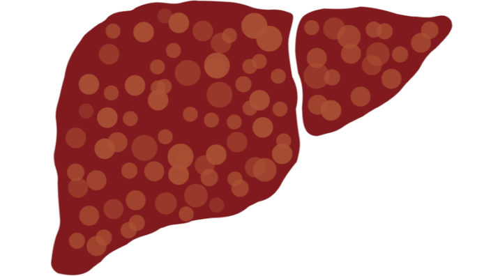
The researchers also reported that niacin improved body composition, and participants saw a decrease in whole-body fat percentage in controls and increased muscle mass in both the control and study groups. After 10 months, participants saw increased muscle strength. They noted that hepatic fat was reduced by half and visceral fat by a quarter; both of these fat deposits are associated with an increased risk of metabolic syndrome.

The researchers also considered the previously mentioned risk of niacin increasing blood glucose levels. The study results showed that niacin did indeed increase fasting glucose levels in both study groups following 4 months of supplementation. However, glycosylated hemoglobin, which reflects long-term glucose levels, was not affected.

Niacin side effects 

A typical side effect of high-dose niacin is the “niacin flush” reaction, which can potentially cause a burning, tingling, and itching sensation on the skin. This flushing is harmless and typically subsides within 30 minutes to an hour. The flush reaction is normally the most intense after the first dose and normally diminishes with continued use of niacin as the body grows used to it. Its severity may also be reduced by starting at a low dose (50-100 mg), taking an aspirin or white willow extract beforehand, and drinking water.

As mentioned previously, slow-release/sustained release niacin carries the risk of liver damage so be careful when purchasing [1]. If you experience any adverse effects, cease taking niacin immediately and consult your doctor.

There is also some concern that niacin can deplete methyl groups [14] and raise homocysteine, an amino acid. Vitamins B12, B6 and folate break down homocysteine to create other molecules but when high homocysteine levels are a risk factor for heart attacks [15]. It may be possible to reduce homocysteine levels by restoring methyl groups using supplements such as trimethylglycine (TMG).

Disclaimer

This article is only a very brief summary, is not intended as an exhaustive guide, and is based on the interpretation of research data, which is speculative by nature. This article is not a substitute for consulting your physician about which supplements may or may not be right for you. We do not endorse supplement use or any product or supplement vendor, and all discussion here is for scientific interest.

We would like to ask you a small favor. We are a non-profit foundation, and unlike some other organizations, we have no shareholders and no products to sell you. All our news and educational content is free for everyone to read, but it does mean that we rely on the help of people like you. Every contribution, no matter if it’s big or small, supports independent journalism and sustains our future.

Literature

[1] Rader, J. I., Calvert, R. J., & Hathcock, J. N. (1992). Hepatic toxicity of unmodified and time-release preparations of niacin. The American journal of medicine, 92(1), 77-81.

[2] Kennedy, D. O. (2016). B vitamins and the brain: Mechanisms, dose and efficacy—A review. Nutrients, 8(2), 68.

[3] Kirkland, J. B. (2012). Niacin requirements for genomic stability. Mutation Research/Fundamental and Molecular Mechanisms of Mutagenesis, 733(1), 14-20.

[4] Li, J., Bonkowski, M. S., Moniot, S., Zhang, D., Hubbard, B. P., Ling, A. J., … & Aravind, L. (2017). A conserved NAD+ binding pocket that regulates protein-protein interactions during aging. Science, 355(6331), 1312-1317.

[5] Brown G, Albers JJ, Fisher LD, et al. Regression of coronary artery disease as a result of intensive lipid-lowering therapy in men with high levels of apolipoprotein B. N Engl J Med. 1990;323:1289–98.

[6] Kamanna VS, Kashyap ML. Mechanism of action of niacin on lipoprotein metabolism. Curr Atheroscler Rep. 2000;2:36–46.

[7] Cashin-Hemphill L, Mack WJ, Pogoda JM, et al. Beneficial effects of colestipol-niacin on coronary atherosclerosis. A 4-year follow-up. JAMA. 1990;264:3013–7.

[8] Grundy, S. M., Mok, H. Y. L., Zech, L., & Berman, M. (1981). Influence of nicotinic acid on metabolism of cholesterol and triglycerides in man. Journal of lipid research, 22(1), 24-36.

[9] Illingworth, D. R., Stein, E. A., Mitchel, Y. B., Dujovne, C. A., Frost, P. H., Knopp, R. H., … & Greguski, R. A. (1994). Comparative effects of lovastatin and niacin in primary hypercholesterolemia: a prospective trial. Archives of internal medicine, 154(14), 1586-1595.

[10] Cashin-Hemphill, L., Mack, W. J., Pogoda, J. M., Sanmarco, M. E., Azen, S. P., & Blankenhorn, D. H. (1990). Beneficial effects of colestipol-niacin on coronary atherosclerosis: a 4-year follow-up. Jama, 264(23), 3013-3017.

[11] Brown, B. G., Zhao, X. Q., Chait, A., Fisher, L. D., Cheung, M. C., Morse, J. S., … & Frohlich, J. (2001). Simvastatin and niacin, antioxidant vitamins, or the combination for the prevention of coronary disease. New England Journal of Medicine, 345(22), 1583-1592.

[12] Goldie, C., Taylor, A. J., Nguyen, P., McCoy, C., Zhao, X. Q., & Preiss, D. (2016). Niacin therapy and the risk of new-onset diabetes: a meta-analysis of randomised controlled trials. Heart, 102(3), 198-203.

[13] Pirinen, E., Auranen, M., Khan, N. A., Brilhante, V., Urho, N., Pessia, A., … & Haimilahti, K. (2020). Niacin cures systemic NAD+ deficiency and improves muscle performance in adult-onset mitochondrial myopathy. Cell Metabolism.

[14] Conze, D., Brenner, C., & Kruger, C. L. (2019). Safety and metabolism of long-term administration of NIAGEN (nicotinamide riboside chloride) in a randomized, double-blind, placebo-controlled clinical trial of healthy overweight adults. Scientific reports, 9(1), 1-13. [15] Chrysant, S. G., & Chrysant, G. S. (2018). The current status of homocysteine as a risk factor for cardiovascular disease: a mini review. Expert review of cardiovascular therapy16(8), 559–565. https://doi.org/10.1080/14779072.2018.1497974