The Blog

Building a Future Free of Age-Related Disease

Diabetes meter

Towards a Possible Solution to Diabetes

A team of researchers led by Dr. Jeffrey Millman at Washington University may be a step closer to a potential solution to diabetes, according to the results of a new study in which replacement beta cells were given to mice.

What is diabetes?

Diabetes is a disease that occurs when blood glucose levels are constantly too high. Blood glucose is the main source of energy for the body and comes from the nutrients we eat. Insulin is a hormone created in the pancreas that allows our cells to use glucose for energy.

Unfortunately, sometimes the body does not make enough or even any insulin, and even when it does, it does not always manage it effectively. The result of this is that the glucose remains in the blood, and, over time, having elevated levels of glucose in the bloodstream can lead to health problems.

There are two main types of diabetes. In type 1, the body does not make insulin at all because the immune system attacks and destroys the beta cells in the pancreas that produce insulin. This form of diabetes is most common in children and young adults, although it can occur later in life also. People with type 1 diabetes must take insulin daily to remain alive.

In type 2, the body does not produce or manage insulin efficiently. Aging, and the resulting metabolic decline and failure, is a common reason why type 2 diabetes develops. Type 2 is most commonly encountered in middle-aged or older people and is the most common form of diabetes.

Replacing lost beta cells

Insulin is produced by the beta cells residing in the pancreas. In type 1 diabetes, these cells are destroyed by the immune system, and in type 2, they do not produce enough insulin. The condition is typically managed by introducing insulin into the bloodstream or using drugs to increase insulin production.

However, in recent years, researchers have been working on ways to replace the missing beta cells and the insulin they produce by converting stem cells into beta cells. These researchers previously demonstrated that giving diabetic mice new beta cells helped to improve the condition [1], and their follow-up study has produced even more promising results by refining the technique [2].

In the past, one barrier to an effective cell therapy has been in the conversion of stem cells to beta cells, as the transformation does not always go as planned. In any given batch of stem cells that are being reprogrammed into other cell types, there are often a number that go wrong and turn into another cell type. The process becomes less efficient the more of these conversion errors there are, and while they are generally not dangerous, it does mean that a larger batch of cells must be used, which makes it more costly. In other words, if a quarter of the batch of stem cells fails to convert properly, it makes the task 25% harder and more expensive.

The research team focused on making the therapy more efficient by reducing the number of off-target cells produced during the conversion process. They targeted the cytoskeleton, the supporting structure that gives cells their shape, to help increase the beta cell yield during conversion. Not only did this reduce the amount of off-target cell types produced in a batch, the beta cells produced were also functionally superior.

Finally, the research team gave diabetic mice these new higher-quality beta cells, which caused their blood sugar to levels normalize and caused their diabetes to be “functionally cured” for a period of up to nine months.

Generation of pancreatic β cells from human pluripotent stem cells (hPSCs) holds promise as a cell replacement therapy for diabetes. In this study, we establish a link between the state of the actin cytoskeleton and the expression of pancreatic transcription factors that drive pancreatic lineage specification. Bulk and single-cell RNA sequencing demonstrated that different degrees of actin polymerization biased cells toward various endodermal lineages and that conditions favoring a polymerized cytoskeleton strongly inhibited neurogenin 3-induced endocrine differentiation. Using latrunculin A to depolymerize the cytoskeleton during endocrine induction, we developed a two-dimensional differentiation protocol for generating human pluripotent stem-cell-derived β (SC-β) cells with improved in vitro and in vivo function. SC-β cells differentiated from four hPSC lines exhibited first- and second-phase dynamic glucose-stimulated insulin secretion. Transplantation of islet-sized aggregates of these cells rapidly reversed severe preexisting diabetes in mice at a rate close to that of human islets and maintained normoglycemia for at least 9 months.

Conclusion

As always with these kinds of studies, the caveat is this is an animal trial, so the results may not necessarily translate to humans, and even if they do, it could take considerable time before they get through human clinical trials.

That said, this is an exciting approach that could be a gamechanger if it can be made to work in humans as it has in mice. Meanwhile, the researchers will be moving forward and testing the approach in larger animals, eventually progressing to human clinical trials.

We would like to ask you a small favor. We are a non-profit foundation, and unlike some other organizations, we have no shareholders and no products to sell you. All our news and educational content is free for everyone to read, but it does mean that we rely on the help of people like you. Every contribution, no matter if it’s big or small, supports independent journalism and sustains our future.

Literature

[1] Millman, J. R., & Pagliuca, F. W. (2017). Autologous pluripotent stem cell–derived β-like cells for diabetes cellular therapy. Diabetes, 66(5), 1111-1120.

[2] Hogrebe, N. J., Augsornworawat, P., Maxwell, K. G., Velazco-Cruz, L., & Millman, J. R. (2020). Targeting the cytoskeleton to direct pancreatic differentiation of human pluripotent stem cells. Nature Biotechnology, 1-11.

Organ chip

Automating Drug Testing With Human Organ Chips

Researchers from the Universities of Harvard and Tel Aviv have succeeded in linking up to 10 “organs-on-a-chip” via an analog of human vasculature. The invention allows for in vitro testing of drug toxicity and action, taking us one step closer to automated clinical trials and the development of personalized drugs.

Chipping away at trial duration

While awaiting the emergence of new anti-aging drugs, we expect their road to the shelves to be long, winding, and frustrating, as life-extending medications take years to go through the pharmaceutical pipeline. One promising way to tackle this problem is the development of “organs-on-a-chip” (or “organ chips”). This “chip” has nothing to do with computer microchips; instead, it is a piece of plastic or other material containing a few thousands of parenchymal (organ-specific) cells that imitate the organ’s action. Basically, it’s a working model of an organ. For example, a “liver-on-a-chip” imitates the liver’s filtration routine, a “heart-on-a-chip” undergoes contraction, and an “intestine-on-a-chip” conducts peristaltic action and absorption.

Organ chips can help to solve numerous problems associated with drug development. New drugs that were successfully tested in animals often fail human clinical trials due to fundamental interspecies differences. Organ chips can potentially replace early animal testing, saving vast amounts of money and effort. As the technology matures, it can also complement Phase 1 clinical trials, which test drug toxicity. Parenchymal cells donated by a single human can undergo hundreds of such trials, each of which costs a fraction of the time and money that an actual human trial does.

These chips can also potentially revolutionize personalized medicine (PM), including the creation of personalized drugs. With organ chips, trials can be conducted on live cells taken from a specific patient without risking the patient’s health. Multiple trials can be run simultaneously to quickly find a life-saving solution.

From organ chips to “human-on-a-chip”

These researchers have made a major leap by developing a sophisticated system that they call a “human-on-a-chip” [1]. One of the hallmarks of this new system is that the organ chips are linked together, in a robotic environment, via a “vascular system” that contains a blood substitute made of vitamins, minerals, hormones, and other essential components. The system transports artificial blood to and from the organ chips to mimic drug distribution and dilution. More importantly, unlike previous models, the new chips include a layer of endothelial cells (cells that line the interior surface of blood vessels) laid along the organ-specific cells. The endothelial barrier emulates a blood vessel and is needed to effectively recapitulate the drug’s pharmacokinetics (PK) – the way it travels through the body.

For example, the system allows the modeling of oral administration, as a drug is delivered to the “intestine-on-a-chip” and then travels to other organs. In one of the experiments, the researchers had modeled the administration of nicotine, and the results were consistent with what we know about nicotine’s PK in vivo. To extrapolate the results received from just a few thousand cells, the researchers used custom-made scaling software.

Theoretically, the system can consist of up to 10 organ chips, but the researchers admit that, currently, the best results are achieved with up to three organs linked together. In one of the two companion papers, the researchers describe a model designed to mimic a first-pass drug absorption. The model consists of gut, kidney, and liver organ chips. First-pass is a phenomenon in which drug concentration is drastically reduced before it reaches systemic circulation, due to metabolism inside the liver and other organs. Studying the first-pass effect on a drug is essential to determining the right dosage. The new device can greatly simplify this task while eliminating any risk to the patients.

Behold the interrogator

The researchers have also constructed a robotic system for conducting experiments on their “human-on-a-chip” [2]. The system, called an interrogator, can run preprogrammed experiments and collect data automatically. It also keeps the tissues inside the organ chips alive and well. The system includes a precision manipulator and an integrated mobile microscope. During the trials, the interrogator successfully maintained the viability and organ-specific functions of eight interlinked organ chips (intestine, liver, kidney, heart, lung, skin, blood-brain barrier and brain) for three weeks.

Conclusion

This is not the first time that automation has been used to drastically reduce the time needed for drug development and testing. Last year, the pharmaceutical start-up Insilico Medicine made headlines by creating a new drug from scratch in 46 days using machine learning algorithms.

By linking several organ chips together and making successful predictions of human responses to drugs, these researchers have cleared another major hurdle on the way to automating certain aspects of drug development. It can result in new drugs being developed and tested in a much faster and safer way – a welcome advancement, considering that currently drug trials take several years.

We would like to ask you a small favor. We are a non-profit foundation, and unlike some other organizations, we have no shareholders and no products to sell you. All our news and educational content is free for everyone to read, but it does mean that we rely on the help of people like you. Every contribution, no matter if it’s big or small, supports independent journalism and sustains our future.

Literature

[1] Herland, A., Maoz, B. M., Das, D., Somayaji, M. R., Prantil-Baun, R., Novak, R., … & Chalkiadaki, A. (2020). Quantitative prediction of human pharmacokinetic responses to drugs via fluidically coupled vascularized organ chips. Nature Biomedical Engineering, 1-16.

[2] Novak, R., Ingram, M., Marquez, S., Das, D., Delahanty, A., Herland, A., … & Calamari, E. (2020). Robotic fluidic coupling and interrogation of multiple vascularized organ chips. Nature Biomedical Engineering, 1-14.

Rejuvenation Roundup February

Rejuvenation Roundup February 2020

Several leaps have been made this February: Leap Year’s Day, leaps in rejuvenation biotechnology development, and a leap of our own in bringing our two websites together into one.

If you want to help us and the world of rejuvenation biotechnology leap into the public consciousness and into the clinic, become a Lifespan Hero today.

LEAF News

Team and activities

Our annual conference series, Ending Age-Related Diseases: Investment Prospects & Advances in Research, will continue for its third year, featuring researchers at the forefront of rejuvenation biotechnology. Hosted at the Mount Sinai Medical Center in New York City on August 20-21, 2020, this event promises to be enlightening and informative for professionals and laypeople alike.

Interviews

Irina & Michael Conboy – Resetting Aged Blood to Restore Youth: The Conboys discuss their past work along with apheresis as a potential method of filtering out the overexpressed factors that occur as the result of aging – and cause further age-related diseases.

Hanadie Yousef – Embryonic Proteins for Tissue Regeneration: Juvena Therapeutics is discovering proteins found in the embryonic environment that cause muscle and other tissues to regenerate, potentially leading to therapies for sarcopenia and age-related brain degeneration.

Lewis Gruber – Senotherapeutics: Alongside his wife Misty, Lewis Gruber discusses what SIWA Therapeutics is doing to address the problem of senescent cells by using monoclonal antibodies, which are used for cancer immunotherapies.

Rejuvenation Roundup Podcast

Ryan O’Shea of Future Grind hosts this month’s podcast, showcasing the events and research discussed here.

Research Roundup

Refining the Allotopic Expression of Mitochondrial Genes: There are significant differences in the codons used by mitochondria and nuclear DNA, and understanding these differences is critical for expressing mitochondrial DNA in the nucleus, which is the core goal of MitoSENS.

Macrophages Become Scars in the Heart: Rather than just summoning fibroblasts to the site of injury, macrophages extrude their own fibers, thus leading to scars forming in place of healthy tissue. A therapy that targets macrophages may be the first step towards giving adults the regenerative capabilities of young children.

What Do DNA Smiley Faces Have to Do With Cancer Research?: A technique called DNA origami can be used to make tiny boxes that open with molecular keys, offering new opportunities for targeted drug delivery and other medical uses.

An epigenetic clock for human skeletal muscle: Focusing on dinucleotide methylation that is specific to human muscle, this clock offers more accuracy than clocks that analyze all tissues.

Two conserved epigenetic regulators prevent healthy ageing: These regulators diminish the nuclear expression of mitochondrial proteins, promoting age-related problems such as cognitive decline.

Hematopoietic stem cell transplants increase lifespan in mice: Avoiding the need for irradiation as in a marrow transplant, this study showed that transplanting HSCs increased healthy lifespan without side effects in a mouse model.

Neural precursor cell transplants promote motor recovery after stroke: This mouse study showed that reprogrammed neural precursor cells help the brain regenerate synapses and restore function.

Depleting microglia improves traumatic brain injury recovery : After a traumatic brain injury, the brain’s immune cells often become neurotoxic. This mouse study showed that killing most of these cells and allowing them to recover alleviates these effects.

Eating a Mediterranean diet for a year promotes a healthy gut microbiome: Featuring 612 human volunteers, this study showed that a Mediterreanean diet promoted the growth of health-associated bacteria and limited the amounts of frailty-associated bacteria.

Caloric Restriction Reprograms the Single-Cell Transcriptional Landscape of Rattus Norvegicus Aging: This atlas shows exactly what happens to the cell during caloric restriction in a rat model.

A Deep Learning Approach to Antibiotic Discovery: A deep learning algorithm identified many compounds that show antibiotic effects, including eight that do not resemble known antibiotics. One of these, halicin, is effective against strains of bacteria that are widely considered dangerous.

Age Reversal and Pluripotency Induced in Supercentenarian Cells: This study shows that it is possible to use OSKM to induce pluripotency in cells from people who are over 110 years old.

News Nuggets

Why Life Expectancy Could Rise Significantly in the Near Future: Originally published by the German magazine Monat, this article offers an introduction to rejuvenation biotechnology.

JangoBio Creating First Organoids for Complete Hormone Restoration: In this press release, JangoBio announces that it has restored hormone production through the use of organoids created with stem cells.

Coming Up

The Longevity Leaders Congress: Held on April 21-22 in London, UK, this event will focus on aging science, assistive technologies, and risks relating to retirement funds. Use the code LEAF15 for a 15% discount.

We would like to ask you a small favor. We are a non-profit foundation, and unlike some other organizations, we have no shareholders and no products to sell you. All our news and educational content is free for everyone to read, but it does mean that we rely on the help of people like you. Every contribution, no matter if it’s big or small, supports independent journalism and sustains our future.
Smiley face

What Do Smiley Faces Have to Do With Cancer Research?

DNA origami, a nano-scale technique used to make very tiny smiley faces out of DNA, is becoming increasingly valuable for drug delivery and other applications, paving the way for true nanomedicine.

What is DNA origami?

Ever since Richard Feynman famously proclaimed in his seminal 1959 talk that “there’s plenty of room at the bottom”, nanotechnology has been hailed as the next technological wonder. In reality, though, its progress has remained slow for decades due to the enormous challenges of building and operating structures on a nanometric scale. Thankfully, in recent years, the pace has quickened, showering us with promising developments. This includes one of the most exciting areas of medical nanotechnology: DNA origami.

Back in 1982, Nadrian Seeman of New York University, inspired by the mind- and space-bending works of M. C. Escher, pioneered the idea of using DNA molecules to build complex three-dimensional structures. Capitalizing on Seeman’s work more than a decade later, Paul Rothemund of CalTech invented a new way of constructing objects from DNA, which he called “DNA origami”. Like an elaborate origami figurine folded from a single sheet of paper, DNA origami objects are built from one long “scaffold” of single-stranded DNA bound in particular places by “staples” that consist of hundreds of shorter strands. As a scaffold, most DNA origami objects use a single strand of DNA, over 7000 base pairs long, that originates from an M13 bacteriophage virus. The staples are man-made.

The staples have a specific base sequence that can only bond with corresponding sequences on the scaffold (thanks to the Watson-Crick base pairing). When two sections of one staple bond with two different regions of the scaffold DNA, they bring those regions together, not unlike real staples, creating a compact and relatively rigid structure. This structure becomes a tiny part of the desired shape.

One of the main advantages and mysteries of DNA origami is the spontaneous process of folding into shape, which is still not fully understood. The building blocks (billions of scaffolds and staples mixed in precise proportion) are subjected to annealing: heating followed by slow cooling. During this process, preprogrammed shapes are formed automatically, although the yield can be low for complex structures. This ability to self-assemble makes DNA origami probably the most powerful and scalable DNA construction technology to date.

To demonstrate the robustness of his method, Rothemund had created numerous objects, including the now-famous smiley faces that are 10 nanometers in diameter. In the following years, the DNA origami technique has been expanded by Rothemund and others to include an impressive array of much more complex two- and three-dimensional objects [1].

How can this help us live longer?

In recent years, researchers have been devising ingenious ways of making the tiny structures exhibit robotic behavior. Some of the resulting designs have demonstrated great potential as delivery vehicles for drug molecules [2]. One example is three-dimensional DNA origami “boxes” made of interconnected two-dimensional plates. Each of these objects has an internal cavity and a door with a molecular lock. Its door can be unlocked by a specific chemical “key” – such as a compound associated with a certain type of cancer cell. DNA origami nanorobots behave like simple logical gates. A robot with two locks, each one reacting to a different compound, functions as an AND gate, opening the internal cavity only when both compounds are present. This allows for more targeted and specialized uses.

The possible applications for targeted action on a cellular scale are innumerable. In 2016, it was demonstrated that DNA nanorobots can be used for diagnostics. A team of scientists used gold nanorods attached to DNA origami structures. After having been absorbed by the tumor cells, the nanorods were used first as contrast agents in optoacoustic imaging (OAI) and then as thermal agents in photothermal therapy. The latter is based on the irradiation of gold particles by a near-infrared laser which leads to their rapid heating. The heat reduces viability of the nearby cancer cells [3].

In 2018, we reported on the successful delivery of thrombin to tumors in mice by DNA origami nanorobots. Thrombin was delivered straight to the tumor in the hope of cutting off its blood supply. The nanorobots succeeded in inhibiting the tumor’s growth and, importantly, were labeled safe after being extensively tested in mice and Bama pigs [4].

Last year, a team from Augmanity Nano and the Wyss Institute for Biologically Inspired Engineering at Harvard reported on nanoscale robots that exhibit quorum sensing (QS). In biology, quorum sensing is the ability of some animals, including bacteria and insects, to adjust their behavior in response to population density fluctuations. The team engineered a QS system to address the problem of drug concentration at the target, as a simpler DNA robot releases its payload immediately. If too few of these simpler robots get to the target simultaneously, drug concentration may not reach the levels necessary for effective treatment. The QS system, however, causes the nanorobots to sense how many of their “siblings” are also in the vicinity of the target and to coordinate a simultaneous payload release [5].

Finally, just last month, a group of Chinese scientists claimed that they were able to create what amounts to a nanoscale precision-guided missile made of DNA (D-PGM) [6]. The robots consist of a warhead loaded with the well-known chemotherapy drug doxorubicin and a guidance system capable of recognizing specific cell subtypes.

The results demonstrate that by mimicking the functionalities of a military precision-guided missile to design the sequential disassembly of the GC system in multistimuli-responsive fashion, our intrinsically biocompatible and degradable D-PGM can accurately identify target cancer cells in complex biological milieu and achieve active targeted drug delivery. The success of this strategy paves the way for specific cell identity and targeted cancer therapy.

This experiment highlights the fact that we already possess capable anti-cancer drugs that may only need targeted delivery systems to unleash their full potential.

Conclusion

While many problems remain, such as the survivability of fragile DNA machines in the body [7], successes continue to mount. Numerous start-ups, such as Tilibit Nanosystems, Gattaquant, Integrated DNA Technologies, and Eurofins Genomics, explore commercial applications and offer custom-built nanostructures for purposes ranging from medicine to quantum computing. A few software tools for designing DNA origami exist, most notably the open-source CadNano. The annual BIOMOD competition features novel DNA origami designs from young teams all over the world. While no clinical trials have been conducted yet, the long-foretold days when we have swarms of nanorobots circulating in our bloodstreams in order to prevent, diagnose, and fight diseases are likely to be on the horizon.

We would like to ask you a small favor. We are a non-profit foundation, and unlike some other organizations, we have no shareholders and no products to sell you. All our news and educational content is free for everyone to read, but it does mean that we rely on the help of people like you. Every contribution, no matter if it’s big or small, supports independent journalism and sustains our future.

Literature

  1. Liu, H., & Fan, C. (2013). DNA Origami Nanostructures. In DNA Nanotechnology (pp. 207-224). Springer, Berlin, Heidelberg.
  2. Udomprasert, A., & Kangsamaksin, T. (2017). DNA origami applications in cancer therapy. Cancer science, 108(8), 1535-1543.
  3. Du, Y., Jiang, Q., Beziere, N., Song, L., Zhang, Q., Peng, D., … & Ntziachristos, V. (2016). DNA‐Nanostructure–Gold‐Nanorod Hybrids for Enhanced In Vivo Optoacoustic Imaging and Photothermal Therapy. Advanced Materials, 28(45), 10000-10007.
  4. Li, S., Jiang, Q., Liu, S., Zhang, Y., Tian, Y., Song, C., … & Chang, Y. (2018). A DNA nanorobot functions as a cancer therapeutic in response to a molecular trigger in vivo. Nature biotechnology, 36(3), 258.
  5. Amir, Y., Abu-Horowitz, A., Werfel, J., & Bachelet, I. (2019). Nanoscale robots exhibiting quorum sensing. Artificial Life, 25(3), 227-231.
  6. Ouyang, C., Zhang, S., Xue, C., Yu, X., Xu, H., Wang, Z., … & Wu, Z. S. (2020). Precision guided missile-like DNA nanostructure containing warhead and guidance control for aptamer-based targeted drug delivery into cancer cells in vitro and in vivo. Journal of the American Chemical Society.
  7. Balakrishnan, D., Wilkens, G. D., & Heddle, J. G. (2019). Delivering DNA origami to cells. Nanomedicine, 14(7), 911-925.
Interview with Lewis Gruber

Lewis Gruber – Senotherapeutics

At the recent Longevity Therapeutics Conference in San Francisco, we had the chance to interview Lewis Gruber of SIWA Therapeutics and discuss his company’s senotherapeutic approach to cancer and senescent cells. He was accompanied by his wife, Misty, who is the CFO of SIWA.

Many of our readers are familiar with CAR-T immunotherapy, which has enjoyed some success, but it’s not without considerable challenges. How does your approach differ?

Lewis: We are using a simpler approach of just manufacturing a monoclonal antibody. Of course, we do that in Chinese Hamster Ovary (CHO) cells and purify and produce a monoclonal antibody product so that we don’t have to modify patient cells or any other cells in order to apply our treatment. It’s just a straight typical monoclonal antibody product, the same sort of immunotherapy that’s used in a variety of cancer therapies.

Misty: Which means that monoclonal antibody goes after a specific marker. In this case, he’s found a marker that’s on cancer cells and senescent cells because of the way the markers are produced, and therefore his monoclonal antibody can zap and remove those cancer cells.

So each antibody is tailored for a specific biomarker, but not for each individual.

Lewis: Yeah, it’s for the one biomarker that’s present in all humans, and actually the marker goes back to yeast.

Misty: We can find antibodies to go after specific markers that we find on certain diseases.

Lewis: Going back to yeast and maybe even back to bacteria.

Can you summarize a bit more how that antibody SIWA 318H works?

Lewis: It binds to proteins on the surface of oxidatively damaged cells that may be senescent or cancerous, or just very dysfunctional. By doing so, it provokes an immune response, initially an innate immune response with the natural killer cells. The bottom line is, the immune response not only destroys and removes the cells to which the antibody binds, but immune cells also secrete factors that promote regeneration. So, while we’re removing cells that are not going to function properly, we’re promoting their replacement with new cells from adult stem cell populations.

Misty: The interesting thing about the markers, it’s a product of glycolysis, and high levels of glycolysis were associated even back before World War Two by Otto Warburg. They were shown on cancer cells. Cancer cells and senescent cells have their peculiarities, and they are high producers of this particular marker; therefore, he can hone in on those two types of cells. There are other markers for other types of diseases, but the key to what he did was find the marker, and then he designed an antibody to go to that specific marker.

Researchers have long been seeking common targetable features on cancer cells, so how’d you discover this particular marker?

Lewis: The process that produces the marker has been reported in the literature. As Misty was mentioning, Otto Warburg back in the 1920s discovered that cancer, virtually all types of cancer cells, have a elevated level of glycolysis, and our marker is a side product of glycolysis. Basically, I didn’t really discover the marker; in that sense, the roots of it go back to the 1920s. It’s only been more recently that the marker has been looked at with respect to aging and aging-related diseases.

So, you rediscovered it, essentially.

Lewis: Rediscovered it, and no one had suggested before using the marker to remove cells that might be dysfunctional. That’s where I came in. I was looking for something to remove dysfunctional cells. I saw the association and literature with cancer and with aging, which also translated to senescent cells, and I said that it would be a good marker.

Misty: So the beauty of it is that both senescent cells and cancer cells have this high level of glycolysis. The Warburg effect is when it’s basically caught on cancer, but for it to also be on senescence cells is really perfect.

Was that a coincidence? Or is there a reason for that?

Lewis: Both types of cells conducted an elevated level of glycolysis and there are various explanations in the scientific literature. Both are highly metabolically active. I didn’t see all the presentations at the conference here, but some people think of senescent cells as almost dead, but, in fact, they are among the most metabolically active of cells, and cancer cells are as well because of high proliferation. Both types of cells have a high need for ATP. One explanation in the literature is that they both resort to glycolysis to get additional ATP.

Misty: The only difference being that cancer cells tend to reproduce rapidly, while senescent cells tend not to reproduce. Senescent cells can be cancer cells too, which is why it’s important to go after both in a cancer drug, for example.

Lewis: The senescent cells put all of their efforts into senescence associated secretory phenotype, so they’re producing a lot of cytokines and other molecules, so they need to use glycolysis, but they’re not using it to divide. A simplified way of looking at it is that senescent cells grow, and they need energy for growth; they basically get to the size that a cell would normally be when it divides, and then they just don’t divide. When you look under the microscope, you see large, flattened cells that are senescent cells, because they’ve grown but they just didn’t divide. Cancer cells, of course, will go ahead and do the division and you’ll have two daughter cells, et cetera. The bottom line is that they both have to grow to that large size.

SIWA 318H is able to likely target both senescent cells and cancer cells, as you mentioned, using a surface marker common at both types of cell. Given their heterogeneity, do all cancer and senescent cells have that marker, and, if so, how’s that been confirmed?

Lewis: It was confirmed for me by looking at the literature for the marker and all of the types of cells. I didn’t dwell on it at the conference, but there might even be a slide in the pack that goes through all of the different types of senescent cells where our marker’s present. Basically, a root cause that they both have is that oxidative damage can lead to three different things. It can lead to senescence, it can lead to cancer, or it can lead to death. The bottom line is that the senescents and cancer share this oxidative damage as part of their history, and all senescent and cancer cells have a history of excess oxidative stress and reactive oxygen species.

Misty: I can give a slightly more pragmatic answer in that we tested it ourselves. We had the animal body, and we did a lot of binding tests. Two of the universities, the big institutions that we’re working with have also done separate studies to show binding. That’s always the first step. You don’t want to go out and do a mouse study unless you know that there’s binding, so we’ve done we did a number of different bindings, including, as Lewis mentioned, a type of glioblastoma that is almost impossible to test. It fits the mold of being a cancer cell that has our marker on it.

Lewis: Basically every type of cancer we’ve tested has bound to our antibody, but I’m relying also on the literature where it’s even reported that our marker is found in association with even more types of cancer. We have yet to find out that there’s a cell that doesn’t have this marker. Again, it goes back to oxidative stress and glycolysis; as I did say in one of my slides, cancer and senescent cells both share having been oxidatively damaged and exhibiting in a high level of glycolysis.

Given that, does that mean a vaccine might be developed for senescent cells or even cancer?

Lewis: Yes, and we are working on what we have. We’ve done preliminary studies in mice, and now we’re looking at expanding it into actually other species, even beyond humans, but with respect to the vaccine, but we do have a candidate vaccine already in the works.

Would that be an inoculation early in life that would manifest later in life, or would it always protect against SCs and cancer cells?

Lewis: It could always protect against SCs and cancer cells. As with any drug, you do want to be careful about certain conditions, pregnancy or other conditions where you don’t want to disrupt any things happening. For example, senescent cells have been found in fetuses.

They have a positive role in organ growth, et cetera.

Misty: In the formation of the hand and the fingers and things like that, they do. They are involved in that.

Lewis: The one common thing, strangely enough, with senescent cells is every situation in which they’re beneficial, they’re removed. After they form the different structures in the fetus, they’re eliminated. The same thing is true in wound healing, which is often given as an example of a beneficial effect. Initially, senescence promotes proliferation of repairing cells, but if that’s allowed to go on too long, the wound tends to produce scar tissue, fibrosis, and the bottom line is that in the natural healing process, senescent cells appear for a time and then are removed. That’s basically what we are: we remove senescent cells. Although you do have to be somewhat careful, you don’t want to interfere with the initial stage of wound healing or with fetal development, otherwise, it’s a good rule of thumb that removing a senescent cell or a cancer cell is not a bad thing.

Cancer is quick to adapt and evolve, so, is it plausible that it could do so against 318H?

Lewis: It would be much harder, I think, than anything else. From the standpoint that our marker is produced by a basic biological process that I mentioned goes back, maybe even back to bacteria, but certainly back to yeast. Evolutionarily, it’s such that it’s hard to imagine, for me anyway, how a cancer cell could evolve around such a basic biological process and come up with something different. Even shifting: for example, some cancer cells conduct more oxidative phosphorylation than others, and so, rather than relying on glycolysis for any ATP, they just do more mitochondrial respiration; however, mitochondrial respiration is a source of reactive oxygen species and oxidative damage. As it turns out, our marker and our antibody would still get them. I don’t mean to belabor all the different ways, but it is a good point, and we have thought about it because it is, as you correctly state, a common feature of cancer to be able to work around drugs, but I think, at least, it would be much harder.

Has your therapy caused a reduction in senescent cells in mice?

Misty: It’s in animal studies that we’ve done, and we’ve shown binding in vitro, which means that the cell is compromised.

Lewis: There’s a reduction of senescent cells in vivo.

Misty: In vivo is where you find that there’s an actual reduction.

In what tissues?

Lewis: In that particular case, we were looking at inguinal adipose tissue and muscle tissue.

Misty: Well, the way that we did a milestone that was the very first thing we did, the first one we looked at was whether we could reduce senescent cells. So we got from a provider, very, very old mice that had not been involved in drug tests, but had been involved only in maze testing and things related to psychology. We took groups: we had very old mice, and we had very young mice. We took two different dosage levels of our antibody, the mouse homologue, and we injected it, and what we found was that there’s a marker called p16Ink4a, which is on senescent cells; everybody’s talked about it, more or less. We showed that in the very old mouse with our upper level of dosage, we reduce the level of p16Ink4a all the way down to the very young mouse controls. We also then looked at a particular muscle tissue to see if we can regenerate muscle tissue because Lewis’ concept was that by removing senescent cells associated with the muscle tissue, the muscle tissue can regenerate. Stem cells were still there, and they do corrective maintenance. In that study, we also got a regrowth of the muscle back to that level. That was the very first study.

Lewis: Also, Mayo Clinic has observed that, even with small molecule senolytics, removing senescent cells can lead to muscle regeneration.

Was that in humans?

Lewis: That was in mice. In humans, we haven’t really been working on that. We’re now using humanized mice, and we’re working toward a clinical trial.

Misty: The benefit of the humanized mice is that we can use the humanized antibody.

Lewis: Since the humanized monoclonal is what we’ll be using in clinical trials and for our pharmaceutical product, we’re anxious to demonstrate that it’s effective in doing the different things that we want to do. We’ve been working with a major institute on cancer studies. They’re looking at a variety of features in the humanized mice, including not only killing the primary tumor cells and senescent cells associated with the tumor but also looking at regeneration of tissues, the formation of desmoplasia, which is scar tissue, and others.

Traditional biomarkers for senescence cells are typically beta-gal and p16Ink4a, but these implementations, so what other ways are you measuring the presence of senescent cells in tissue and any changes to their levels?

Lewis: Well, we have looked at both p16 and beta-gal, and wherever we find them, we find binding of our antibody.

Misty: So, those are confirming our antibodies.

Lewis: We’ve also looked at the large, flat morphology; we look for cells that morphologically look senescent, as well. The final way is that we put them in situations where the histone H2AX is present. We’ve looked for beta-gal, p16, H2AX, and the morphology.

One reason we age is the inhibition of our stem cells due to inflammation, which prevents regeneration of tissue. Do you believe that the SASP from senescent cells plays a key role in stopping healthy tissue regeneration, and what studies do you feel support this?

Lewis: As far as the supporting students are concerned, we could certainly provide those; we have something like 800 pages to maintain. It’s also true that oxidative stress is involved with inflammation. In fact, it has been considered as a cause of inflammaging, which has been related to senescent cells as well. So what we’re looking for is that our antibody removes cells that are promoting inflammation. There’s senescent or not, but presumably they would be senescent cells, and, again, I have seen papers support what you’re saying.

Misty: Even the Mayo Clinic looked at, in the normally old animals, muscle regeneration, and what that involves is removal of senescent cells; the way those muscles regenerate would be if the stem cells were allowed to start regenerating the tissue again, so that has been done in vivo in that sense.

Lewis: In terms of interefering with stem cells.

Misty: The stem cells were limited by the senescent cells: that’s the general theory of us and others who have done the same tests.

Lewis: We do have papers in our bibliography.

You’ve been testing your senolytic approach to combat sarcopenia in naturally aged mice. What have your results been so far?

Lewis: Well, as Misty mentioned, the result was that we had regeneration of muscle tissue in the mouse model that we use. I should qualify; you mentioned that we were senolytic. We certainly are senotherapeutic, we certainly remove senescent cells, but the reason I’m cautioning you is that, at least in some papers, senolytics are defined as small molecules, not biologics.

You’re biologics, purely. Xenobiologics, is that a good term?

Lewis: I think we make senotherapeutics, which can also though be blockers, but the bottom line is that I would certainly qualify it as something that can remove senescent cells but not call it a senolytic. I don’t know if it fits the strict definition that was proposed in some of the early studies.

Do you expect cancer or senescent cell human trials to begin first?

Lewis: That is tough because we are working with this major institute. They’re very far along in the pancreatic cancer model and are working with humanized mice, so they could be the first prepared. We’re separately doing toxicology and other studies so that the combination could mean that we could go into the clinic with them.

Misty: They have the safety studies and dosing already done.

Lewis: And they have organized clinical trials before, so they’re expressed an interest in doing so.

You can’t say what the group is?

Misty: No, we have a confidentiality agreement.

Lewis: It’s an educational institution, and in my career, I’ve represented a lot of educational institutions, and they’re very nervous about having anyone use their name. It’s not just this particular one; in general, I’ve found that they don’t like that. In our case, we do have a contractual agreement where we are not supposed to use their name.

If you do make it to trials, either for senescent cell removal or cancer, is there an approximate time frame you’re looking at right now?

Misty: Probably 12 to 15 months. Part of the issue is we have to keep making the antibody, and we’re still doing relatively small batches, we haven’t yet moved to large batch amounts, so that was a little bit of a limitation.

Lewis: That’s the phase that we’re at now. We have a large contract organization that makes very large quantities for pharmaceutical companies as well.

Misty: Which we’ll switch to when we get closer to this, but right now we’re using small quantities.

Lewis: We’re about ready to switch over.

Do you pronounce that “see-wa”, is that right?

Lewis: Yes. Actually, we’re named after an oasis in Egypt. It was the location of the oracle of the god Amun where Alexander the Great went in Egypt to ask questions about his descent from a god, which they confirmed, conveniently for him. One thing I like to point out is that these ancient oracles, not just Siwa, the Delphi and a lot of the well-known ones, people think of them as places of religious inspiration, but, because they had customers from around the ancient world, they were really intelligence services collecting information. The reason that our company’s named after them is that we look at ourselves as being like them, as being collectors of all the information and providing the best synthesis of that into a satisfying answer.

We would like to ask you a small favor. We are a non-profit foundation, and unlike some other organizations, we have no shareholders and no products to sell you. All our news and educational content is free for everyone to read, but it does mean that we rely on the help of people like you. Every contribution, no matter if it’s big or small, supports independent journalism and sustains our future.
Interview with Hanadie Yousef

Hanadie Yousef – Embryonic Proteins for Tissue Regeneration

At the recent Longevity Therapeutics Conference in San Francisco, we had the chance to interview Hanadie Yousef, the co-founder and CEO of Juvena Therapeutics. Juvena is isolating proteins from embryonic stem cells that promote tissue regrowth.

Can you describe in more detail Juvena’s approach to developing protein therapeutics that promote tissue regeneration in the elderly?

We are utilizing the secretome of human embryonic stem cells. We know that human embryonic stem cells have the capability to develop every tissue in the body, an entire human being. I and my colleagues discovered, nearly a decade ago, that by isolating a sub-fraction of the proteins that they themselves secrete and produce in order to signal to stem cells to develop every tissue in the body, concentrating these proteins, and then adding them directly onto old muscle precursor cells isolated from humans over the age of 65, we could enhance their regenerative potential. When we injected this cocktail of proteins into injured old mice, we saw muscle regeneration returned to levels of younger animals, two-month old mice that are like people in their 20s, and this is a cocktail of human proteins. The way that Juvena Therapeutics is taking this discovery into the clinic is by establishing a very efficient identification, high-throughput screening, and preclinical development pipeline, which has become ever more predictive and accelerated with the use of AI tools in order to identify what proteins in this original cocktail are actually driving the rejuvenation process, which ones are master regulators of signal transduction and key regulatory pathways involved in tissue differentiation and regeneration. By identifying those proteins and their sequences and exactly what they are compositionally, we can then test them individually and in combinations for their ability to promote human muscle precursor cell function and promote tissue regeneration in mouse models of human aging and human diseases.

Was this based on your PhD work?

It was based on some of my PhD work done during the five and a half years that I spent in the labs of Profs. Irina Conboy and David Schaffer at UC Berkeley. It’s also based on my team’s unique approach and the platform that we have built, and based on the expertise that I’ve gained over the last decade, focused on the biology of aging and mechanisms of aging, including both my work at UC Berkeley and then the five-year postdoc I did at Stanford. Basically, a postdoc, if you want it to be successful, is just another PhD. It’s five or six years, because that’s how long it takes to get a major publication, and my first authored publication in Nature Medicine came out in May of last year.

How do your other key discoveries made during your PhD and postdoc, in particular the mechanisms underlying the inhibitory effects of old blood and other discoveries using parabiosis research, fit into Juvena’s unique drug discovery approach?

Essentially, in order to really understand the aging process and the mechanisms underlying decline of tissue function at the level of loss of stem cell function, loss of strong vasculature, and a change in the signaling that happens, we really need to understand, at a systems biology level, what are the key drivers of aging and communication of cells and stem cells that allow your body to heal and repair after injury or develop new tissues, which I’ve dedicated the last decade of my life to understanding. I’ve been doing research for 17 years, but particularly the last decade, elucidating the mechanisms by which stem cells in both brain and muscle decline in function with age as well as the mechanisms by which blood interacts with our organs, specifically in the brain, in order to actually change signaling and drive the aging process when you age: the inhibitory effects of older blood. Based on the discoveries that myself and my colleagues made, we now understand what pathways are deregulated with aging, what increases with aging such as inflammatory factors or signaling on endothelial cells that can cause inhibitory effects that can prevent tissue regeneration, cause you to lose homeostasis, cause undesired inflammation that can really attack, become pathogenic, to your tissues. We also figured out the mechanisms by which you can stimulate and rejuvenate tissues, both at the levels of reducing the chronic low-level inflammation that happens with age, which was one of my last discoveries before launching Juvena Therapeutics, or by taking advantage of stimulatory pathways that we know can activate stem cells, can cause them to divide properly and regenerate and make new tissue but which we also now know decrease with age or are just inhibited. By understanding how we lose homeostasis in the environment surrounding our stem cells, we can then use that understanding to actually target the mechanisms underlying the aging process to reverse the process, to enhance tissue regeneration by revitalizing own-body stem cells.

Why did you choose to focus on muscle cell regeneration?

Interesting fact about muscle: It’s the largest internal tissue organ in the body. One of the first hallmarks of aging is the fact that once we hit our 30s, everybody, for the rest of our lives, heads downhill. We’re losing muscle strength and mass every year, but it accelerates with every decade so that by the time we’re in your 60s, everyone has some form of muscle wasting, some people more severe than others, so severe enough, in fact, that it prevents their daily functions and daily living and can be so severe that they can be clinically diagnosed with the disease of sarcopenia. Because there is now an ICD-10 code for sarcopenia, which was only issued at the end of 2016, meaning it’s an age-related disease that has a clinical indication, we can actually make therapies to target it. There’s zero FDA approved therapies, so it’s a huge unmet need and a huge market. Excitingly, one of the best experimental models that we have today to really understand how stem cells decline and function with age is the muscle system. Key discoveries made by my former co-thesis advisor, Irina Conboy, and other pioneers in the field, really paved the way for us to understand mechanistically how stem cells decline and function with age in muscle and develop methods to repair and rejuvenate them, so it’s a great first tissue to focus on. Juvena will use this as a way to then launch into other tissue types. Laser-like focus on muscle first; once we find the proteins that are secreted by human embryonic stem cells that can drive muscle regeneration, we’ll then apply our platform and our technology and our approach to identifying therapeutics, approaching candidates that can act as therapeutics to promote the brain and prevent things like dementia, really targeting degenerative diseases, as well as go after other tissue types, such as the heart, the skin, and other ones that are really affected with age and decline in function in part by loss of stem cell function.

Our blood has dozens if not hundreds of factors that influence aging; you touched on this today in your talk. How many have you identified so far in your research?

Myself and my colleagues have done a thorough analysis of blood, and there are so many proteins that change with age. There’s a bunch of recent papers, in fact some that I’m a co-author on, that describe how proteins change in expression with age and can be used to create a chronological clock of potential biomarkers and targets, but when it comes to using blood to develop cures, you’re looking for needles in a haystack. There are thousands of proteins that are circulating that are responsible for everything that happens, mainly immune cell interactions and immune regulation; until you’ve defined which ones are actually acting specifically in stem cells to rejuvenate them, it would be a challenging process, not that it’s not possible. There are a few companies that are doing that, and in particular, a company that my former postdoc mentor, professor Tony-Wyss Coray, co-founded, is, right now, developing therapeutics based on the blood. Alkahest, the company that he co-founded, is doing that, but Juvena Therapeutics, we’re actually using an even more youthful and pro-regenerative source, which is the secretome of human embryonic stem cells that we already know are made and produced to regenerate tissues, to stimulate stem cells to signal to them to develop every tissue in the body. So, rather than going for a needle in a haystack, we have a much greater likelihood, in our opinion, of finding and mining this gold mine pro-regenerative protein library for leads that can, on their own or in simple combinations, enhance stem cell function and regenerate tissues.

You mentioned iPSCs a second ago, and you talked about ESCs in your talk, so you’re using both?

Right now, we’re really focused on the secretome, which is nearly identical whether it’s a secretome of human embryonic stem cells, which just means that they were isolated from inner cell mass blastocysts and cultured, or utilizing somatic cells that were reprogrammed to become pluripotent, able to develop every tissue in the body like embryonic stem cells. They’re different in the fact that any reprogrammed cell isn’t perfectly like an embryonic stem cell. At Juvena Therapeutics, we really focus on the embryonic stem cell secretome, but we believe that we would find a very similar protein library if it was iPSCs instead of hESCs.

While there could be a large number of factors involved in aging that are present in old blood, is it likely that there are probably only a limited number of primary factors sitting at the top of the process and regulating everything? Have you found evidence supporting that?

The way I see it is that there are so many proteins circulating in the blood that are often very redundant. They will all signal through some key morphogenic signaling pathways, immune signaling pathways, integrated signaling pathways that can find receptors in many ways, but then the signal transduction converges inside the cell. While I wouldn’t call it a master regulator, I would say that we can identify some proteins that can act to stimulate key regulatory signaling pathways. Instead of needing ten different kinds of ligands that use the same pathway, we could hopefully just use one that can do what all ten do. It gets more complex outside the cell then really converges for master regulatory pathways inside the cell, so we can find key proteins that can work in concert to converge. That’s what I would call maybe a master regulator.

We’re using this term cocktail, which is pretty commonly used for what you’re talking about. Do you think it is logistically plausible that a therapy could be created that delivers this ideal cocktail of rejuvenation factors?

That was something that I really sat on for a few years during my postdoc while I was also, on the side, devising my plan for Juvena Therapeutics and my blueprint and searching for my incredible, world-class proteomic expert co-founder. I was worried about the same question, but essentially in order to really bring this cocktail to market, we needed a safe route. It is actually not feasible to try to mass produce a secretome of human embryonic stem cells that are actually, it turns out, 1000 proteins, the sub-fraction that we specifically isolated that are the most pro-regenerative, and to just inject that into people could be dangerous. It could potentially be not dosed properly, it could lead to toxic side effects, things that we don’t want. We want to actually bring it to market in a highly controlled and regulated manner so that we can safely administer it to people at the correct dose during the correct time frames for the correct levels of aging that you’re at to rejuvenate your tissues. To do that, we had to put a really efficient preclinical development pipeline together where we identify key proteins in the mixture. Then, either on their own, or in combinations of at most two to three proteins, they can mimic what the cocktail is doing. Individual proteins will not really mimic what the cocktail is doing, because it’s not hitting up multiple signaling pathways that need homeostatic rebalancing.

So, a simple cocktail, if it’s any kind of cocktail.

We have actually found and discovered that simple cocktails of just two to three proteins can work nearly as well as the original cocktail of 1000 proteins. I wouldn’t say they’re master regulators; I would just say that there are a lot of redundant proteins that are produced that you don’t really need as much of. You could use a few in the correct dose and combination to mimic the effect of the original 1000.

With that kind of mini-cocktail approach, how do you calibrate that cocktail to suit the individual but still avoid the rising costs associated with tailored, personalized approaches in each case?

At Juvena Therapeutics, our actual approach is to first bring to market a single-protein therapeutic clinical candidate, which will be able to much more easily get an investigational drug FDA approval and into clinical trials to show safety and efficacy, and then our hopeful approach will be to bring other proteins to market which eventually could be used in combination. It is plausible to actually bring, from the get-go, a combination approach to market. In fact, we have various leads that are acting in a two-protein combination that will actually be even more efficacious and act synergistically in a lower dose than just one or the other on their own. Eventually, we’ll bring those to market, but to bring them to market together, you’d have to do safety profiles, Phase 1 of each one individually to show its safety, before combining them, so it just adds a few layers of complexity, which is often difficult for an early-stage startup to really afford. Ultimately, I think it is going to be the most efficacious approach.

How do you plan to mass produce these factors to keep costs down and ensure wide access?

Another reason why I was so excited to develop protein therapeutics to bring to market is because protein therapeutics, biologics, are actually the most lucrative and successful category of therapeutics. The industry has done all the kind of hardware and labor for us; there’s incredible technology in place. Recombinant DNA technology gives us the ability, once we know what a sequence or protein is, to mass produce it and distribute to millions if not billions of people, in a very cost-effective manner. What we envision is targeting a disease such as sarcopenia, which affects so many people. Everyone has some degree of muscle wasting, but it’s estimated by the ICD that as many as 25% of people over the age of 65 have such bad muscle wasting that they can be clinically diagnosed with sarcopenia. By the time you’re in your 80s, it’s actually over 50%, it’s that bad. You look around, see someone in their 80s, chances are, even if they didn’t get a diagnosis from a doctor, they’re clinically diagnosable with sarcopenia. What we envision is bringing a safe protein therapeutic to market that can be administered the way that we administer insulin: subcutaneously at home, and cheaply as well, so that everyone has access to it, everyone who wants it can use it. The reimbursement, even if it’s cheap for something like that, has a massive potential market because it’s something that everybody needs, just like so many people need insulin.

You mentioned AI and a proprietary AI platform. How does that work? What are the benefits of that approach?

It is proprietary, but what I can share right now is that we are taking advantage of Juvena Therapeutics’ expert proteomics and gold mine pro-regenerative library of proteins, our unique approach and amazing proprietary training data sets we are generating through computer vision, proteomics and deep sequencing. We have featurized and labeled our datasets properly in order to build models to predict which proteins in our pro-regenerative protein library have the capacity to drive the rejuvenation process of specific tissue types. We quantify, through computer vision analysis and high-throughput microscopy, our aged primary human precursor cells that have been treated with our therapeutic protein candidates in order to determine how likely they are to be more youthful and more pro-regenerative and discover what cell state they are in, if they resemble more youthful stem cells, and their functionality and protein expression. Our AI-enabled platform allows us to accelerate our drug discovery and development and become more predictive in the proteins that we do select as potential lead candidates to translate to the clinic.

Where are you in your development process at this point?

We have established our platform and pipeline of pro-regenerative protein therapeutic candidates. We have several lead candidates that have shown, individually or in simple combinations, to be able to rejuvenate old human muscle precursor cells and reverse hallmarks of sarcopenia and frailty in preclinical models. We are now taking our lead muscle regeneration candidate first to an investigational new drug status, so we’re going to raise the funds needed to perform IND-enabling studies and expand our AI-enabled drug discovery and development platform.

We would like to ask you a small favor. We are a non-profit foundation, and unlike some other organizations, we have no shareholders and no products to sell you. All our news and educational content is free for everyone to read, but it does mean that we rely on the help of people like you. Every contribution, no matter if it’s big or small, supports independent journalism and sustains our future.

Optimism

Life Expectancy Could Rise Significantly in the Near Future

Let’s face it, getting older sucks, and not because of all the extra candles on the birthday cake. Getting cake and presents every year is great, but the loss of health and independence isn’t a particularly good birthday present. (Wow, what’d I get this year? Just what I didn’t want: sarcopenia and hearing loss!)

Given the downsides of aging, it really is surprising how little people talk about it beyond the odd grumble or even as a joke. Normally, it’s to complain about the aches and pains that gradually appear as the years roll by, as we find it harder to walk up the stairs and “bright-eyed and bushy-tailed” turns into “cloudy-eyed and with an aching back”.

That’s not even the serious side of aging, which involves the gradual loss of independence and the age-related diseases that first rob us of our quality of life before they get around to killing us. The serious part is the horror of Alzheimer’s and the loss of self that it brings, the heart disease that cripples us, the frailty that steals our independence, and the lurking threat of cancer that rises dramatically as we age.

Many people make light of aging and pass it off as simply being part of life, believing that there is nothing they can do about it. That has certainly been true for the entirety of human history, but how we consider and treat aging may be very different in the near future, as could life expectancy.

The sometimes bizarre search for the fountain of youth

While our understanding of what aging is and how it works is advancing rapidly, this has not always been the case, and history is filled with some hilarious examples of how people tried to defeat aging or even death using some really crazy ideas that, in hindsight, were never going to work.

For example, the first emperor of China, Qin Shi Huang, wanted to live forever. Born in 259 B.C., he is perhaps best known for unifying China and was buried in a huge underground palace complex surrounded by an army of terracotta warriors to serve him in the afterlife.

However, he was known to have ordered an official search for ways to achieve immortality and even went as far as taking mercury pills to live forever. Of course, modern science knows that mercury is highly toxic, and it is likely that his own “immortality pills” killed him at age 39 – while the three assassination attempts did not!

Another strange attempt was tried in 1889 by a prestigious scientist by the name of Charles Édouard Brown-Séquard. At the time, scientific knowledge was still very limited, especially our understanding of aging and how the body worked. Charles announced that he had discovered an elixir of youth that could restore youthful vitality to anyone using it.

The source of this “elixir” was, in fact, extracts of dog and guinea pig testicles that he had been injecting into his thigh, and he claimed that using it made him stronger and more vital and that it had even cured his impotence! Even with the limited knowledge of the time, his claim was viewed with skepticism, especially by medical professionals. However, given his prior standing as a scientist, it started a craze, and members of the public eagerly began injecting this testicular extract into themselves.

Of course, the elixir did not actually work, and tests versus a placebo showed that it had no effect nor did it contain significant amounts of active ingredients that could possibly explain the claimed results. Needless to say, Brown-Séquard, who had patented and was selling his solution by then, ended up discredited, and he and his business were effectively run out of town on a rail. Unfortunately, for Brown-Séquard, injecting extracts from animal testicles was not a ticket to eternal life; it was just a fast way to ruin his reputation and live the rest of his life in quiet anonymity.

Seeking eternal life versus reality

People have done a lot of wild and wacky things in their search for eternal youth, but thankfully, science has come a long way since then, and we know a lot more about what causes us to age than some of these ill-fated experimenters did.

However, before we take a look at some of the exciting, real-world therapies in development that might change how we age, we should probably clear up a common misconception.

Real aging research is not about achieving “eternal life” or “immortality”. The goal is to find effective cures for Alzheimer’s, Parkinson’s, cancer, heart disease, and so on. Put simply, this is medicine with the ultimate goal of ending age-related diseases and the suffering that they bring.

Of course, if all age-related diseases were brought under effective medical control, this would almost certainly lead to substantially longer and healthier lives. However, health, independence, and longevity are good things, as they give us more time to spend enjoying the company of loved ones, traveling, and partaking in hobbies – which is always something worth striving for. This is the ultimate goal of the very real therapies under development that could potentially help us to actually live longer and healthier lives.

A new way of treating age-related diseases

While aging was once thought to be a one-way street, a process that could not be reversed, more and more scientists are now convinced of the plausibility of delaying or reversing aging itself in order to prevent age-related diseases.

According to the widely cited 2013 paper The Hallmarks of Aging, aging consists of nine individual but linked processes that cause the familiar diseases of old age to develop.

  • Genomic instability: Damage to nuclear and mitochondrial DNA by free radicals, radiation, and mutagens.
  • Epigenetic alteration: Modifications in gene expression, turning on pro-aging genes and shutting down youthful ones, leading to system-wide loss of function.
  • Telomere attrition: Wearing down of the protective caps on chromosomes.
  • Loss of proteostasis: Deregulation of the mechanisms responsible for protein folding and recycling, leading to the accumulation of harmful by-products.
  • Deregulated nutrient sensing: Deterioration of the cell’s response to nutrients, leading to impairments in energy production, cell growth, and other essential functions.
  • Mitochondrial dysfunction: Damage to the DNA stored in the mitochondria, resulting in reduced efficiency in energy (ATP) production, an increase in oxidative stress, and the contamination of other mitochondria in a chain reaction.
  • Cellular senescence: Accumulation of senescent (non-dividing) cells in the body, impairing tissue function and increasing inflammation.
  • Stem cell exhaustion: Depletion of stem cell reserves, leading to a weaker immune system and inadequate tissue repair.
  • Altered intercellular communication: Deregulation of the communication channels between cells, causing chronic inflammation and tissue damage.

Researchers are now developing therapies that might address these hallmarks directly at their roots, thus preventing those diseases from developing at all.

This approach is in contrast to most current medicine, in which we generally wait until we get sick and then treat the symptoms. This is perfectly fine when the disease in question is an infectious one, such as a cold, but it is of limited use when it comes to age-related diseases, because the underlying problems are still there and treating the symptoms will not cure them.

The other tantalizing thing to consider with this new approach is that it could also potentially treat or prevent multiple age-related diseases at once because they have a common source: the hallmarks of aging. For example, senescent cells have been implicated in multiple diseases, so their successful removal has very broad implications for health.

So, how close are therapies that target aging?

So, after separating science fact from science fiction, what’s the actual current state of progress in aging research? Most importantly, when might we see the first therapies based on this new approach?

There are multiple therapies in different stages in development, and some of them may arrive in the next few years, assuming all goes well; let’s take a look at some of these potentially transformative therapies.

Boosting the aged immune system

The immune system keeps us safe from the constant invasion of viruses, bacteria, and other pathogens, helping us to stay healthy and free from diseases. However, the immune system begins to break down as we get older and we become ever-more vulnerable to diseases and infections. To solve this problem, scientists are exploring therapies to regenerate the immune system so that it is better able to fight back against diseases as it did in youth.

One company, resTORbio, Inc. has been busy developing a drug called RTB101, an oral medication that helps boost the performance of the aged immune system by targeting the mammalian target of rapamycin (mTOR) pathway, which acts as a regulator of cell metabolism, growth, and survival. The mTOR pathway is divided into two parts, TORC1 and TORC2. In various animal studies, blocking the function of TORC2 appears to reduce lifespan, which is definitely something you don’t want in any drug!

However, reducing TORC1 activity appears to have some beneficial effects, including improved brain function, a reduction of fatty tissue, and delaying the appearance of age-related diseases.

With this in mind, resTORbio has been testing a combination of RTB101 and another TORC1-inhibiting drug, everolimus, to treat respiratory tract infections in the elderly. This combination improves the immune system, giving it the ability to effectively fight back, rather than attempting to treat individual infectious agents.

The company is already well into human trials of RTB101. In 2018, the company ran a phase 2b trial that returned positive results, showing an improvement in the immune systems of elderly patients in response to respiratory infections. 652 older persons with an increased risk of respiratory infections took part in the trial, and after 16 weeks, there were significantly less people suffering from respiratory infections compared to the untreated control group.

Unfortunately, not everything in medicine is simple, and despite the initial positive results, RTB101 was not successful in phase 3, as it failed to reduce the incidence of respiratory infections in a larger patient group. However, all is not lost, and ResTORbio is conducting trials with RTB101 as a therapy for Parkinson’s disease, urinary tract infections, and heart failure using a different dosage and endpoint. There is plenty of reason to believe that its approach could work, given the correct disease target and dosage.

Thymic regeneration

One of the most vital parts of our immune system is the thymus, the training ground for our T cells, which patrol the body and destroy unwelcome guests seeking to invade.

The thymus does a great job at keeping us healthy and free from disease, but there is a problem. Shortly after puberty, the thymus steadily begins to shrink as the T-cell producing tissue turns into fat and slowly wastes away.

This is a real concern because it means that the older we get, the weaker our immune system becomes, leaving us wide open to diseases. The shrinking of the thymus correlates directly with the risk of cancer; this is why beyond age 50 or so, the risk of cancer rises dramatically. Also, diseases such as flu and other respiratory infections can quickly become life-threatening with a compromised immune system.

Recently, a nine-person human trial demonstrated that it is possible to rejuvenate the thymus, causing it to regrow and produce T cells again like it does in a younger person. Dr. Greg Fahy of Intervene Immune led the study, and he showed that in all the patients, the thymus did regrow, with T-cell producing tissue increasing and fatty tissue being reduced. Obviously, this is still early-stage research, but this is the first demonstration in people that the thymus can be regrown to boost the immune system.

Wnt – Samumed

As we get older, our ability to recover from injuries becomes increasingly worse, and our tissues struggle to repair themselves once damaged. The Wnt pathway is a vitally important pathway that controls the behavior and function of the stem cells in our body, and it ensures that our tissues are supplied with fresh cells to replace those that are lost due to injury, disease, and simply wearing out.

Unfortunately, with advancing age, the Wnt pathway, like many other bodily systems, becomes dysfunctional and stops working properly, leading to increasingly poor tissue repair. Falls and other injuries can often prove lethal or lead to life-threatening complications for older people, and tissue and organ failures are all too common as we get older.

Samumed is a biotechnology company that is developing drugs that target the Wnt pathway to restore it to youthful, healthy function. If successful, this approach would allow the body to resume efficient repair of tissues, and it would replenish aged and failing tissues and organs with fresh, healthy cells supplied by the rejuvenated stem cells.

The company has multiple Wnt-targeting drugs in its development pipeline, including several in human clinical trials right now. Two of its more advanced human trials, which are focused on knee osteoarthritis and androgenetic alopecia, have already returned positive results from phase 2 testing and are now in phase 3; if they are successful, it is likely that these drugs will be available in the next few years.

Samumed is also testing drugs to address Alzheimer’s disease, cancerous tumors, tendinopathy, degenerative disc disease, and idiopathic pulmonary fibrosis. Given that the Wnt pathway is fundamentally involved in all our tissue regeneration, these therapies could potentially address a very wide range of diseases.

NAD+ repletion

We rely on a supply of nutrients from the food and drink we consume, as our bodies convert them into the fuel that cells need to function. However, as we get older, our cells become worse at making energy and sensing nutrients, and they become dysfunctional, which can lead to metabolic disorders such as T2 diabetes and obesity.

Nicotinamide adenine dinucleotide (NAD+) is an important molecule found in every cell in our body. It plays a critical role in energy metabolism, DNA repair, cell signaling and a myriad of other cellular functions; quite simply, life would be impossible without NAD+. While NAD+ can be created by our own bodies using amino acids, the majority of it comes from the food we eat.

Unfortunately, from middle age onwards, the level of NAD+ begins to decline, and our cells become less able to repair DNA damage and produce energy; this opens the door for various diseases to develop, and we lack the energy we once had in youth. Obviously, we’d like to put the spring back into our step.

Researchers at the Sinclair Lab at Harvard have shown that restoring the levels of NAD+ in aged mice reversed many aspects of aging in their cells after a week of treatment with a NAD+ boosting compound known as NMN. Following therapy, the mice became closer to younger mice in their ability to repair DNA damage, and they showed improvements in cognition and physical performance.

NMN is currently undergoing human clinical trials at Brigham and Women’s Hospital to see if the benefits observed in animal studies will translate to humans. If successful, NMN therapy has the potential to delay various age-related diseases and could help address metabolic disorders such as T2 diabetes.

Senolytics

We need a constant supply of healthy, new cells to keep our tissues and organs working, and we have an efficient recycling system that removes damaged and old cells from circulation, allowing fresh, new cells to replace them. However, as we get older, this disposal system begins to falter, and an increasing amount of cellular garbage begins to build up as does an accompanying background of chronic inflammation.

Senescent cells have reached the end of their useful and beneficial life: they cease to divide and no longer function properly, but do not dispose of themselves as they should. Instead, they resist removal and produce inflammatory signals that can damage nearby healthy cells and prevent organs and tissues from effectively repairing themselves. The more of these cells that accumulate, the worse our repair mechanisms become as they struggle to cope with the rising tide of inflammation.

Senescent cells have been linked to a plethora of diseases, including diabetes, cancer, osteoartritis, age-related macular degeneration, glaucoma, idiopathic pulmonary fibrosis, chronic obstructive pulmonary disease, kidney disease, and many more. Researchers are now developing ways to remove senescent cells from the body using a new class of drugs known as senolytics. These drugs and therapies encourage senescent cells to destroy themselves, and their removal appears to be beneficial for health and longevity, as shown in multiple animal studies.

Two human trials of senolytics have already been successfully conducted at the Mayo Clinic with some promising results, and biotech companies such as Unity, Cleara, Senolytic Therapeutics, and Oisin have been further developing and refining senolytics, getting them ready for more clinical trials soon. Senolytics have the potential to be truly game-changing in healthcare, and with the initial results in humans looking positive, there is plenty of reason to be optimistic about their arrival in the near future.

If we can eradicate age-related diseases, how long might we live?

This common question is not easy to answer. Some insurance actuaries have attempted to calculate how long someone might live if age-related diseases were no longer a problem, and based on current known and projected risk factors, the estimate is somewhere around 500 years.

Of course, calculating these things is highly challenging, and with the advance of technology, it is unlikely that risk factors will remain the same as they are now. For example, car accidents are a risk, but safety measures in cars have been constantly improved over the past few decades alongside technology. Things like air bags, autonomous braking, side impact bars, and other innovations that protect the driver have dramatically reduced your chances of being killed in a car accident, and these innovations are continuing to improve.

Given that technology and risk factors are constantly shifting, it is essentially impossible to really put a hard number on how long we might live if age-related diseases were no longer a factor. Trying to work this out is a bit like trying to count ants by looking at an anthill.

Some people suggest that it might even be possible to live indefinitely, given sufficiently advanced medical technology. However, while there is no biological reason why this could not happen, the universe is a dangerous place, and the odds are very much stacked against anyone living forever. A meteorite to the face is fairly lethal, and there are plenty of risks in even just stepping out the door to go to the store.

That said, most people would probably not be opposed to a few more decades of healthy life, more time with loved ones, and more time to do the things we enjoy. While it is impossible to really say how long we might live, given advances in medical technology, and it is fun to speculate on how long we might live if aging were brought under medical control, we should probably focus on ending age-related diseases for now and just see how things go regarding lifespans.

So, when is the future?

Right now, there are many more exciting therapies under development. While many of them are in the early stages and it’s likely that some will fail in human clinical trials, others could overcome these hurdles and become standard medical treatment. While we should remain grounded about these advances, the results so far leave us with plenty of room for optimism.

How medicine thinks about aging and treats age-related diseases is likely to change significantly in the next decade or two. A world without these diseases is a world worth striving for, and in such a world, we might just have the good fortune to enjoy longer and healthier lives. Health is the greatest wealth, so the sooner that world arrives, the better.

This article originally appeared in the German magazine Schweizer Monat: https://schweizermonat.ch/die-wege-zur-langlebigkeit/
We would like to ask you a small favor. We are a non-profit foundation, and unlike some other organizations, we have no shareholders and no products to sell you. All our news and educational content is free for everyone to read, but it does mean that we rely on the help of people like you. Every contribution, no matter if it’s big or small, supports independent journalism and sustains our future.
Macrophage

Macrophages Become Scars in the Heart

A paper published in Nature Communications reveals that, rather than just summoning fibroblasts, inflammatory macrophages themselves form fibrous tissue that is the basis of scars.

The dual role of macrophages

Macrophages perform two critical roles in the immune system. After an injury, M1 macrophages spur local inflammation, causing immune cells to converge on the area in order to destroy foreign matter, such as harmful bacteria. M2 macrophages spur healing, encouraging stem cells to divide and replace lost tissues such as skeletal muscle, thus restoring the injured part to health [1].

However, this balance can be upset, with serious consequences. A constant state of inflammation has previously been shown to lead to the accumulation of scar tissue in the heart, leading to life-threatening conditions.

Macrophages form fibrous tissue themselves

It has been known that M1 macrophages spur myofibroblasts, a specialized cell type, to deposit collagen at the site of injury. What was not previously known was that macrophages themselves are responsible for collagen deposits of their own.

Perhaps even more interestingly, the researchers took macrophages from adult mice and placed them into newborn mouse hearts, which have substantially greater regenerative ability; however, the presence of these adult macrophages caused scarring rather than regeneration. This is because these macrophages, even in a regenerative environment, are “fixed” towards forming collagen rather than allowing for other tissues to regrow. The researchers suggest that this may be one of the reasons why adult mammals do not regenerate in the same way as newborn mammals or zebrafish.

The opposite experiment, taking macrophages from neonatal mice and placing them into the bodies of adults, was not considered feasible by this team.

Abstract

Canonical roles for macrophages in mediating the fibrotic response after a heart attack include extracellular matrix turnover and activation of cardiac fibroblasts to initiate collagen deposition. Here we reveal that macrophages directly contribute collagen to the forming post-injury scar. Unbiased transcriptomics shows an upregulation of collagens in both zebrafish and mouse macrophages following heart injury. Adoptive transfer of macrophages, from either collagen-tagged zebrafish or adult mouse GFPtpz-collagen donors, enhances scar formation via cell autonomous production of collagen. In zebrafish, the majority of tagged collagen localises proximal to the injury, within the overlying epicardial region, suggesting a possible distinction between macrophage-deposited collagen and that predominantly laid-down by myofibroblasts. Macrophage-specific targeting of col4a3bpa and cognate col4a1 in zebrafish significantly reduces scarring in cryoinjured hosts. Our findings contrast with the current model of scarring, whereby collagen deposition is exclusively attributed to myofibroblasts, and implicate macrophages as direct contributors to fibrosis during heart repair.

What does this have to do with aging?

As we age, pro-inflammatory factors in our blood increase, many of which originate from the senescence-associated secretory phenotype (SASP) that is emitted by senescent cells. This leads to the systemic inflammation called inflammaging, which is known to prevent stem cell growth and harm regenerative capacity in other tissues as well, and it can contribute to heart attacks [2] – and then the same process causes further damage in the form of excessive scar tissue, which we now know to be, at least partially, left by macrophages themselves.

Conclusion

Now that researchers know that macrophages are directly responsible for scar formation, they are better poised to develop therapies that ameliorate this scarring and prevent it from forming. Furthermore, it may one day be possible to go even farther: If adult macrophage population types could be reverted back to youthful ones, preventing them from forming scar tissue, it could be an additional step towards allowing humans to regain the same regenerative capacity that we had in youth.

We would like to ask you a small favor. We are a non-profit foundation, and unlike some other organizations, we have no shareholders and no products to sell you. All our news and educational content is free for everyone to read, but it does mean that we rely on the help of people like you. Every contribution, no matter if it’s big or small, supports independent journalism and sustains our future.

Literature

[1] Dort, J., Fabre, P., Molina, T., & Dumont, N. A. (2019). Macrophages are key regulators of stem cells during skeletal muscle regeneration and diseases. Stem cells international, 2019.

[2] Freund A, Orjalo AV, Desprez PY, Campisi J. Inflammatory networks during cellular senescence: causes and consequences. Trends Mol Med (2010) 16(5):238–46. doi: 10.1016/j.molmed.2010.03.003

Irina and Michael Conboy

Irina & Michael Conboy – Making Old Blood Young Again

We were at the recent Longevity Therapeutics Conference in San Francisco, and we had the opportunity to interview Irina and Michael Conboy from UC Berkeley about their pioneering work on blood factors and aging.

Irina is a member of our Scientific Advisory Board, and the Conboys’ work is well-known to rejuvenation advocates, as they are helping to develop next-generation apheresis (the removal of certain components from blood) as a way to filter out pro-aging factors in blood so that the blood returns to a youthful state, encouraging tissues and organs to repair themselves properly as they do in youth.

The Conboys have also done quite a lot of research on parabiosis (the joining of two organisms’ circulatory systems) and blood factors in the context of altered intercellular communication, a hallmark of aging that, among other things, can prevent stem cells from functioning – and which they discuss in depth in this interview.

You stated that aging is not just progression of time; it seems to be a highly regulated process with a great deal of plasticity, and by understanding that regulation, it means aging is something we might slow or reverse. Despite this evidence, why do you think the world, and even academia, have been slow to accept that aging is not a one-way process?

Irina: I think that lack of cure goes hand in hand with inability to accept that this is disease. For example, there was some resistance to accept tuberculosis as the actual disease. When there was no antibiotics or cure against it, people tended to discard it and said, oh, it’s just nerves, you need to go to a sanatorium and relax. Perhaps it’s a subjective human feeling; if we cannot cure it, let’s just say it just happens, you cannot deal with that. That is my kind of feeling about it. Instead, it goes hand in hand that as human beings, you just want to say okay, I am going to die from old age, but it’s okay, it’s just a normal process.

Do you think we can reverse it at this point; we have visibility enough to know we will be able to reverse it?

Irina: At some point. Right now, we cannot, clearly, still, but we are making great strides over the situation where we can reverse it, like for example tuberculosis, or meningitis, bacterial meningitis. It used to be that, please do not diagnose that there’s bacterial meningitis, because there is no cure. Whatever else you can come up with, do it first. Now, diagnose it as fast as possible, so we can put patients on antibiotics immediately. My prediction is that the same will happen to aging; right now, we say it’s no disease. Then, okay, just take this thing or do these things, and then you will stop deterioration; of course, we will say, yeah, it’s bad, and this is good. Why not?

Has your attitude changed in the last few years in terms of rejuvenation and its likelihood, or has that attitude developed for some time now?

Irina: I don’t know, what do you think about my attitude?

Michael: I think it’s just as optimistic, but it’s becoming more focused on short-term practical steps towards that goal.

So incremental change, not a watershed moment, “Now we can do it.”

Irina: No, it’s not a watershed moment, “Now we can do it.” Personally, Mike and I always knew that we can do it, and we knew the general strategic approach to how to do it, but now there are more tactical steps. The strategy has already been accomplished, so now how do you go about doing the rejuvenation therapy?

Your work suggests that stem cells do not suffer the ravages of time that somatic cells do, as even older cells utilize telomerase to maintain telomeres. They don’t appear to accumulate DNA and other forms of damage as fast if at all, and activated stem cells produce young, healthy tissue, even in an aged body, so what is preventing stem cells from working properly as we age?

Michael: We and others have demonstrated that you can, from the outside, either by some signal or blood therapy, parabiosis, something like that, some intervention, jump-start the aged resident stem cells in the tissue and get them to behave as, by whatever means you’re measuring it, young or a lot closer to young than they would normally be. The intrinsic capacity of them to act that way is there.

Why is it being suppressed if they’re using telomerase?

Michael: That is getting into everybody’s personal theory of why we age. I look at aging as just development. You’re young, you’re growing, then you mature, and you differentiate. If you were a very simple organism, that’s kind of like what it would look like. If you’re human, it’s a lot more complicated, but the same thing is going on. We wear out and have all these problems with dysregulation and die because there hasn’t been enough selection to extend our healthspan longer intrinsically, although it’s my understanding that Homo Sapiens have the capacity to live a lot longer than any of our nearest ape relatives or any of our ancestral hominid species. By chance or whatever, maybe it has something to do with a delayed development to get a big brain, we differentiate over a longer period of time and have longer to live. By differentiate, I mean achieve the adult, mature form, whether that’s an organism or a tissue. I think it’s a byproduct of that that the process is, you need to differentiate cells in a tissue and differentiate the tissue into a functional organ and get all that to work together in a mature way. You don’t have the grow over the damage phenomenon, the exceptional repair that you do when you’re young and growing.

There’s been a debate for a long time now about programmed aging versus stochastic processes. Do you think aging is programmed, or is it just entropic breakdown?

Irina: Basically, to add to what Mike said, even if stem cells in our bodies and tissues have telomerase, they’re not going to activate this enzyme or divide unless they’re instructed by their environment. Because if they did, that would be horrible, we would not maintain our size and shape, we’d just become amorphous blobs of flesh. So instead, we have a regulated process, and as we grow old, the environment of differentiated niche stem cells does not provide productive instruction. It provides counterproductive instruction, which, overall, tells them just to remain quiescent and do nothing. So, they have potential to produce healthy tissue, but the potential is not realized because they simply never divide to produce the next cell that will produce the next cell. Whether it is programmed in general or is stochastic, I used to argue with Mike back and forth because I thought that it is programmed and he thought that it’s stochastic; now we came to a mutual understanding that it’s kind of neither or both. What is programmed is our limited lifespan; even though now we live longer on average than we used to live like 200 years ago, but the people who live a maximum lifespan are still around 90 or so; regardless if you were Archimedes or a millennial, still about 90. So that is programmed, and it’s programmed by multiple steps. It’s not just one gene you can reset.

Michael: It’s not a program to kill you.

Irina: Exactly. It’s not a program to kill you. It’s the lack of a program to keep you young and healthy for longer than 90 years.

You are a believer in the disposable soma theory of aging. Is that right?

Irina: Yes. Again, with all of these theories, I want to just caution people that there is no absolute.

Michael: When I think of programmed aging, I think of the salmon and the mayfly. Perfectly good fish, perfectly good bug. It just ups and dies.

Irina: We are not salmon.

Michael: No, and we have programs and the program is programmed growth and maturity.

Irina: And what to do when you’re a damaged differentiated cell. If your program was that whenever you’re a damaged, differentiated cell, you simply trigger apoptosis and activate stem cells to make new cells, we would live much longer and healthy. The program right now is to resist being dead and replaced as much as you can for as long as you can, so cells produce too much TGF beta because it helps them to keep functioning even when they’re damaged. That too much TGF beta, ironically, inhibits resident stem cells, so they are not replacing old cells with new ones. It’s almost like, I give this metaphor so many times, like you have old bureaucrats that are running an organization and do not want to be replaced.

That’s a great metaphor. So we’re not programmed literally, we are “programmed” through negligence or poor design.

Irina: Poor design, not efficient. It was designed because what is better, to immediately replace a damaged cell or to make it function as long as possible? There are two options, and the design was to keep making it function as long as possible.

The dominance of intrinsic aging over extrinsic aging has become a popular topic recently. What studies do you think best support the dominant role of extrinsic aging?

Irina: Well, the best one is when you have, which I showed today in a talk, when you have a completely young mouse, with a young brain and young behavior, and you exchange it with old blood once, and several days later, it becomes old by numerous parameters, and there is no chronological time to impose any age-related damage. We did not start with intrinsic, we started with extrinsic, then the program of what cells are doing and how they behave.

Michael: But you could argue that there’s a certain intrinsic component, of course, too.

Irina: Of course, in all of this, right, there is an unannounced prevalence. It’s not absolute.

What are your thoughts on partial cellular reprogramming as a potential therapy?

Irina: We reviewed that in Journal Club, a paper, a couple of papers. Once again, what I want to mention is that our thoughts are probably different from most people, because we go to the data and the data show that they’re not really fully what authors wrote in the abstract or conclusion. When you look at that, my thought is that much more work needs to be done before it could be even thought to be commercialized.

Michael: Some of the things we’ve tested on mice, are we partially reprogramming?

Irina: We’re not programming them with iPS to the embryonic stage. We are resetting their epigenetics to younger cells.

Using Yamanaka factors?

Irina: Not using Yamanaka factors.

What are you using?

Irina: Anything, absolutely.

Michael: We published TGF beta inhibitor.

Irina: Oxytocin, heterochronic blood exchange.

Michael: Fibroblast growth factor, insulin-like growth factor. Stem cells, we respond to those in the tissue.

Irina: Diet and exercise. How do you think that diet and exercise work? They work through epigenetic resetting of many things in your body.

Michael: Systemic signaling.

Irina: People are saying that we need to go all the way to the chromosome by strange gene therapy, and that’s how you become younger. This is, first of all, impractical because gene therapy doesn’t work for much simpler diseases than aging. One genetic defect is not cured by gene therapy. The multiple genetic defects in aging are not going to be cured by gene therapy. More importantly, it is not needed because we can treat cells or animals with a number of growth factors, or a combination of those, to reset the epigenome to younger states.

Using Yamanaka factors to reprogram using young factors, you think that therapy is not at a point where we can say this has been effective or is an effective therapy for other interventions? It’s too soon?

Irina: Too soon.

Michael: And complicated. It’s a big spectrum, you can take Yamanaka factors, you can take some of these fibroblasts and reprogram them and make an embryonic cell, grow it up, maybe throw it in a damaged tissue. Typically, the introduced cells die, but before that, they produce some factors that rejuvenate the local tissue. Then, instead of reprogramming Yamanaka factors, you can treat that with mRNA from Yamanaka factors or even proteins like Sox, Klf4, that kind of stuff, and get some reprogramming. You can expose cells to chemical cocktails that demethylate and happen to activate certain promoters in cells just to get partial reprogramming, and then, if you go that far, then it doesn’t seem like a whole big jump from “Okay, what if you could give me some cell signaling molecules and have them reprogram their genome to a more stemmy or, if it’s a stem cell, to tell to wake up and start doing its job, even though it’s otherwise getting signals in an old environment saying to stay asleep.” We’re all differentiated, we’re all mature.

Irina: We need to remember that Yamanaka factor reprogramming is to an imperfect embryonic life state. There are numerous problems with cloning animals using Yamanaka factors. You don’t want it to cause reprogramming of any of our cells to an embryonic state, because even one cell will form a tumor and you will die very fast. So, there is a threshold, how much of the Yamanaka factors do you need to activate to become younger but not get horrible cancer? If you think about sustainable therapy, can you do it year after year after year after year? Through cumulative errors, you will start forming tumors. Absolutely. The thing is not that it will not be robust. The thing that will never be robust is hundred percent robust, and only one cell will kill you.

There’s plenty of evidence to suggest that intrinsic aging damage also plays a key role in aging. Do you believe that repair strategies will still be required for intrinsic aging even if extrinsic aging is reset, and why?

Irina: At some point, yes. I think that the first jump on longevity and health will come from an intervention on extrinsic aging. At some point, the next problem will be accumulative DNA damage, so extrinsic alone does not work. Basically, the therapies that we’re working on right now will probably buy you 30, 40, or 50 more years of productive life to figure out what to do about the next step, which is improved DNA repair.

Back in 2018, we discussed with you the idea that the rejuvenation of aged tissue was due to the dilution of pro-aging factors in old blood. Do you think that the data since then has supported the idea that this dilution is more important than adding youthful factors from young blood?

Irina: Yes, I think it supports this idea, but it is still preliminary and in progress, so we will see exactly how much support will come from it. We don’t yet have a straightforward experiment.

Michael: Next year.

Irina: What we can probably say is about mixing young and old serum.

Michael: We need to publish that.

Irina: We have published that.

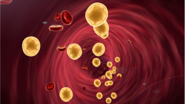
Michael: The experiment we’ve done, in culture, if you have cells, they usually require some sort of serum, and they tend to grow much better in young serum from a given animal more than they do in an old serum from a given animal. If you do the experiment, “Okay, what if I mix the young and old serum together?” If there’s good stuff in the young serum that’s dominant over the pro-aging or negative stuff in the old serum, you’d think that the young serum would win. If there’s bad stuff in the old serum that’s dominant and more pro-aging than the good stuff, then the old serum would win. We’ve done that experiment; the old serum wins every time we do that.

Every time?

Irina: Every time.

Michael: Every time I did it, which was just a few times.

Irina: If you contrast that with parabiosis, which has young animal veins right next door, the young animal has a young liver and kidneys that are capable of physically removing old blood constituents, but when you don’t have that, when you simply mix them 50/50 in a culture experiment for cells, then old serum wins. These and many other things are in progress that may not just focus on this question so much, but dilutions are in progress.

Your research suggests that by using extrinsic cues, it’s possible to quickly and robustly rejuvenate aged tissues and stem cells and also that it’s possible to rapidly age young cells simply by introducing these extrinsic cues. This supports the idea that calibrating the levels of blood factors to a more youthful profile can encourage youthful regeneration of organs and tissues. You’ve already demonstrated this in mice; could you tell us a bit more about what you have achieved so far using this approach?

Irina: We have demonstrated this in mice, and we have funding for Phase 2b clinical trials as initial tests to see if the same approach will work in people. The trials should be conducted this year as soon as we have the results of the study, so nothing yet to tell beyond the work in mice, but soon, there will be; when we know things, we publish them, and we are all set to go, so in one year, everybody should know.

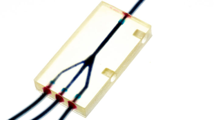
You mentioned apheresis today in your talk as a potential way to calibrate these various factors in aged blood to restore productive signaling in stem cells and surrounding tissues. The signaling is controlled by known networks and a combination could reset the interactions between cells to younger levels. How are you adapting apheresis to this task?

Irina: We have not adapted it yet, but we have an NIH grant to work on next-generation apheresis for small animals where we could test numerous interlocking modules and the precise administration of select factors. That is really very much in progress, I’m just working on proper support of that grant.

You’ve been using BONDCAT to identify the target blood factors. Can you explain how that works and how you have been working out what combination of factors are to be targeted?

Michael: People have been doing metabolic labeling for for hundreds of years now. You feed your favorite cell or organism some radioactive isotope for amino acid, it takes it up in the protein, you can detect it later by the radioactivity. BONDCAT works in a similar way, but instead of using radioactivity, the amino acid has a certain chemical modification to it that’s then easily and accurately detected chemically in a preparation of proteins later. How this is useful for our studies is that the gene that encodes the enzyme to help link that amino acid to the protein chain has been engineered to accept this non-natural amino acid. Because it’s a gene, you can make it transgenic, so we have mice that if you give them this, basically this amino acid substrate, it’ll put it into the product of protein and, therefore, label all the proteome of that particular mouse. You can even put this under genetic control so you can play with just the brain proteome, play with just the muscle proteome, whatever. That gives us the ability then to take that animal and say what in that animal is rejuvenative. We take the signaling or the blood or wherever your favorite theory is for what’s rejuvenative and expose another animal’s cells or tissue to that. We can see then which proteins are sticking and which proteins are being activated by that, and vice versa; we can say which is old and progeric.

So, you suggested a few years ago that you thought only a handful of primary factors and their interactions would need to be targeted for calibration. Do you still believe this is the case, and what factors are they?

Irina: We still believe it’s the case because, typically, we have only a few limited factors to build organs during embryogenesis. Through a number of our papers, we found that the same combination of the same factors work in a similar fashion to repair organs when we are adults. The same factors fail to work when we grow old. It will be limited; it will not be 70, 100, 4,000, or 50,000. Which ones, of course, we cannot tell you until the work is published.

What are your primary factors that you guys have identified right now, if any?

Michael: We and others have identified growth factors in the MAP kinase pathway.

Irina: MAP kinase pathway, TGF beta, those are the primary ones. Oxytocin in the map kinase part. Interleukin six, SASP, senescent cells’ secretory phenotype.

Michael: The hierarchy and cross talk can be signaling pathways among all the other hundreds of players. They’re nodes, and you try to go to their nodes.

Along the same lines, if it turns out that there actually are hundreds of factors to calibrate, how could apheresis be adapted to handle that kind of complexity?

Irina: So apheresis actually can be, it’s an engineering program and a problem that can be solved by engineering approaches. We don’t need 100 factors, but let’s say this 75 meters or 50 factors, they could be precisely measured and they could be calibrated precisely in a computer-operated way. It is a new combination of a few already existing engineering platforms. That is the part of the work that is partially funded by the NIH and partially by other sources that we are undertaking

Are you using deep learning?

Irina: We’ll use deep learning and computer learning AI eventually; the first step would be to develop enough of the algorithms and patterns from which to start the computer learning.

Apheresis is already approved by the FDA, so this should make adapting the technology and getting it approved easier. What are the challenges that you’ve had so far in getting it approved?

Irina: We don’t, so far, there are really zero challenges, it’s relatively easy. It’s more like a time challenge to make administrators to work on approvals that you need, rather than any blow-up or pushback.

I think you might have already answered this, but do you believe, assuming things go well, that this therapy might be in clinical trials in the next five years?

Irina: Human clinical trials this year.

Michael: The only thing that allows it to be open to the masses is that it’s already FDA approved; any practicing physician.

Irina: Under our guidance.

That’s “our” meaning your lab?

Michael: That’s the value added part to make it work.

Irina: So that’s bringing in our collaborator, but what Mike was saying was correct, that any approved physician can potentially prescribe it, but the danger would be in the details; if you want to calibrate something that if you do not calibrate correctly, it will cause you to stop producing red blood cells, you certainly will cause more harm, and no, no improvement at all.

Given that apheresis is available, why is there a continued fixation on plasma transfusion for rejuvenation, and is it really practical or plausible?

Michael: Well, that’s already got some momentum right now to steam for the past few years. Competing theories have to ramp up and still prove themselves. We might say something, but until people believe it or somebody else reproduces some experiments, then that gets some momentum too.

Irina: I can tell you this much, some people, not many, but some people justify plasma transfusions with our original 2005 parabiosis paper, and there is no justification in that paper that taking young plasma to an old person, old mammal, will do anything. There is none. There is a scientist in charge as the first author of that paper and last senior author of a heterochronic blood exchange paper published in Nature in 2017 or 16; there is no justification in his papers. If people would like to use young plasma, they should justify that with something else, not my work.

So how does your work compared to that being done by for example, Alkahest?

Irina: My work? My work shows that if you mix 50% of young blood with 50% of old blood, old blood dominates and there is no positive effect of young blood.

So the Alkahest approach doesn’t have good data to support it?

Irina: My data does not support that approach. I don’t know what data they might have, but it would not be my data.

You collaborate with Judy Campisi, who works with senescent cells and senolytics. You’ve mentioned we should be cautious and that simply destroying p16-expressing cells can be problematic, since we’re yet to fully understand the heterogeneity of senescent cells. Can you explain your concerns some more?

Irina: That’s actually not my concern; it’s a concern shared by numerous researchers in the senescence field, including Judy Campisi, who is a wonderful collaborator and a good friend, that p16 cannot be used as one single marker of cells that are bad and should be destroyed, that you need to understand much more about what senescent cells are, what markers identify the bad ones from the good ones before we use therapeutic approaches. That’s one concern. The second concern is that p16 is upregulated even in lung tissues when cells differentiate. It’s one of the hallmarks of cell differentiation, not just aging. The third concern is one that even Judy published, now more people are publishing, cells that are considered senescent are actually also good for some things. For example, they did an injury model in skin, and if you don’t have senescent cells, it doesn’t heal very well. There are numerous caveats, and the general theme is that whether we discuss Alkahest, young plasma, or senescent cells, there is no simplistic cure against age-related diseases, and simplistic viewpoints will be erroneous and could cause harm, particularly in people who are not scientists, who are not educated in that field.

So, do you think that senolytics could be developed into a suitable cotherapy alongside blood calibration? If so, what challenges do we have to overcome that to succeed?

Irina: We cannot comment on that because we have not really thought about it at all.

Michael: My thoughts about senolytics are that it seems clear that there might be some good, effective senolytics, but you wouldn’t want to senolyze yourself continuously, so then it becomes dosage and administration.

Do you think senolytics could work once or twice to address more macro symptoms, but then after that they can be risky, or are you saying, in general, don’t even go there?

Michael: Don’t really know yet. We’re trying to figure that out

Irina: My main point is that, for a young plasma infusion or senolytics, is not to prevent people from using them commercially or proposing to use them commercially, it’s to prevent people from starting to use them on themselves before they understand all of the side effects and basically killing themselves.

So, D&Q self-experimentation, you think, should be off the table?

Irina: Exactly. That’s my whole point. Or even if you offer some services, for example, “I am going to inject plasma or senolytics.” I think that should be done in a very conscientious way such as that it should be not just approved by the FDA but condoned by the FDA as highly based in fact. Some of those statements are dangerous, and people could seriously damage their health.

Michael: I don’t think it necessarily should be. I was happy that the FDA weighs in on occasion on this kind of stuff. I don’t think it necessarily should be illegal. In the early days of flight, people would throw on their jacket and put the scarf around their neck and jump into some, some rickety plane and fly off and crash and die.

For science.

Michael: It could be for vanity, it could be for science. It could be just to be cool, or it can be whatever. And I don’t disrespect that. It’s just that you’ve gotta realize that you’re self-experimenting. You might die from from some of this stuff, you don’t know. Then they get the FAA, and the FAA says, “Okay, we got some rules and regulations. You gotta follow this and then maybe you increase your chances of living,” and some people say, “I don’t like those rules and regulations.” Well, you can go fly off and be a bush pilot someplace or an ultralight, but you can’t argue with physics.

Irina: Yeah, exactly. That brings this point about the placebo effect, and what causes my disappointment and fear for people’s safety, is that many people who would like to use senolytics or GDF-11 or things of that nature that are not yet rigorously tested, young plasma, they do it because they believe that this is something that will rescue them, that this is something that is not allowed for them to be used, but if they find the secret way to use it, they will be better. I think we should somehow notify them saying this is not the case. Like Mike said, it is like jumping in an airplane made out of cardboard and trying to fly someplace as the first test pilot of that airplane. If that could be communicated to people somehow, then perhaps I would feel better about biohacking or using this technology. If they understand the dangers, fine; if they don’t understand the dangers, I would like, perhaps, the dangers to be explained better.

Now we’re going to ask you about the possibility of creating your own biotech company that you revealed today; you are, in fact, starting to create IMU. So, tell us more about IMU, your new startup.

Irina: So, we started IMU in November of last year, so quite recently. We have legal representation, a corporate law firm that comes recommended.

Which firm?

Irina: Gunderson, and they come highly recommended by one of my former students who started his successful company. So, we have three or four more scientists of the kind of laboratories that have successful companies or unsuccessful companies, so I feel really sheltered, protected and provided with a lot of advice in this regard. So did my former collaborators from Berkeley, for example, Professor Dave Schaffer, who started 4D. So they recommended Gunderson. Our law firm Gunderson specializes in very early startups, and they’re helping us to move along to launch. Right now, we are building a very strong team of senior as well as junior scientists. I don’t have my mission statement in front of me, but basically to paraphrase, we do not plan to make you live longer; we plan to make you be much healthier and safeguard you from the devastation of aging for additional decades.

Through apheresis?

Irina: Through a couple of innovative technologies.

Will you be abandoning academia to pursue this or would this be a side venture?

Irina: We have a very supportive for entrepreneurship environment at Berkeley. Numerous faculty start companies as founders and then continue as consultants and step back once the company has been successful and the rest of it is doing well. We’ll be in a similar situation. I’m starting it as a co-founder, there will be another senior co-founder, we have a board and Chief Medical Officer already, and a couple of people who have interviewed for CEO positions. Once it’s on track, and we have the rejuvenative services in place and being proud and blessed and so forth, then I will step back. Then, there are a few innovative rejuvenation technologies that are based on our IPs but are not identical to our presented work or published work.

Well, congrats and good luck. Is there a question interviewers never ask you that you’d like us to ask today?

Michael: Should you write your congressperson or state legislature to encourage them to support aging researchers?

Do you think the public’s not engaged enough?

Michael: Yes.

On the policy front for longevity science?

Michael: We’re a bunch of fringe nut jobs as far as the general public is concerned.

It’s changing fast; it seems like a lot of people nowadays are recognizing that we are in a real kind of cusp on the science.

Michael: Among the cognoscenti, it’s changing.

Irina: That is true. What is the percent of the population that understands that there is rejuvenation research and that it could be successful? When we go to these sorts of conferences, it seems that they’re all of us, but if you compare it to the general population, it’s probably less than 1%. It will be very important to have better outreach to people, also safeguarding them from using things that are unsafe and improving their understanding of what is in the pipeline. If you know that a better airplane, safer airplane, is being built to take you from one coast to another coast, are you going to fly in a cardboard airplane? Probably not. You’d probably say, “Okay, I can wait a couple of weeks.”

We would like to ask you a small favor. We are a non-profit foundation, and unlike some other organizations, we have no shareholders and no products to sell you. All our news and educational content is free for everyone to read, but it does mean that we rely on the help of people like you. Every contribution, no matter if it’s big or small, supports independent journalism and sustains our future.
Mitochondria

Refining the Allotopic Expression of Mitochondrial Genes

Researchers from SENS Research Foundation, including Matthew O’Connor and Amutha Boominathan, have published a new study showing how codons play an important role in getting copies of mitochondrial genes placed in the cellular nucleus to express themselves correctly [1].

A possible solution to mitochondrial diseases

Mitochondrial disease is not a single disease; in fact, it is a group of rare and related conditions that are thought to affect perhaps 1 in 5000 people. These are caused due to mutations in the genes involved in the process of aerobic respiration, one of the main functions of our mitochondria.

The majority of these altered and defective genes are located in the cell nucleus within our own DNA, but there are 13 genes that are located outside the safety of the nucleus in the mitochondria themselves, and they are much more vulnerable to damage from free radicals, which are a byproduct of their own energy production.

The reason these 13 genes are outside the nucleus is a legacy of the bacterial lineage of the mitochondria; rather than being naturally part of us, they are symbiotic organelles with their own DNA, as they had descended from bacteria that infiltrated the cells of our distant ancestors hundreds of millions of years ago. These bacteria had become fully symbiotic with our cells long ago, and, slowly but surely, their mitochondrial DNA (mtDNA) has moved, one gene at a time, into our own cellular nuclei.

Mutations to the mitochondrial genes that facilitate aerobic respiration are always bad news, but mutations to the 13 genes that still reside in the mitochondria can often have severe consequences. The researchers who have published this study have been studying the mitochondria in the context of aging and disease for many years, and they have a proposed solution to the problem of mutations to these 13 genes: allotopic expression via gene therapy.

Allotopic expression is the expression of genes in the cell nucleus that are normally expressed only by the mitochondrial genome. In other words, it delivers copies of the 13 mitochondrial genes, in this case using gene therapy, into the cell nucleus and finishes the job that nature started millennia ago. The aim is for the required gene to express itself normally, thus correcting the mitochondrial disease.

So far so good, but one problem

Unfortunately, the mitochondria does not use the same genetic code that our own mammalian DNA does. Instead it utilizes its own code, which uses codon frequencies more akin to the bacteria from which it is thought to have evolved. There have been various attempts, dating back to the 1980s, to recode the mitochondrial genes so that they use the same code we do, and it was the result of these early studies that allotopic expression was first suggested. Originally used as a research tool to examine mitochondrial proteins and their interactions, this method is now being developed as a potential way to address mitochondrial disease by creating copies of genes to replace the ones mutated in the mtDNA.

This new study explores the role of codons, a sequence of three DNA or RNA nucleotides that relate to a specific amino acid or stop signal during protein synthesis. Importantly, this paper also sheds light on how optimizing codons to communicate with the mitochondria more efficiently could help allotopic expression become a workable therapy in humans.

Mutations in mitochondrial DNA can be inherited or occur de novo leading to several debilitating myopathies with no curative option and few or no effective treatments. Allotopic expression of recoded mitochondrial genes from the nucleus has potential as a gene therapy strategy for such conditions, however progress in this field has been hampered by technical challenges. Here we employed codon optimization as a tool to re-engineer the protein-coding genes of the human mitochondrial genome for robust, efficient expression from the nucleus. All 13 codon-optimized constructs exhibited substantially higher protein expression than minimally-recoded genes when expressed transiently, and steady-state mRNA levels for optimized gene constructs were 5–180 fold enriched over recoded versions in stably-selected wildtype cells. Eight of thirteen mitochondria-encoded oxidative phosphorylation (OxPhos) proteins maintained protein expression following stable selection, with mitochondrial localization of expression products. We also assessed the utility of this strategy in rescuing mitochondrial disease cell models and found the rescue capacity of allotopic expression constructs to be gene specific. Allotopic expression of codon optimized ATP8 in disease models could restore protein levels and respiratory function, however, rescue of the pathogenic phenotype for another gene, ND1 was only partially successful. These results imply that though codon-optimization alone is not sufficient for functional allotopic expression of most mitochondrial genes, it is an essential consideration in their design.

Conclusion

The MitoSENS team at SENS Research Foundation has made great strides in bringing allotopic expression via gene therapy closer to the clinic, and while there are certainly more challenges ahead to get all 13 mitochondrial genes copied to the nucleus and expressed optimally, there is plenty of room for optimism. Recently, the MitoSENS team successfully funded its second project, MitoMouse, with us at lifespan.io, which builds on the findings in this paper to take the next step and pursue mitochondrial gene therapy in an animal model. We were delighted to have been involved in supporting this study and we wish the team the best of luck in developing allotopic expression into a workable therapy, as it has big implications for mitochondrial disease as well as aging.

We would like to ask you a small favor. We are a non-profit foundation, and unlike some other organizations, we have no shareholders and no products to sell you. All our news and educational content is free for everyone to read, but it does mean that we rely on the help of people like you. Every contribution, no matter if it’s big or small, supports independent journalism and sustains our future.

Literature

[1] Lewis, C. J., Dixit, B., Batiuk, E., Hall, C. J., O’Connor, M. S., & Boominathan, A. (2020). Codon optimization is an essential parameter for the efficient allotopic expression of mtDNA genes. Redox Biology, 101429.

Rejuvenation Roundup January

Rejuvenation Roundup January 2020

2020 has been a common year to define the beginning of “the future”, so now’s a great time to discover just how close real biotechnology has come to popular expectations. To kick off the first month of the new year, we have plenty of research to talk about, along with announcements, three X10 episodes, and one more video from last year’s EARD conference.

LEAF News

Team and activities

Brian Kennedy Joins the LEAF Scientific Advisory Board: Professor Kennedy, a Distinguished Professor in the Department of Biochemistry and Physiology at the University of Singapore, offers his insight and expertise as part of our SAB.

Journal Club A Human Trial for Skin Aging: Rapamycin is a common research topic for aging researchers, and this month, Oliver hosted a discussion on a human trial of its effects on skin aging,

Rejuvenation Roundup Podcast

Ryan O’Shea of Future Grind hosts this month’s podcast, showcasing the events and research discussed here.

Research Roundup

Treating Glaucoma With Senolytics: In an animal model of glaucoma, ganglion cell death was halved by removing the inflammation caused by the senescence-associated secretory phenotype (SASP) – which required the removal of senescent cells.

Fisetin: This explanatory article offers a broad look at this common supplement, identifying its sources, effectiveness as a senolytic, and clinical studies.

Mitochondria Found Independently Living in Blood: This insightful study shows mitochondria living in independent structures outside of cells. Why these mitochondria are there, and what purpose they serve for the body, are facts not yet discovered.

Dasatinib plus quercetin prevents uterine age-related dysfunction and fibrosis in mice: Dasatinib and quercetin are a well-known combination of senolytics, and this study shows that they remove the senescent cells that are responsible for uterine problems in older mice.

Cellular senescence changes extracellular vesicles’ cargo and function: This study outlines a direct link between cellular senescence and altered intercellular communication, two of the hallmarks of aging; in this case, the intercellular communication consists of extracellular vesicles.

Depletion of senescent astrocytes protects cognitive function in mice: Radiotherapies for cancer cause brain aging, but this study shows that senolytics reverse some of this damage in mice.

FOXO4-DRI restores testosterone production in mice: Another senolytic study shows that removing senescent cells ameliorates late-onset hypogonadism, at least in mice.

Good diets: In line with previous research on the subject, three separate studies show that the Mediterranean diet and caloric restriction are connected with healthy longevity.

Exercise: Again entirely unexpectedly, researchers have documented how exercise can extend healthy lifespan, whether that exercise involves marathons or sprints.

An extended keto diet leads to obesity in mice: An unrestricted diet in which most calories are from fat rather than carbohydrates causes obesity and harms metabolic health in this mouse study.

High-protein diets increase cardiovascular risk: This study shows that excessive protein causes mitochondrial dysfunction in macrophages, which leads to their greater accumulation in cardiovascular plaque.

CRISPR/Cas9 discovers the secret of regeneration in the axolotl: This is a small salamander with a large genome, and researchers have used modern gene editing technologies to discover why it can regenerate.

Well-maintained splicing may cause negiglible senescence in mole rats: This study has found that naked mole rats control their DNA splicing more effectively than in other species, preventing adverse events.

NAD+ supplementation prevents hearing loss in prematurely aged mice: In a mouse model of Cockayne syndrome, a condition that causes premature aging and hearing loss, nicotinamide riboside (NR), a source of NAD+, was shown to be effective.

Dill extract produces elastin fibers in the ascending aortae of aged mice: After ingesting dill extract for three months in drinking water, aged mice have been shown to produce elastin fibers that restore the function of their critical arteries.

News Nuggets

Revel Pharma to Develop Glucosepane Breakers: Starting with research funded by Yale Lab with the cooperation of SENS Research Foundation, the biotechnology company Revel Pharma has begun to develop a method of removing glucosepane, the principal source of dangerous age-related collagen crosslinks in human beings.

Three Groundbreaking Longevity Startups: This article discusses Underdog Pharmaceuticals, Juvena Therapeutics, and Revel Pharmaceuticals, three startups that are likely to make waves in the future of rejuvenation biotechnology development.

Building an Atlas of Senescent Cells: A team of researchers led by Prof. Judy Campisi has created an atlas that describes senescenent cells and the proteins they produce as part of the senescence-associated secretory phenotype (SASP).

We would like to ask you a small favor. We are a non-profit foundation, and unlike some other organizations, we have no shareholders and no products to sell you. All our news and educational content is free for everyone to read, but it does mean that we rely on the help of people like you. Every contribution, no matter if it’s big or small, supports independent journalism and sustains our future.

Results From a Human Trial for Skin Aging

The January edition of Journal Club, hosted by Dr. Oliver Medvedik, took a look at the results of a recent human trial in which the drug rapamycin was used to treat skin aging with some promising results.
We would like to ask you a small favor. We are a non-profit foundation, and unlike some other organizations, we have no shareholders and no products to sell you. All our news and educational content is free for everyone to read, but it does mean that we rely on the help of people like you. Every contribution, no matter if it’s big or small, supports independent journalism and sustains our future.
Independent in blood

Mitochondria Found Independently Living in Blood

The mitochondria are well known as being the powerhouses of the cell, as they convert nutrients into the energy that our cells need in order to function and remain alive. Until recently, it was believed that they remain within our cells all their lives, but a new discovery by researchers at the Montpellier Cancer Research Institute has turned our understanding on its head.

Introducing the mitochondria

Mitochondria, which are often called the powerhouses of cells, act like miniature factories, converting the food we eat into usable energy in the form of a chemical called adenosine triphosphate (ATP). ATP provides energy to fuel a myriad of cellular processes, such as muscle contraction, nerve impulse propagation, and protein synthesis. ATP is common to all forms of life and is often referred to as the “molecular unit of currency” of intracellular energy transfer.

Interestingly, mitochondria did not originate as part of multicellular life; they appear to be stowaways in our cells and have their own unique DNA, which is separate from our own. It is widely thought that they merged with a very early ancestor of all multicellular life to form a symbiotic relationship. Mitochondria become dysfunctional as we age and are host to their own separate (though similar) forms of damage.

The age-related failure and dysfunction of mitochondria that occurs due to damage to their DNA is one of the hallmarks of aging.

How on Earth did we miss this?

The research team has published a new study, which shows that aside from the usual mitochondrial populations living inside our cells, there are also wandering mitochondria floating around in our bloodstreams. From time to time, mitochondria are found outside the cells, but only in the context of debris within platelets, so what are these intrepid mitochondria doing out there alone in the bloodstream?

The seven-year study has confirmed that there are intact and fully functional mitochondria in the blood plasma living inside highly stable structures [1]. The researchers used electron microscopy to examine plasma samples from approximately 100 people. They discovered that there were up to 3.7 million of these lone mitochondria living inside these structures for every milliliter of plasma.

Given the amount of plasma samples that are taken and tested daily, it begs the question “How could we have missed such an important discovery?” The researchers had a hunch that this might be the case by reading previous studies, which had discovered that a typical person’s blood plasma could contain up to 50,000 times more mitochondrial DNA (mtDNA) than regular DNA. The multitude of circulating mitochondria riding around in these protective bubble structures explains why.

While it is mind-boggling, given how many of these mitochondria there are circulating in our bloodstreams, these researchers specialize in DNA analysis in the bloodstream and have worked with fragmented mtDNA in particular.

The researchers suggest that these fully working mitochondria are not merely there by chance but are perhaps playing a role in cell signaling, helping to facilitate communication between cells and regulating immune and inflammatory responses.

Mitochondria are considered as the power-generating units of the cell due to their key role in energy metabolism and cell signaling. However, mitochondrial components could be found in the extracellular space, as fragments or encapsulated in vesicles. In addition, this intact organelle has been recently reported to be released by platelets exclusively in specific conditions. Here, we demonstrate for the first time, that blood preparation with resting platelets, contains whole functional mitochondria in normal physiological state. Likewise, we show, that normal and tumor cultured cells are able to secrete their mitochondria. Using serial centrifugation or filtration followed by polymerase chain reaction-based methods, and Whole Genome Sequencing, we detect extracellular full-length mitochondrial DNA in particles over 0.22 µm holding specific mitochondrial membrane proteins. We identify these particles as intact cell-free mitochondria using fluorescence-activated cell sorting analysis, fluorescence microscopy, and transmission electron microscopy. Oxygen consumption analysis revealed that these mitochondria are respiratory competent. In view of previously described mitochondrial potential in intercellular transfer, this discovery could greatly widen the scope of cell-cell communication biology. Further steps should be developed to investigate the potential role of mitochondria as a signaling organelle outside the cell and to determine whether these circulating units could be relevant for early detection and prognosis of various diseases.

Conclusion

The discovery that there are populations of mitochondria outside of the cells raises a whole host of questions and turns our understanding of mitochondria upside down. It is going to require more research in order to understand why they are there and how their protective structures are formed, but this is what makes biology so exciting. There are always new discoveries to be made, and this one is particularly intriguing.

We would like to ask you a small favor. We are a non-profit foundation, and unlike some other organizations, we have no shareholders and no products to sell you. All our news and educational content is free for everyone to read, but it does mean that we rely on the help of people like you. Every contribution, no matter if it’s big or small, supports independent journalism and sustains our future.

Literature

[1] Al Amir Dache, Z., Otandault, A., Tanos, R., Pastor, B., Meddeb, R., Sanchez, C., … & El Messaoudi, S. (2019). Blood contains circulating cell-free respiratory competent mitochondria. The FASEB Journal.

Three buildings

Three Groundbreaking Longevity Startups

There are lots of exciting companies working in the aging field, and it’s a great time to tell you about three of the more interesting ones. Most of these companies are a while away from human trials yet, but their innovations could possibly be truly game changing.

Underdog Pharma

Underdog Pharmaceuticals is a spin-off company of SENS Research Foundation and is developing a novel approach to treating atherosclerosis.

Atherosclerosis is the number one killer worldwide, and it currently has no totally effective solution. There are three ways in which current medicine tries to address it: Lifestyle changes, including diet and exercise; drugs that slow down the rate of cholesterol accumulation; and interventions such as stents and bypass surgery.

None of these approaches really addresses the root problem nor cures the disease, which is why Underdog Pharmaceuticals’ proposal is so compelling.

Underdog is developing a therapeutic approach that could potentially prevent or reverse atherosclerosis by removing a harmful lipid known as 7-ketocholesterol (7KC) from the arterial walls.

What is 7-ketocholesterol?

7KC is a harmful, oxidized byproduct of cholesterol. It is highly toxic with no redeeming qualities; it serves no purpose in our bodies aside from allowing heart disease to develop. Once 7KC becomes lodged in the arterial wall and an atherosclerotic lesion begins to form, the immune system responds and sends specific immune cells, known as macrophages, as the cleanup crew to remove it.

Unfortunately, macrophages are unable to metabolize 7KC, but they keep absorbing it until they become bloated. At this point, they become “foam cells”, huge bloated cells that are unable to process waste at all and form a large part of what make up atherosclerotic plaques.

The bad news is that as these engorged foam cells become stuck in the arterial walls and slowly die, they call other macrophages to the scene; and these reinforcements become trapped as well. Ironically, in trying to fix the problem, our own cells become part of it.

Underdog is developing a therapy that could potentially remove 7KC from the bloated macrophages, allowing them to return to healthy function, by delivering them a molecule that can extract the oxidized waste.

The therapy is based on a variant of cyclodextrin, an existing drug that is already approved by the FDA and has a good safety profile. This should cut down on the time it takes to get the new therapy approved, and the company has a plan to reach human trials in the next three or four years. If successful, this would offer a way to reverse atherosclerosis and put an end to the number one global killer.

Juvena Therapeutics

Juvena Therapeutics is a company focused on discovering and developing pluripotent stem cell-derived protein therapeutics; to put it another way, pluripotent stem cells secrete signaling proteins to other cells, causing them to regenerate and function like younger cells.

Rejuvenation using blood factors

While there are likely hundreds of signaling proteins involved in rejuvenation of tissue, the current evidence suggests that there are only a handful of primary signals that act like master regulators and orchestrate the entire process.

Juvena is working out which signaling factors are the most important to achieve this. Once the key signals that regulate rejuvenation are known, they could then be mass produced and used as a therapy in older people to spur the healthy regeneration of tissues and organs.

The company is focusing initially on sarcopenia, the age-related loss of muscle mass that leads to frailty and can result in lethal falls and serious injury in the elderly. Juvena is also looking at using these therapies for acute injury, trauma and wound healing for the elderly, who generally have poor tissue repair.

The unique drug discovery platform that the company has developed could allow it to target multiple tissues for whole body rejuvenation and to address things like neurodegeneration, cardiovascular disease, and lung fibrosis.

Revel Pharmaceuticals

Revel Pharmaceuticals is another spin-off biotech company based on years of research funded by the SENS Research Foundation and conducted at the Spiegel Lab at Yale. This company is developing a therapy that can break down glucosepane cross-links in our tissues which form as we age and cause our arteries to stiffen.

What are cross-links?

A proposed reason why we age is the accumulation of sugary metabolic wastes known as advanced glycation end-products (AGEs). AGEs are harmful compounds that are created when proteins or fats combine with sugars in the bloodstream in a process known as glycation. AGEs can also be encountered in foods, particularly foods that have been exposed to high temperatures, such as in grilling or frying.

AGEs can cause random damage by altering protein structure and function, but the inflammation they cause is thought to be a primary contributor to the background of chronic, age-related inflammation typically seen in older people. AGEs contribute to a variety of microvascular and macrovascular complications via the formation of cross-links between molecules in the basement membrane of the extracellular matrix and by interacting with the receptor for advanced glycation end products (RAGE).

This interaction with RAGE leads to oxidative stress and the activation of protein complex NF-?B, a master regulator of inflammation, DNA transcription, and cell survival. This activation leads to excessive levels of NF-?B activity and is thought to be responsible for AGE-associated inflammation and cellular damage.

High levels of AGEs are linked to a number of age-related diseases, including diabetes, heart disease, and Alzheimer’s. There is also evidence to support that people who have high blood sugar, such as people with diabetes, have a higher risk of producing more AGEs that can accumulate in the body faster than they can be cleared, thus contributing to the decline of multiple organs.

Our bodies have ways to eliminate these harmful compounds, including antioxidants and enzymes; however, these mechanisms have their limits, and if we consume too many AGEs, or too many appear through the normal operation of metabolism, they can begin to accumulate, leading to increasing numbers of cross-links, oxidative stress, and inflammation.

Glucosepane

There are many types of AGEs, each with its own unique chemical composition. The body is able to remove some of them but lacks the biological tools needed to break down others; unfortunately, glucosepane falls into the latter category.

Glucosepane is the most abundant AGE in the body, and this particular AGE and its resulting cross-links cannot be broken down by the body at all. Instead, they continue to build up throughout our lives.

The Spiegel lab has been working on ways to break down AGEs, particularly glucosepane, for many years. This molecule has proven to be a considerable challenge, initially because it was hard to manufacture the substance outside the body in order to test interventions against it, and because doing so is simply difficult in general.

In 2015, this all changed when, thanks to funding from SENS Research Foundation, the Spiegel lab successfully developed a way to create glucosepane on demand. From there, the lab identified bacterial enzymes that could break down glucosepane cross-links, and now things have reached the point at which those enzymes are ready to be developed into a therapy.

Now that Revel Pharmaceuticals has been created, the race to develop glucosepane breakers and take them to market has finally begun. Of all the companies in this article, this one is likely to have the longest road to achieve its goal, but it is a goal that could be transformative in healthcare.

These three biotech companies are merely a fraction of the companies now working in this space, but we find them to be the most exciting because of what they are trying to achieve.

Collagen

Revel Pharma to Develop Glucosepane Breakers

Some pleasant news has recently arrived: Revel Pharmaceuticals has successfully completed a seed round in order to begin developing therapeutics that target glucosepane crosslinks, which are a proposed reason why we age, develop diseases such as diabetes, and suffer from stiffened arteries and hypertension.

In the business of startups, a seed round refers to a series of related investments in which 15 or fewer investors “seed” a new company with funds, typically ranging between $50,000 to $2 million. This money is often used to support initial research and early development. Investors are typically offered equity, convertible notes, or preferred stock options in exchange for investing.

What are crosslinks?

A proposed reason why we age is the accumulation of sugary metabolic wastes known as advanced glycation end-products (AGEs). AGEs are wastes that are, in some cases, hard for our metabolisms to break down quickly enough; they may even be impossible for us to break down at all, as our cells lack the tools to do so.

There are various AGEs present in the body, although none are as common as glucosepane, which is the most abundant by a huge margin. Glucosepane is very hard for the body to break down (if indeed it can at all), and it can last several decades once formed.

These AGEs form crosslinks, binding together important proteins, such as those making up the supporting extracellular matrix scaffold, and preventing them from moving.

The elastic properties of skin and blood vessel walls are due to the extracellular matrix having a particular structure, and crosslinks degrade that structure, preventing it from functioning correctly. The presence of AGEs is thought to contribute to blood vessel stiffening with age and is implicated in hypertension and diabetes.

The SENS solution to dealing with AGEs is to find ways to break down the crosslinks, thus freeing up the trapped proteins and restoring tissue elasticity.

Finally, things are shifting up a gear

Revel Pharmaceuticals is a biotech company that has been established to develop drugs that can break harmful glucosepane crosslinks and is built on the work carried out by the Spiegel Lab at Yale over many years.

This research, funded by SENS Research Foundation, has steadily been progressing: initially, the researchers discovered how to synthesize glucosepane in order to facilitate testing, then they discovered bacterial enzymes that could break down glucosepane cross-links.

Those enzymes are now under development at the new company and will hopefully become the jumping-off point for their development into a therapy that can break down glucosepane crosslinks.

For the past 10 years, Yale Professors David Spiegel and Jason Crawford have been working on tools to enable the development of glucosepane-cleaving drugs. Kizoo Technology Capital investors say now is the time to advance this groundbreaking research toward the clinic and are leading funding of a new company, Revel Pharmaceuticals Inc., founded by Drs. David Spiegel, Jason Crawford, and Aaron Cravens. Kizoo leads the seed financing round at Revel, with Oculus co-founder Michael Antonov participating. SENS Research Foundation provided funding to the Yale GlycoSENS group for several years.

The long-lived collagen proteins that give structure to our arteries, skin, and other tissues are continuously exposed to blood sugar and other highly reactive molecules necessary for life. Occasionally, these sugar molecules will bind to collagen and form toxic crosslinks that alter the physical properties of tissues and cause inflammation. As a result, tissues slowly stiffen with aging, leading to rising systolic blood pressure, skin aging, kidney damage, and increased risk of stroke and other damage to the brain.

Perhaps the most important of these Advanced Glycation End-product (AGE) crosslinks is a molecule called glucosepane. Revel is developing therapeutics that can cleave glucosepane crosslinks thus maintaining and restoring the elasticity of blood vessels, skin, and other tissues, and preventing the terrible effects of their age-related stiffening.

The Yale group’s first major milestone – the first complete synthesis of glucosepane – was highly recognized when published in Science. Since then progress has been rapid, with development of glucosepane binding antibodies and discovery of therapeutic enzyme candidates capable of breaking up glucosepane crosslinks. Revel will build upon this progress by advancing the first GlycoSENS therapeutics into the clinic.

“This is truly a first. We are proud to help Revel open an entirely new category in repairing a significant damage of aging – crosslinking of collagen. Glucosepane crosslinks may cause not only wrinkles on your face but also lead to age-related rising blood pressure and possibly stroke.” says Frank Schueler, Managing Director of Kizoo Technology Capital.

David Spiegel, MD, PhD, Professor of Chemistry at Yale University and Revel founder says: “We are delighted to join Kizoo in building a world-class team to advance crosslink-breaking therapeutics into the clinic. These first-in-class agents have enormous potential to help patients suffering from a wide range of diseases”

“Collagen is the infrastructure of our bodies – in every tissue, supporting cellular function and health – but with aging, this critical molecular infrastructure accumulates damage. By clearing out this damage, we can restore tissue function and repair the body. Revel is one of only a few companies taking a repair-centric approach to treat diseases of aging and one day our AGE-cleaving therapeutics will undo this damage at the molecular level,” says Aaron Cravens, co-Founder of Revel Pharmaceuticals.

Conclusion

Finally, after many years, the research is now at the point at which a biotech company can be spun off to take the technology to market, which is where the real battle begins. Glucosepane breakers have been lagging behind most of the SENS categories, so to see a biotech company finally developing a product based on this research is very welcome news.

On a related note, it is great that Kizoo is once again supporting cutting-edge biotech companies that are focused on repairing the damage of aging. Kizoo and its associated non-profit, Forever Healthy Foundation, have had a huge impact on the industry in the last few years, and we at LEAF greatly appreciate their contribution to the cause.