The Blog

Building a Future Free of Age-Related Disease

Mouse with prominent whiskers

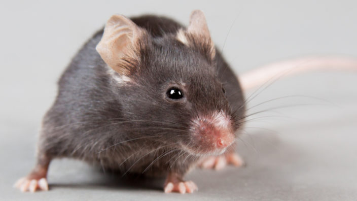
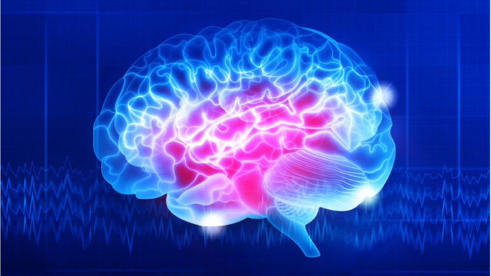
NMN Restores Brain Function In Old Mice

A group of researchers has demonstrated that treatment with NMN, a precursor of NAD+, restores neurovascular coupling (NVC) in aged mice [1]. Since NVC deficiency seems to be a major factor in the age-related decline of cognitive and motor functions, this discovery presents exciting new possibilities for longevity research.

Neurovascular coupling

While the human brain is the evolutionary advantage that brought us to where we are today, operating this machine requires considerable resources. Our cerebral blood flow (CBF) accounts for 15% of cardiac output and 20% of resting total oxygen consumption, even though the brain itself comprises just 2% of body mass. CBF has to be constantly redirected to the regions of the brain that are currently active, and NVC is the mechanism in charge of this complex operation. Importantly, the CBF/cardiac output ratio decreases with age [2].

NVC works mostly by vasodilation – i.e. by widening blood vessels in regions where augmentation of blood flow is required [3]. The cellular mechanism behind this involves production of nitric oxide (NO) by the endothelial cells that line blood vessels in the brain. This is just the first step in an intricate multi-stage process that eventually leads to cerebral vasodilation and allows the brain to perform at its best.

There is growing evidence that NVC function declines with age [4] and that this decline plays an important role in the development of cognitive impairment and dementia in old people. Some motor functions, such as gait performance, are affected as well.
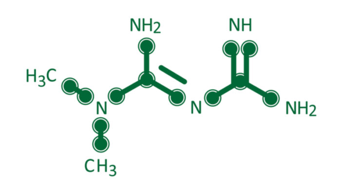
Why is NMN needed?

NAD+ is important for numerous cellular processes, but it is known best for its role in the electron transport chain, the main intracellular energy-producing process that takes place in mitochondria. The decline of NAD+ levels with age corresponds to impaired mitochondrial function. Their replenishment through NMN intake has been shown to reverse age-related dysfunction in multiple organs and to improve healthspan in mice.

Mitochondrial degeneration leads to increased mitochondrial oxidative stress, which, in turn, contributes to cardiovascular dysfunction [5] and age-related neurovascular impairment [6]. Hypothesizing that the decline in NVC function can also be attributed to mitochondrial degeneration, the researchers set out to determine whether this effect can be reversed by NMN supplementation.

NMN increases NO production via mitochondrial restoration

During the experiment, aged mice were treated with NMN for two weeks and then evaluated for cognitive and motor functions that are known to be affected by NVC response.

First, CBF responses were measured following whisker stimulation that is known to trigger a cascade of brain activity in mice. Initial CBF responses were much fainter in aged mice, indicating age-related NVC impairment. NMN treatment significantly increased CBF responses to whisker stimulation in aged mice to the levels observed in young mice. MRI analysis showed the same picture: CBF was weaker in aged mice compared to young ones, but NMN treatment increased it significantly.

To establish that these effects were primarily due to increased NO production, the researchers administered the NO synthase inhibitor L-NAME to the same three groups of mice (young, untreated aged, and treated aged). As a result, NVC responses were greatly decreased in young mice but not in the old, untreated ones, indicating that the latter group produced much less NO. Aged mice treated with NMN demonstrated restored L-NAME response.

Experiments in vitro showed that NMN treatment attenuates age-related mitochondrial oxidative stress in cerebromicrovascular endothelial cells (CMVECs). To determine whether this mechanism influences NO production, the scientists also performed a series of tests using mtROS (mitochondrial reactive oxygen species) inhibitors/scavengers that are structurally different from NMN: SS-31 peptide, resveratrol, and MitoTEMPO. These compounds elicited an increased NO response as well, which led the researchers to conclude that the restoration of mitochondrial function through NMN was indeed the underlying cause for the increased NO production. The authors also tried to determine the role of sirtuins that are known to be activated by NAD+. Knocking out SIRT1/SIRT2 genes in CMVECs prevented the beneficial effects of NMN, lending weight to the hypothesis that sirtuins are an important intermediary of this pathway.

Several cognitive and motor skills tests demonstrated substantial improvement following NMN treatment, such as the water maze test, in which mice are required to find a floating platform to get out of the water as fast as possible. This test evaluates working memory that is involved in immediate conscious perception and thus requires surges of CBF. Aged mice with NMN supplementation showed restoration of working memory to levels observed in young mice. Gait performance was yet another aspect in which significant improvement was achieved. Gait alterations have been linked to neurovascular dysfunction both in older humans and in animal models of aging. Age-related gait issues are a leading cause of falls and walking impairment in older people.

Conclusion

NMN supplementation has been consistently showing promising results in many areas of longevity research. The current study is the first to demonstrate that this method can be used to restore NVC and to attenuate cognitive and motor problems associated with its age-related decline. As NVC is also compromised in patients with Alzheimer’s disease, the authors suggest that their findings can contribute to the ongoing fight against this disease. Since mitochondrial dysfunction is one of the hallmarks of aging, further research of the apparent beneficial effects of NMN supplementation on mitochondrial metabolism is imperative.

We would like to ask you a small favor. We are a non-profit foundation, and unlike some other organizations, we have no shareholders and no products to sell you. All our news and educational content is free for everyone to read, but it does mean that we rely on the help of people like you. Every contribution, no matter if it’s big or small, supports independent journalism and sustains our future.

Literature

[1] Tarantini, S., Valcarcel-Ares, M. N., Toth, P., Yabluchanskiy, A., Kiss, T., Ballabh, P., … & Ungvari, Z. (2020). Nicotinamide mononucleotide (NMN) supplementation rescues cerebromicrovascular endothelial function and neurovascular coupling responses and improves cognitive function in aged mice. The FASEB Journal34(S1), 1-1.

[2] Xing, C. Y., Tarumi, T., Liu, J., Zhang, Y., Turner, M., Riley, J., … & Zhang, R. (2017). Distribution of cardiac output to the brain across the adult lifespan. Journal of Cerebral Blood Flow & Metabolism37(8), 2848-2856.

[3] Girouard, H., & Iadecola, C. (2006). Neurovascular coupling in the normal brain and in hypertension, stroke, and Alzheimer disease. Journal of applied physiology100(1), 328-335.

[4] Lipecz, A., Csipo, T., Tarantini, S., Hand, R. A., Ngo, B. T. N., Conley, S., … & Csiszar, A. (2019). Age-related impairment of neurovascular coupling responses: a dynamic vessel analysis (DVA)-based approach to measure decreased flicker light stimulus-induced retinal arteriolar dilation in healthy older adults. Geroscience41(3), 341-349.

[5] Rosca, M. G., & Hoppel, C. L. (2010). Mitochondria in heart failure. Cardiovascular research88(1), 40-50.

[6] Petrozzi, L., Ricci, G., Giglioli, N. J., Siciliano, G., & Mancuso, M. (2007). Mitochondria and neurodegeneration. Bioscience reports27(1-3), 87-104.

Telomere shortening

Restoring Telomerase Activity in Telomere Biology Diseases

Researchers have demonstrated that it is possible to restore telomerase activity in stem cells affected by telomere biology diseases, which prevent them from producing telomerase and repairing their telomeres.

Telomeres and telomerase

Each chromosome that stores our genetic information has a protective cap at each end known as a telomere, a specific DNA sequence that is repeated thousands of times. This sequence has two purposes: it protects the coding regions of the chromosome and prevents it from being damaged, and it acts as a clock that controls the number of replications a cell can make; this is known as the Hayflick limit.

With each cell division, this protective cap is worn away, gradually wearing down and shortening the telomere. The telomeres suffer this steady attrition due to the inability of DNA polymerases to fully restore the sequence of DNA.

Once telomeres reach a critically short length, cellular senescence is initiated as a way of preventing DNA double-strand breaks from being repaired, which can result in fused chromosomes.

This is the case for the majority of our regular somatic cells; however, stem cells are a bit different and have a way to maintain replicative capacity by expressing telomerase reverse transcriptase (TERT). When TERT is combined with the non-coding RNA (ncRNA) template telomerase RNA component (TERC), it creates telomerase ribonucleoprotein (RNP), which is able to replenish the protective telomere repeats. This is what allows our stem cells to keep functioning sometimes for decades or even our entire lives.

However, there are multiple telomere biology diseases in which this telomere maintenance is disrupted in the stem cells, leading to syndromes such as dyskeratosis congenita, a genetic form of bone marrow failure resulting in the inability of the marrow to produce sufficient blood cells. It can also lead to conditions such as aplastic anemia, liver cirrhosis, and pulmonary fibrosis.

The researchers of a new study have demonstrated that it is possible to restore lost telomerase activity in stem cells that have been affected by telomere biology diseases [1]. They show that by using PAPD5 inhibitors, it is possible to restore lost telomere length both in vitro and in mice.

Genetic lesions that reduce telomerase activity inhibit stem cell replication and cause a range of incurable diseases, including dyskeratosis congenita (DC) and pulmonary fibrosis (PF). Modalities to restore telomerase in stem cells throughout the body remain unclear. Here, we describe small-molecule PAPD5 inhibitors that demonstrate telomere restoration in vitro, in stem cell models, and in vivo. PAPD5 is a non-canonical polymerase that oligoadenylates and destabilizes telomerase RNA component (TERC). We identified BCH001, a specific PAPD5 inhibitor that restored telomerase activity and telomere length in DC patient induced pluripotent stem cells. When human blood stem cells engineered to carry DC-causing PARN mutations were xenotransplanted into immunodeficient mice, oral treatment with a repurposed PAPD5 inhibitor, the dihydroquinolizinone RG7834, rescued TERC 3′ end maturation and telomere length.

Conclusion

These findings have the potential to support the development of therapies for treating conditions such as dyskeratosis congenita and pulmonary fibrosis. Also, as telomere attrition and the resulting stem cell exhaustion are hallmarks of aging, this research may also have utility for the treatment of other age-related diseases.

We would like to ask you a small favor. We are a non-profit foundation, and unlike some other organizations, we have no shareholders and no products to sell you. All our news and educational content is free for everyone to read, but it does mean that we rely on the help of people like you. Every contribution, no matter if it’s big or small, supports independent journalism and sustains our future.

Literature

[1] Nagpal, N., Wang, J., Zeng, J., Lo, E., Moon, D. H., Luk, K., … & Lindsley, R. C. (2020). Small-Molecule PAPD5 Inhibitors Restore Telomerase Activity in Patient Stem Cells. Cell Stem Cell.

Blood Vessel

Refining Senolytic Drugs to Be Less Toxic and More Effective

Researchers have developed a way to modify an existing cancer drug with toxic side effects into something that is less toxic to blood platelets and more effective at removing harmful and inflammatory senescent cells, one of the reasons we age, from mice.

What are senescent cells?

As you age, increasing numbers of your cells enter into a state known as senescence. Senescent cells do not divide or support the tissues of which they are part; instead, they emit a range of potentially harmful chemical signals that encourage nearby healthy cells to enter the same senescent state. Their presence causes many problems: they reduce tissue repair, increase chronic inflammation, and can even eventually raise the risk of cancer and other age-related diseases.

Senescent cells normally destroy themselves via programmed cell death known as apoptosis, and they are also removed by the immune system; however, the immune system weakens with age, and increasing numbers of senescent cells escape this process and begin to accumulate in all the tissues of the body.

By the time people reach old age, significant numbers of these senescent cells have built up, causing chronic inflammation and damage to surrounding cells and tissue. Senescent cell accumulation is proposed to be one of the nine reasons we age and develop the age-related diseases that eventually kill us.

Senolytics

Senolytics are a new class of drugs that are known to be able to induce cell death. One of the first existing drugs to be identified as having senolytic potential was the cancer drug navitoclax (also known as ABT263), an orally active cancer drug, which works by inhibiting the Bcl-2 (B-cell lymphoma 2) pathway. The Bcl-2 gene is the founding gene of the Bcl-2 family of proteins that regulate cell death, by either inhibiting or inducing apoptosis.

Some types of cancers and senescent cells are able to manipulate the Bcl-2 pathway so that it no longer induces apoptosis and allows them to survive and remain at large in the body. Navitoclax targets both Bcl-2 and Bcl-xl as part of its activity and is therefore of interest in the context of destroying senescent cells, but there are concerns about using it.

The main issue with navitoclax, which was identified during the early senolytic experiments, was that it has some serious side effects that limited its use. Chief among these side effects was its dose-limiting platelet toxicity, and using too much runs the risk of causing thrombocytopenia. Thrombocytopenia is a condition that is characterized by abnormally low levels of platelets, known as thrombocytes, in the blood. Platelets are colorless blood cells that help the blood to clot, and they stop bleeding by clumping and forming plugs in blood vessel injuries.

The researchers of a new study have demonstrated that this platelet toxicity can be reduced by the use of proteolysis-targeting chimera (PROTAC) technology to convert the navitoclax into PZ15227, which is far less toxic to platelets and only inhibits Bcl-xl, which is more relevant to senescent cells [1].

The researchers also report that not only can its toxicity be reduced by this conversion process, its effectiveness against senescent cells can be improved as well, leading to rejuvenation of tissue stem and progenitor cells in aged mice. Presumably, this restoration of tissue upkeep and more efficient repair is due to the removal of harmful senescent cells and the resulting reduction of inflammation, which normally inhibits stem cell activity and tissue repair.

Small molecules that selectively kill senescent cells (SCs), termed senolytics, have the potential to prevent and treat various age-related diseases and extend healthspan. The use of Bcl-xl inhibitors as senolytics is largely limited by their on-target and dose-limiting platelet toxicity. Here, we report the use of proteolysis-targeting chimera (PROTAC) technology to reduce the platelet toxicity of navitoclax (also known as ABT263), a Bcl-2 and Bcl-xl dual inhibitor, by converting it into PZ15227 (PZ), a Bcl-xl PROTAC, which targets Bcl-xl to the cereblon (CRBN) E3 ligase for degradation. Compared to ABT263, PZ is less toxic to platelets, but equally or slightly more potent against SCs because CRBN is poorly expressed in platelets. PZ effectively clears SCs and rejuvenates tissue stem and progenitor cells in naturally aged mice without causing severe thrombocytopenia. With further improvement, Bcl-xl PROTACs have the potential to become safer and more potent senolytic agents than Bcl-xl inhibitors.

Conclusion

The refinement of existing senolytic drugs to be more selective and have fewer off-target effects is a solid step forward to preparing these drugs for eventual wide human use in the fight against age-related diseases. If systems such as PROTAC can improve these cancer drugs to destroy senescent cells more efficiently and reduce the negative side effects, then this is wonderful news indeed.

It is almost certain now that senolytics will be the first repair-based therapeutic approach to aging to arrive in humans. Given there are now several human trials and attempts to refine these senolytic drugs underway, it’s really only a matter of time before senescent cell-clearing therapies arrive to the clinic. Let us hope that progress and refinements make that day sooner rather than later.

We would like to ask you a small favor. We are a non-profit foundation, and unlike some other organizations, we have no shareholders and no products to sell you. All our news and educational content is free for everyone to read, but it does mean that we rely on the help of people like you. Every contribution, no matter if it’s big or small, supports independent journalism and sustains our future.
Mouse eye

Fibroblasts Reprogrammed Into Photoreceptors Restore Vision

A group of researchers has succeeded in directly reprogramming fibroblast cells into photoreceptors and transplanting them into mice, which resulted in partial restoration of vision [1]. This achievement can potentially lead to the development of cheap and effective treatments for retinopathies such as age-related macular degeneration (AMD), the leading cause of vision loss in older people.

Rod cells and cone cells are the two types of retinal photoreceptor cells, and their degeneration leads to the impairment or loss of vision. Currently, vision loss caused by photoreceptor degeneration is considered irreversible.

Methods to produce photoreceptor cells by reprogramming stem cells have previously been developed [2], but these methods are complicated and time-consuming, as they rely on induced pluripotent stem cells (iPSCs), which, themselves, have been produced from regular cells. This new method, however, entirely foregoes the stem cell stage and generates chemically induced photoreceptor-like cells (CiPCs) directly from non-pluripotent cells.

The authors chose fibroblasts as their source cells, as they are the most abundant cells in our connective tissue, charged with synthesizing the extracellular matrix and collagen. Three types of fibroblasts were used in the experiment: mouse embryonic fibroblasts (MEFs), human adult dermal fibroblasts (HADFs), and human fetal lung fibroblasts.

Chemically induced reprogramming

Chemically induced reprogramming of non-pluripotent cells has been around since 2006, when Dr. Shinya Yamanaka and his colleagues discovered that a cocktail of four chemicals, now collectively known as the Yamanaka factors and OSKM, could induce reprogramming into a pluripotent state by upregulating certain genes. After experimenting with different combinations of chemicals that were previously known to convert fibroblasts into neurons, the researchers discovered a formula that transformed them into CiPCs in only 10 days.

In order to confirm the success of the transformation, the MEFs were sourced from a transgenic Nrl-GFP mouse. Nrl is a transcription factor expressed in rod photoreceptors, while GFP stands for green fluorescent protein and is frequently used as a marker of expression [3]. Cells from these transgenic mice are programmed to produce GFP in the event of Nrl activation. Consequently, the presence of green fluorescence in the differentiated cells provided the necessary proof that the differentiation indeed went as planned. This result was backed by transcriptome analysis – a test that confirmed that the new cells expressed genes in a photoreceptor-like manner, while fibroblast-specific genes became downregulated.

The researchers then proceeded to investigate the mechanism of this chemically induced differentiation. One of their major findings was that this multi-step process was orchestrated by mitochondria. This was the first time mitochondria-to-nucleus signaling [4] was implicated in direct chemical reprogramming. Interestingly, one of the steps involved the increase in the production of mitochondrial reactive oxygen species (mROS). Until a few years ago, ROS were considered just a harmful byproduct of cellular metabolism (and one of longevity’s archenemies), but, since then, it has been shown to take part in numerous important cellular processes [5].

Transplantation successful in some cases

Fourteen transgenic mice of a different type, programmed to develop rapid retinal degeneration, were used as transplantation targets. Following the transplantation of CiPCs into the subretinal space, six of these mice demonstrated improved pupillary constriction (a marker of photoreceptor function) three to four weeks later. Predictably, none of the mice in the control group showed any improvement. To avoid the possibility that the improvement happened due to the recovery of the mice’s own photoreceptors, the researchers chose to transplant CiPCs relatively late: 31 days after the mice were born. At this point, due to the pace of the retinal degeneration, no native rod photoreceptors could exist.

The scientists then performed a light-avoidance test. Being nocturnal animals, mice naturally shun well-lit places and try to hide in the darkness. All CiPC mice who demonstrated pupillary constriction were also found to spend substantially more time in the dark than mice in the control group or CiPC mice that had not shown improvement in pupillary action.

To determine whether the transplanted photoreceptors continued to function and had connected to deeper neural cells, the researches followed up on the mice three months after the transplantation. They found that the mice that had exhibited improved visual function retained it. Moreover, CiPCs were found to develop synaptic terminals that apparently transmit light signals into the inner retinal neurons.

Conclusion

Retinopathies, often caused by the death of photoreceptors, inflict extreme suffering by inducing vision impairment and blindness. At least two of these diseases are age-related: AMD and diabetic retinopathy. Therefore, chemical reprogramming of non-pluripotent cells into transplantable rod photoreceptors represents an important milestone in treating some forms of age-related vision loss. The researchers contend that the low rate of conversion of fibroblasts into photoreceptors and the lack of proliferation of the transplanted cells may impede the translation of their discovery into clinical use, but they have high hopes for the future optimization of their technique.

We would like to ask you a small favor. We are a non-profit foundation, and unlike some other organizations, we have no shareholders and no products to sell you. All our news and educational content is free for everyone to read, but it does mean that we rely on the help of people like you. Every contribution, no matter if it’s big or small, supports independent journalism and sustains our future.

Literature

[1] Mahato, B., Kaya, K. D., Fan, Y., Sumien, N., Shetty, R. A., Zhang, W., … & Mohanty, S. (2020). Pharmacologic fibroblast reprogramming into photoreceptors restores vision. Nature, 1-6.

[2] Gonzalez-Cordero, A., Kruczek, K., Naeem, A., Fernando, M., Kloc, M., Ribeiro, J., … & Sampson, R. D. (2017). Recapitulation of human retinal development from human pluripotent stem cells generates transplantable populations of cone photoreceptors. Stem cell reports9(3), 820-837.

[3] Chalfie, M., Tu, Y., Euskirchen, G., Ward, W. W., & Prasher, D. C. (1994). Green fluorescent protein as a marker for gene expression. Science263(5148), 802-805.

[4] Eisenberg‐Bord, M., & Schuldiner, M. (2017). Ground control to major TOM: mitochondria–nucleus communication. The FEBS journal284(2), 196-210.

[5] Shadel, G. S., & Horvath, T. L. (2015). Mitochondrial ROS signaling in organismal homeostasis. Cell163(3), 560-569.

gut brain axis diagram

Inflammaging Links Alzheimer’s and Changes to the Microbiome

A new review sheds light on the connection between chronic age-related inflammation, modern lifestyles, and the development of Alzheimer’s disease.

Inflammaging is the link between the microbiome, aging, and Alzheimer’s disease

Aging is accompanied by a chronic, smoldering background of inflammation known as inflammaging. This backdrop of low-grade inflammation contributes significantly to mortality risk in the elderly and has a number of sources, such as cell debris, the presence of senescent cells, gut microbiome changes, and immunosenescence.

There is a clear link between inflammaging and age-related disease, and as more data comes in, that association is becoming increasingly appreciated. Some researchers propose that changes to the gut microbiome is actually the origin point of inflammaging and that the detrimental changes to our gut microbiome are major drivers of systemic inflammation.

The gut microbiome is a complex and diverse environment populated by vast numbers and types of archaea, eukarya, viruses, and bacteria. Four microbial phyla, Firmicutes, Bacteroides, Proteobacteria, and Actinobacteria, make up 98% of the intestinal microbiome.

Essentially, the microbiome is an ecosystem that regulates various aspects of gut function along with the immune system, the nutrient supply, and metabolism. It also helps to control the growth of pathogenic bacteria, protects from invasive microorganisms, and maintains the intestinal barrier.

While the microbiome remains in a state of balance, known as homeostasis, it facilitates the healthy and efficient function of the body. However, poor lifestyle and aging cause detrimental changes to the bacterial populations in the gut, causing behavioral changes that have harmful consequences.

The researchers of this new review explore the link between the age-related changes to the gut microbiome and the development of Alzheimer’s disease [1]. They also examine these changes in the context of our modern and often sedentary lifestyles, which can also cause harmful changes to the microbiome.

Gut microbiome is a community of microorganisms in the gastrointestinal tract. These bacteria have a tremendous impact on the human physiology in healthy individuals and during an illness. Intestinal microbiome can influence one’s health either directly by secreting biologically active substances such as vitamins, essential amino acids, lipids et cetera or indirectly by modulating metabolic processes and the immune system. In recent years considerable information has been accumulated on the relationship between gut microbiome and brain functions. Moreover, significant quantitative and qualitative changes of gut microbiome have been reported in patients with Alzheimer’s disease. On the other hand, gut microbiome is highly sensitive to negative external lifestyle aspects, such as diet, sleep deprivation, circadian rhythm disturbance, chronic noise, and sedentary behavior, which are also considered as important risk factors for the development of sporadic Alzheimer’s disease. In this regard, this review is focused on analyzing the links between gut microbiome, modern lifestyle, aging, and Alzheimer’s disease.

Conclusion

Given the evidence thus far, it seems very likely that inflammaging is the link between the changes in the gut microbiome that create systemic inflammation and the inflammation driving the onset and development of Alzheimer’s disease.

We would like to ask you a small favor. We are a non-profit foundation, and unlike some other organizations, we have no shareholders and no products to sell you. All our news and educational content is free for everyone to read, but it does mean that we rely on the help of people like you. Every contribution, no matter if it’s big or small, supports independent journalism and sustains our future.

Literature

[1] Askarova, S., Umbayev, B., Masoud, A. R., Kaiyrlykyzy, A., Safarova, Y., Tsoy, A., … & Kushugulova, A. (2020). The Links Between the Gut Microbiome, Aging, Modern Lifestyle and Alzheimer’s Disease. Frontiers in Cellular and Infection Microbiology, 10, 104.

Old man walking on a clock

The Challenges of Developing Aging Biomarkers

A new study reviews the state of the art of aging biomarkers and explores the future development of even better ways of measuring biological age.

The need for better aging biomarkers

Human life expectancy has been increasing throughout the 20th and 21st centuries due to improvements such as better access to healthcare and sanitation, lower child mortality, reduction of poverty, and better education access.

This steady increase in life expectancy results in a larger population of older people. However, with that increase also comes a rising prevalence of age-related diseases and disabilities. Current medicine does a great job at keeping people alive for longer, but that extra time does not always come with accompanying good health or quality of life.

It is becoming ever more apparent and critical that healthcare should shift focus towards developing preventative strategies for monitoring and maintaining health as well as therapies that can directly address the various aging processes to delay or prevent the onset of age-related diseases.

One of the most important ways in which science can achieve this is through the development of effective ways to measure how someone is aging. This involves developing accurate aging biomarkers that can determine biological age, the true measure of how much someone has aged; this is distinct from chronological age, which is simply how many years a person has lived.

Using chronological age is not really very useful as it gives a poor indication of how fast or slow someone is aging, and it is not an accurate way to determine an individual’s risk factor for developing various age-related diseases. This is quite simply because everyone ages at different speeds, and even the organs and tissues in a single person can age at different rates. While everyone ages and that aging is driven by the same aging processes, the speed at which these different processes occur can vary considerably between people.

In fact, a study this year showed how this varied aging rate across multiple tissues and organs means that people all have unique aging profiles, known as ageotypes [1]. The results of their study and the biomarkers they used show how your ageotype shows how you age.

Essentially, when it comes to developing aging biomarkers that are clinically relevant, the challenge has always been in determining what exactly to measure in order to give the most accurate picture of a person’s biological age.

Today, we want to draw your attention to a new review published in the journal Science. it includes researchers João Pedrode Magalhães, Vadim N. Gladyshev, and Alex Zhavoronkov, names our regular readers are no doubt familiar with [2]. The review takes an in-depth look at the current generation of “aging clocks”, biomarker panels that measure the gene expression profiles of mice and people to calculate their biological ages. The review also considers the limitations of these clocks along with the future development of biomarkers, including the ever-increasing use of deep learning.

The aging process results in multiple traceable footprints, which can be quantified and used to estimate an organism’s age. Examples of such aging biomarkers include epigenetic changes, telomere attrition, and alterations in gene expression and metabolite concentrations. More than a dozen aging clocks use molecular features to predict an organism’s age, each of them utilizing different data types and training procedures. Here, we offer a detailed comparison of existing mouse and human aging clocks, discuss their technological limitations and the underlying machine learning algorithms. We also discuss promising future directions of research in biohorology — the science of measuring the passage of time in living systems. Overall, we expect deep learning, deep neural networks and generative approaches to be the next power tools in this timely and actively developing field.

Conclusion

The more high-quality and reliable aging biomarkers we have access to, the better, especially given that a number of therapies that target aging are now near or even in human trials. The need for accurate ways to measure biological aging and its potential reversal via therapies has never been more critical.

We would like to ask you a small favor. We are a non-profit foundation, and unlike some other organizations, we have no shareholders and no products to sell you. All our news and educational content is free for everyone to read, but it does mean that we rely on the help of people like you. Every contribution, no matter if it’s big or small, supports independent journalism and sustains our future.

Literature

[1] Sara Ahadi, Wenyu Zhou, Sophia Miryam Schüssler-Fiorenza Rose, M. Reza Sailani, Kévin Contrepois, Monika Avina, Melanie Ashland, Anne Brunet & Michael Snyder. Personal aging markers and ageotypes revealed by deep longitudinal profiling. Nature Medicine, 2020 DOI:

[2] Galkin, F., Mamoshina, P., Aliper, A., de Magalhães, J. P., Gladyshev, V. N., & Zhavoronkov, A. (2020). Biohorology and biomarkers of aging: current state-of-the-art, challenges and opportunities. Ageing Research Reviews, 101050.

Metformin structure

Metformin Reverses Myotonic Dystrophy Symptoms in Cells

A new study published in Aging shows that metformin, a drug that has been studied for its effects on longevity, restores mitochondrial function and repairs metabolic defects in cells from people with myotonic dystrophy, a condition that shares many of the same characteristics as aging.

A genetic disease that causes age-related symptoms

Myotonic dystrophy is a genetic disease that causes muscular weakness and atrophy along with multiple other disorders. As the researchers explain, it has two main types: DM1 and DM2. DM1 is both more severe and more common, and the researchers note that its effects share many similarities with the hallmarks of aging, including cellular senescence, genomic instability, telomere attrition, and deregulated nutrient sensing.

DM1 also harms the ability of mitochondria to perform their most fundamental task, which is the conversion of food into ATP; it causes one major pathway, oxidative phosphorlyzation (OXPHOS), to lose half of its ability to do this. DM1 cells also respond poorly to metabolic stresses, being largely unable to increase production when needed.

Restoring respiration

As a potential treatment for this disease, the researchers of this study turned to metformin, a drug that is known to have a beneficial influence on mitochondria [1].

The results of the study were clear. As the graphs show, the use of metformin resulted in increases of cellular respiration and ATP production close to the level of the control group, and it substantially reduced production of hydrogen peroxide, a reactive oxygen species (ROS) that leads to oxidative damage.

Results of metformin in a DM1 cell study

The study also showed that metformin increases cell division and viability in DM1 cells, decreasing markers of senescence such as p16Ink4a and p21CIP.

Abstract

Myotonic dystrophy type 1 (DM1; MIM #160900) is an autosomal dominant disorder, clinically characterized by progressive muscular weakness and multisystem degeneration. The broad phenotypes observed in patients with DM1 resemble the appearance of a multisystem accelerated aging process. However, the molecular mechanisms underlying these phenotypes remain largely unknown. In this study, we characterized the impact of metabolism and mitochondria on fibroblasts and peripheral blood mononuclear cells (PBMCs) derived from patients with DM1 and healthy individuals. Our results revealed a decrease in oxidative phosphorylation system (OXPHOS) activity, oxygen consumption rate (OCR), ATP production, energy metabolism, and mitochondrial dynamics in DM1 fibroblasts, as well as increased accumulation of reactive oxygen species (ROS). PBMCs of DM1 patients also displayed reduced mitochondrial dynamics and energy metabolism. Moreover, treatment with metformin reversed the metabolic and mitochondrial defects as well as additional accelerated aging phenotypes, such as impaired proliferation, in DM1-derived fibroblasts. Our results identify impaired cell metabolism and mitochondrial dysfunction as important drivers of DM1 pathophysiology and, therefore, reveal the efficacy of metformin treatment in a pre-clinical setting.

Conclusion

This is a cell study, not a clinical trial. While the cells used are from human beings and not mice, the results of cell studies cannot always be replicated in clinical trials. Additionally, as myotonic dystrophy is a genetic disease, the only potential true cure is a genetic one; while drugs such as metformin may be instrumental in treating the symptoms, they cannot treat the cause.

That said, however, it may be possible that metformin will offer new hope for sufferers of myotonic dystrophy or other disorders that negatively impact metabolism, and this study also helps put together disparate hallmarks of aging, such as mitochondrial dysfunction and cellular senescence.

We would like to ask you a small favor. We are a non-profit foundation, and unlike some other organizations, we have no shareholders and no products to sell you. All our news and educational content is free for everyone to read, but it does mean that we rely on the help of people like you. Every contribution, no matter if it’s big or small, supports independent journalism and sustains our future.

Literature

Wang Y, An H, Liu T, Qin C, Sesaki H, Guo S, Radovick S, Hussain M, Maheshwari A, Wondisford FE, O’Rourke B, He L. Metformin Improves Mitochondrial Respiratory Activity through Activation of AMPK. Cell Rep. 2019; 29:1511–1523.e5.

Aging is the foremost risk factor for COVID-19

The Top Risk Factor For COVID Is Your Age

There are many lessons to be learned from the current COVID-19 pandemic, particularly ones about how our society, healthcare system, and socioeconomic system can be better positioned and equipped to cope with future pandemics and other catastrophic events. One important takeaway, however, is already self-evident: age is one of the most significant risk factors for COVID-19.

According to the CDC, the risk of death from the disease starts rising sharply after age 40, rising dramatically with every additional decade of life.

Covid-19 Mortality Rate by Age

While COVID-19 is a novel, unexpected, and rapidly developing threat, aging has always been the leading risk factor for numerous diseases. Aging is the most important risk factor for cancer, for example, as the median age of a diagnosis is 66. The prevalence of cardiovascular diseases, the leading cause of death globally, steadily rises with age until it becomes a near certainty rather than a probability, as around 85% of octogenarians are affected. The same is true for influenza and pneumonia, chronic lower respiratory diseases, diabetes, and, of course, Alzheimer’s disease. The cumulative risk of death from these causes increases 100- to 1000-fold between the ages of 35 and 85; furthermore, they are also comorbidity factors in COVID-19.

Mortality Rate by Age for Cancer, Alzheimers, Influenza, and Cardiovascular Diseases

The elderly are also more susceptible to infectious diseases due to the gradual decline of the immune system with age. The dysregulation of immune response leads, among other things, to chronic inflammation, which, in turn, leads to even more diseases.

This makes aging a leading cause of mortality and suffering in the world, an observation that seems banal but is often overlooked as “a fact of life” that is given a free pass. While it is true that scientists and doctors all over the world are trying to stave off the onslaught of age-related diseases, the current approach of attacking each disease separately clearly has serious limitations. It has also failed to keep global lifespan on the rise – the increase has been minuscule in recent years, and these additional years of life are still blighted by age-related diseases.

Change in global life expectancy over time
 

Aging as a disease

The longevity research community, on the other hand, believes that aging itself should be treated as a disease, using the whole arsenal of modern science. This is sometimes called an upstream approach, as it attacks the fundamental causes of aging rather than the symptoms. The question, then, is whether or not it is possible to use this approach to substantially expand lifespan and healthspan, the part of our life that is largely disease-free.

Nature itself reveals that aging has no “natural” pace but can unfold at different rates. Supercentenarians – people who are in their 110s – tend to suffer much less from age-related diseases and stay healthy and active almost up to their deaths. Their remarkable healthspan is followed by a quick decline rather than a long agony; this is known as compression of morbidity. Researchers are currently trying to understand and mimic these rare individuals’ biological machinery.

It is even more evident in some animals. One rodent species, the naked mole rat, has been described by scientists as a non-aging mammal. Its average lifespan is a whopping 30 years, five times longer than that of any other rodent. These animals seem to defy the Gompertz-Makeham law of mortality by not experiencing any significant increase in mortality risk with age. Studies of this and other long-lived species suggest that they are endowed with superior self-repair mechanisms. It is possible that we could recreate those mechanisms, as there seems to be no fundamental biological law that prohibits us from living much longer.

Living longer pays off

Still, some people express reservations about the quest for longevity, which usually revolve around the potential societal impact of a significant increase in lifespan and healthspan, such as its environmental effects, the risk of overpopulation, and its effects on economies that are already struggling to support their rapidly aging populations.

These questions are not without merit, but deeper analysis shows that most turn out to have compelling answers in support of increasing healthy lifespan. If you are mostly worried about the economic impact of prolonging life, remember that the current pandemic is on course to become the biggest economic catastrophe since the Great Depression. The expected costs are in the trillions of dollars. In a world where we all had the adaptive immune systems of young children but the immune memory of adults, however, COVID-19 might not have even made the headlines. We can decrease the threat of pandemics even more by delaying the onset of age-related diseases that are comorbidity factors for infections – which is what longevity research is all about.

Even beyond the context of this or any pandemic, research shows that extending lifespan and healthspan could be hugely beneficial for the economy. According to a prominent study, delaying the negative effects of aging would be economically superior to the existing approach of addressing each aging-related disease separately. Even a modest 2.2-year increase in lifespan, provided that people spend these additional years mostly in good health, would bring in 7.1 trillion dollars in economic value over 50 years. Directly addressing aging  in this way would also thereby make our healthcare and social safety-net programs more sustainable and allow people to stay productive longer. This is a rare case in which moral and economic considerations coalesce.

The successes

Longevity research has made great strides in recent decades. Researchers have succeeded in increasing lifespan, restoring cellular function, and restoring fertility in mice; in rejuvenating human cells; in lowering the levels of senescent cells that promote chronic inflammation; and in many other key areas.

Recently, a group of scientists successfully reversed aging of the human thymus. The thymus, a small gland that produces T cells and is the backbone of our adaptive immune system, begins to shrink in size during childhood, in a process known as involution. As the thymus shrinks, it also loses its productivity, producing fewer and lower-quality T cells. Given that the thymus is at its largest and most efficient in children, this is likely a reason why younger children seem to be highly resistant to COVID-19.

By reversing this thymic involution, the researchers succeeded in reversing the biological age of all the patients. This change in biological age was measured using an epigenetic clock, a very accurate way of measuring the age of a person based on gene expression. The clock they used in the study is named the Horvath Clock, after its inventor. Over the course of the experiment, which lasted a year, patients became an average of a year and a half younger than they were at the beginning, for a net gain of 2.5 years – a small step for them, a giant leap for longevity research.

Additionally, stem cell therapy is a vast and vibrant field of study that has begun to produce some impressive anti-aging results. Stem cells can differentiate into more specialized cell types, replenishing the cell population in tissues or boosting our immune system. Just a few weeks ago, stem cells were successfully used in a medical study to treat COVID-19 patients by helping to regulate a major factor of COVID-19 mortality: the cytokine storm, an immune overreaction that is especially deadly to old people. This shows how relevant and serendipitously life-saving longevity research can be.

What next?

Success in extending human lifespan and healthspan will undoubtedly bring challenges to our society. However, human life and health should hold the utmost value, and we should not weigh unknown future challenges above the alleviation of the suffering of millions in the present – particularly when they are susceptible to a global pandemic. Yes, if we succeed in defeating aging, we will have to adapt. Our world will change profoundly, as it has done many times in the past, and there is no nobler reason for the world to change.

If we want to live longer and healthier lives, unburdened by diseases and better protected from epidemics, much more needs to be done. Most aging research is fundamental science. The private sector, driven by material incentives, can only take us so far. Governments and charitable foundations must pick up the gauntlet too. Enormous resources have been poured into fighting the consequences of aging, sometimes to no avail. It’s time to start fighting its causes.

We would like to ask you a small favor. We are a non-profit foundation, and unlike some other organizations, we have no shareholders and no products to sell you. All our news and educational content is free for everyone to read, but it does mean that we rely on the help of people like you. Every contribution, no matter if it’s big or small, supports independent journalism and sustains our future.

Time-restricted fasting

Intermittent Fasting Has Multiple Health Benefits

Intermittent fasting is a common topic in the longevity community, and a new study suggests that there could be some benefits from adopting this lifestyle.

Intermittent fasting has health benefits

A new study published in the Journal of Proteomics builds on previous studies and suggests that intermittent fasting has a host of benefits in humans, not just mice.

This study offers evidence that intermittent fasting between dawn and sunset for over 14 hours daily for 30 days improves multiple critical health biomarkers, including circadian clock rhythm and cognition.

The blood biomarker data also suggests that fasting this way is protective against many well-known age-related conditions, such as cancer and obesity, along with Alzheimer’s disease.

During the study, the participants ate a predawn breakfast, fasted throughout the day, and had dinner at sunset for a period of 30 consecutive days. The fasting regimen was conducted without eating or drinking between these two meals, so no snacks or drinks were allowed. Participants were allowed to follow their usual diet during non-fasting hours.

The subjects of the study were included based on the following criteria:

  • 18 years old or older
  • Planned to fast during the month of Ramadan
  • In excellent general health, do not take daily medication for any condition, and report no acute illnesses or symptoms at the time of enrollment.

Subjects were excluded if they had any of the following:

  • Body mass index equal to or over 30 kg/m2
  • History of acute, sub-acute, or chronic disease
  • Use of any daily medications other than occasional use of over-the-counter medications to relieve pain, such as acetaminophen or ibuprofen, in minimal to moderate amounts
  • Use of alcohol or recreational substances

It is noteworthy that fasting in this manner did not include caloric restriction, another popular approach to longevity; it simply meant the participants changed their eating times. It appears based on other studies that fasting and caloric restriction work via different pathways and, as the participants did not reduce their caloric intake, it can be ruled out of influencing the data.

Murine studies showed that disruption of circadian clock rhythmicity could lead to cancer and metabolic syndrome. Time-restricted feeding can reset the disrupted clock rhythm, protect against cancer and metabolic syndrome. Based on these observations, we hypothesized that intermittent fasting for several consecutive days without calorie restriction in humans would induce an anticarcinogenic proteome and the key regulatory proteins of glucose and lipid metabolism. Fourteen healthy subjects fasted from dawn to sunset for over 14 h daily. Fasting duration was 30 consecutive days. Serum samples were collected before 30-day intermittent fasting, at the end of 4th week during 30-day intermittent fasting, and one week after 30-day intermittent fasting. An untargeted serum proteomic profiling was performed using ultra high-performance liquid chromatography/tandem mass spectrometry. Our results showed that 30-day intermittent fasting was associated with an anticancer serum proteomic signature, upregulated key regulatory proteins of glucose and lipid metabolism, circadian clock, DNA repair, cytoskeleton remodeling, immune system, and cognitive function, and resulted in a serum proteome protective against cancer, metabolic syndrome, inflammation, Alzheimer’s disease, and several neuropsychiatric disorders. These findings suggest that fasting from dawn to sunset for 30 consecutive days can be preventive and adjunct therapy in cancer, metabolic syndrome, and several cognitive and neuropsychiatric diseases.

Conclusion

These results certainly offer food for thought; however, there were only 14 participants with strict participation requirements, so we should take the findings with a pinch of salt, as the criteria for inclusion cannot be considered representative of the general population. While the data makes for intriguing reading, a larger-scale study that includes a wider array of people with diverse health and lifestyles would be the ideal follow-up to this initial study.

That said, given that the price point of trying an intermittent fasting regimen such as this is effectively zero, some people might consider trying it themselves and monitoring changes to their biomarkers. Like exercise, fasting is a low cost method of potentially improving health and could help people reach the time in history when more robust rejuvenation technologies become available.

We would like to ask you a small favor. We are a non-profit foundation, and unlike some other organizations, we have no shareholders and no products to sell you. All our news and educational content is free for everyone to read, but it does mean that we rely on the help of people like you. Every contribution, no matter if it’s big or small, supports independent journalism and sustains our future.

Literature

[1] Mindikoglu, A. L., Abdulsada, M. M., Jain, A., Choi, J. M., Jalal, P. K., Devaraj, S., … & Jung, S. Y. (2020). Intermittent fasting from dawn to sunset for 30 consecutive days is associated with anticancer proteomic signature and upregulates key regulatory proteins of glucose and lipid metabolism, circadian clock, DNA repair, cytoskeleton remodeling, immune system and cognitive function in healthy subjects. Journal of Proteomics, 103645.

Lungs

Treating Asthma by Removing Senescent Cells

Could senolytic therapies designed to remove senescent cells, which accumulate with age, potentially be used to treat asthma? The researchers of a recent review certainly believe this could be the case, and there is plenty of reason to think they could be right.

Senescent cells and aging

As we grow older, increasing amounts of our cells enter into a state known as senescence. Senescent cells no longer divide to create new healthy cells, and they stop supporting the tissues and organs of which they are part.

If that was not bad enough, senescent cells also secrete a range of harmful pro-inflammatory chemical signals (cytokines) that can cause nearby cells to become senescent too; these secreted signals are collectively known as the senescence-associated secretory phenotype (SASP). A relatively small number of senescent cells can cause widespread problems if left to linger in the body.

Their presence causes many problems: they degrade tissue function, increase levels of chronic inflammation, and can even eventually raise the risk of cancer. Senescent cells normally destroy themselves via a programmed process called apoptosis, and they are also removed by the immune system; however, the immune system weakens with age, and increasing numbers of these senescent cells escape this process and build up.

By the time people reach old age, significant numbers of these senescent cells have accumulated in the body, causing inflammation and damage to surrounding cells and tissues. The accumulation of senescent cells is a proposed cause of aging and plays a key role in the onset and progression of age-related diseases as well as other conditions.

One proposed solution to the problem is the removal of these harmful senescent cells with drugs capable of causing them to self-destruct; such drugs are known as senolytics.

Senolytics and asthma

Today, we want to highlight a recent review that looks at the role of senescent cells in the context of asthma [1]. Its researchers discuss how senescent cells play a role in the development of age-related diseases such as chronic obstructive pulmonary diseases (COPD) and idiopathic pulmonary fibrosis (IPF). They then go on to explore the association between asthma and senescent cells.

The review also discusses potentially using senolytics for the treatment of asthma and touches upon azithromycin as a potential candidate. Azithromycin is an antibiotic originating from erythromycin, and it has bactericidal properties, appears to be anti-inflammatory, and is capable of regulating inflammatory responses [2]. The researchers discuss the potential of this particular antibiotic including the fact that in 2018, azithromycin showed its potential as a senolytic drug by removing around 97% of senescent human lung fibroblasts during an in vitro study [3]. This could mean that azithromycin is a senolytic with the potential to reduce the SASP in asthmatic lungs. The review also touches upon other senolytic drugs and compounds and is well worth reading.

Cellular senescence is a complicated process featured by irreversible cell cycle arrest and senescence-associated secretory phenotype (SASP), resulting in accumulation of senescent cells, and low-grade inflammation. Cellular senescence not only occurs during the natural aging of normal cells, but also can be accelerated by various pathological factors. Cumulative studies have shown the role of cellular senescence in the pathogenesis of chronic lung diseases including chronic obstructive pulmonary diseases (COPD) and idiopathic pulmonary fibrosis (IPF) by promoting airway inflammation and airway remodeling. Recently, great interest has been raised in the involvement of cellular senescence in asthma. Limited but valuable data has indicated accelerating cellular senescence in asthma. This review will compile current findings regarding the underlying relationship between cellular senescence and asthma, mainly through discussing the potential mechanisms of cellular senescence in asthma, the impact of senescent cells on the pathobiology of asthma, and the efficiency and feasibility of using anti-aging therapies in asthmatic patients.

Conclusion

It is highly likely that senescent cells play a significant role in the development and progression of asthma as they do in many other conditions, both age-related and otherwise. These researchers have done a great job at compiling some interesting information and in exploring the potential of senolytics to treat yet another disease.

Today, there are many companies developing senolytic drugs, and it is highly likely that this particular approach will be the first available repair-based therapy that directly targets one of the aging processes in order to prevent, delay, or even reverse age-related diseases.

We would like to ask you a small favor. We are a non-profit foundation, and unlike some other organizations, we have no shareholders and no products to sell you. All our news and educational content is free for everyone to read, but it does mean that we rely on the help of people like you. Every contribution, no matter if it’s big or small, supports independent journalism and sustains our future.

Literature

[1] Wang, Z. N., Su, R. N., Yang, B. Y., Yang, K. X., Yang, L. F., Yan, Y., & Chen, Z. G. (2020). Potential Role of Cellular Senescence in Asthma. Frontiers in Cell and Developmental Biology, 8, 59.

[2] Kanoh, S., & Rubin, B. K. (2010). Mechanisms of action and clinical application of macrolides as immunomodulatory medications. Clinical microbiology reviews, 23(3), 590-615.

[3] Ozsvari, B., Nuttall, J. R., Sotgia, F., & Lisanti, M. P. (2018). Azithromycin and Roxithromycin define a new family of “senolytic” drugs that target senescent human fibroblasts. Aging (Albany NY), 10(11), 3294.

Foods containing resveratrol

Resveratrol and Other Polyphenols Support Genomic Stability

New research shows that resveratrol, a chemical found in red wine, contributes to genomic stability by reducing the occurrence of DNA double-strand breaks and prolongs lifespan in genetically modified mice that are prone to carcinogenic mutations [1].

DSBs and genomic instability

Genomic instability, one of the hallmarks of aging, is a condition characterized by frequent mutations within the genome, and it has long been associated with cancer [2]. The authors of this study state that one of its major causes is the erroneous repair of DNA double-strand breaks (DSBs). High numbers of DSBs have been found in pre-cancerous cells, and DNA lesions caused by unrepairable DSBs accumulate with time, both in organisms and in cultured cells. One of the possible culprits is the degradation of DNA repair mechanisms in aged cells [3].

DSBs are among the most detrimental factors affecting genomic stability. While various instances of DNA damage can happen as often as 100,000 times a day in a single cell [4], DSBs are much rarer and more dangerous, and they can occur due to replication stress, irradiation, or other factors. Replication stress is a complex and not fully defined phenomenon that includes the slowing or stalling of replication fork progression (the process of unzipping the double strand for duplication) and/or DNA synthesis [5].

A DSB occurs when both of the strands of DNA break in close proximity, typically within 10-20 base pairs. If not repaired quickly, DSBs can cause cellular death, chromosomal aberrations, mutations, and cancer. Cells utilize some intricate machinery to repair DSBs, much of which is controlled by certain histones (proteins around which DNA is wrapped). The initial message that a DSB has occurred is generated by the phosphorylation of a histone of this type, called H2AX, at the breakage site. This attracts various elements of the DNA damage response (DDR), which arrive at the scene to commence the repair. γH2AX (the phosphorylated version of H2AX) helps them by holding the broken ends together and halting cellular division to make time for the repair. It also seems to regulate the end result: if the repair was successful, cellular division can proceed. Otherwise, the cell may die through apoptosis or cease to divide [6].

Several years ago, γH2AX histones were found to be an extremely accurate cellular marker of DSBs, since they appear only when a break occurs and disappear shortly after the event’s resolution. One appearance of a γH2AX histone corresponds to a single DSB.

The two experiments

Resveratrol is a polyphenol that is found in many edible plants. Resveratrol and its cousin pterostilbene, which was not part of this study, are probably the most well-known polyphenols. The first is commonly associated with red wine and the second with berries, although both are also found in other edible products. Numerous animal studies have shown that polyphenols, when consumed regularly, can contribute to cancer suppression and lifespan extension. Resveratrol is known to activate sirtuins, proteins implicated, among other things, in DNA repair [7]. The current study measures γH2AX to determine whether the beneficial effects of resveratrol and some other polyphenols can be linked to the prevalence of DSBs.

In the first experiment, conducted in vitro, the researchers grew mouse embryonic fibroblast cells (MEFs) under a protocol that induces abnormal cellular stress and eventually leads to senescence and immortalization with high levels of genomic instability. As expected, MEFs grown under the standard protocol became immortalized with tetraploidy, a pathology in which a cell ends up with four sets of chromosomes instead of two. MEFs that were regularly treated with resveratrol had maintained genomic stability and were protected against immortalization. The authors monitored γH2AX and found that γH2AX foci were significantly reduced in number after the cells were treated with resveratrol. The experiment also showed that resveratrol causes a transient rise in the levels of H2AX – which is probably the mechanism behind the increased efficacy of DSB repair.

The authors repeated the experiment with chlorogenic acid, a polyphenol found in coffee, and melinjo resveratrol, a mixture of several resveratrol-associated polyphenols found in melinjo seeds, and saw largely similar results. It is worth noting, though, that only one of the three major polyphenols found in melinjo seeds (gnetin C) showed a positive effect.

Melinjo resveratrol was then used in the second, in vivo, experiment, in mice with a knocked-out Msh2 gene, which causes them to exhibit high rates of mutation and cancer. Such mice fed a diet containing melinjo resveratrol exhibited a significantly longer lifespan than the control group. This does not necessarily prove that resveratrol can extend lifespan in healthy animals, although such results have been achieved in other studies [8], but this study shows that it significantly contributes to the survivability of mice prone to carcinogenic mutations.

Conclusion

The authors suggest that maintaining genomic stability would likely prevent the formation of mutations and suppress cancer development. Some cancer types are associated with mutations randomly induced during DNA replication, which are probably unavoidable. However, others have been specifically linked to genomic instability, a condition that can potentially be treated by reinforcing cellular repair mechanisms. The degradation of such mechanisms with age adds weight to the well-established link between cancer and aging. Resveratrol and other similar compounds have long been known for their cancer-mitigating and life-prolonging qualities. The current research provides an intriguing possible explanation for these qualities that can be relevant for future cancer and longevity research.

We would like to ask you a small favor. We are a non-profit foundation, and unlike some other organizations, we have no shareholders and no products to sell you. All our news and educational content is free for everyone to read, but it does mean that we rely on the help of people like you. Every contribution, no matter if it’s big or small, supports independent journalism and sustains our future.

Literature

[1] Matsuno, Y., Atsumi, Y., Alauddin, M., Rana, M. M., Fujimori, H., Hyodo, M., … & Nakatsu, Y. (2020). Resveratrol and its Related polyphenols contribute to the Maintenance of Genome Stability. Scientific Reports10(1), 1-10.

[2] Schmitt, M. W., Prindle, M. J., & Loeb, L. A. (2012). Implications of genetic heterogeneity in cancer. Annals of the New York Academy of Sciences1267, 110.

[3] Sedelnikova, O. A., Horikawa, I., Zimonjic, D. B., Popescu, N. C., Bonner, W. M., & Barrett, J. C. (2004). Senescing human cells and ageing mice accumulate DNA lesions with unrepairable double-strand breaks. Nature cell biology6(2), 168-170.

[4] Hoeijmakers, J. H. (2009). DNA damage, aging, and cancer. New England Journal of Medicine361(15), 1475-1485.

[5] Zeman, M. K., & Cimprich, K. A. (2014). Causes and consequences of replication stress. Nature cell biology16(1), 2-9.

[6] Mah, L. J., El-Osta, A., & Karagiannis, T. C. (2010). γH2AX: a sensitive molecular marker of DNA damage and repair. Leukemia24(4), 679-686.

[7] Mei, Z., Zhang, X., Yi, J., Huang, J., He, J., & Tao, Y. (2016). Sirtuins in metabolism, DNA repair and cancer. Journal of Experimental & Clinical Cancer Research35(1), 182.

[8] Bhullar, K. S., & Hubbard, B. P. (2015). Lifespan and healthspan extension by resveratrol. Biochimica et Biophysica Acta (BBA)-Molecular Basis of Disease1852(6), 1209-1218.

Intestinal villi

Enzyme Prevents Frailty and Intestinal Barrier Decline

A new study suggests that the enzyme intestinal alkaline phosphatase (IAP) appears to help to prevent age-related loss of intestinal barrier integrity in mice, fruit flies, and potentially humans.

Improving intestinal barrier integrity

There can now be little doubt that the decline of intestinal barrier integrity and the resulting inflammation play an important role in aging. In fact, some researchers suggest that inflammaging, the low-grade chronic background of inflammation seen in older people, has its origin point in the microbiome, the ecosystem of bacteria living in our guts.

The emerging theory is that aging causes changes to the microbiome that lead to the loss of intestinal barrier integrity and an increase in gut-derived systemic inflammation, as bacterial products pass through the intestinal barrier in greater numbers.

The way this process is regulated is still somewhat unsolved, but a group of researchers from Massachusetts General Hospital have published a study that may help shed light on this mystery using the naturally produced gut enzyme Intestinal alkaline phosphatase (IAP) [1].

IAP is secreted by enterocytes, epithelial cells that line the inner surface of the small and large intestines. It seems to play a key role in intestinal homeostasis, the balance that allows gut bacteria to function in a healthy rather than harmful pro-inflammatory way.

For the experiment, the team studied both aged mice and fruit flies and found that IAP helped to prevent the loss of intestinal barrier integrity and thus reduced the incidence of gut-derived systemic inflammation. This led to a reduced incidence of frailty and an increase in lifespan.

They tested blood taken from the portal venous system, which connects the GI tract to the liver prior to its journey deeper into the body. This gave them a much more focused measure of what was traveling through the intestinal barrier than blood from elsewhere in the body would give.

The researchers reported that the oral administration of IAP in aged mice helped to improve intestinal barrier integrity and that it had a significant influence in the preservation of healthy gut microbiome function against aging.

They believe that as IAP is a naturally occurring enzyme that is found almost exclusively in the gut, it should prove to be safe for human use and supplementation by people who have low levels of the enzyme, which can often occur with age. They think that as IAP has systemic anti-inflammatory properties, it may help address various conditions such as Crohn’s, ulcerative colitis, and metabolic disorders such as diabetes and obesity.

The research team is now taking the required steps to develop IAP as a supplement to help maintain gut microbiome health.

Gut barrier dysfunction and gut-derived chronic inflammation play crucial roles in human aging. The gut brush border enzyme intestinal alkaline phosphatase (IAP) functions to inhibit inflammatory mediators and also appears to be an important positive regulator of gut barrier function and microbial homeostasis. We hypothesized that this enzyme could play a critical role in regulating the aging process. We tested the role of several IAP functions for prevention of age-dependent alterations in intestinal homeostasis by employing different loss-of-function and supplementation approaches. In mice, there is an age-related increase in gut permeability that is accompanied by increases in gut-derived portal venous and systemic inflammation. All these phenotypes were significantly more pronounced in IAP-deficient animals. Oral IAP supplementation significantly decreased age-related gut permeability and gut-derived systemic inflammation, resulted in less frailty, and extended lifespan. Furthermore, IAP supplementation was associated with preserving the homeostasis of gut microbiota during aging. These effects of IAP were also evident in a second model system, Drosophilae melanogaster. IAP appears to preserve intestinal homeostasis in aging by targeting crucial intestinal alterations, including gut barrier dysfunction, dysbiosis, and endotoxemia. Oral IAP supplementation may represent a novel therapy to counteract the chronic inflammatory state leading to frailty and age-related diseases in humans.

Conclusion

If these results translate to humans, IAP would be a useful aid in maintaining microbiome health and gut homeostasis while combating age-related loss of intestinal membrane integrity and chronic inflammation

We would like to ask you a small favor. We are a non-profit foundation, and unlike some other organizations, we have no shareholders and no products to sell you. All our news and educational content is free for everyone to read, but it does mean that we rely on the help of people like you. Every contribution, no matter if it’s big or small, supports independent journalism and sustains our future.

Literature

[1] Kühn, F., Adiliaghdam, F., Cavallaro, P. M., Hamarneh, S. R., Tsurumi, A., Hoda, R. S., … & Vasan, R. (2020). Intestinal alkaline phosphatase targets the gut barrier to prevent aging. JCI insight, 5(6).

Rejuvenation Roundup March

Rejuvenation Roundup March 2020

Today might be April Fool’s Day, but COVID-19 is no joke, aging isn’t either, and the combination of the two definitely isn’t – and the people attempting to reverse the hallmarks of aging in order to give us longer, healthier lives aren’t messing around. Here’s what’s happened in the world of rejuvenation in March.

LEAF News

Team and activities

Note for our MitoMouse Backers: All of the swag items have been ordered; however, we will not be able to physically ship anything until we are allowed into the building. As of now, the shelter-in-place situation will last until April 7, but it could very well extend for an undefined amount of time. Due to the pandemic, shipping has been halted until the California shelter-in-place order is rescinded, as health is our first priority.

Rejuvenation Roundup Podcast

Ryan O’Shea of Future Grind hosts this month’s podcast, showcasing the events and research discussed here.

Research Roundup

Towards a Possible Solution to Diabetes: Washington University researchers discovered a method of restoring beta cells to the pancreas; this method may prove effective in the treatment of diabetes.

Our Immune Cells Damage Our Brains During Aging: Chronic, systemic inflammation (inflammaging) can cause our own immune systems to attack our brains, resulting in damage to fundamental cognitive abilities.

Success in Reprogramming a Supercentenarian’s Cells: At AgeX Therapeutics, researchers used a technique to reprogram cells from a 114-year-old woman, causing them to act like young cells again.

AgeGuess, a Methylomic Prediction Model for Human Ages: Using an algorithm to determine the relationship between epigenetic changes and aging, a team of researchers has built upon previous work to develop a more accurate epigenetic clock.

The senotherapeutic drug ABT-737 disrupts aberrant p21 expression to restore liver regeneration in adult mice: Liver is one of the best regenerative tissues in the body, but excessive p21 expression harms this process – and is linked to cellular senescence. This drug has been shown to interfere with excessive p21 expression.

Reduced caloric intake and periodic fasting independently contribute to metabolic effects of caloric restriction: In this study, the researchers conclude that some, but not all, of the metabolic benefits of caloric restriction are achieved through periodic fasting.

DNA methylation clocks as a predictor for aging and age estimation in naked mole rats: Naked mole rats are among the longest-lived mammals and a frequent target of aging research efforts. They now have an epigenetic clock in order to support further studies on mammalian aging.

Probiotics modulate the microbiota–gut–brain axis and improve memory deficits in aged SAMP8 mice: The gut and brain are connected through the gut-brain axis, and our gut bacteria have been shown to have significant effects on the functioning of our brains. This study shows that restoring the microbiome through a cocktail of probiotics is beneficial to cognition.

CRISPR/Cas9-Mediated miR-29b Editing as a Treatment of Muscle Atrophy in Mice: Overexpression of miR-29b leads to muscle atrophy, and this mouse study shows that gene therapy is an effective treatment against this condition.

News Nuggets

Automating Drug Testing With Human Organ Chips: Mimicking human vasculature, Tel Aviv researchers linked together up to 10 tiny organoids in order to automate drug testing.

We would like to ask you a small favor. We are a non-profit foundation, and unlike some other organizations, we have no shareholders and no products to sell you. All our news and educational content is free for everyone to read, but it does mean that we rely on the help of people like you. Every contribution, no matter if it’s big or small, supports independent journalism and sustains our future.
Dividing cells

Success in Reprogramming a Supercentenarian’s Cells

Researchers from AgeX Therapeutics and other organizations have proved the feasibility of reprogramming banked cells derived from a supercentenarian. Their discovery portends exciting new possibilities for aging research.

What is cellular reprogramming?

Cellular reprogramming is the process of reverting mature, specialized cells into induced pluripotent stem cells (iPSCs), which can develop into any cell type found in the human body. Cellular reprogramming technology was pioneered in 2006 by Drs. Takahashi and Yamanaka, who achieved this impressive result by overexpressing just four genes, Oct4, Sox2, Klf4, and c-Myc (OSKM), which became collectively known as the Yamanaka factors. For this breakthrough, Yamanaka was awarded the Nobel Prize in 2012. Fun fact: Yamanaka called these cells iPSCs – with a small “i” – as a nod to the iPod and similarly named devices.

No upper age limit on reprogramming

AgeX Therapeutics was founded by Dr. Michael West, a true pioneer of longevity research who founded his first company in the field, Geron Corporation, in 1990. We interviewed Dr. West at last year’s Ending Age-Related Diseases conference, which we will be holding this year as well.

This research [1] was conducted with cells derived from a single 114-year-old female. People near her age are extremely rare: only 0.15% to 0.25% of centenarians (100 years or older) become supercentenarians (110 years or older). Supercentenarians exhibit even greater resistance to age-related degenerative diseases than centenarians; therefore, they have an extreme “compression of morbidity” – an especially long healthspan followed by a rapid decline into death. Scientists reasonably believe that centenarians’ and supercentenarians’ enviable health can be at least in part attributed to their genomic and epigenomic characteristics, which makes them highly valuable subjects for longevity research.

The researchers used banked lymphoblastoid cell lines (LCLs), which are a popular source of cells for reprogramming and other areas of cellular research. LCLs are a derivative of B lymphocytes that have been immortalized (conditioned for continuous reproduction). This technology provides a virtually endless supply of person-specific cells.

LCLs, taken from this 114-year-old supercentenarian (SC), a 43-year-old healthy disease-free control (HDC), and an 8-year-old with a rapid aging disease (Hutchinson-Gilford progeria syndrome, HGPS) were then reprogrammed to give rise to iPSCs. The researchers report not detecting any negative impact of extreme donor age on their ability to obtain iPSC clones. One substantial difference did arise: in iPSCs derived from all three donors, telomere length, which declines with age (or as a result of progeria) was completely reset to nearly embryonic levels. While virtually all HDC-iPSCs and HGPS-iPSCs exhibited this effect, telomere length regeneration only occurred in 1 in every 3 SC-iPSCs. This shows that extreme age still poses some limitations on successful cell regeneration. The process of telomere resetting is not entirely understood, and even less is known about the factors behind impaired telomere resetting in extremely old people. The researchers suggest that it might be the “epigenetic memory” of old cells that affects the reset of telomere length.

The next step was to determine whether the newly-obtained pluripotent stem cells could be differentiated into mesenchymal progenitor cells (MPCs), which are themselves multipotent (but not pluripotent), as they are able to differentiate further into one of the several types of cells belonging to our skeletal tissues, such as cartilage, bone and fat. MPCs, being more specialized cells, exhibited donor-dependent differences in gene expression. The HDC-MPCs and the SC-MPCs were more closely related to each other than to the HGPS-MPCs, which exhibited overexpression of genes commonly associated with aging. In short, the genetic causes of early aging had survived the reprogramming, which may limit this technology’s prospects of curing progeria. Yet another interesting finding was that many genes that were differentially expressed in SC-MPC compared to HDC-MPCs are known to play a role in glucose metabolism and fat regulation. These genes, the researchers suggest, mimic caloric restriction. It may be possible to harness these genetic and epigenetic mechanisms for use in longevity therapies.

Conclusion

In this study, the scientists tried “to assess the upper limit for reprogramming and to potentially provide means of modelling extreme healthspan”. Although experiments with centenarians’ cells have been conducted before [2], this is the first time that supercentenarian-derived cells were used in a study of this kind. The scientists were able to prove that there is apparently no upper age limit for cellular reprogramming, even though telomere length recovery seems to be affected by age. This research suggests that even extremely aged people can potentially benefit from reprogramming therapies. It also offers new opportunities for studying supercentenarians’ remarkable healthspan in vitro.

We would like to ask you a small favor. We are a non-profit foundation, and unlike some other organizations, we have no shareholders and no products to sell you. All our news and educational content is free for everyone to read, but it does mean that we rely on the help of people like you. Every contribution, no matter if it’s big or small, supports independent journalism and sustains our future.

Literature

[1] Lee, J., Bignone, P. A., Coles, L. S., Liu, Y., Snyder, E., & Larocca, D. (2020). Induced pluripotency and spontaneous reversal of cellular aging in supercentenarian donor cells. Biochemical and Biophysical Research Communications.

[2] Lapasset, L., Milhavet, O., Prieur, A., Besnard, E., Babled, A., Aït-Hamou, N., … & Lehmann, S. (2011). Rejuvenating senescent and centenarian human cells by reprogramming through the pluripotent state. Genes & development, 25(21), 2248-2253.

Brain inflammation

Our Immune Cells Damage Our Brains During Aging

New research suggests that the very immune cells that protect our brain can eventually start attacking it due to chronic systemic inflammation, something that is typically present in older people.

The blood-brain barrier

The blood-brain barrier (BBB) surrounds most of the blood vessels in the brain and forms a protective barrier between the bloodstream and the brain. This barrier is highly selective, which means it only allows certain substances to pass through into the brain and vice versa. This serves to protect the brain from harmful toxins, pathogens, and other harmful molecules that could damage the tissues of the brain.

The microglia are specialized brain tissue-resident macrophages that help to control the permeability of the blood-brain barrier in response to systemic inflammation levels. A research team led by Prof. Hiroaki Wake of Nagoya University Graduate School of Medicine shows that during systemic inflammation, microglia first act in a protective way to maintain the barrier’s integrity, but the new study shows that, over the long term, that behavior can also be reversed, reducing the barrier’s integrity [1].

During aging, systemic inflammation typically becomes a permanent feature, and older people tend to have a persistent low-grade background of smoldering inflammation pretty much all the time. The sources of this chronic inflammation are varied and include inflammatory signals secreted by accumulating senescent cells, cell debris, cellular waste building up, and dysfunctional immune cells triggering inappropriate inflammation.

The researchers knew that the microglia become activated during systemic inflammation, but they wanted to explore exactly how the microglia regulate permeability of the BBB. Using fluorescent labeling to mark proteins within the mouse microglia, the research team was able to use a technique known as two-photon imaging to observe the interactions between the microglia and the BBB.

The mice were also given fluorescent molecules that are normally too large to pass through the BBB but can do so if its permeability is reduced enough. This allowed the researchers to study how the microglia influenced the permeability of the BBB under different conditions.

Perhaps the most interesting aspect of the study is how inducing systemic inflammation in the mice using an inflammatory molecule caused the microglia to move to the blood vessels. Initially, the microglia acted to prevent an increase of permeability of the BBB; however, as the inflammation continued unabated, the microglia became dysfunctional and started to attack the BBB, which increased its permeability. This led to inflammatory molecules crossing the BBB and entering the brain, and these molecules have the potential to cause inflammation in the brain tissue and damage the neurons.

Microglia survey brain parenchyma, responding to injury and infections. Microglia also respond to systemic disease, but the role of blood–brain barrier (BBB) integrity in this process remains unclear. Using simultaneous in vivo imaging, we demonstrated that systemic inflammation induces CCR5-dependent migration of brain resident microglia to the cerebral vasculature. Vessel-associated microglia initially maintain BBB integrity via expression of the tight-junction protein Claudin-5 and make physical contact with endothelial cells. During sustained inflammation, microglia phagocytose astrocytic end-feet and impair BBB function. Our results show microglia play a dual role in maintaining BBB integrity with implications for elucidating how systemic immune-activation impacts neural functions.

Conclusion

This study really highlights just how significant chronic inflammation is to aging. Indeed, many researchers describe this smoldering background of inflammation as “inflammaging”, and with good reason. Given enough inflammation, even the very cells of our immune system, which are there to protect and defend us, become damaged and dysfunctional and ultimately contribute to aging.

The researchers’ next steps will be to identify therapeutic targets within the microglia that regulate BBB permeability. There is plenty of evidence supporting the idea that chronic inflammation and inappropriate inflammatory responses in the brain can lead to cognitive decline and support the onset of neurodegenerative conditions. If researchers can successfully target inflammation and prevent microglia from attacking the BBB, such a therapy may be a basis for treating neurodegenerative conditions.

We would like to ask you a small favor. We are a non-profit foundation, and unlike some other organizations, we have no shareholders and no products to sell you. All our news and educational content is free for everyone to read, but it does mean that we rely on the help of people like you. Every contribution, no matter if it’s big or small, supports independent journalism and sustains our future.

Literature

[1] Haruwaka, K., Ikegami, A., Tachibana, Y., Ohno, N., Konishi, H., Hashimoto, A., … & Moorhouse, A. J. (2019). Dual microglia effects on blood brain barrier permeability induced by systemic inflammation. Nature Communications, 10(1), 1-17.