The Blog

Building a Future Free of Age-Related Disease

The Plasma Proteome Reveals a New Target for Osteoporosis

Recently, a group of South Korean scientists has analyzed the blood proteome of mice to determine how it changes with age and to identify aging-related proteins that have previously eluded researchers. After identifying several such proteins, the group focused on a specific protein, cadherin-13, that seems to prevent age-related bone loss [1].

The plasma proteome changes with age

As we age, our tissues significantly change the ensemble of proteins that they secrete into our blood. Studying these changes can potentially elucidate many mechanisms of aging, detect new therapeutic targets, and provide more reliable methods of assessing biological age. The importance of plasma composition for aging has been proven time and again. For instance, some studies have shown that adding young plasma to the blood causes various anti-aging effects [2], rejuvenating the brain, muscles, bones, liver, and heart.

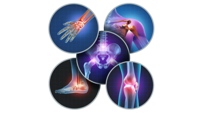
Osteoporosis: age-related and deadly

One of the unmistakable signs of aging is the loss of bone density. It causes osteoporosis, a dangerous disease that can go unnoticed until a fracture occurs. Hip fractures in the elderly result in up to 20-24% mortality in the first year alone. Bone homeostasis depends on both estrogen and testosterone, but women are disproportionately affected by osteoporosis, since estrogen levels drop sharply during menopause and they have lower bone density to begin with. Therefore, 75% of hip fractures in people after 50 happen to women, and a 50-year-old woman has a 2.8% risk of dying from the consequences of a hip fracture during her remaining lifetime – roughly the same as of dying of breast cancer.

Bone homeostasis depends, in part, on the intricate balance of osteoclastic and osteoblastic activity [3]. Osteoblasts are cells that differentiate from mesenchymal stem cells and are responsible for bone deposition, the creation of the bone extracellular matrix through the secretion of dense collagen and other proteins. Osteoclasts, derived from macrophages, are large, multinucleated cells that play a central role in the opposite process: bone resorption. Osteoclasts secrete acid and collagenase that disassemble bone tissue. This seemingly destructive activity is critical for maintenance, repair, and remodeling of bones, but with age, osteoclast-induced bone resorption outpaces osteoblast-induced bone deposition, leading to a gradual loss of bone mass.

Identifying the target

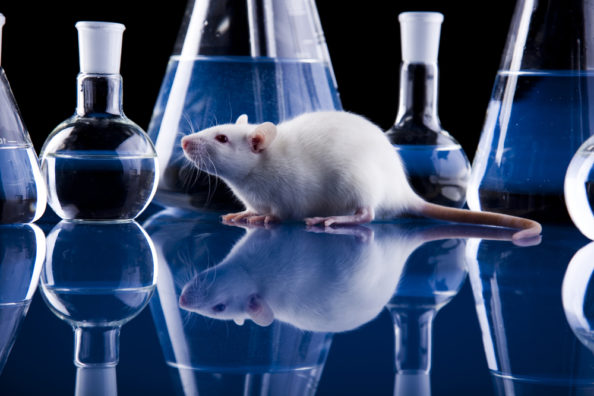
The researchers analyzed blood plasma taken from 12 young and 12 aged mice. They were able to identify a total of around 600 proteins that were present in all the samples, and some of them varied considerably in concentration between young and old mice, indicating that the plasma proteome does change with age. From among these proteins, seven candidates were selected, as they were downregulated in aged mice and had not been previously studied by longevity researchers.

To test if any of those proteins played a role in bone mineral density (BMD) decline, the scientists applied them to bone marrow-derived macrophages that were undergoing an induced differentiation into osteoclasts. Among the candidates, cadherin-13 was the only one that demonstrated significant ability to inhibit osteoclast differentiation while neither being toxic to the cells nor impairing osteoblast differentiation.

Cadherin-13: promising but mysterious

Cadherin-13 is an odd member of the cadherin superfamily. As it lacks the transmembrane domain, it cannot participate in cell-cell adhesion, which seems to be the main purpose of other cadherins. Instead, cadherin-13 has been shown to play a role in cell migration and phenotype changes, and it acts as an LDL (low density lipoproteins) receptor. Elevated levels of cadherin-13 have been associated with ADHD, depression, and other psychiatric disorders as well as with some cardiovascular diseases, such as atherosclerosis [4]. Nevertheless, cadherin-13’s functions and mechanisms of action remain mainly undetermined.

Osteoclast formation requires the presence of two chemicals: receptor activator of nuclear factor κβ ligand (RANKL) and Macrophage colony-stimulating factor (M-CSF). The researchers hypothesized that cadherin-13 might be causing disruption in one of these pathways. The tests revealed that cadherin-13 did substantially obstruct signaling down the RANKL pathway by inhibiting RANKL phosphorylation of several signaling molecules.

After these encouraging findings, the researchers moved on to experimenting in vivo. Female mice were injected with cadherin-13 for 4 months, beginning at 15 months of age. The BMD of the mice’s femur bones had been periodically monitored in vivo using micro-computer tomography. Over the course of 16 weeks, BMD of the control group had declined dramatically. In mice that were being treated with cadherin-13, this decline was attenuated, while two other bone health indicators, bone volume fraction and trabecular thickness, increased.

Conclusion

Declining bone density is the immediate cause of osteoporosis, a potentially deadly aging-related disease that disproportionately affects women. Since current treatments leave much to be desired, new approaches to osteoporosis are desperately needed. This research opens an interesting novel possibility for intervention. It also illustrates how comparing plasma proteomes of young and old organisms can be used to discover new therapeutic targets for longevity research. On the other hand, the target that was identified, cadherin-13, remains largely unresearched, and various safety concerns might yet block its path to clinical use.

We would like to ask you a small favor. We are a non-profit foundation, and unlike some other organizations, we have no shareholders and no products to sell you. All our news and educational content is free for everyone to read, but it does mean that we rely on the help of people like you. Every contribution, no matter if it’s big or small, supports independent journalism and sustains our future.

Literature

[1] Yang, Y. R., Kabir, M. H., Park, J. H., Park, J. I., Kang, J. S., Ju, S., … & Lee, K. P. (2020). Plasma proteomic profiling of young and old mice reveals cadherin-13 prevents age-related bone loss. Aging, 12.

[2] Baht, G. S., Silkstone, D., Vi, L., Nadesan, P., Amani, Y., Whetstone, H., … & Alman, B. A. (2015). Exposure to a youthful circulation rejuvenates bone repair through modulation of β-catenin. Nature communications6, 7131.

[3] Tanaka, Y., Nakayamada, S., & Okada, Y. (2005). Osteoblasts and osteoclasts in bone remodeling and inflammation. Current Drug Targets-Inflammation & Allergy, 4(3), 325-328.

[4] Philippova, M., Suter, Y., Toggweiler, S., Schoenenberger, A. W., Joshi, M. B., Kyriakakis, E., … & Resink, T. J. (2011). T-cadherin is present on endothelial microparticles and is elevated in plasma in early atherosclerosis. European heart journal, 32(6), 760-771.

Circadian Rhythm Sleep

NAD+ and the Circadian Rhythm

A new study may shed some light on how aging and circadian rhythms may be linked by discovering a role for nicotinamide adenine dinucleotide (NAD+) in preventing the age-related disruption of circadian rhythms in mice.

A circadian rhythm is an automatic process that regulates a cycle of behavioral changes and repeats roughly every 24 hours. The most well-known example of a circadian rhythm is sleeping at night and being awake during the day, but it can refer to any biological process that has an internal, entrainable rhythm based around a 24-hour period.

It is well documented that as we age, our circadian rhythms can become disrupted, which may contribute towards the development of age-related diseases. While researchers have been aware of circadian rhythms for a long time now, the actual molecular workings and mechanisms linking them with aging are still somewhat of a mystery.

Thankfully, new research may shed some light on how the two could be linked, and this research may also have relevance to humans.

What is NAD+?

Nicotinamide adenine dinucleotide is a coenzyme found in all living cells. It is a dinucleotide, which means that it consists of two nucleotides joined through their phosphate groups. One nucleotide contains an adenine base, and the other contains nicotinamide.

In metabolism, NAD facilitates redox reactions, carrying electrons from one reaction to another. Therefore, NAD is found in two forms in the cell; NAD+ is an oxidizing agent that takes electrons from other molecules in order to become its reduced form, NADH. NADH can then become a reducing agent that donates the electrons it carries. The transfer of electrons is one of the main functions of NAD, though it also performs other cellular processes, including acting as a substrate for enzymes that add or remove chemical groups from proteins in post-translational modifications.

NAD+ biology has seen a great deal of interest in the last few years, partially due to the discovery of two precursors of NAD+ biosynthesis, nicotinamide riboside (NR) and nicotinamide mononucleotide (NMN), which both increase NAD+ in multiple tissues. NAD+ is also a cofactor for sirtuin deacetylases, which are known to promote healthspan and lifespan.

As we age, cellular levels of NAD+ decline, largely (if not wholly) due to the chronic systemic age-related inflammation known as inflammaging. Previous animal studies have shown that the use of NAD+ precursors can promote more youthful behavior in aged mice as well as reduce some aspects of aging.

The NAD+ and circadian rhythm connection

Researchers have published the results of a new study suggesting a role for NAD+ in resisting age-related circadian rhythm disturbance using the NAD+ precursor nicotinamide riboside [1].

The mice were given NR in their drinking water for a period of 4 months; they then examined circadian-regulated gene expression in the mouse livers. The results showed that the gene expression patterns of around half of the circadian-regulated liver genes were altered in a beneficial manner when NAD+ levels were increased using NR.

The connection that NAD+ and circadian rhythms have are through sirtuins, of which sirtuin 1 (SIRT1) regulates circadian rhythms. When there is sufficient NAD+ available, the body is able to regulate circadian rhythm properly, but, inevitably, as aging steadily creeps in and the level of NAD+ production declines, SIRT1 can no longer work with it to regulate circadian rhythm. The paper is worth a read as it explains the exact mechanism by which this works, but this is a simple summary of what happens:

Disrupted sleep-wake and molecular circadian rhythms are a feature of aging associated with metabolic disease and reduced levels of NAD+, yet whether changes in nucleotide metabolism control circadian behavioral and genomic rhythms remains unknown. Here, we reveal that supplementation with the NAD+ precursor nicotinamide riboside (NR) markedly reprograms metabolic and stress-response pathways that decline with aging through inhibition of the clock repressor PER2. NR enhances BMAL1 chromatin binding genome-wide through PER2K680 deacetylation, which in turn primes PER2 phosphorylation within a domain that controls nuclear transport and stability and that is mutated in human advanced sleep phase syndrome. In old mice, dampened BMAL1 chromatin binding, transcriptional oscillations, mitochondrial respiration rhythms, and late evening activity are restored by NAD+ repletion to youthful levels with NR. These results reveal effects of NAD+ on metabolism and the circadian system with aging through the spatiotemporal control of the molecular clock.

Conclusion

There is a great deal of enthusiasm and energy going into developing therapies that can restore NAD+ levels in aged mice and people, and that may prove useful in helping older people improve their health and quality of life, which includes sleep quality.

However, another avenue for restoring declining NAD+ levels is exercise; for example, a recent study showed that resistance training in middle age doubles muscle NAD+ levels. This is a practical measure that we can all take at little to no expense to slow down the loss of NAD+ and slow down the rate of aging somewhat.

Does that mean that exercise is superior to NAD+ boosting therapies? There is no doubt it is beneficial up to the point at which the aged body stops responding to exercise efficiently, and so there may be a case that exercise and NAD+ boosting therapies could be used together to good effect.

We would like to ask you a small favor. We are a non-profit foundation, and unlike some other organizations, we have no shareholders and no products to sell you. All our news and educational content is free for everyone to read, but it does mean that we rely on the help of people like you. Every contribution, no matter if it’s big or small, supports independent journalism and sustains our future.

Literature

[1] Levine, D. C., Hong, H., Weidemann, B. J., Ramsey, K. M., Affinati, A. H., Schmidt, M. S., … & Brenner, C. (2020). NAD+ Controls Circadian Reprogramming through PER2 Nuclear Translocation to Counter Aging. Molecular Cell.

Pills

SYK Inhibitors May Be a New Class of Senolytics

At this moment in history, the study of the biology of aging frequently brings us new discoveries, and today is no exception. Researchers have discovered a new drug that appears to destroy lingering senescent cells, and it achieves this without using any of the previously known mechanisms or pathways normally associated with inducing cell destruction.

What are senescent cells?

As you age, increasing numbers of your cells enter into a state known as senescence. Senescent cells do not divide or support the tissues of which they are part; instead, they emit a range of potentially harmful chemical signals that encourage nearby healthy cells to enter the same senescent state. Their presence causes many problems: they reduce tissue repair, increase chronic inflammation, and can even eventually raise the risk of cancer and other age-related diseases.

Senescent cells normally destroy themselves through a self-destruct process called apoptosis, and they are also removed by the immune system; however, the immune system weakens with age, and increasing numbers of senescent cells escape this process and begin to accumulate in all the tissues of the body.

By the time people reach old age, significant numbers of these senescent cells have accumulated in our tissues and organs, causing chronic inflammation and damage to surrounding cells and tissue. The accumulation of senescent cells is thought to be one of the reasons we age.

The proposed solution to the problem of senescent cell accumulation is to cause them to stop resisting and initiate apoptosis; drugs that can tip senescent cells over the edge and destroy themselves are known as senolytics.

SYK inhibitors appear to destroy senescent cells, but we don’t know how

In the journal Aging, the researchers of a new study have published their discovery of a new senolytic compound, which, intriguingly, does not use any of the currently known mechanisms or pathways to induce apoptosis [1].

The current generation of senolytic compounds focus on known pathways such as p53 and the BCL-2 family in order to cause death-resistant senescent cells to self-destruct. While these researchers do not fully understand the mechanism, they show that R406, an SYK inhibitor, does appear to induce apoptosis.

Spleen tyrosine kinase (SYK) is mainly expressed by hematopoietic cells, a population of bone marrow stem cells that give rise to other blood cells during a process called haematopoiesis. SYK plays a key role in the B-cell receptor signaling pathway and is also an important component in signal transduction from other immune receptors, such as Fc and adhesion receptors.

There are currently a number of SYK inhibitors, including the thrombocytopenia drug fostamatinib, the cancer drug entospletinib, and the cancer drug cerdulatinib. R406 is an orally bioavailable SYK inhibitor and has been shown in early mouse studies and human studies to reduce Immune system-mediated inflammation; in human trials, it was shown to inhibit activation of basophils and thus reduce inflammation [2]. The data also suggest that it can also influence other types of immune cells, such as macrophages and B-cells.

The selective removal of senescent cells by senolytics is suggested as a potential approach to reverse aging and extend lifespan. Using high-throughput screening with replicative senescence of human diploid fibroblasts (HDFs), we identified a novel senolytic drug R406 that showed selective toxicity in senescent cells. Using flow cytometry and caspase expression analysis, we confirmed that R406 caused apoptotic cell death along with morphological changes in senescent cells. Interestingly, R406 altered the cell survival-related molecular processes including the inhibition of phosphorylation of the focal adhesion kinase (FAK) and p38 mitogen-activated protein kinase (MAPK) in senescent cells. This pattern was not observed in other known senolytic agent ABT263. Correspondingly, apoptotic cell death in senescent cells was induced by simultaneously blocking the FAK and p38 pathways. Taken together, we suggest that R406 acts as a senolytic drug by inducing apoptosis and reducing cell attachment capacity.

Conclusion

The exact method by which R406 appears to have a senolytic effect is currently unknown; however, its ability to reduce immune system-mediated inflammation may be improving the function of the immune system, thus preventing the excessive and detrimental activation of immune cells.

The immune system is known to become dysfunctional during aging at least in part due to excessive exposure to the chronic, systemic inflammation known as inflammaging, so it could be reducing senescent cell burden by making the immune system better at locating and destroying senescent cells more efficiently.

Like all things in biology, it is just a matter of time before the exact pathways and mechanism are discerned, but the discovery of a potential new group of senolytic drugs is great news.

We would like to ask you a small favor. We are a non-profit foundation, and unlike some other organizations, we have no shareholders and no products to sell you. All our news and educational content is free for everyone to read, but it does mean that we rely on the help of people like you. Every contribution, no matter if it’s big or small, supports independent journalism and sustains our future.

Literature

[1] Cho, H. J., Yang, E. J., Park, J. T., Kim, J. R., Kim, E. C., Jung, K. J., … & Lee, Y. S. (2020). Identification of SYK inhibitor, R406 as a novel senolytic agent. Aging, 12.

[2] Braselmann, S., Taylor, V., Zhao, H., Wang, S., Sylvain, C., Baluom, M., … & Wong, B. R. (2006). R406, an orally available spleen tyrosine kinase inhibitor blocks fc receptor signaling and reduces immune complex-mediated inflammation. Journal of Pharmacology and Experimental Therapeutics, 319(3), 998-1008.

Arthritic joint pain

New Possible Treatment for Osteoarthritis Reported

Researchers have successfully reversed the course of osteoarthritis in rats by combining two molecules, one of which, a-Klotho, has long been a popular subject of anti-aging research.

A classic age-related disease

Osteoarthritis (OA) is a classic example of a wear-and-tear disease. Its manifestations are in the slow degeneration of articular cartilage, the appearance of bone spurs around joints, and inflammation. While genetics, obesity, and other factors can increase susceptibility to OA, this disease remains mainly age-related.

By increasing inflammation and limiting motility, OA contributes to multiple other age-related pathologies and represents an important part of the vicious cycle of aging. Currently, no methods of reversing OA exist. Treatment may include pain alleviation and physical therapy in earlier stages, but more serious cases demand drastic surgical measures, such as joint replacement.

Better together?

Recently, a group of researchers from the Salk Institute for Biological Studies in California decided to test the joint therapeutic effect of two molecules that, according to earlier research, might play a role in the processes that are responsible for OA [1].

The first molecule is a-Klotho, the main variation of the Klotho enzyme. Upon its discovery in 1997, this molecule was suspected of possessing anti-aging properties, which is why it was named after one of the Moirai, creatures in Greek mythology that control the thread of life of every mortal from birth to death. a-Klotho can promote cardiovascular health by suppressing oxidative stress and inflammation [2], and its deficiency has been tied to obesity, chronic kidney failure, bone loss, and cognitive deficits. Genetic overexpression of Klotho has been shown to extend lifespan in mice [3]. Importantly for this research, a-Klotho is downregulated in cartilage affected by OA [4] and may act as an inhibitor of extracellular matrix (ECM) degradation.

The second compound is TGFßR2, a receptor that binds TGFß1 (transformative growth factor beta, isoform 1). TGFß1 is a cytokine that regulates various cellular functions, such as cell proliferation, adhesion, and migration. On one hand, TGFß1 seems to have some cartilage-repairing properties, including chondrocyte proliferation [5]. On the other, there is substantial evidence that it also contributes to OA progression [6]. TGFß1 levels seem to be low to nonexistent in healthy joints but drastically elevated in joints with OA. This led the researches to hypothesize that maintaining the right balance in the TGFß pathway by binding excessive TGFß1 would be beneficial for cartilage homeostasis.

To test their hypotheses, the researchers first induced OA in rats. Four weeks after the intra-articular injection of papain, an enzyme known to cause OA, the affected rats exhibited clear signs of early-stage OA. Then, the expression of the two molecules in the rats’ OA-affected joints was ramped up using an AAV-based delivery system. AAV (adeno-associated virus) is a popular vehicle for the delivery of DNA into cells, particularly because it elicits little immune response. Importantly, prior to the main experiment, the researchers had established in vitro that the combined beneficial effect of the two molecules on OA markers was indeed greater than the effect of each one separately. Therefore, only the combination of the two molecules advanced to the in vivo testing stage.

Four groups were established: the healthy control group, the OA control group, the treatment group, and the sham group. Rats in the latter group had been treated with AAV minus the payload to control for any standalone effects that the viral DNA might have.

KT vs OA: 1:0

The research group showed improvement six weeks after the injection, with significant recovery of cartilage structure and thickness versus the OA control. The sham group exhibited even more severe deterioration compared to the OA control group, leading to the conclusion that the improvement in the treatment group happened despite, rather than due to, the presence of the viral DNA.

RNA sequencing revealed that the treatment resulted in the downregulation of some genes that are known to promote inflammatory reaction and excessive immune response in patients with OA. These results are compatible with our previous knowledge of TGFß1’s contributions to inflammation during OA and of a-Klotho’s ECM-protective qualities. The Klotho + TGFßR2 (KT) treatment eventually resulted in the reclassification of the rats in the treatment group from OA grade 2 to grade 1, which represented a partial reversal of the course of the disease. The authors speculate that KT treatment can potentially fully heal OA, which would be a major improvement over current treatment methods.

Conclusion

Osteoarthritis, though not as lethal as some other age-related diseases, can cause extensive physical and psychological suffering. Moreover, it is the most prevalent musculoskeletal disorder among the elderly and the leading cause of disability in the US [7]. Current treatment options are either only partially effective or highly invasive. The research in question offers a new possible solution, though much more must be done to elucidate the detailed mechanism behind the treatment and to ensure its effectiveness and safety. The research also contributes to our understanding of the age-defying properties of Klotho and of the various, and at times contradictory, effects of TGFß.

We would like to ask you a small favor. We are a non-profit foundation, and unlike some other organizations, we have no shareholders and no products to sell you. All our news and educational content is free for everyone to read, but it does mean that we rely on the help of people like you. Every contribution, no matter if it’s big or small, supports independent journalism and sustains our future.

Literature

[1] Martinez-Redondo, P., Guillen-Guillen, I., Davidsohn, N., Wang, C., Prieto, J., Kurita, M., … & Lezaki, T. (2020). aKLOTHO and sTGFßR2 treatment counteract the osteoarthritic phenotype developed in a rat model. Protein & Cell, 1-8.

[2] Lim, K., Halim, A., Lu, T. S., Ashworth, A., & Chong, I. (2019). Klotho: A Major Shareholder in Vascular Aging Enterprises. International journal of molecular sciences, 20(18), 4637.

[3] Kurosu, H., Yamamoto, M., Clark, J. D., Pastor, J. V., Nandi, A., Gurnani, P., … & Shimomura, I. (2005). Suppression of aging in mice by the hormone Klotho. Science, 309(5742), 1829-1833.

[4] Pásztói, M., Nagy, G., Géher, P., Lakatos, T., Tóth, K., Wellinger, K., … & Pálóczy, K. (2009). Gene expression and activity of cartilage degrading glycosidases in human rheumatoid arthritis and osteoarthritis synovial fibroblasts. Arthritis research & therapy, 11(3), R68.

[5] Varela-Eirin, M., Loureiro, J., Fonseca, E., Corrochano, S., Caeiro, J. R., Collado, M., & Mayan, M. D. (2018). Cartilage regeneration and ageing: targeting cellular plasticity in osteoarthritis. Ageing research reviews, 42, 56-71.

[6] Scharstuhl, A., Glansbeek, H. L., van Beuningen, H. M., Vitters, E. L., van der Kraan, P. M., & van den Berg, W. B. (2002). Inhibition of endogenous TGF-ß during experimental osteoarthritis prevents osteophyte formation and impairs cartilage repair. The Journal of Immunology, 169(1), 507-514.

[7] Zhang, W., Ouyang, H., Dass, C. R., & Xu, J. (2016). Current research on pharmacologic and regenerative therapies for osteoarthritis. Bone research, 4(1), 1-14.

Oriel College

Jim Mellon Donates £1 Million to Aging Research

Investment mogul Jim Mellon has donated a record-breaking 1 million British pounds to Oriel College in the United Kingdom, offering a ray of light for aging research.

Supporting fundamental research on aging

In recent years, one of the real dynamos of our community has been British investor and philanthropist Jim Mellon. Jim is perhaps best known for his investment support of promising aging-focused biotech companies through his company Juvenescence, which has invested in rising stars such as Lygenesis, AgeX, and Insilico Medicine.

Jim has also been actively involved in the community and is frequently seen attending various aging research conferences, rallying investors to help build the fledgling but steadily growing rejuvenation industry, lobbying government, and supporting basic research on aging.

Today’s story is an example of the latter, as Jim has stepped up to support the fundamental aging research happening in the United Kingdom, which has been an important hub for this sort of research. We are happy to learn that Jim has generously donated £1 million for the support of longevity research at Oriel College, which is part of Oxford University. This donation establishes the Mellon Longevity Science Program at Oriel College with the aim of advancing research into health resilience in the elderly.

Jim’s donation will directly support the research of Professor Lynne Cox, a George Moody Fellow in Biochemistry at Oriel and a principal investigator within the Department of Biochemistry. The Cox lab is studying the molecular basis of human aging with the goal of reducing age-related morbidity and frailty though improving health resilience. This could include researching therapies for strategies such as boosting the immune systems of older people in order to help them maintain health as well as reducing the effects of sarcopenia and frailty with advancing age to improve quality of life.

Jim’s stated reasons for supporting Oriel College are as follows:

There has never been a more important time to address the frailty of human health. The COVID-19 pandemic has highlighted the huge economic and social costs connected to the lack of immune resilience in our increasingly ageing population and the need for greater scientific research into this area.

Boosting immunoresilience among the most vulnerable in society and advancing healthspan are critical to helping more people reach their potential as well as, more urgently, improving our collective resilience in the face of future pandemics. Oxford’s leadership in the field of research and understanding of the ageing process makes it a natural home to advance longevity science and support the growth of the longevity industry, and I am proud to support this work.

In addition, the donation will also allow the creation of a DPhil scholarship in Aging and Cell Senescence at Oriel College. The plan is that the first recipient of the DPhil scholarship will join the College for the 2020-21 academic year and will help foster a future generation of aging researchers to join the existing leaders in the field.

Conclusion

This donation is a record breaker in the context of aging research at a UK university, making Oriel, and Oxford in general, a real hub for this line of scientific inquiry. Oxford University has long had links to the rejuvenation research community, as it was the scene of a 2012 debate between Dr. Aubrey de Grey and Professor Colin Blakemoor over the desirability of increasing healthy human lifespans, the conclusion of which was somewhat undecided at the time.

We cannot help but wonder how the audience would vote now given the passage of eight years and much more progress in the field.

Obviously, we welcome this news with enthusiasm, and we once again congratulate Jim for his continued support and generosity in support of the field. At a time when many economies are facing great challenges, this money will provide a real shot in the arm for aging research.

We would like to ask you a small favor. We are a non-profit foundation, and unlike some other organizations, we have no shareholders and no products to sell you. All our news and educational content is free for everyone to read, but it does mean that we rely on the help of people like you. Every contribution, no matter if it’s big or small, supports independent journalism and sustains our future.

Metformin Enhances Autophagy and Alleviates Inflammaging

A recent study published in Cell Metabolism has shown that metformin, a drug that has been previously shown to be effective against some aspects of aging, ameliorates inflammaging by promoting autophagy, the cellular recycling of damaged components.

Mitochondrial dysfunction, autophagy, and TH17

Mitochondrial dysfunction is one of the hallmarks of aging, the root causes that cause us all to age. The mitochondria are the powerhouses of the cell, but as we age, our mitochondria have their DNA damaged by sources such as reactive oxygen species, harming their ability to perform their fundamental job. This gradual increase in dysfunctional mitochondria causes our cells to lose their ability to utilize energy, leading to a panoply of age-related diseases.

Autophagy is the consumption of damaged or dysfunctional organelles by intracellular processes. Through autophagy, cells are able to recycle and renew their internal components, and, as the researchers of this paper explain, CD4+ (helper) T cells that do not properly perform autophagy behave like older cells.

Cytokines are known for causing inflammation, and, as the researchers demonstrate, the TH17 subset of cytokines is strongly associated with the chronic, age-related inflammation known as inflammaging, a key driver of multiple age-related diseases.

In order to demonstrate the relationship between autophagy, mitochondrial dysfunction, and TH17, the researchers used RNA silencing to disrupt the ability of cultured cells to engage in autophagy. Such cells that were taken from a young subject had their mitochondrial function decreased to the level of an older person. This RNA silencing also set the TH17 cytokine profile of these cells to become one associated with age and diabetes.

Obviously, what we want is the reverse of this process, and the researchers found that metformin, a commonly researched drug in the rejuvenation biotechnology field, was able to accomplish this in human cell cultures, spurring autophagy and reversing the TH17 cytokine profile.

Summary

Age is a non-modifiable risk factor for the inflammation that underlies age-associated diseases; thus, anti-inflammaging drugs hold promise for increasing health span. Cytokine profiling and bioinformatic analyses showed that Th17 cytokine production differentiates CD4+ T cells from lean, normoglycemic older and younger subjects, and mimics a diabetes-associated Th17 profile. T cells from older compared to younger subjects also had defects in autophagy and mitochondrial bioenergetics that associate with redox imbalance. Metformin ameliorated the Th17 inflammaging profile by increasing autophagy and improving mitochondrial bioenergetics. By contrast, autophagy-targeting siRNA disrupted redox balance in T cells from young subjects and activated the Th17 profile by activating the Th17 master regulator, STAT3, which in turn bound IL-17A and F promoters. Mitophagy-targeting siRNA failed to activate the Th17 profile. We conclude that metformin improves autophagy and mitochondrial function largely in parallel to ameliorate a newly defined inflammaging profile that echoes inflammation in diabetes.

Conclusion

This is a cell culture study, not a human trial, so the usual caveat applies: it may fail in this respect for reasons as of yet unknown. However, as metformin has a well-known safety profile and is already approved by the FDA for other conditions, including diabetes itself, conducting a human trial to assess its effectiveness in stimulating autophagy is easier than testing a novel drug would be. We look forward to such a trial and hope for its success.

We would like to ask you a small favor. We are a non-profit foundation, and unlike some other organizations, we have no shareholders and no products to sell you. All our news and educational content is free for everyone to read, but it does mean that we rely on the help of people like you. Every contribution, no matter if it’s big or small, supports independent journalism and sustains our future.

Weightlifting

Resistance Training in Middle Age Doubles Muscle NAD+ Levels

A new study suggests that we can actively fight back against age-related decline with resistance training to restore our levels of NAD+, a critical molecule used by the mitochondria in our cells to produce energy.

NAD+ and mitochondria

The mitochondria are the powerhouses of the cell, and they use the nutrients we consume in order to produce the energy that our cells need to function. Mitochondria rely on a supply of nicotinamide adenine dinucleotide (NAD+) in order for them to produce adenosine triphosphate (ATP), the form of chemical energy universally used by cells. NAD+ is a coenzyme found in all living cells; without it, life would be impossible.

In metabolism, NAD+ facilitates redox reactions, carrying electrons from one reaction to another. This process causes NAD+ to be found in two forms in the cell. NAD+ is an oxidizing agent that takes electrons from other molecules in order to become its reduced form, NAD+H. NAD+H can then become a reducing agent that donates the electrons it carries.

The transfer of electrons is one of the main functions of NAD+, though it also performs other cellular processes, including acting as a substrate for enzymes that add or remove chemical groups from proteins in post-translational modifications, performing cellular signaling, regulating metabolism, facilitating DNA repair, and engaging in many other functions within the cell.

Unfortunately, NAD+ declines with age due at least in part to the chronic systemic inflammation often referred to as inflammaging. This inflammation comes from various sources, including, but not limited to, senescent cell accumulation and the proinflammatory signals it produces, cell debris, immunosenescence, and changes to the gut microbiome.

NAD+ is created from simple building blocks, such as the amino acid tryptophan, and it is created in a more complex way via the intake of food that contains nicotinic acid (niacin) or other NAD+ precursors. These different pathways ultimately feed into a salvage pathway, which recycles them back into the active NAD+ form.

Combating age-related NAD+ decline

There is currently a great deal of research focused on increasing levels of NAD+ in older people as well as a myriad of supplements and other kinds of therapies, not all of which are equal or proven to work in humans. The two most notable precursors of NAD+ biosynthesis, nicotinamide riboside (NR) and nicotinamide mononucleotide (NMN), have been of great interest to researchers as they both increase NAD+ directly and in multiple tissues.

While it may be the case that these approaches may prove useful, there is another way in which we all might offset NAD+ decline now: resistance training. Studies have already shown that exercise reverses age‐related decline in NAD+ salvage capacity, thus making more NAD+ available, and the research we want to cover today builds on this.

The results of a new study show that ten weeks of resistance training in middle-aged, overweight, untrained individuals can significantly influence NAD+ levels. A ten-week period of supervised resistance training was initiated as part of the study and included leg/hip sled movements, leg extensions, lying leg curls, barbell bench presses, and cable pull-downs.

During the training, the participants, aged 55-63, were asked to rate the difficulty of the exercises from 0-10, with 0 being easy and 10 being hard. If an individual reported that an exercise was 10 or failed to complete an exercise session, then weight was reduced to make it easier; if the participant indicated that the difficulty was 7 or lower, more weight was added in order to increase the challenge. In this manner, the researchers were able to present an exercise routine that was challenging but not too punishing and that would adjust as people improved on an individual basis.

The results after ten weeks showed that NAD+ in muscle cells was more than doubled, making them similar to the levels present in a college-aged test group also part of the study.

We examined if resistance training affected muscle NAD+ and NADH concentrations as well as nicotinamide phosphoribosyltransferase (NAMPT) protein levels and sirtuin (SIRT) activity markers in middle-aged, untrained (MA) individuals. MA participants (59±4 years old; n=16) completed 10 weeks of full-body resistance training (2 d/wk). Body composition, knee extensor strength, and vastus lateralis muscle biopsies were obtained prior to training (Pre) and 72 hours following the last training bout (Post). Data from trained college-aged men (22±3 years old, training age: 6±2 years old; n=15) were also obtained for comparative purposes. Muscle NAD+ (+127%, p<0.001), NADH (+99%, p=0.002), global SIRT activity (+13%, p=0.036), and NAMPT protein (+15%, p=0.014) increased from Pre to Post in MA participants. Additionally, Pre muscle NAD+ and NADH in MA participants were lower than college-aged participants (p<0.05), whereas Post values were similar between cohorts (p>0.10). Interestingly, muscle citrate synthase activity levels (i.e., mitochondrial density) increased in MA participants from Pre to Post (+183%, p<0.001), and this increase was significantly associated with increases in muscle NAD+ (r2=0.592, p=0.001). In summary, muscle NAD+, NADH, and global SIRT activity are positively affected by resistance training in middle-aged, untrained individuals. Whether these adaptations facilitated mitochondrial biogenesis remains to be determined.

Conclusion

The health benefits of exercise have long been known, and yet many of us do not exercise enough, which is a real problem in our increasingly sedentary world. Our ancestors were, generally speaking, considerably more physically active than the average person in today’s technological world.

There is an absolute raft of studies covering the effects of exercise and its relation to health; needless to say, we should all try to move more and engage in physical activity as much as we can as part of a healthy lifestyle. Even if you are middle aged, adopting healthy habits by 50 may add a decade to healthy lifespan. It may not sound like a lot, but a decade is a long time in the research and biotechnology world, and it could mean the difference between living long enough to see true anti-aging therapies arrive or not.

We would like to ask you a small favor. We are a non-profit foundation, and unlike some other organizations, we have no shareholders and no products to sell you. All our news and educational content is free for everyone to read, but it does mean that we rely on the help of people like you. Every contribution, no matter if it’s big or small, supports independent journalism and sustains our future.

Literature

[1] Lamb, D. A., Moore, J. H., Mesquita, P. H. C., Smith, M. A., Vann, C. G., Osburn, S. C., … & Goodlett, M. D. (2020). Resistance training increases muscle NAD+ and NADH concentrations as well as NAMPT protein levels and global sirtuin activity in middle-aged, overweight, untrained individuals. Aging, 12.

Hand holding drug

A Senolytic Prodrug For Multiple Cell Types

A group of scientists from China has successfully created an effective senolytic prodrug that targets the enzyme β-galactosidase [1]. This prodrug, called SSK1, has demonstrated impressive results across various cell types and advantages over some existing senolytics.

Cellular senescence is a phenomenon in which cells stop dividing and supporting the tissues of which they are part. It is a major hallmark of aging, and it is associated with chronic inflammation, fibrosis [2], and other detrimental effects. Senescent cells accumulate in the body, secreting damaging chemicals and inducing senescence in nearby cells as well. Chemicals released by senescent cells, collectively known as the SASP (senescence-associated secretory phenotype), are thought to significantly contribute to aging and cancer. Senescence itself may be caused by various factors, such as telomere erosion, irradiation, and oxidative stress. The removal of senescent cells has been shown to attenuate various age-related conditions and to lengthen healthspan in mice [3].

Researchers have developed a class of drugs called senolytics in order to remove senescent cells. Some of them have shown potential and are currently undergoing human trials. Senolytics are supposed to cause a long-overdue death by apoptosis in senescent cells. However, according to the authors of this study, creating a senolytic that excels in both efficacy and selectivity may be challenging due to the diversity of senescence mechanisms across various cell types.

One of the characteristics shared by all senescent cells is the greatly increased production of the enzyme β-galactosidase (β-gal). β-gal is one of about 60 various enzymes found in lysosomes – cellular organelles charged with breaking down obsolete or unused biomolecules from both inside and outside the cell. It is not entirely understood why senescence causes a spike in β-gal levels, but this phenomenon presents a convenient target for intervention.

The researchers of this new study set out to create a prodrug – a compound that is therapeutically neutral until it encounters a trigger chemical in the body. In this case, the trigger is β-gal, and the prodrug is expected to be cleaved by the enzyme, releasing an apoptosis-inducing compound. After screening hundreds of FDA-approved drugs in search of such a compound, the researchers settled on gemcitabine, a known chemotherapy medication. Gemcitabine had been previously shown to effectively induce apoptosis in both human and murine cells. It also has little systemic toxicity due to its short circulation time in the body.

The resulting prodrug, SSK1, was tested first on mouse embryonic fibroblasts (MEFs), both on healthy cells and on their counterparts rendered senescent by replication. The authors report that SSK1 was found to effectively and selectively kill β-gal-positive senescent cells within a wide therapeutic window (i.e. across a wide spectrum of dosages).

To establish that the prodrug indeed targets β-gal, the researchers administered another gemcitabine-based chemical that lacked the β-gal-sensitive component. This second chemical had indiscriminately killed both senescent and non-senescent cells, as expected of an apoptosis-inducing drug. The scientists then used genetically modified cells in which GLB1 gene, the one responsible for β-gal encoding, was significantly downregulated. Senescent cells with the gene knocked down demonstrated regular levels of other senescence markers and were unharmed by SSK1. This was yet another way to demonstrate that SSK1 works by specifically targeting β-gal.

To examine the specificity of SSK1 for both murine and human cells, the authors performed a battery of additional tests. First, they used the prodrug on MEFs in which senescence had been induced by three different factors: ionizing radiation, oncogene overexpression and genotoxic stress. In all three cases, SSK1 successfully eliminated senescent cells while causing little to no damage to the healthy ones. Similar results were later recreated in mice that had artificially induced lung fibrosis. SSK1 succeeded in removing senescent cells from the affected lungs and in lowering various senescence and inflammation markers.

Other in vitro tests involved human embryonic fibroblasts (HEFs), human umbilical vein endothelial cells (HUVECs) and human preadipocytes (the precursors of mature fat cells). Yet again, SSK1 demonstrated the ability to selectively kill senescent cells and mostly spare healthy ones across various cell types and senescence stimuli.

Since some non-senescent cells also tend to express higher levels of β-gal, the researchers tested SSK1 on tissues from the two organs with the highest concentrations of such cells: the kidney and the submandibular gland. Following treatment with SSK1, only a negligible number of healthy cells underwent apoptosis, leading the researchers to conclude that the prodrug, overall, is extremely safe.

The authors also compared SSK1 to three other senolytic candidate drugs (ABT263, dasatinib plus quercetin, and fisetin) on HEFs, HUVECs, and human preadipocytes. They report that although all drugs showed senolytic abilities, SSK1 proved superior in the combined categories of potency, specificity and non-toxicity.

There was another interesting facet to this research. Macrophages are normally an important element of our immune system. However, activated macrophages that accumulate in aging tissues induce chronic inflammation [4] and exhibit elevated levels of β-gal, not unlike senescent cells. Building on this latter quality, the researchers tested SSK1 on macrophages and discovered that the prodrug successfully lowered the number of activated macrophages in the livers of aged mice.

Finally, upon discovering that SSK1 significantly reduces chronic inflammation locally and systemically, the researchers decided to run functional tests on aged mice treated with the novel prodrug. The tests showed improvements in motor function, balance ability, exercise endurance, skeletal muscle capacity, and spontaneous exploration compared to the untreated mice. Here, SSK1 yet again proved to be superior overall to other senolytics, even though they also demonstrated beneficial effects.

Conclusion

Cellular senescence is a major hallmark of aging, contributing to age-related chronic inflammation and other detrimental phenomena. Fortunately, senolytic drugs may be our best hope of dealing a serious blow to aging in the near future, and this prodrug strategy has yielded highly promising results in terms of efficacy, specificity, and non-toxicity.

We would like to ask you a small favor. We are a non-profit foundation, and unlike some other organizations, we have no shareholders and no products to sell you. All our news and educational content is free for everyone to read, but it does mean that we rely on the help of people like you. Every contribution, no matter if it’s big or small, supports independent journalism and sustains our future.

Literature

[1] Cai, Y., Zhou, H., Zhu, Y. et al. Elimination of senescent cells by β-galactosidase-targeted prodrug attenuates inflammation and restores physical function in aged mice. Cell Res (2020).

[2] Schafer, M. J., White, T. A., Iijima, K., Haak, A. J., Ligresti, G., Atkinson, E. J., … & Mazula, D. L. (2017). Cellular senescence mediates fibrotic pulmonary disease. Nature communications8(1), 1-11.

[3] Baker, D. J., Childs, B. G., Durik, M., Wijers, M. E., Sieben, C. J., Zhong, J., … & Khazaie, K. (2016). Naturally occurring p16 Ink4a-positive cells shorten healthy lifespan. Nature530(7589), 184-189.

[4] Oishi, Y., & Manabe, I. (2016). Macrophages in age-related chronic inflammatory diseases. NPJ aging and mechanisms of disease2(1), 1-8.

Lab rats are an important animal for research purposes.

Blood Factors Reverse Epigenetic Age by Half in Rats

Researchers have demonstrated that epigenetic age can be halved in rats by using signals commonly found in the blood.

Epigenetic changes

One of the proposed reasons we age are the changes to gene expression that our cells experience as we get older; these are commonly called epigenetic alterations. These alterations harm the fundamental functions of our cells and can increase the risk of cancer and other age-related diseases.

Gene expression is modified by the addition of epigenetic markers to the DNA, which change the pattern of gene expression in a cell, suppressing or enhancing the expression of certain genes in a cell as the situation demands.

You might think of DNA as the building blocks and epigenetics as the instruction manual that explains how to assemble those blocks in order to make a certain structure to suit a particular situation.

This is how a cell in the liver knows that it needs to be a liver cell: the epigenetic instructions make sure that it is given the right guidance to become the correct cell type. At a basic level, these epigenetic instructions make sure that the genes needed to develop into a liver cell are turned on while the instructions specific to other types of cells are turned off.

However, as we age, our cells are exposed to damage from environmental factors and are subject to negative genomic changes through epigenetic mechanisms. Such changes accumulate over time and have been correlated with the decline observed in aging cells.

Epigenetic alterations in aging include changes to methylation patterns, and, in general, these correlate with a decrease in the amount of heterochromatin and an increase in chromosome fragility and transcriptional alterations (variance in gene expression), remodeling of chromatin (a DNA support structure that assists or impedes its transcription), and transcriptional noise.

Some researchers believe that these harmful epigenetic alterations could be reset to youthful levels in humans and, in doing so, reverse some aspects of aging, particularly improving the repair and rejuvenation of tissues and organs. There are a number of potential ways we might achieve this; today, we are going to focus on resetting the epigenetic age of cells using the signal molecules in our blood.

The internet of the body

Like the internet, the bloodstream is an information superhighway; it transmits signals between cells across huge distances, allowing remote parts of the body to communicate and regulate their activities. This network of blood vessels delivers nutrients, oxygen, and various kinds of cellular signals around the body to cells, so it is highly likely that it is a principal regulator of aging on a systemic level.

Researchers such as Dr. Dongsheng Cai have long proposed that certain elements of aging are controlled by the hypothalamus, a small area of the brain located at the base of the brain near the pituitary gland. While it’s very small, the hypothalamus plays a crucial role in many important functions, including releasing hormones and regulating body temperature. Dr. Cai and his team have discovered evidence that specialized stem cells living in the hypothalamus regulate systemic aging via secreted miRNAs [1].

As we age, these specialized cells die off and the hypothalamus becomes increasingly dysfunctional and unable to properly control the various functions it once did, in this manner, stem cell exhaustion in the hypothalamus seems to accelerate aging. This then leads to it secreting signals that are either not enough or too much, depending on the type. This then contributes to the shift in the environment of the bloodstream, as do other sources of harmful signaling, such as senescent cell secretions, changing it to a pro-aging rather than pro-youthful environment.

As we get older, there is typically a build-up of pro-inflammatory signals known as cytokines, which in low levels are helpful in healing and regeneration but when present in excessive amounts harm tissue upkeep and repair by blocking stem cell activity, cause immune cells to become dysfunctional, and contribute to a rise in systemic inflammation levels that support the onset of various age-related diseases. Typical cytokines that increase in concentration in the blood as we age are transforming growth factor beta 1 (TGF-ß1), interleukin 6 (IL6), and tumour necrosis factor alpha (TNFa).

Resetting epigenetic age with blood factors

Drs. Irina and Michael Conboy have long suggested with their research that certain elements of aging and tissue rejuvenation can be spurred by the calibration of signaling molecules in the blood. Indeed, their experiments have shown that youthful tissue function can be restored in aged mice by selectively reducing the level of key cytokines in the blood [2]. Doing so has the result of releasing stem cells, normally blocked from working by inflammatory signals typically present in old age, and allowing them to resume their activity. This return to activity allows the stem cells to begin repairing tissue and creating replacement cells as they do when the host is young.

Other researchers, such as Dr. Amy Wagers, Dr. Tony Wyss-Coray, and Dr. Hanadie Yousef, have also conducted research that strongly supports the idea that the epigenetic element of aging might at least be partially reset to a youthful level by calibrating, removing, or adding various signaling factors in blood.

While there are likely hundreds of factors in blood that may play a role in aging, the evidence is increasingly supporting the idea that a handful of key factors sit at the top of the process and regulate the hundreds of factors below. In other words, targeting these key factors may reverse epigenetic aging and restore youthful tissue function in aged individuals.

The results from a new study in rats suggest once again that the rejuvenation of aged organs and tissues is possible by resetting the epigenetic state of old cells back to a more youthful one using blood factors. It should be noted at this point that the results have been published as a preprint on bioRxiv, a site on which authors are able to make their research immediately available to the scientific community and receive feedback from their colleagues before they are submitted to journals for peer review and publication. With that caveat out of the way, let’s take a look at the data.

The researchers delivered Elixir, an undisclosed mixture of plasma fractions presumably isolated from young rat plasma, to aged rats. They state that their technique is based on heterochronic parabiosis but forgoes the need to physically join an old and young animal together so that they share circulatory systems.

The researchers claim that their results are averaged based on measurements taken using multiple epigenetic clocks.

Crucially, plasma treatment of the old rats reduced the epigenetic ages of blood, liver and heart by a very large and significant margin, to levels that are comparable with the young rats. According to the six epigenetic clocks, the plasma fraction treatment rejuvenated liver by 73.4%, blood by 52%, heart by 52%, and hypothalamus by 11%. The rejuvenation effects are even more pronounced if we use the final versions of our epigenetic clocks: liver 75%, blood 66%, heart 57%, hypothalamus 19%. According to the final version of the epigenetic clocks, the average rejuvenation across four tissues was 54.2%.

If these results are correct, then the epigenetic age of these rats has essentially been halved and returned to a level seen in young rats. On top of these epigenetic clock measurements, a number of other aging biomarkers saw improvement:

  • Pro-inflammatory cytokines IL-6 and TNFa were reset to a lower, more youthful, level
  • Blood triglycerides were reduced to a youthful level
  • HDL cholesterol were increased to a youthful level
  • Blood glucose levels dropped to more youthful levels
  • Cognition became more youthful according to tests in a Barnes Maze
  • Glutathione (GSH), superoxide dismutase (SOD), and some other antioxidant levels were reset to more youthful levels

Unanswered questions

These impressive results were apparently achieved with just four injections; however, it remains to be seen if these changes are persistent and for how long.

Given that there are many feedback loops in the system; it could be the case that once reset, the changes will persist for considerable time; after all, it takes decades for your system to break down and reach the point where aging becomes apparent.

There is also the question with blood factor-based rejuvenation experiments as to if it is what you put into aged blood or what you take out that is key to rejuvenation.

The Conboys have, in the past, suggested that reducing the excessive levels of proinflammatory cytokines in aged blood is likely more critical than adding anything found in young blood. In other words, it’s more about calibration of aged blood and the cytokines therein rather than there being any “special sauce” in young blood that spurs rejuvenation.

Of course, if could just as easily be both, and the past results of the Conboys, which showed that reduced levels of TGF-ß1 resulted in tissue rejuvenation, support this notion. Equally, there have been other experiments by researchers such as Dr. Amy Wagers and Dr. Tony Wyss-Coray in which factors found in young blood were added to aged blood and nothing was removed, which also spurred some level of rejuvenation.

In this particular experiment, the researchers only added replacement young blood plasma factors without removing anything, which suggests that what they did was enough to have a significant effect. It does appear in this case that whatever was added was able to encourage some level of rejuvenation in the context of epigenetic age. This could be more effective than the calibration and removal of harmful cytokines via filtering as proposed by the Conboys.

It could be that adding enough key factors is enough to spur rejuvenation without the need to carefully calibrate factors in aged blood. That remains to be seen, and at this early stage, it is too soon to say if what you take out is more important than what you put in or even if doing both will produce an even better result.

Conclusion

What needs to happen next is a lifespan study to see if resetting epigenetic age in this manner leads to an increase in healthy lifespan in rats. Also, this experiment used a small number of rats, so a greater number of rats used in such a follow-up lifespan study would be ideal. It would also be interesting to see if these results can be replicated in mice as a step towards moving this to human trials.

Taken as a whole, these results and similarly focused studies further support that methylation patterns in nuclear DNA are not merely the hands of a clock indicating cellular age but are also the workings of that clock. This is further evidence that epigenetic aging is one of the primary reasons we age and that its reversal could have big ramifications for human aging and healthy longevity if successfully translated.

We would like to ask you a small favor. We are a non-profit foundation, and unlike some other organizations, we have no shareholders and no products to sell you. All our news and educational content is free for everyone to read, but it does mean that we rely on the help of people like you. Every contribution, no matter if it’s big or small, supports independent journalism and sustains our future.

Literature

[1] Zhang, Y., Kim, M. S., Jia, B., Yan, J., Zuniga-Hertz, J. P., Han, C., & Cai, D. (2017). Hypothalamic stem cells control ageing speed partly through exosomal miRNAs. Nature, 548(7665), 52-57.

[2] Yousef, H., Conboy, M. J., Morgenthaler, A., Schlesinger, C., Bugaj, L., Paliwal, P., … & Schaffer, D. (2015). Systemic attenuation of the TGF-ß pathway by a single drug simultaneously rejuvenates hippocampal neurogenesis and myogenesis in the same old mammal. Oncotarget, 6(14), 11959.

The Challenge of Including Aging in the ICD

Few things in aging research are ever straightforward, and today’s topic is no exception, as two researchers enjoy some back and forth regarding whether and how the World Health Organization (WHO) should recognize the aging processes as a disease.

Proposing a new framework to facilitate development of rejuvenation therapies

Back in November 2019, an international group of researchers published a paper proposing a new healthcare framework in the journal Science in order to help older people stay healthier for longer by improving the development of therapies that target age-related diseases [1].

The proposal encouraged WHO, governments, and the medical science community to work together to develop a classification and staging system that uses a new framework as the foundation for the diagnosis and treatment of age-related diseases.

The publication came as a response to the fact that many age-related diseases currently lack sufficient diagnostic criteria and clinical-severity staging. This then presents a considerable barrier to the development of therapies that aim to address those diseases.

Currently, the classification and severity staging of age-related diseases is limited in effectiveness due to it being inconsistent, incomplete, and non-systematic. For example, some diseases can develop in several organs but are only classified in a single organ. This is the case for intrinsic organ aging, which can affect multiple organs.

In order to solve this problem, the researchers, including bench scientists and medical practitioners guided by Dr. Stuart Calimport and Dr. João Pedro de Magalhães, created a position statement to serve as the foundation for properly and comprehensively classifying and staging the severity of age-related diseases. The statement includes aging at both the tissue and organ level, organ atrophy, the pathological remodeling and calcification of tissues, and age-related systemic and metabolic diseases.

The aim of this publication is for WHO to adopt its suggested framework, which would potentially speed up the development of therapies that target the aging processes directly.

A drug development researcher begs to differ

As is the norm in academia, there is often a great deal of back and forth between researchers, and this is especially true with a project of this kind. There has been considerable discussion around this proposal, including a response letter from Dr. Alexander Zhavoronkov of Insilico Medicine, published in the journal Science in response to this original publication.

One of Dr. Zhavoronkov’s concerns is the timing, and as a year has passed since the latest version of the International Classification of Diseases (ICD), which included sarcopenia (age-related muscle wastage) in its codes, the proposal might seem a bit late to the game.

ICD-10, the previous version, was first endorsed by the World Health Assembly in 1990, and it was first used by member states in 1994, which offers some idea on how infrequently major updates occur.

Changing the ICD between these periodic updates is, of course, possible, but it would require considerable effort, including a comprehensive review of the literature by medical researchers from multiple countries. The next revision of the ICD is anticipated to be in 2028 or later, and there is a lot of basic research on aging to be done before then.

Dr. Zhavoronkov suggests that the proposal also overestimates our understanding of aging in the context of classifying age-related diseases. There is evidence that the detrimental effects of aging begin from a very early age, making the staging of most aging processes a massive undertaking requiring large-scale longitudinal studies in humans and animals.

Also, the concept of staging is not consistent with the current ICD code system, which is designed to provide a standardized system for disease reports and cause of death. Implementing a staging system as proposed into the ICD would require some serious levels of retooling that would need a huge international effort.

Dr. Zhavoronkov believes that we are still in the early days for aging research in the pharmaceutical drug discovery field. Indeed, the majority of the industry is still currently focused on the common biological pathways linking aging and age-related diseases and are doing it within the current healthcare model; in other words, they are mostly treating symptoms and not the root causes of aging.

He suggests that in order to change the focus of drug discovery as a whole, we must now demonstrate that basic research on aging can provide us with useful therapeutic targets that can deliver demonstrable results in treating or preventing age-related diseases.

This is a good point, and, really, until there is some very clear demonstrable evidence that anti-aging is plausible in humans, things will not change. Let us hope that human trials for senolytics and other approaches that directly target aging will change things in the near future and that the pharmaceutical drug discovery industry will put its full weight behind developing drugs that seek to restore youthful function.

Finally, he also points out that there is still yet to be a consensus on which biomarkers of aging are optimal for measuring senescence. This is true and something that we have written about many times here at lifespan.io: there is not only an urgent need to develop a range of accurate biomarkers of aging but also a need to agree, as a scientific community, which biomarkers together best constitute a panel able to measure changes to biological age resulting from therapeutic interventions. If we cannot measure those changes accurately, then we cannot demonstrate a therapy works and get it approved for human use.

An author’s response

One of the original authors, Dr. Stuart Calimport, also responded to these concerns in the same edition of Science.

Dr. Calimport argues that while indeed the major updates of the ICD are infrequent, WHO does accept submissions and proposed updates on an ongoing basis. Given that the ICD sees regular revision, Dr. Calimport believes that this would not be a major barrier.

Regarding the concern of our current knowledge of aging and the processes of senescence being barriers to their addition to the ICD, Calimport suggests otherwise. He counters that classification and staging consensus are things that the ICD would benefit from now. He also suggests that as senescence at the level of our organs is already in the ICD, in the case of “intrinsic aging of the skin” and “photo-aging of the skin”; with a corresponding staging scale, it should be equally valid to apply that to all organs and tissues of the body in the same manner as well as to age-related diseases.

The author accepts that there is indeed still much to learn about age-related diseases; however, he counters that there are also diseases currently defined in the ICD which are not fully understood either, and yet they have codes. In the case of skin aging, for example, the level of knowledge grew following its classification in the ICD. He also uses the example of Alzheimer’s disease and its inclusion in the ICD-9; prior to its classification, it was not acknowledged as a leading cause of age-related death. Essentially, Dr. Calimport argues that if a disease or a disorder can be identified, is distinct enough to be recognized, and has sufficient supporting research behind it, then it should be included.

He also begs to differ for the case for staging in the context of senescence in the ICD. He argues that it is quite common for diseases to be classified and included in the ICD before they have clinically accepted biomarkers.

Dr. Calimport suggests that macroscopic and histologic measures, similar to the Glogau scale used for skin, could be used to determine the staging of organismal senescence, at least initially. Longitudinal studies in animals and humans could then be used to further develop and refine staging of organismal senescence in tissues and organs and be used in the context of clinical endpoints and personalized medicine.

The author also accepts that senescence does begin from an early point in life but suggests that the ICD classification would be able to distinguish between this early stage of aging and the harmful changes and decline aging causes later in life.

Dr. Calimport also welcomes the development of effective aging biomarkers and a comprehensive staging system that would support the creation of interventions against the aging processes and thus against age-related diseases.

Conclusion

Considering aging as a disease in its own right, and how it could be included in a way that supports the development of effective therapies, is currently a hotly debated topic. The exchange described above is fairly typical of such discussions surrounding this topic, and hopefully sooner, rather than later, a solution that satisfies the majority of the academic community will be found.

We would like to ask you a small favor. We are a non-profit foundation, and unlike some other organizations, we have no shareholders and no products to sell you. All our news and educational content is free for everyone to read, but it does mean that we rely on the help of people like you. Every contribution, no matter if it’s big or small, supports independent journalism and sustains our future.

Literature

[1] Calimport, S. R., Bentley, B. L., Stewart, C. E., Pawelec, G., Scuteri, A., Vinciguerra, M., … & Fleming, G. A. (2019). To help aging populations, classify organismal senescence. Science, 366(6465), 576-578.

Apollo logo

The Moonshot Venture Fellowship

We had the opportunity to speak to Apollo Health Ventures about the Moonshot Venture Fellowship and about the aging research field in general from the perspective of a venture capital firm.

Apollo Health Ventures is a life science venture capital firm and life science company builder working across Europe and North America. Apollo is unique among investment firms due to its focus on a specific field of scientific research – the basic biology of aging. Apollo helps scientists working on understanding how and why we age to set up companies based on their discoveries, then builds those companies together with scientists and biotech professionals.

The Moonshot Venture Fellowship is a 12-month program designed to give scientists the experience and support to create, launch, and build a venture-backed life science company based on outstanding science. For a scientist with a passion for translating research into medicines that make a difference for patients, the Moonshot Fellowship is an accelerated path to equip the Fellow with the skills to be a leader in life science companies.

We had the chance to speak with Dr. Ole Mensching from Apollo Health Ventures about the Moonshot Venture.

Can you tell us a little bit about yourself, your background, and how you got interested in this field?

I’m a serial entrepreneur who has built fast-scaling companies in different industries over the last 9 years. I started with tech companies in the early 2010s. Among them was an international headhunting group I founded and sold to a private equity fund last year.

Almost 5 years ago, my business partner Nils Regge and I decided that we want to solve a very big and important problem in the future. We rented a room without windows in the basement of our office, met once a week, and brainstormed on what we want to do, looking at many different things. We were fascinated by the fact that we were the first generation to understand what makes us age at a biochemical level – extending healthspan was the most appealing mission to us. At the same time, investments in this space are the biggest business opportunity out there.

Today, I’m responsible for the recruiting and team building at Apollo. I have the privilege to apply the expertise I’ve built up over the last decade at a topic I’m truly passionate about.

The idea that something might be done about age-related diseases using a repair-based approach targeting the root causes of aging was, for the most part, not taken seriously just a decade ago. However, that has changed in recent years, and now significant investment money has started to flow into the field. What has happened in the last few years to increase acceptance and confidence in the idea?

I think the biggest change is the progress in aging science – over the last 10 years, scientific knowledge has evolved very quickly and reached a point where we finally understand what aging means on a molecular level and how we can fight it. Also, accelerating technologies like CRISPR and AI have catalyzed the entire longevity industry. At Apollo, we are leveraging this scientific progress to build the companies that will finally target the root cause of age-related diseases.

Can you tell us a little bit about the Moonshot Venture Fellowship?

Our industry is a very young one. Thus, we believe that company building is needed to build up our industry. In the Moonshot Venture Program, either very senior pharma executives or biotech entrepreneurs are coming to us with an specific idea for a new company or smart and ambitious postdocs who don’t have a specific idea for a company but unique insides and expertise in a specific area of the longevity field.

Traditionally, there has been somewhat of a disconnect between basic research and spinning that off into a biotech company capable of developing and delivering a therapy to market. How exactly is the Moonshot Venture Fellowship helping to bridge that gap?

The postdocs that we hire do exactly that. Most of them are coming out of universities and have significant expertise in aging science when they join our program. They have about 6 months of time to speak with everybody in their field, visit conferences, and read papers. Together with us, they evaluate the best technologies and ideas they find during their research. Our team’s expertise and long biotech experience is a great source to come up with a promising development plan. We help with tasks like IP evaluation, technology licensing, indication selection, and drug development plans.

When all evaluations are positive, we found a company jointly with our fellows. Our support does not stop with the foundation of the company. We continue to be deeply engaged in the development of the company. Fundraising in our broad co-investor network and recruiting are two good examples where we can be really helpful for young companies.

Many promising startups fall foul of the “valley of death” before they can deliver to market and become profitable. How can programs such as the Moonshot Venture Fellowship help to mitigate this issue?

A program like ours can help to mitigate that issue, because apart from the scientific evaluation, we have the expertise, manpower and capital to commercialize such technologies. We are convinced that a clear clinical strategy and simply knowing who are the right co-investors for a project keep promising technologies from “drying out”. Our team has co-invested with all the experienced biotech VCs. We actively fundraise for our companies in our network so that the team can focus on what they are good at, i.e. developing promising therapeutics for age-related diseases.

What do you see based on your experience as being the greatest bottleneck to getting rejuvenation biotechnology based approaches to aging from the bench to the bedside?

It is definitely, first and foremost, money. However, the situation is getting better quickly, as more and more VCs and institutional investors realize that the longevity field is developing real technologies, solving a very important problem and the biggest business opportunity out there.

Is there anything else you would like to share with us about the Moonshot Venture Fellowship or Apollo Health Ventures in general?

This program is perfect for everyone who wants to work entrepreneurial on frontier technologies and make a difference to the world. At Apollo, everybody works on aging by conviction. If you share the same mission, the Moonshot Fellowship will be the greatest program you can ever join.

If you are a scientist and are interested in participating in the Moonshot Venture Fellowship program, we urge you to visit the Apollo website to learn more and apply.

Finally, we would like to thank Dr. Ole Mensching for taking the time to speak with us today and want to remind our readers that Dr. Alexandra Bause, Co-Founder and Investment Director at Apollo Health Ventures, will be speaking at our online conference, Ending Age-Related Diseases 2020 on August 20-21, and you can learn more about the event here.

We would like to ask you a small favor. We are a non-profit foundation, and unlike some other organizations, we have no shareholders and no products to sell you. All our news and educational content is free for everyone to read, but it does mean that we rely on the help of people like you. Every contribution, no matter if it’s big or small, supports independent journalism and sustains our future.

The Paper That Helped Change WHO’s Position on Aging

We had the opportunity to interview Daria Khaltourina about a review written by her team, which recommended giving aging its own disease classification in the ICD-11.

Back in 2018, an international group of scientists filed a proposal with the World Health Organization to introduce aging as a disease in the new iteration of the International Classification of Diseases (ICD-11), which was in the making at the time. The proposal was accompanied by and based on a comprehensive review of the current research of age-related diseases and longevity, which has recently been published as a paper and makes fascinating reading for anyone interested in the field [1]. To match the criteria of a disease outlined in ICD-11, this roundup deals with such aspects of aging as symptomatology, etiology, course and outcome, treatment response, and linkage to genetic and environmental factors. It is also based mainly on human research and trials, per WHO standards. This restrictive approach, although necessary, does leave out several major developments that are still undergoing animal trials.

The paper arrives at the conclusion that aging fits the WHO definition of a disease from numerous standpoints. First, several key pathological processes behind aging have been identified, such as low-grade systemic inflammation, replicative cellular senescence, proteostasis failures, immune senescence, mitochondrial dysfunctions, increased fibrotic propensity, insulin resistance, body composition changes, and hormonal changes. Second, some interventions have been proved effective, including lifestyle changes and approved drugs and supplements. Last but not least, aging has been firmly linked to certain genetic and environmental factors.

The proposal resulted in a partial success, with the WHO introducing a new extension code – “Aging-related” – in the “Causality” section of ICD-11. We decided to ask the leading scientist behind the review, Daria Khaltourina, a few questions about this important initiative.

Why should aging be considered a disease? Why is it important?

Aging is, of course, the number one killer, the culprit behind most deaths in the world. Our study shows that aging fits the disease criteria of the International Classification of Diseases (ICD) of the World Health Organization, so it can be considered a disease. There is a practical need to include aging in some way into the ICD to facilitate the development of effective anti-aging therapies. The fact that aging was not included into the ICD has been an obstacle for attracting investment into research and development in the field of longevity. I know at least two biomedical companies that had developed geroprotector candidate drugs and had to file applications to register them for conditions other than aging. As a result, these drugs have not been tested as systemic geroprotectors, and important opportunities have been missed. To avoid such situations in the future, aging should be officially designated as a disease.

Your success was important, yet partial: aging was introduced to the ICD not as a disease but as the “Aging-related” extension code. What is the difference?

Our goal was for aging to be included in the ICD-11 as a disease. Our proposal was backed up by a detailed review of aging based on medical research literature. But the ICD-11 Joint Task Force informed us that since many of the aspects of what we call aging are already included in the ICD, they just introduced a new extension code, “Aging-related”. This code can be added to other conditions listed in the ICD-11. Some of the conditions containing this code have already been incorporated into the ICD-11. The code can be used to designate new conditions as “aging-related”, such as aging-related primary immunodeficiencies or aging-related lung atrophy. This creates an opportunity to develop more therapies for specific pathologies that constitute aging. For example, if an effective treatment for immunosenescence is created, it will fall under “Aging-related acquired immunodeficiencies” and be recommended to every person older than 60, just like pneumococcal vaccines are today.

Some of the hallmarks of aging, such as cellular senescence and proteostasis failure, are present on the map of the major pathogenic processes of aging that is included in your paper, while others, such as genomic instability, are not. Can you explain the rationale behind this?

Our review of aging is clearly incomplete. We left out a few important aspects of aging, such as DNA integrity and repair failures, stem cell maintenance and epigenetic aging, since there was not enough clinical data from human trials, especially from the standpoint of interventions. As more clinical data emerges, we will eventually present WHO with an updated review advocating for broader incorporation of aging into the ICD.

Your article is an impressive review of what we know today about aging. Had your own views on the subject evolved as you worked on it?

There were many insights that we gained during this work. For me, one of them was the importance of the extracellular matrix. For example, fibrotic processes constitute an important and dangerous aspect of aging, because they may be irreversible. Greater emphasis should be put on the early proactive prevention of fibrosis in blood vessels, heart, kidneys, liver, and lungs. Yet another issue is the fragility of the elastic layer [a layer of elastic tissue that forms the outer part of the innermost layer of blood vessels]. This layer is formed mostly in utero and soon after birth and then degenerates for the rest of our lives. Elastin degradation is a major factor in vascular and pulmonary aging, so we need to develop ways to regenerate and protect the elastic layer and fiber. The third major takeaway is how many good sets of biomarkers of aging are available. The problem is no longer the lack of such sets, it is developing a consensus among the medical community as to which sets are to be used in clinical trials.

In a hypothetical situation in which the importance of combating aging is suddenly realized by humankind, and longevity research gets all the resources it needs, how should we proceed? What should be done first?

What really stands between us and effective anti-aging therapies is the lack of clinical trials. I am sure that anti-aging therapies that could extend lifespan by many years have already been discovered. They just need to be tested on humans in well-controlled medical experiments with the best possible safety profile. I cannot overstate the importance of this. Such trials do not have to be very expensive. They could be conducted not just in the West but also in countries where it costs less, while satisfactory standards are still achievable.

You list dietary changes and exercise as the most effective anti-aging therapies to date. Is this indeed true?

Healthy lifestyle habits, such as avoiding smoking and alcohol abuse, healthy diet, and physical activity, could extend life by 14-17 years compared to some of the worst possible lifestyles, according to studies from Germany and the US. Clearly, we should not forego this opportunity to live longer and survive until better anti-aging therapies are available. Smoking is by far “the best way to age early”. Healthy diet and exercise, when done wisely, have many other beneficial effects – for instance, exercise acts as an anti-depressant. I see it as a win-win choice that is hard not to make.

Would it be correct to say that there are numerous symptoms that are considered diseases when they happen earlier in life but not when they occur due to aging?

Pathological changes should not be considered normal whenever they occur. Modern medicine may not always have enough diagnostic and therapeutic power to control and cure such changes, but this should be the ultimate goal. The right to enjoy the highest attainable level of health is guaranteed to everyone by UN documents and national constitutions.

We would like to ask you a small favor. We are a non-profit foundation, and unlike some other organizations, we have no shareholders and no products to sell you. All our news and educational content is free for everyone to read, but it does mean that we rely on the help of people like you. Every contribution, no matter if it’s big or small, supports independent journalism and sustains our future.

Literature

[1] Daria, K., Yuri, M., Aleksey, A., Franco, C., & Anca, I. (2020). Aging Fits the Disease Criteria of the International Classification of Diseases. Mechanisms of Ageing and Development, 111230.

Back pain

Modulating Macrophages to Prevent Disc Degeneration

A paper published in Aging has shown that it is possible to treat spinal disc degeneration through altering the epigenetics of macrophages.

Macrophages and inflammaging

Macrophages are the clean-up crew of the immune system, engulfing and destroying harmful germs, but their polarization is a double-edged sword. On one hand, M1 macrophages serve on the front line against infection and invasion by foreign matter, spurring inflammation in order to summon additional immune cells and deal with immediate threats. Macrophages that are polarized towards any of the M2 subtypes focus on healing and repair, reducing inflammation and spurring healing processes.

Under normal circumstances, inflammation is a temporary response to a temporary problem. However, when the balance of M1 and M2 macrophages is imbalanced towards M1, inflammation becomes chronic, and this is part of inflammaging, the systemic inflammation that leads to multiple age-related diseases.

Changing macrophages to treat the spine

Lumbar disc degeneration (LDD), which leads to intense pain and crippling disability, is one of the many diseases caused by immune dysregulation. The researchers of this new paper cite prior work showing that macrophages are the only immune cells that infiltrate into the nucleus pulposus [1], which makes up the shock absorbers of the spinal column, and that dysregulated macrophages cause a decline in the extracellular matrix, which also contributes to this disease [2]. However, M2 macrophages are not entirely positive in this regard; the M2c subtype of macrophages produces transforming growth factor beta 1 (TGFβ1), which has a negative effect on LDD.

Therefore, in order to treat this disease, the researchers used one adeno-associated virus (AAV) to deplete DNA methyltransferase 1 (DNMT1) from macrophages, as this depletion has been shown to encourage them to polarize towards M2 [3]. The team used another AAV to inhibit TGFβ1.

The results in this mouse model were stark. As the researchers explain, cellular suicide through apoptosis is the central pathology of LDD, and apoptosis was decreased dramatically through suppression of DNMT1 and further decreased through suppressing TGFβ1.

Graph showing effects of AAVs on disc degeneration

Abstract

Inflammation plays an essential role in the development of lumbar disc degeneration (LDD), although the exact effects of macrophage subtypes on LDD remain unclear. Based on previous studies, we hypothesized that M2-polarization of local macrophages and simultaneous suppression of their production of fibrotic transforming growth factor beta 1 (TGFβ1) could inhibit progression of LDD. Thus, we applied an orthotopic injection of adeno-associated virus (AAV) carrying shRNA for DNA Methyltransferase 1 (DNMT1) and/or shRNA for TGFβ1 under a macrophage-specific CD68 promoter to specifically target local macrophages in a mouse model for LDD. We found that shDNMT1 significantly reduced levels of the pro-inflammatory cytokines TNFα, IL-1β and IL-6, significantly increased levels of the anti-inflammatory cytokines IL-4 and IL-10, significantly increased M2 macrophage polarization, significantly reduced cell apoptosis in the disc degeneration zone and significantly reduced LDD-associated pain. The anti-apoptotic and anti-pain effects were further strengthened by co-application of shTGFβ1. Together, these data suggest that M2 polarization of macrophages induced by both epigenetic modulation and suppressed production and release of TGFβ1 from polarized M2 macrophages, may have a demonstrable therapeutic effect on LDD.

Conclusion

Disc degeneration in the spine is commonly thought of as a wear-and-tear process, something that simply results from the mechanical effects of living life. However, this research shows that, instead, it is influenced by a well-known inflammatory process and that we might possibly be able to do something about it.

Of course, the potential of this line of research is not limited to disc degeneration. Many other age-related conditions are caused by inflammatory processes, and having AAVs in our toolkit against them, encouraging our macrophages to fight back against inflammation instead of spreading it, is very likely to lead to future treatments that directly affect this critical aspect of aging.

We would like to ask you a small favor. We are a non-profit foundation, and unlike some other organizations, we have no shareholders and no products to sell you. All our news and educational content is free for everyone to read, but it does mean that we rely on the help of people like you. Every contribution, no matter if it’s big or small, supports independent journalism and sustains our future.

Literature

[1] Yang C, Cao P, Gao Y, Wu M, Lin Y, Tian Y, Yuan W. Differential expression of p38 MAPK α, β, γ, δ isoforms in nucleus pulposus modulates macrophage polarization in intervertebral disc degeneration. Sci Rep. 2016; 6:22182.

[2] Koike Y, Uzuki M, Kokubun S, Sawai T. Angiogenesis and inflammatory cell infiltration in lumbar disc herniation. Spine. 2003; 28:1928–33.

[3] Wang, X., Cao, Q., Yu, L., Shi, H., Xue, B., & Shi, H. (2016). Epigenetic regulation of macrophage polarization and inflammation by DNA methylation in obesity. JCI insight, 1(19).

Rejuvenation Roundup April

Rejuvenation Roundup April 2020

As the COVID-19 pandemic rages on throughout the world, so does aging – and the two of them are heavily connected. Fortunately, research into dealing with both continues apace. Here’s what happened in the world of rejuvenation biotechnology in April.

LEAF News

Ending Age-Related Diseases 2020 is Going Online to #FlattenTheCurve and Aging!

Due to the ongoing COVID-19 pandemic, Ending Age-Related Diseases: Investment Prospects and Advances in Research is going online in 2020. We will host our conference on August 20-21 as planned, and our main event will feature more than 16 hours of discussion panels and presentations for you to enjoy. Interviews and online poster sessions will be available, and we intend to have plenty of networking features available to get you in touch with the major players of the longevity industry. Access to presentation materials and videos will be available, so don’t worry about missing anything this year if you choose to attend.

Team and activities

Aging Is the Foremost Risk Factor for COVID-19. Let’s Fight It.: Not only is aging the main risk factor for this current pandemic, it is the main risk factor for many other diseases as well.

Rejuvenation Roundup Podcast

Ryan O’Shea of Future Grind hosts this month’s podcast, showcasing the events and research discussed here.

Research Roundup

NMN Restores Brain Function by Improving Neurovascular Coupling: Neurovascular coupling gets blood to where it needs to be in the brain, and mitochondrial dysfunction impedes this process – but the NAD+ precursor NMN has been shown to restore it in mice.

Restoring Telomerase Activity in Telomere Biology Diseases: Telomere biology diseases prevent cells from maintaining their telomeres, leading to serious disorders. PAPD5 inhibitors have been shown to restore them in mice and cell cultures..

Refining Senolytic Drugs to Be Less Toxic and More Effective: Navitoclax has strong senolytic effects, but it has the serious side effect of disrupting blood platelets. A new technology directs this drug towards senescent cells and away from platelets.

Fibroblasts Reprogrammed Into Photoreceptors Restore Vision: Instead of inducing pluripotency and transforming fibroblasts into immature cells before further alterations, a new technique transforms them directly into photoreceptors, restoring vision in mice.

Inflammaging Links Alzheimer’s Disease and Changes to the Microbiome: Age-related inflammation is associated with changes to the microbiome, and this systemic inflammation may also lead to proteostasis diseases such as Alzheimer’s.

The Challenges of Developing Aging Biomarkers: Chronological age is not the best measure of biological age, and many of the existing clocks have their own limitations. Deep learning and other approaches may offer better methods.

Metformin Reverses Myotonic Dystrophy Symptoms in Cells: Myotonic dystrophy, which causes muscle atrophy among other disorders, is caused by a lack of ATP production by dysfunctional mitochondria. Metformin has been shown to restore this ability.

Intermittent Fasting Shows Multiple Health Benefits: Intermittent fasting is not the same as caloric restriction, but it has its own health benefits, as this human study shows.

Treating Asthma by Removing Senescent Cells: Senescent cells have been implicated in asthma, and the antibiotic and anti-inflammatory drug azithromycin has been shown to remove the particular cells involved.

Resveratrol and Other Polyphenols Support Genomic Stability: In models that are prone to cancer, resveratrol has been shown to maintain the stability of the genome through improved repair of double-strand breaks.

Gut Enzyme Prevents Frailty and Intestinal Barrier Integrity Loss: Intestinal alkaline phosphatase, an enzyme secreted by cells in the guts, promotes intestinal health and protects the integrity of the intestinal barrier separating the gut microbiome from the bloodstream.

Elimination of senescent cells attenuates inflammation and restores physical function in aged mice: Lysosomal ß-galactosidase is a primary characteristic of senescent cells, and these researchers have developed SSK1, a prodrug that targets cells expressing this enzyme.

Systematic age-, organ-, and diet-associated ionome remodeling and the development of ionomic aging clocks: The ionome is the composition of chemical elements in tissue, and it changes with age – and not for the better. However, this means that it can be used as a biomarker to determine biological age.

A human-origin probiotic cocktail ameliorates aging-related leaky gut and inflammation via modulating microbiota-taurine-tight junction axis: Cultures taken from the human infant microbiome were shown to reduce inflammatory problems in adults.

Dimeric Thymosin ß4 Loaded Nanofibrous Interface Enhanced Regeneration of Muscular Artery in Aging Body: Stem cells that regenerate arteries are located around the fat surrounding these vessels, and dimeric thymosin ß4 promotes their activity along with M2 healing-type macrophage differentiation.

The neuronal receptor tyrosine kinase Alk is a target for longevity: Reducing the amount of Alk has been shown to extend the lifespan of fruit flies, making it a candidate for further study.

Long-term treatment with spermidine increases health span of middle-aged Sprague-Dawley male rats: Spermidine, a compound found in multiple foods, has been shown to improve the healthspan of rats through improvements in brain function.

Spermidine and spermine delay brain aging by inducing autophagy in SAMP8 mice: This study shows similar results as the previous study, and it explains the biochemical processes that promote autophagy, the consumption of older cellular components.

Senolytics Could Cause a Sea Change in How We Treat Aging: As this review explains, once we reach the point at which age-related diseases are considered treatable, more attention will be devoted to treating age-related diseases.

Coming Up

The 1st Metchnikoff’s Day Online Conference: Focused on the relationship between COVID-19 and aging, this conference will be conducted through Zoom on May 16 at 11 PM EDT. Participation is free, but pre-registration is required.

Longevity Leaders Congress: Just like EARD2020, the LSX Longevity Leaders Conference is going virtual this year. Taking place from May 19 to May 22, this four-day event will offer both live and on-demand content along with tremendous networking opportunities among the leaders of the longevity industry.

We would like to ask you a small favor. We are a non-profit foundation, and unlike some other organizations, we have no shareholders and no products to sell you. All our news and educational content is free for everyone to read, but it does mean that we rely on the help of people like you. Every contribution, no matter if it’s big or small, supports independent journalism and sustains our future.
Researcher using a microscope

Senolytics Could Cause a Sea Change in How We Treat Aging

Today, we want to highlight a new review that takes a look at senescent cell removal therapies and their potential to treat and reverse age-related diseases by reversing one of the aging processes.

The current approach of medicine to treating age-related disease is broken

The current approach to treating age-related diseases is predominately to treat the symptoms rather than the cause, essentially following an infectious disease model of treatment, which works perfectly well in that context but is an exercise in futility and diminishing returns when it comes to the chronic diseases of aging. In order to cure age-related diseases, we should instead focus on the foundation of these diseases: aging.

The idea that directly targeting the aging processes to prevent age-related diseases is now slowly gaining more traction as time passes, but gaining that acceptance has been and remains a hard slog. However, with a number of repair-based therapies either already in human trials right now or getting closer to that point, things may be poised to change.

Make no mistake: the only thing that will realistically change the current medical paradigm and convince the masses is large-scale positive results in people. We have seen dozens and dozens of examples in which aging can be reversed and healthy lifespans extended in various animal models such as worms, rats, and mice, but despite these promising results, the idea is yet to gain mass support.

While this is, of course, the foundational research that must happen on the road to translating these therapies to humans, the bottom line is that in order to convince the majority that directly treating aging is the right path to take, we must have that first irrefutable evidence in people.

Of all of the proposed therapeutic pathways to address age-related diseases using a repair-based approach that targets an aging process directly, senescent cell removal is arguably the most advanced in terms of near-future human application.

What are senescent cells?

As we age, increasing numbers of our cells enter into a state known as senescence. Senescent cells do not divide or support the tissues of which they are part; instead, they emit a range of potentially harmful chemical signals that encourage nearby healthy cells to enter the same senescent state. Their presence causes many problems: they reduce tissue repair, increase chronic inflammation, and can even eventually raise the risk of cancer and other age-related diseases.

Senescent cells normally destroy themselves via a programmed process called apoptosis, and they are also removed by the immune system; however, the immune system weakens with age, and increasing numbers of senescent cells escape this process and begin to accumulate in all the tissues of the body.

By the time people reach old age, significant numbers of these senescent cells have built up, causing chronic inflammation and damage to surrounding cells and tissue. These senescent cells are a key process in the progression of aging.

Senescent cells only make up a small number of total cells in the body, but they secrete proinflammatory cytokines, chemokines, and extracellular matrix proteases, which, together, form the senescence-associated secretory phenotype, or SASP. The SASP is thought to significantly contribute to aging and cancer; thus, targeting senescent cells and removing them has been suggested as a potential solution to this problem.

Therapies that can remove problematic senescent cells are known as senolytics and have been a hot topic in the research world for a few years now. It is likely that senolytics will be the first therapy in the repair model to deliver those long-sought after results and hopefully turn the trickle of support for the field into a tide.

Reviewing senolytics and their clinical potential

The paper we want to highlight today is another review of current knowledge of cellular senescence and its complexities as well as an exploration of the clinical potential of senolytic therapies to remove senescent cells [1].

Abstract

Cellular senescence is the dynamic process of durable cell-cycle arrest. Senescent cells remain metabolically active and often acquire a distinctive bioactive secretory phenotype. Much of our molecular understanding in senescent cell biology comes from studies using mammalian cell lines exposed to stress or extended culture periods. While less well understood mechanistically, senescence in vivo is becoming appreciated for its numerous biological implications, both in the context of beneficial processes, such as development, tumor suppression, and wound healing, and in detrimental conditions, where senescent cell accumulation has been shown to contribute to aging and age-related diseases. Importantly, clearance of senescent cells, through either genetic or pharmacological means, has been shown to not only extend the healthspan of prematurely and naturally aged mice but also attenuate pathology in mouse models of chronic disease. These observations have prompted an investigation of how and why senescent cells accumulate with aging and have renewed exploration into the characteristics of cellular senescence in vivo. Here, we highlight our molecular understanding of the dynamics that lead to a cellular arrest and how various effectors may explain the consequences of senescence in tissues. Lastly, we discuss how exploitation of strategies to eliminate senescent cells or their effects may have clinical utility.

Conclusion

These researchers appear to concur with our view that the current medical philosophy of treating symptoms rather than causes is fundamentally flawed in the context of treating the chronic diseases of old age. It is refreshing to see such views being openly expressed and is a sign that things are indeed beginning to steadily change as the weight of evidence in support of directly treating aging accumulates.

With a number of human trials currently underway for senolytics, we are confident that, sooner or later, we will see the positive results needed to really start to change hearts and minds. At the point when there is solid human data, we expect that the greater medical and funding communities will finally get on board, and, ultimately, so should the general public.

We would like to ask you a small favor. We are a non-profit foundation, and unlike some other organizations, we have no shareholders and no products to sell you. All our news and educational content is free for everyone to read, but it does mean that we rely on the help of people like you. Every contribution, no matter if it’s big or small, supports independent journalism and sustains our future.

Literature

[1] Prieto, L. I., Graves, S. I., & Baker, D. J. (2020). Insights from In Vivo Studies of Cellular Senescence. Cells, 9(4), 954.