The Blog

Building a Future Free of Age-Related Disease

David Furman Interview

Dr. David Furman on Inflammation and Aging

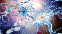
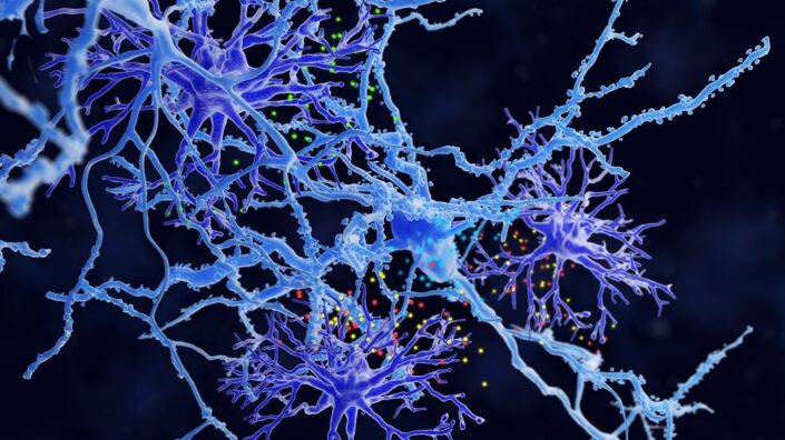
The longevity field hasn’t been very good at naming things, but one notable exception is “inflammaging”: the low-grade chronic inflammation that correlates with aging. Recognition has been growing that inflammation, the ubiquitous reaction of the immune system to various stressors, is a major driver of many age-related diseases and possibly one of the limiting factors for our species’ maximal lifespan.

Dr. David Furman, who has been studying inflammation for many years at Stanford and the Buck Institute for Research on Aging, might be the best authority to talk to about inflammation and aging. Recently, his team developed an inflammation aging clock that he aims to commercialize. David also leads with a personal example by minimizing environmental exposures that cause inflammation.

Tell me about your journey to where you currently are: a renowned geroscientist working at two top-tier institutions on inflammation in the context of aging.

I’m from Argentina. My journey started very early on when I realized I wanted to create a strong positive impact on humanity and decided to try and go for med school. Seeing how some can benefit from the best medicines and others simply have no access to clean water was shocking. I wanted to change that and help people live better and longer. Then, I had a conversation with my dad, who convinced me to study biology or biochemistry, and that way, I could have a much profound impact than being a physician.

I studied biology and then focused on immunology. This taught me how important inflammation is for fighting viruses and other pathogens. In the early 2000s, we first heard the idea that inflammation and the immune system also participated in age-related diseases. It sounded very surprising at the time. We know that inflammation and the immune system protect us from infectious diseases. We also know that if inflammation or an autoinflammatory condition goes awry, you can develop autoimmune diseases, but the idea that non-communicable diseases of aging, like cardiovascular disease or Alzheimer’s, had an immune root was very appealing to me.

That brought me to Stanford. I was recruited by Mark Davis, who I’m sure at some point will get the Nobel Prize for the discovery of T-cell receptors that recognize viruses and cancer cells. A few years after I joined the Stanford community in 2008 as a postdoc at Mark’s lab, he asked me to lead and be more involved with the Thousand Immunomes project, which was just starting at that time.

I became the director of that project, which looked at the immune system at large. We were doing multi-omics before it was even called multi-omics. We were analyzing a few hundred proteins, whereas today, we’re looking at over 10,000, but the premise is the same: by looking at many parameters in human cohorts, we can learn from humans and then apply those learnings directly to humans, skipping animal models entirely. We know they’re broken, right?

So, by analyzing this massive amount of data, I sort of became a data scientist by brute force. I learned from Rob Tibshirani, Trevor Hastie, and Daphne Koller, who essentially invented machine learning and AI at Stanford. Bridging computational sciences and immunology led to many findings and publications, and all of them had this aging component. It was striking that when looking at the immune system, the strongest signal by far was aging signatures.

That put me in a position to focus more on aging and longevity. In 2019, Eric Verdin recruited me as an Associate Professor and to lead the AI platform at the Buck Institute for Research on Aging.

Everything you’ve just said resonates with me strongly. I’ve also been fascinated with the role of inflammation in aging. I agree that its importance appears to be massive and has probably been overlooked. Can you tell me more about the connection between these two things?

Let me give you the historical perspective. We’ve been studying the immune system with decent technologies for about a hundred years, and we all understand that it protects us from infectious diseases, but the idea that aging is partly due to derangements in the immune system only started around the year 2000. It’s a very recent concept. Only 25 years ago, the first paper was published by Claudio Franceschi, who basically said that inflammation resulting from a number of environmental exposures will accelerate aging rates.

That was absolutely shocking to everybody. As a community, we started looking at the pathways, but at that time, we didn’t know what markers or cells were implicated in aging or age-related diseases. It turns out that inflammation not only affects the molecular hallmarks of aging, it can also drive particular diseases of aging.

Take cancer, for instance. I was shocked to see that if you take cancer cells and deprive the media of interleukin-6, they don’t grow, but if you put IL-6 in the media, they start proliferating like crazy. We now know that cancer – from the very early transformation of cells to metastasis and late stage four cancers – is largely dependent on inflammation.

Then we have the relationship between cardiovascular disease and inflammation. We’ve published several papers in major journals showing that inflammation in older adults is largely associated with a higher risk of heart attacks and other heart-related events, like arterial stiffening and ventricular remodeling.

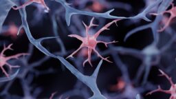
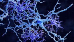
Depending on the specific protein of the inflammatory process you’re looking at, you’re going to see a different effect. For example, eotaxin is a protein typically elevated in older adults that is associated with neurodegeneration. Who would have thought the immune system could drive Alzheimer’s disease? This is changing the paradigm in Alzheimer’s research, in cancer research, and in every single disease of aging as we understand more about the causal relationships between immune system cells and molecules and derangement at the organ level.

That naturally brings up the idea of intervening early. This seems to be an early type of accumulating damage, so theoretically, if we intervene soon enough, we could have a substantial impact on aging.

That’s very well put. I spent 17 years of my career on the idea of identifying early, preclinical signs of disease – molecular changes that are already happening in the body. Using these molecular and cellular changes, you can predict diseases and mortality rates in people who may not even show any symptoms yet.

If you can identify these changes, you can do more than just intervene; you can intercept these diseases. I like to call it the molecular interception of a disease that is developing very slowly and is not yet clinically observable. The whole premise of what we’re doing is not just predicting or understanding biomarkers. It’s about preventing disease and extending the healthspan of the population by intervening early in its course.

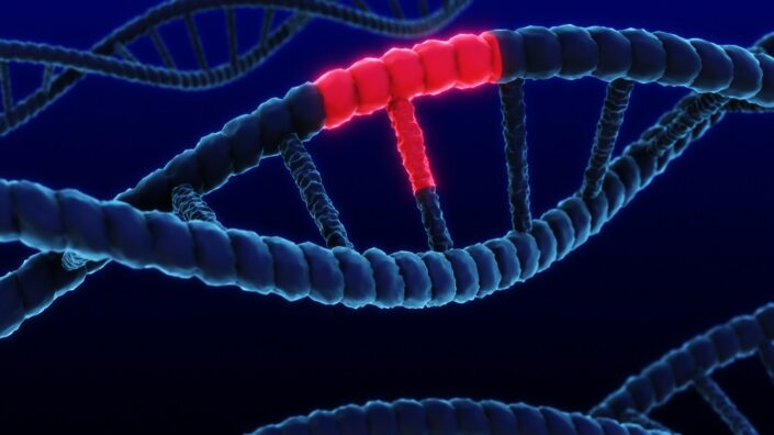
It sounds similar to epigenetic clocks, which work because we accumulate epigenetic damage from very early on. Epigenetic aging starts in the womb. Is that also the case with what we might call “inflammaging” or inflammatory aging? How early do the changes you track begin?

That’s an area of research that hasn’t flourished yet; it’s extremely early, so I can’t substantiate claims with strong science. The reason is a lack of sufficient data sets. The theory, however, is that inflammation may start two or three generations before an individual is born.

The concept of transgenerational epigenetic modifications also applies to inflammation. We’ve seen in some studies that the inflammatory state of new generations is impacted by what past generations have suffered from – things like psychological trauma or famine. These events can impact the epigenetic landscape and make individuals more susceptible to an increased inflammatory load later in life.

To give you an example, if an expectant mother has excessive inflammation – perhaps due to smoking or obesity – this can reflect on the growing fetus. Those children will have a higher risk of developing inflammation-related diseases like autism, early diabetes, cardiovascular disease, and even suicidal thoughts. In 2014, we published a paper in Molecular Psychiatry where we studied 500 individuals with major depression disorder and 500 controls. The inflammation levels were all over the place in those who suffered from the disorder. So yes, this starts very early on, possibly even before conception.

You used your 1000 Immunomes Project to build an aging clock that you believe is superior in some ways to existing clocks. If I understand correctly, it uses just a handful of proteins. Can you tell me more about it?

Yes. We leveraged the 1000 Immunomes dataset, which, at the time we built the clock, had data from over 12 years. We built a deep neural network to analyze the data. Protein networks have a lot of redundancy, and human data is typically very noisy. We addressed this using a specific type of neural network called a deep guided autoencoder, which is very different from what most people use for building clocks.

The beauty of autoencoders is that they can effectively deal with redundancy, non-linearity, and noise: the three main challenges in our data. So, it was the ideal tool. Using it, we predicted a person’s calendar age. What I really like about this clock is that it’s not perfectly accurate at predicting calendar age. That inaccuracy gives you room for biological interpretation as to why some people score so much higher or lower than the rest of the population.

So, this clock is trained on calendar age, not on intrinsic capacity?

You’re probably talking about a different aging clock we have that is trained on intrinsic capacity, from a collaboration with a group in France. The two clocks are interrelated, though. High intrinsic capacity, which is a very positive thing to have, correlates strongly with a low inflammatory age. We can explore that later, but for the inflammatory clock, which we call iAge, we predict chronological age. The model is “guided” because it’s trained on two target variables: the immune protein data you feed the algorithm with, and calendar age.

The output is the closest thing to an “immune age” out there. We then use the residual – the difference between a person’s calendar age and their predicted immune age – to see if the clock has clinical validity, and it does. We could associate an increase in the inflammatory clock with having multiple diseases at once (multimorbidity). We also saw a strong prediction of frailty; if I measure your inflammatory age today, I can predict with high accuracy whether you will become frail seven years from now. We then created a gene expression proxy for this protein clock and validated it in external datasets, like the Framingham Heart Study, where we were able to predict mortality in 2,500 people.

I remember you saying that your immune clock singles out centenarians as having a very different immune profile. I think this is amazing because it suggests that inflammation and immune system exhaustion might be what kill the oldest old and that centenarians are people who can somehow defend against this. What can you tell me about that?

Yes, that was one of our clinical validations. We looked at individuals with extreme longevity: centenarians and supercentenarians. We took a cohort of about 20 individuals from the Bologna area in Italy, all of them 100 years or older, and ran our iAge analysis on them. On average, their inflammatory age was 40 years younger than their calendar age.

There was one super-healthy 105-year-old male who had almost never seen a doctor. His inflammatory age was 25. That’s 80 years below his calendar age. He is an outlier, a really interesting person. What is it about his immune system that allows him such a level of control over inflammation? We don’t know yet. Other studies by Nir Barzilai, for example, show that the immune systems of centenarians are very different from their 80-year-old counterparts. They have peculiar CD4 T cells and a very different microbiome. Perhaps the explanation is that their microbiome and immune cell proportions are just shaped differently, but the fact remains: in supercentenarians and centenarians, their inflammatory age is dramatically lower than their calendar age.

Mimicking the immune system of centenarians can help us compress mortality. However, they also eventually die, and immune exhaustion is emerging as a central cause. By doing something about that, we might even be able to go one step further and extend maximum human lifespan.

That’s a hypothesis, right? We can think of multiple ways to try and push the healthspan of the population to, say, 120 years old, so people can be super healthy and then die quickly. But, I want to stress one thing about the difference between healthspan and lifespan. We all want to live healthier, for sure, but many people think of death as a very negative thing. Why? Because most people lack a humanitarian purpose and achievement in life, something that impacts more than themselves and their families. I think it is pretty scary to die and not leaving a legacy behind. Living is great, and dying shouldn’t be the worst thing that could ever happen to you.

Let’s pivot to something more down-to-earth. You said your inflammation clock gave you clues about what works in terms of diet, exercise, and other interventions.

I’m not sure if you’ve seen an article that is coming out in Business Insider about my experience reducing my own inflammation by modifying my environment and lifestyle. I was at a longevity investors meeting in Switzerland and told a reporter my story. She found it amazing and wanted to write a piece on it.

For 15 years, I’ve been studying the social and lifestyle determinants of inflammation, and I decided to start testing these principles on myself. It significantly changed my family’s life. We moved from the Palo Alto area to an off-the-grid cabin in a small valley called San Gregorio. There, we decided to apply the principles of evolutionary medicine to control inflammation.

The idea comes from a paper I published in Nature Medicine in 2019, which basically states that many environmental and lifestyle factors drive inflammation. I hate to call them “choices,” because someone in Fresno breathing polluted air has no choice. I dislike when people say, “lifestyle choices,” because for 90% of the population, there is no choice.

Anyway, I was guided by the following principle: if you move any species to a new environment that it has not evolved with or adapted to, it will develop inflammation as a natural response. The corollary is that the more distant a person’s life is from our species’ evolutionary experience, the more inflamed that person will be.

The immune system acts a sensory system of your environment, much like hearing capacity or vision; the only difference with classical sensory systems is that the output (inflammation) doesn’t reach your consciousness. You can measure this, and you can start thinking about how to implement this principle in your day-to-day life. It’s everywhere: the workplace, your household, your city. There are external and internal factors, some you can control and some you can’t. It applies to water quality, air quality, the food you eat, and the household products you use.

If your body hasn’t seen something during its two-million-year evolution, you probably shouldn’t be exposed to it, because it will cause inflammation. This applies to plastic containers with phthalates, and the microplastics and nanoplastics we are breathing that accumulate in our organs. Everything converges on inflammation and reactive oxygen species. When you read the literature, it becomes obvious that every one of these insults signals to your body through inflammation. That’s what causes issues in the brain, the heart, and the joints.

So, we made changes. We cut out wheat; humans haven’t been exposed to it for more than 8,000 years. Same with dairy products; we only started domesticating cows a few thousand years ago. Then there are hyper-processed foods. There’s a very long list of things you can start tweaking. And then you have to measure the effect. The problem is that the canonical markers of inflammation mostly work for acute inflammation.

You mean markers such as C-reactive protein (CRP)? It probably doesn’t tell you much about chronic inflammation.

Right, it doesn’t tell you anything. It’s worthless for this purpose. People look at CRP because there’s nothing else, but for predicting cardiovascular disease, its accuracy is about the same as flipping a coin. Paul Ridker built part of his career around CRP and now IL-1β, and he loves the idea of drugging these things.

High-sensitivity CRP is widely used, but a savvy cardiologist will tell you they don’t find it that useful for predicting risk. They use it now for suspected acute infections, of course. But it’s an acute-phase reactant; it goes up, but then it comes back down. IL-1β is similar, sometimes chronic, sometimes acute. Some proteins reflect chronic states, while others just change for a short period.

In terms of other interventions, simply not moving around will increase your inflammation. Your body will interpret a sedentary state as a sign that you’re sick, it’s actually called sickness behavior in psychology.

Basically, inflammation is a lifelong, adaptive reaction that can also be very destructive.

Exactly. It can be. Inflammation is built for repair and protection. Your skin and microbiome interact with inflammation all the time, but it becomes very detrimental if it’s sustained and doesn’t resolve. There’s remarkable work from Charles Serhan at Harvard on the biomarkers of inflammation resolution. That’s a whole other area of research that is super interesting and could be pivotal for finding solutions.

I want to ask you about one of your companies, Edifice Health. Is this how you’re commercializing the iAge clock?

Yes, exactly, and let me give you the high-level answer for why. The system for academic research is not ideal; it’s really broken. Think of the amount of money that goes from the government to academia. It’s incredibly inefficient. Why? Because in the academic setting, there is no incentive or training to start a commercial entity. If you don’t do that, the findings end up in a drawer, in the trash, or just as a publication. They don’t translate to the bedside, to households, to solving people’s problems in the marketplace. Federal money does not equal translation. The pathway is not from bench to bedside; it’s from bench to company to bedside.

It seems like the new agency ARPA-H is taking a different approach, and you have applied for one of their programs, correct?

Exactly, they are following this principle. A program we just applied for, called PROSPR, requires us to have FDA approval and a working commercial entity by year five. It’s very pro-startup; a commercialization strategy is a requirement for this ARPA-H funding. It’s a beautiful, dream program. It’s what everyone should be doing: putting their efforts into early diagnosis or interventions for aging but with a translational lens.

We would like to ask you a small favor. We are a non-profit foundation, and unlike some other organizations, we have no shareholders and no products to sell you. All our news and educational content is free for everyone to read, but it does mean that we rely on the help of people like you. Every contribution, no matter if it’s big or small, supports independent journalism and sustains our future.
TransVision Summit

Madrid Set to Become the Longevity Capital of Europe

We are thrilled to announce the second edition of the International Longevity Summit (www.TransVisionMadrid.com) in beautiful Madrid after the major success in 2024.

Organized by the Illustrious College of Medical Doctors of Madrid (ICOMEM) and the International Longevity Alliance (ILA), this year we expect over 500 participants with more international experts, including George Church (Professor, Harvard Medical School and MIT), Mehmood Khan (CEO, Hevolution Foundation), Steve Horvath (Principal Investigator, Altos Labs), Michael Ringel (COO, Life Biosciences), Aubrey de Grey (CEO, LEV Foundation: Longevity Escape Velocity), Joao Pedro de Magalhaes (Chair of Molecular Biology, University of Birmingham), Andrea B. Maier (Founding President, Healthy Longevity Medicine Society), Phil Newman (Founder, Longevity Technology), Liz Parrish (CEO, BioViva), David Wood (Executive Director, LEV Foundation: Longevity Escape Velocity), Natalie Coles (Director, Supercentenarians Project, R3 Bio), Ken Scott (Founder, Human Longevity and Rejuvenation Syndicate), Tina Woods (Executive Director, International Institute of Longevity), Juan Carlos Mendez (President, SOVEMAL), Sabinije von Gaffke (Director of Partnerships, Super Human Network), Roberto Grau (Cofounder, Juventas4Life), Sajidxa Mariño (Founder & CEO, Respira Libre), Jose Angel Olalde (Founder, Centro de Medicina Regenerativa), Jose Cordeiro (Director, International Longevity Alliance) and others to be announced soon, plus a select group of Spanish experts, from scientists and doctors all the way to the world of economics and politics, starting with the Mayor of Madrid: José Luis Martínez Almeida.

Madrid is the most longevous capital in the European Union, with a current life expectancy of 86.1 years at birth, and Spain is also the most longevous country in the region, according to Eurostat. Some of the oldest people in the world have been Spanish, like María Branyas Morera who was the oldest living person until she died last year at the age of 117 years and 168 days. In fact, life expectancy in Madrid is higher than in the so-called Blue Zones of Sardinia in Italy, Ikaria in Greece, Okinawa in Japan, Nicoya Peninsula in Costa Rica or Loma Linda in California. Therefore, Madrid can be considered as a Blue Zone 2.0, beating all the old Blue Zones, and with a much higher and pleasurable standard of living. Yes, it is the Mediterranean diet, and more, much more that makes Madrid the most longevous city in the EU!

Come to Madrid and discover the beauty of life and longevity in Spain, the second most longevous country in the world, among large nations, only after Japan. Thus, just before the International Longevity Summit (October 1-2), we will start that week organizing 2 days of pre-conference tours (September 29-30) to visit the UNESCO World Heritage sites around Madrid (Avila, Segovia, Aranjuez, Toledo, El Escorial and Alcala de Henares). You are more than welcome to join us for one or both tours so that you can really enjoy the beauty of Castilla La Mancha, the land of Cervantes and Don Quixote, the land of fiesta and siesta, the land of Picasso and Dali, the land of vino and sangria, the land of tapas and picoteo, the land of flamenco and fandango, the land of Plus Ultra as the Romans called Hispania over two millennia ago.

During October 1st, which corresponds to the International Longevity Day, we will announce the Madrid Longevity Declaration and will award the Madrid Longevity Prizes. Thus Madrid reinforces its position as the longevity capital of Europe, and Spain as the most longevous country in the EU. All these activities will be in the historic Great Amphitheater of the Illustrious College of Medical Doctors of Madrid (ICOMEM), the same place where Spanish Nobel laureate Santiago Ramon y Cajal gave his master lectures, and the most impressive Classical theater of Spain, rich in history. We will also premiere an award-winning longevity documentary and will walk the Madrid March for Longevity, between the world-renowned Puerta del Sol (Kilometer 0.0 of Madrid and Spain) and the famous Cibeles Fountain (where world champion Real Madrid celebrates its football victories). Come to Madrid, let´s all write history and create the future together, it will be an immortal experience, very literally!

For more information, contact: info@TransVisionMadrid.com
We would like to ask you a small favor. We are a non-profit foundation, and unlike some other organizations, we have no shareholders and no products to sell you. All our news and educational content is free for everyone to read, but it does mean that we rely on the help of people like you. Every contribution, no matter if it’s big or small, supports independent journalism and sustains our future.
Doctor holding brain

Fixing Sugar Metabolism Shows Promise Against Dementia

Scientists have shown that aberrant metabolism of glycogen in neurons is linked to the accumulation of harmful tau protein. Caloric restriction, genetic interventions, and small molecules might help [1].

Glycogen and the brain

Aberrant aggregation of microtubule-associated protein tau (MAPT), or simply tau protein, is a hallmark of several neurodegenerative diseases [2]. The most famous of them is Alzheimer’s disease, in which tau accumulation in the form of hyperphosphorylated neurofibrillary tangles (NFTs) damages neurons.

Another, less-known, characteristic of many of these diseases is abnormal glycogen metabolism and accumulation [3]. Glycogen is a stored form of glucose, used by the body as an energy source when nutrient levels are low. It is mostly found in the liver and muscle, but brain cells (predominantly astrocytes but also neurons) also contain small amounts of it.

Impaired glycogen metabolism in neurons hurts learning and memory, while dietary restriction (DR) is known to extend lifespan and delay neurodegeneration in animal models of neurodegenerative diseases. In this new study published in Nature Metabolism, scientists from the Buck Institute for Research on Aging tried to understand how these two facts might be connected.

Diet restriction rescues lifespan

The authors started with two Drosophila fly models. One exhibited accelerated accumulation of wild-type tau protein, while the other included a known mutation in MAPT (R406W), which, in humans, causes a severe familial disease called frontotemporal lobar degeneration with tau inclusions (FTLD-tau).

The flies were either freely fed or restricted in calories. DR significantly increased lifespan even in healthy controls. In the two disease models, the effect was even more dramatic. DR rescued lifespan in flies with aberrant accumulation of wild-type tau almost completely, and in mutation-carrying flies, the difference was highly significant. Accordingly, in DR flies, levels of neuronal death fell dramatically.

Tauopathies and dietary restriction

Proteomic analysis of the flies’ brains revealed that pathways related to fat and glycogen metabolism were among the most drastically changed by DR, and glycogen levels were indeed elevated in the brains of tauopathic flies.

Interestingly, however, DR did not seem to alter overall levels of glycogen, despite clearly having a strong beneficial impact. The researchers suspect that what might be important is the rate of glycogen turnover. The enzymes involved in this turnover, including glycogen phosphorylase (GlyP), were upregulated in mutant flies on DR. Overexpression of GlyP increased the lifespan of mutant flies by almost 70% and drastically reduced neuronal death.

More antioxidants!

The researchers used metabolomics and RNA sequencing to study the molecular effects of GlyP upregulation. Surprisingly, the pathways for energy production, namely glycolysis and the citric acid cycle, were actually downregulated. Instead, the glucose from the broken-down glycogen was being shunted into the pentose phosphate pathway (PPP). Its primary function is to generate antioxidants: molecules that combat oxidative stress. Reactive oxygen species (ROS) were indeed significantly reduced in the brains of the flies with enhanced glycogen breakdown.

According to the researchers, this might at least partially explain the benefits of DR and GlyP upregulation. In line with this hypothesis, blocking the PPP with a small molecule abolished the protective effects of glycogen breakdown. The team also successfully recreated the effects of genetic GlyP overexpression by using another small molecule, 8-Bromo-cAMP, to activate the GlyP-producing pathway.

A vicious cycle?

The team then ran experiments in vitro on human neurons derived from induced pluripotent stem cells (iPSCs) that were obtained from patients with FTLD-tau. Genetically corrected cells from the same donors were used as controls. The researchers demonstrated increased glycogen accumulation in FTLD-tau cells and also tested the rescue mechanism by overexpressing the human version of the glycogen breakdown enzyme (PYGB) in the diseased human neurons. This reduced abnormal glycogen accumulation and restored mitochondrial abundance, which declines with this disease.

Importantly, using these human neurons, the team showed that tau protein and glycogen co-localize within cells and physically interact, supporting the hypothesis that a direct interaction between the two might be part of the problem. The authors hypothesize that this may create a detrimental vicious cycle in which tau binding promotes glycogen accumulation, which, in turn, exacerbates tau pathology and oxidative stress.

“Our findings suggest that glycogen is more than just a metabolic reservoir – it may act as a sticky trap for tau, creating a dangerous feedback loop where tau promotes glycogen buildup, and glycogen in turn fuels tau aggregation,” said Dr. Pankaj Kapahi, the corresponding author of the study, to lifespan.io. “Breaking this cycle could open a new therapeutic front in the fight against Alzheimer’s disease.”

We would like to ask you a small favor. We are a non-profit foundation, and unlike some other organizations, we have no shareholders and no products to sell you. All our news and educational content is free for everyone to read, but it does mean that we rely on the help of people like you. Every contribution, no matter if it’s big or small, supports independent journalism and sustains our future.

Literature

[1] Bar, S., Wilson, K. A., Hilsabeck, T. A., Alderfer, S., Dammer, E. B., Burton, J. B., … & Kapahi, P. (2025). Neuronal glycogen breakdown mitigates tauopathy via pentose-phosphate-pathway-mediated oxidative stress reduction. Nature Metabolism, 1-17.

[2] Goedert, M., Eisenberg, D. S., & Crowther, R. A. (2017). Propagation of tau aggregates and neurodegeneration. Annual review of neuroscience, 40(1), 189-210.

[3] Mann, D. M. A., Sumpter, P. Q., Davies, C. A., & Yates, P. O. (1987). Glycogen accumulations in the cerebral cortex in Alzheimer’s disease. Acta neuropathologica, 73, 181-184.

Healthy elderly

Researchers Connect Cellular Markers to Physical Well-Being

In Aging Cell, a team of researchers has described how the health of skin fibroblasts relates to physical and functional ability.

Frailty, capacity, and skin cells

This paper begins with an explanation of how frailty and capacity are defined. The World Health Organization defines it as an overall state of reduced strength, endurance, and physiogical functions that increase the risk of adverse health outcomes and lead to dependency [1]. Intrinsic capacity, while difficult to measure, refers to a collection of functional attributes that define overall health [2]. These metrics lead to a better understanding of how generally healthy someone is than the simple number of chronological age.

However, there is a gap between other clocks, such as epigenetic clocks, and biomarkers of functionality. The authors of this paper focus on a ‘gerophysical’ approach that links the two [3].

This study focuses on skin cells (fibroblasts) for several good reasons. Skin, of course, is easy to measure compared to other parts of the body, and fibroblasts preserve both the function and structure of multiple tissue types [4]. Previous work has found that fibroblasts play a significant role in immune responses [5] and metabolic regulation [6]. These cells have been heavily studied in the context of aging, including epigenetics and transcriptomics such as age-related mRNA strands [7]. However, these researchers note that no one had previously linked cellular aging biomarkers to intrinsic capacity metrics.

Some biomarkers are more telling than others

This study used skin samples from 133 volunteers of both sexes in the INSPIRE-T cohort. Their ages ranged from 20 to 96, encompassing healthy, pre-frail, and frail states.

The first part of this study cultured these fibroblasts in vitro, comparing chronological age to various biomarkers. As expected, the proliferation rate of the fibroblasts slowed with age, and markers of DNA damage increased along with the senescence marker p16 and the inflammatory factor IL-6. Interestingly, this study did not show any statistically significant correlation between chronological age and many other senescence markers, including SA-β-gal. However, older cells did express more SA-β-gal when exposed to the stressor doxorubicin.

In the next part of this study, the researchers focused on three key aspects of fibroblast function: tissue structure, immune responses, and metabolic regulation (SIM) along with senescence. This was a biomarker-based analysis, focusing on several key biomarkers in each of these domains: 31 in total.

SIM framework

The researchers then used a statistical measurement called Mahalanobis distance to quantify homeostatic dysregulation: how different a person’s biomarkers are from a normal baseline. As expected, this measurement was strongly correlated with chronological age, and the researchers suggest that it can be used as a biomarker of aging.

The researchers took a closer look at how the S, I, and M indices correlate with one another. They found that while many aspects of structure, such as MMP1, did not seem to be closely related with aging, others did, including Periostin and TIMP1. The researchers suggest that these metrics are closely tied to age-related changes in the extracellular matrix.

Cytokine production, as expected, is significantly upregulated with aging, including IL-6. Other well-known inflammatory compounds, such as TGF-β, were undetectable in most samples. Interestingly, aging increases the responsiveness of inflammation to viral infection, although this inflammation’s effect can be negative.

The examination of metabolism revealed a decrease in mitochondrial respiratory efficiency with aging, and antioxidant genes were found to be largely upregulated in response to increased oxidative stress. Once more, there were some interesting negative results: SIRT1 and the NRF genes were not found significantly correlated with aging in this study.

Putting it all together

Directly comparing each of these biomarkers to intrinsic capacity yielded crucial findings. Periostin was once more singled out as a significant contributor to functional aging, as were CD36 and mitochondrial respiration markers. The researchers believe that their overall SIM analysis allows for a holistic approach that provides a detailed analysis of multiple aspects of aging.

Periostin is known in the literature as being crucial to wound healing [8], but it has been little discussed in the context of aging. While the researchers do not directly suggest that it is a suitable target for future interventions, and they cannot demonstrate that it has a causal relationship in this paper, later work may investigate whether or not it is a valid target.

The researchers note this study’s limitations: there were a substantial number of analyses made from a limited number of skin cells, and the biopsies may have been poorly representative of the people from whom they were taken. External factors such as lifestyle and environment could not be accounted for. Larger cohorts would need to be utilized to further refine this SIM analysis.

We would like to ask you a small favor. We are a non-profit foundation, and unlike some other organizations, we have no shareholders and no products to sell you. All our news and educational content is free for everyone to read, but it does mean that we rely on the help of people like you. Every contribution, no matter if it’s big or small, supports independent journalism and sustains our future.

Literature

[1] Beard, J. R., Officer, A., De Carvalho, I. A., Sadana, R., Pot, A. M., Michel, J. P., … & Chatterji, S. (2016). The World report on ageing and health: a policy framework for healthy ageing. The lancet, 387(10033), 2145-2154.

[2] Gonzalez-Bautista, E., & Beard, J. R. (2023). The Challenge of Measuring Intrinsic Capacity. The journal of nutrition, health & aging, 27(10), 806-807.

[3] Kemoun, P. H., Ader, I., Planat-Benard, V., Dray, C., Fazilleau, N., Monsarrat, P., … & Casteilla, L. (2022). A gerophysiology perspective on healthy ageing. Ageing research reviews, 73, 101537.

[4] Plikus, M. V., Wang, X., Sinha, S., Forte, E., Thompson, S. M., Herzog, E. L., … & Horsley, V. (2021). Fibroblasts: Origins, definitions, and functions in health and disease. Cell, 184(15), 3852-3872.

[5] Haniffa, M. A., Wang, X. N., Holtick, U., Rae, M., Isaacs, J. D., Dickinson, A. M., … & Collin, M. P. (2007). Adult human fibroblasts are potent immunoregulatory cells and functionally equivalent to mesenchymal stem cells. The Journal of Immunology, 179(3), 1595-1604.

[6] Zhao, X., Psarianos, P., Ghoraie, L. S., Yip, K., Goldstein, D., Gilbert, R., … & Liu, F. F. (2019). Metabolic regulation of dermal fibroblasts contributes to skin extracellular matrix homeostasis and fibrosis. Nature metabolism, 1(1), 147-157.

[7] Tsitsipatis, D., Martindale, J. L., Mazan‐Mamczarz, K., Herman, A. B., Piao, Y., Banskota, N., … & Gorospe, M. (2023). Transcriptomes of human primary skin fibroblasts of healthy individuals reveal age‐associated mRNAs and long noncoding RNAs. Aging Cell, 22(11), e13915.

[8] Elliott, C. G., Wang, J., Guo, X., Xu, S. W., Eastwood, M., Guan, J., … & Hamilton, D. W. (2012). Periostin modulates myofibroblast differentiation during full-thickness cutaneous wound repair. Journal of cell science, 125(1), 121-132.

Ordinary lab mouse

Healthspan Effects of an Anti-Aging Vaccine on Mice

The researchers of a recent study published in Aging Cell described their novel CD38 peptide vaccine, which improved many measurements of physical health and prevented cognitive decline in aged mice [1].

A long-term anti-aging vaccine

Many anti-aging strategies, such as supplementation, require regular, daily intake. A vaccine, on the other hand, is a therapeutic approach that, once administered, can have lasting effects.

Anti-aging vaccination approaches have already been undertaken previously, and they were designed to target, among others, senescent T cells and the cells that line blood vessels (vascular endothelial cells) [2, 3]. These researchers chose a different target: the widely researched CD38 protein expressed in several types of immune cells and tissues, which has multifunctional enzymatic activities related to NAD metabolism.

The age-dependent increase in CD38 levels has been linked to NAD+ decline and mitochondrial dysfunction [4]. Previous research on targeting CD38 with small molecules for therapeutic purposes showed benefits in glucose intolerance, physical dysfunction, and neuroinflammation [5, 6], while CD38 antibody research showed benefits against age-related syndromes, such as fibrosis, NAD+ deficiency, and cardiotoxicity [7-9], making it a promising target for a vaccine.

First, the researchers needed to find which part of CD38 would induce the strongest response from the immune system, thus making it the best candidate for a vaccine. While the researchers encourage future studies to test different sequences, they limited themselves to three amino acid sequences with which to create three peptide vaccines. After a few weeks, they tested how the immune systems of mice responded to the vaccines and chose the one that spurred the strongest response.

Physically stronger and cognitively younger

The researchers immunized a group of 12-month-old mice with the chosen vaccine. At 15-18 months old, the researchers analyzed the mice’s healthspan, then immunized them again at 18 months and 3 weeks of age and euthanized them at 20 months to collect tissues for further assessment.

The mice showed a strong immune response to the vaccination, translating into physical and cognitive health improvements in aged mice. The researchers observed that vaccination prevented a decline in total walking distance, maximal walking speed, grip strength, and hanging endurance in both male and female animals. Similarly, measuring physical fitness by placing mice in an open arena and observing their behavior showed better measures of total movement distance and average exploring velocity. Aged vaccinated mice also scored better on frailty scores and generally appeared younger than controls.

Cognitive skills were tested using a classic Morris water maze experiment and a novel object recognition model. The results suggested that the CD38 vaccine prevented cognitive decline in mice.

Beyond physical and cognitive tests, the researchers noted that vaccination improves metabolic features, which tend to deteriorate with age. They reported improvements in glucose tolerance, insulin sensitivity, and different measures of body energy consumption, such as energy expenditure and oxygen consumption, in aged mice compared to controls.

Decreasing senescence

These results prompted the researchers to investigate the underlying molecular mechanisms. Therefore, they analyzed both gene expression and the full suite of liver proteins (the proteome). This focus on the liver stems from its essential role in metabolism, including glucose and NAD+ metabolism, and its role as a target for anti-aging therapeutics, such as metformin and NAD+ precursors [10, 11].

First, the researchers confirmed that the vaccine specifically targeted CD38. Both gene expression and proteome analysis confirmed reduced CD38 levels.

Among the aging-related processes impacted by CD38 was senescence, and CD38 vaccination led to decreased senescence in aged mice’s solid organs. The researchers observed a decrease of the senescence marker p21 but not p16 in the liver along with decreased senescence-associated secretory phenotype (SASP) factors. Similarly, the activity of another senescence marker, SA-β-gal, was reduced in the liver and spleen following vaccination.

In previous experiments, the researchers reported CD38 vaccination’s impact on metabolic processes. This was also reflected at the molecular level.

In the liver, there was decreased expression of some glucose metabolism-related genes, specifically key glycolytic enzyme genes, in vaccinated animals compared to controls, which was confirmed by the proteome analysis. The proteome analysis also identified upregulation of fatty acid metabolic processes, breakdown of complex molecules (catabolism), and peroxisome proliferator-activated receptor (PPAR) signaling pathways, which, among other roles, regulate metabolic processes and are closely associated with oxidative phosphorylation (OXPHOS), which generates most cellular energy.

Reversing the decline

Previous research linked a specific group of immune cells, CD38-expressing macrophages, to age-related NAD+ decline [12]. NAD+ metabolism is an important player in aging. Anti-aging approaches aimed at NAD+ repletion have shown some beneficial anti-aging and healthspan-improving results.

In this study, vaccination reduced the levels of CD38-expressing macrophages in the liver and increased the NAD+/NADH ratio in the aged mice’s liver and brain tissue, partly restoring the age-related NAD+ decline.

In total, these results suggest ‘that the CD38 peptide vaccine ameliorated abnormal metabolism-related proteome shifts’ and rejuvenated the protein profile.

Proof of concept

This proof-of-concept study showed the anti-aging potential of the CD38 vaccine. The observed promising effects are most likely caused by the elimination of CD38-positive cells; however, different yet unidentified mechanisms may exist.

Since this research was conducted on mice, further studies are necessary to confirm its effect in people. The researchers also speculate that future studies should address its potential in different age-associated diseases, such as Alzheimer’s disease.

Peptide vaccines generally have high specificity and safety. Based on these results, this vaccine was not an exception from the rule, as the researchers did not detect any side effecs in the vaccinated mice. However, since CD38 plays a role in infection response, it might lead to increased infection rates, so researchers should monitor for possible infections in future studies.

Additionally, the activation of inflammatory responses might cause side effects. Inflammation is linked to aging and is thought to drive senescence, and in the short term, activation of inflammation might accelerate aging processes. Indeed, the researchers observed increased IL-6, one of the SASP factors, following vaccination in middle-aged and older mice, but it decreased later. These changes in inflammatory response warrant further investigation into how this CD38 vaccine impacts the immune system.

We would like to ask you a small favor. We are a non-profit foundation, and unlike some other organizations, we have no shareholders and no products to sell you. All our news and educational content is free for everyone to read, but it does mean that we rely on the help of people like you. Every contribution, no matter if it’s big or small, supports independent journalism and sustains our future.

Literature

[1] Yu, S., Li, Z., Tang, Y., Chen, Y., Ma, Y., Du, K., Zong, Z., Feng, K., Wei, Y., Chen, L., & Deng, H. (2025). CD38-Targeting Peptide Vaccine Ameliorates Aging-Associated Phenotypes in Mice. Aging cell, e70147. Advance online publication.

[2] Yoshida, S., Nakagami, H., Hayashi, H., Ikeda, Y., Sun, J., Tenma, A., Tomioka, H., Kawano, T., Shimamura, M., Morishita, R., & Rakugi, H. (2020). The CD153 vaccine is a senotherapeutic option for preventing the accumulation of senescent T cells in mice. Nature communications, 11(1), 2482.

[3] Suda, M., Shimizu, I., Katsuumi, G., Yoshida, Y., Hayashi, Y., Ikegami, R., Matsumoto, N., Yoshida, Y., Mikawa, R., Katayama, A., Wada, J., Seki, M., Suzuki, Y., Iwama, A., Nakagami, H., Nagasawa, A., Morishita, R., Sugimoto, M., Okuda, S., Tsuchida, M., … Minamino, T. (2021). Senolytic vaccination improves normal and pathological age-related phenotypes and increases lifespan in progeroid mice. Nature aging, 1(12), 1117–1126.

[4] Camacho-Pereira, J., Tarragó, M. G., Chini, C. C. S., Nin, V., Escande, C., Warner, G. M., Puranik, A. S., Schoon, R. A., Reid, J. M., Galina, A., & Chini, E. N. (2016). CD38 Dictates Age-Related NAD Decline and Mitochondrial Dysfunction through an SIRT3-Dependent Mechanism. Cell metabolism, 23(6), 1127–1139.

[5] Roboon, J., Hattori, T., Ishii, H., Takarada-Iemata, M., Nguyen, D. T., Heer, C. D., O’Meally, D., Brenner, C., Yamamoto, Y., Okamoto, H., Higashida, H., & Hori, O. (2021). Inhibition of CD38 and supplementation of nicotinamide riboside ameliorate lipopolysaccharide-induced microglial and astrocytic neuroinflammation by increasing NAD. Journal of neurochemistry, 158(2), 311–327.

[6] Tarragó, M. G., Chini, C. C. S., Kanamori, K. S., Warner, G. M., Caride, A., de Oliveira, G. C., Rud, M., Samani, A., Hein, K. Z., Huang, R., Jurk, D., Cho, D. S., Boslett, J. J., Miller, J. D., Zweier, J. L., Passos, J. F., Doles, J. D., Becherer, D. J., & Chini, E. N. (2018). A Potent and Specific CD38 Inhibitor Ameliorates Age-Related Metabolic Dysfunction by Reversing Tissue NAD+ Decline. Cell metabolism, 27(5), 1081–1095.e10.

[7] Peclat, T. R., Agorrody, G., Colman, L., Kashyap, S., Zeidler, J. D., Chini, C. C. S., Warner, G. M., Thompson, K. L., Dalvi, P., Beckedorff, F., Ebtehaj, S., Herrmann, J., van Schooten, W., & Chini, E. N. (2024). Ecto-CD38-NADase inhibition modulates cardiac metabolism and protects mice against doxorubicin-induced cardiotoxicity. Cardiovascular research, 120(3), 286–300.

[8] Shi, B., Amin, A., Dalvi, P., Wang, W., Lukacs, N., Kai, L., Cheresh, P., Peclat, T. R., Chini, C. C., Chini, E. N., van Schooten, W., & Varga, J. (2023). Heavy-chain antibody targeting of CD38 NAD+ hydrolase ectoenzyme to prevent fibrosis in multiple organs. Scientific reports, 13(1), 22085.

[9] Ugamraj, H. S., Dang, K., Ouisse, L. H., Buelow, B., Chini, E. N., Castello, G., Allison, J., Clarke, S. C., Davison, L. M., Buelow, R., Deng, R., Iyer, S., Schellenberger, U., Manika, S. N., Bijpuria, S., Musnier, A., Poupon, A., Cuturi, M. C., van Schooten, W., & Dalvi, P. (2022). TNB-738, a biparatopic antibody, boosts intracellular NAD+ by inhibiting CD38 ecto-enzyme activity. mAbs, 14(1), 2095949.

[10] Radziuk, J., Bailey, C. J., Wiernsperger, N. F., & Yudkin, J. S. (2003). Metformin and its liver targets in the treatment of type 2 diabetes. Current drug targets. Immune, endocrine and metabolic disorders, 3(2), 151–169.

[11] Mitchell, S. J., Bernier, M., Aon, M. A., Cortassa, S., Kim, E. Y., Fang, E. F., Palacios, H. H., Ali, A., Navas-Enamorado, I., Di Francesco, A., Kaiser, T. A., Waltz, T. B., Zhang, N., Ellis, J. L., Elliott, P. J., Frederick, D. W., Bohr, V. A., Schmidt, M. S., Brenner, C., Sinclair, D. A., … de Cabo, R. (2018). Nicotinamide Improves Aspects of Healthspan, but Not Lifespan, in Mice. Cell metabolism, 27(3), 667–676.e4.

[12] Wu, S., & Zhang, R. (2020). CD38-expressing macrophages drive age-related NAD+ decline. Nature metabolism, 2(11), 1186–1187.

Rabbit ears

Study Discovers a Mammalian Mechanism of Tissue Regeneration

Scientists have analyzed the differences between mammalian species that can regrow ear tissue after injury and those that cannot. Their findings can pave the way for novel regenerative therapies [1].

The lost art of regeneration

Many animal species have amazing regenerative abilities. On the one side of the spectrum sit planarian worms: slice them up, and every slice will grow into a fully developed animal. Even more complex animals, such as amphibians, can fully regenerate limbs. Mammals, however, have largely lost this ability. If only we could “teach” human tissues how to regenerate, this would open completely new horizons for anti-aging therapies.

Interestingly, some mammals have retained certain regenerative potential. For instance, rabbits can fully regenerate damaged outer ear (“ear pinna”) tissue, while mice and rats cannot. In a new study published in Science, a team of Chinese researchers set out to discover what sets those species apart when it comes to regeneration.

One master regulator

First, they punched holes in the ears of three regenerating species (rabbits, goats, and African spiny mice), and two non-regenerating ones (laboratory mice and rats). As expected, in the first group, the injured ears fully regenerate, including the cartilage. In mice and rats, however, only the wound boundaries heal, leaving the hole.

However, the team made a crucial observation: the initial stages were surprisingly similar. Both species formed a blastema: a mass of formerly specialized cells, such as skin and muscle cells, that dedifferentiated into a more stem-like state to facilitate regeneration and initially showed robust cell proliferation. The difference was that in non-regenerative species, the process was weaker and soon petered out.

This showed that the failure in mice and rats wasn’t an inability to start the regenerative process but an inability to sustain it. Next, the researchers focused on finding the molecular causes for those differences in regenerative capacity.

Using state-of-the-art techniques, including single-cell RNA sequencing (scRNA-seq) and spatial transcriptomics, on the healing ear tissue, the team ran a deep comparison between rabbits and mice. The most significant differences in gene expression were found in wound-induced fibroblasts (WIFs), a subpopulation of fibroblasts that appeared only after tissue damage in both species.

The researchers ultimately zeroed in on nine regeneration-associated genes (RAGs) that were differentially expressed in the WIFs of regenerative and non-regenerative species and ran a series of experiments, overexpressing some of the top candidates in mice using viral delivery. One gene, Aldh1a2, was sufficient to fully restore ear pinna regeneration. The team then confirmed through RNA analysis that Aldh1a2 was strongly activated following injury in rabbits, goats, and African spiny mice but barely detectable in mice and rats.

The gene produces aldehyde dehydrogenase 1 family member A2, a rate-limiting enzyme in the synthesis of retinoic acid (RA), a known regeneration factor. Retinoic acid’s precursor is vitamin A (retinol).

Systemically treating mice with retinoic acid boosted ear regeneration. Retinol, on the other hand, did not have that effect, because in retinoic acid synthesis, it lies upstream of ALDH1A2. Conversely, blocking RA synthesis in rabbits impaired their natural ability to regenerate.Mammalian regeneration

The evolutionary trade-off

The investigation then shifted from a biological question to an evolutionary one: why did mice and rats lose the ability to activate Aldh1a2 during evolution? The researchers found that rabbits have several active enhancers that boost Aldh1a2 transcription after injury. While the DNA for these enhancers exists in mice and rats, it has accumulated mutations over time, rendering them non-functional.

Finally, the researchers created a line of transgenic mice by inserting a single functional rabbit enhancer into the mouse genome to control the mouse’s own Aldh1a2 gene. Reactivation of the RA pathway “transformed the nonregenerating response into a rabbit-like response and directed WIFs to form new tissues,” the paper states.

The authors propose an interesting evolutionary hypothesis as to why the ability to activate Aldh1a2 following injury was lost in some mammalian species. The retinoic acid pathway has multiple jobs. It is crucial both for regeneration and for the normal development and function of sensory systems, particularly hearing and vision.

The authors suggest that the need to build a highly specialized organ, like a high-performance ear, can create an evolutionary trade-off with the ability to regenerate it. In some lineages, like mice and rats, the genetic changes required to tightly control developmental pathways for building this complex ear resulted in the permanent disabling of those same pathways for regeneration after injury.

This might be a common theme in mammals. As the authors note, “recent evidence also suggests that the acquisition of endothermy and the metabolic shift from glycolysis to fatty acid oxidation contributed to cardiomyocyte cell-cycle arrest in adult mammals incapable of heart regeneration.” [2]

By identifying the dormant RA pathway as a master switch, this study provides a clear and actionable target for regenerative medicine. It suggests that reactivating these latent abilities in human tissues may one day be a practical therapeutic strategy.

We would like to ask you a small favor. We are a non-profit foundation, and unlike some other organizations, we have no shareholders and no products to sell you. All our news and educational content is free for everyone to read, but it does mean that we rely on the help of people like you. Every contribution, no matter if it’s big or small, supports independent journalism and sustains our future.

Literature

[1] Lin, W., Jia, X., Shi, X., He, Q., Zhang, P., Zhang, X., … & Wang, W. (2025). Reactivation of mammalian regeneration by turning on an evolutionarily disabled genetic switch. Science, 388(6754), eadp0176.

[2] Hirose, K., Payumo, A. Y., Cutie, S., Hoang, A., Zhang, H., Guyot, R., … & Huang, G. N. (2019). Evidence for hormonal control of heart regenerative capacity during endot1acquisition. Science, 364(6436), 184-188.

Rejuvenation Roundup June 2025

Rejuvenation Roundup June 2025

This month, in between reporting on new methods of affecting aging at its most basic levels, we caught up with AI developers and a longevity fund. Here’s what’s happened in June.

LEAF News

EditorialSpringtime for the Longevity Industry: If you are in the Northern Hemisphere, then spring is well underway and the weather is warming up. This is the season of renewal and growth. With that in mind, let’s take a look at what the Lifespan and LRI team has been up to.

Interviews

Rejuve.AI: Just Another App or a Longevity Research Network?: On its website, Rejuve.AI, a company co-founded by its dynamic CEO, Jasmine Smith, and a renowned AI researcher, Ben Goertzel, promises a lot of things: to “democratize longevity, globally,” to enable you to “take control of your data, and harness its earning potential,” and to “unite against aging.”

Boyang Wang InterviewBoyang Wang on Targeting Underfunded Longevity Projects: In this interview, Boyang Wang of Immortal Dragons discusses the kinds of projects he wants to fund, ways in which the industry can be encouraged to develop, relationships between the East and West in longevity research and development, and what got him involved in longevity.

Advocacy and Analysis

Longevity Policy, Advocacy in the Spotlight at Vitalist Bay: While the conference did not turn up huge crowds, it was an important first attempt to kick-start a discussion about how the longevity movement can take over the global agenda.

Research Roundup

Disappearing brainBlunting an Inflammatory Pathway Slows Alzheimer’s in Mice: Scientists have demonstrated that knocking out part of the cGAS-STING DNA-sensing pathway slows disease progression in a mouse model of Alzheimer’s, calming down microglia and protecting neurons.

Educated ‘Night Owls’ Might Have More Cognitive Decline Risk: A recent analysis of over 20,000 middle-aged and older adults showed an association between a later chronotype (‘night owls’) and cognitive decline among highly educated people.

NeuronsNew Insights Into How Neural Stem Cells Age: Researchers publishing in Aging Cell have used single-cell transcriptomics to discover new insights into how neural stem cells (NSCs) change with aging.

Younger Cohorts Show Less Dementia at the Same Age: While the overall prevalence of dementia might be rising due to population aging, a study has found that today’s older people seem to be less prone to dementia than in the past.

DNA Pasta ClockResearchers Find Age-Modulatory Perturbations at Scale: Scientists have developed a new open source transcriptomic aging clock and published their work as a pre-print. The newly identified rejuvenating drugs and gene perturbations could be applied in regenerative medicine and longevity therapies.

New Study Could Pave the Way for Better Cancer Vaccines: Scientists have found that only about 1% of presented tumor antigens come from oncogenic mutations. The remaining 99%, previously overlooked, may offer better treatment targets.

Gut bacteria close upHow Gut Microbiota Impact Endothelial Cell Senescence: In a recent study that included data from humans, mice, and cell culture experiments, researchers demonstrated that gut microbes and their metabolites can profoundly influence the senescence of endothelial cells.

Preventing CRISPR From Causing Senescence: Researchers publishing in Cell Reports Medicine have taken a look at what causes CRISPR/Cas9 gene-editing technology to drive cells senescent and investigated a potential method of preventing it.

Blood vessel cellsA Popular Sweetener Adversely Affects Blood Vessels: A new study has lent more support to previous epidemiological data that ties the popular sugar substitute erythritol to elevated cardiovascular risk.

A New Method of Modifying Stem Cells: Researchers have investigated a never-before-used method of transfecting senescent mesenchymal stem cells (MSCs) and published their results In the Cell journal Molecular Therapy Nucleic Acids.

Older people with financial problemsFinancial Behavior Might Help Diagnose Dementia Early: By analyzing a large UK dataset, scientists have identified various financial behaviors that might point to dementia years before it leads to loss of financial capacity.

Longer Reproductive Spans Linked to Younger Brains in Women: Analysis of over a thousand postmenopausal women suggests that women with longer reproductive spans, earlier first period (menarche), and later menopause experience slower brain aging.

Glial cellEngineering Microglia to Deliver an Anti-Alzheimer’s Drug: In Cell Stem Cell, researchers have described how genetically engineered microglia can be used to deliver therapeutic proteins to the brain.

Senolytics May Treat Some Long-Term Viral Lung Damage: In Aging Cell, researchers have published their findings that mice exposed to influenza experience long-term consequences that can be partially ameliorated with senolytics.

Elderly muscleLipid Metabolite Rejuvenates Muscle Stem Cells in Mice: A recent study investigated the effect of a single treatment of prostaglandin E2 on improving muscle strength and rejuvenating muscle stem cells in mice.

Targeting an Inflammatory Pathway Fights Alzheimer’s: Scientists have discovered that a rare mutation protects against Alzheimer’s disease by dampening a central inflammatory pathway. They recapitulated these results using a small molecule.

ScutellariaHow Part of the Krebs Cycle Affects Senescence: Researchers have discovered how and why α-ketoglutaric acid (AKG) affects cellular senescence and how a small molecule may be useful in affecting this process.

Subcutaneous and Visceral Fat React Differently to Obesity: Scientists have found that visceral fat and subcutaneous fat produce different responses to obesity in male mice and human patients and identified an important regulator of these processes.

AstrocyteResearchers Identify a New Dementia Target: Researchers have ascertained that excessive amounts of complement C3, an essential immune protein that increases with aging, are responsible for causing dementia in a mouse model.

The molecular impact of cigarette smoking resembles aging across tissues: This multi-tissue and multi-omic analysis of the effects of cigarette smoking provides an extensive characterization of the impact of tobacco smoke across tissues.

Long-term fasting and its influence on inflammatory biomarkers: A comprehensive scoping review: Results differ based on study design, fasting protocol, participant health status, and inflammatory baseline.

Enhancing active aging through exercise: a comparative study of high-intensity interval training and continuous aerobic training benefits: Future research should focus on longitudinal studies to assess the durability of these benefits and explore combining HIIT and CAT for optimal outcomes.

Coffee Consumption Is Associated With Later Age-at-Onset of Parkinson’s Disease: This relationship appears to be causal, although there is no evidence of an association with Parkinson’s risk or progression.

Green tea consumption and dementia risk in community-dwelling Japanese people aged 40–74 years: Higher consumption of green tea is independently associated with a lower risk of dementia.

Daily low-dose aspirin halves incident type 2 diabetes in elderly subjects with prediabetes: Daily treatment with 100 mg aspirin was associated with approximately a 50% reduction in the incidence of new-onset Type 2 diabetes, but also with an increased risk of gastrointestinal bleeding, in elderly individuals with prediabetes.

Nicotinamide Riboside Supplementation Benefits in Patients With Werner Syndrome: A Double-Blind Randomized Crossover Placebo-Controlled Trial: NR may be beneficial for preventing atherosclerosis, skin ulcers, and kidney dysfunction in patients with Werner syndrome.

Rapamycin, Not Metformin, Mirrors Dietary Restriction-Driven Lifespan Extension in Vertebrates: A Meta-Analysis: Overall, this study suggests that rapamycin and dietary restriction confer comparable lifespan extension across a broad range of vertebrates.

Senotherapy as a multitarget intervention in chronic obesity: In this study, sulforaphane was more broadly effective than the well-known combination of dasatinib and quercetin.

A non-genotoxic stem cell therapy boosts lymphopoiesis and averts age-related blood diseases in mice: These results suggest that non-genotoxic hematopoietic stem cell ransplantation could fundamentally change the clinical management of age-associated hematological disorders

Intrinsic health as a foundation for a science of health: These researchers provide a definition of intrinsic health as a quantifiable property of individuals that declines with age and interacts with context.

News Nuggets

Longevity Manhattan Project Launches: San Francisco, June 20: Viva.city and BerlinHouse have opened Viva Frontier Tower, a 6-week popup village in the newly purchased 16-floor Frontier Tower in downtown San Francisco.

Coming Up

6th TimePie Forum6th TimePie Longevity Forum: As China’s population ages rapidly, the development of the longevity industry has been prioritized to the level of national strategy since 2024. To drive its growth, government policies support a broad spectrum of longevity innovations, ranging from preventive wellness to nutritional supplements.

We would like to ask you a small favor. We are a non-profit foundation, and unlike some other organizations, we have no shareholders and no products to sell you. All our news and educational content is free for everyone to read, but it does mean that we rely on the help of people like you. Every contribution, no matter if it’s big or small, supports independent journalism and sustains our future.

Astrocyte

Researchers Identify a New Dementia Target

Researchers have ascertained that excessive amounts of complement C3, an essential immune protein that increases with aging, are responsible for causing dementia in a mouse model.

A necessary complement

The complement system is a key immune system component that is involved in removing misfolded proteins, infectious diseases, and various cellular fragments from the brain [1]. Deficiencies in C3 or its receptor lead to severe cognitive deficits in model mice [2]. However, another study found that C3-deficient mice, unlike their normal counterparts, do not suffer from age-related deterioriation of the hippocampus [3].

Just like in monkeys and mice, C3 increases with age in human beings [4]. In frontotemporal dementia patients, increased C3 is correlated with decreased volume of the frontal lobe [5]. Those studies led these researchers to inquire into the fundamental biochemistry of how and why a critical immune system protein might lead to damage.

Too much of a good thing

The researchers’ first experiment used genetically engineered mice that overexpress C3. After confirming that their modification worked, they performed behavioral tests on 16 C3-overexpresing mice and an equal number of wild-type controls. The C3-overexpressing mice were less likely to examine novel objects and showed a reduced ability to learn fear responses, and they performed worse on a rotarod test. These negative effects were only found in mature mice; 1-month-old mice did not seem affected by this overexpression.

These findings were confirmed with a more direct examination of brain synapses. PSD95, a biomarker of postsynaptic density, and NeuN, a biomarker of neuronal activity, were both decreased in the C3-overexpressing mice compared to the control group, although these effects appared to be limited to a specific area of the hippocampus. The number of astrocytes was also significantly increased in the C3-overexpressing mice, and dopamine release was decreased.

These negative effects were found to be related to impaired insulin signaling. Diabetes and obesity have been found to lead to cognitive problems [6], and a lack of functional insulin receptors has been found to make Alzheimer’s worse in model mice [7].

C3 is naturally cleaved into C3a and C3b. Introducing more C3a to astrocytes in cellular culture impaired insulin signaling, decreasing their survivability. It also had negative effects on the mitochondria: reactive oxygen species (ROS) were increased, while fundamental genes in the mitochondria became less strongly expressed. Matching the murine results, dopamine release was also significantly decreased.

A path to a potential treatment

The researchers then utilized SAMP8 mice, which age faster than normal mice and have accompanying cognitive decline. This was found to be accompanied by an increase in C3. Introucing C3 antibodies directly into the brains of these mice improved their performance on some memory tests, including the Y maze test, compared to a control group; however, it had no significant effects on the Barnes maze test, which examines spatial learning and memory.

This study did not examine wild-type mice, nor did it test a therapy that could be potentially used in human beings; the C3 antibody was introduced through a brain cannula. However, it did set the stage for further studies. If this immune system component is responsible for significant cognitive decline in older people, it may be possible to treat it directly or to determine and treat the root cause of its increase.

We would like to ask you a small favor. We are a non-profit foundation, and unlike some other organizations, we have no shareholders and no products to sell you. All our news and educational content is free for everyone to read, but it does mean that we rely on the help of people like you. Every contribution, no matter if it’s big or small, supports independent journalism and sustains our future.

Literature

[1] Stephan, A. H., Barres, B. A., & Stevens, B. (2012). The complement system: an unexpected role in synaptic pruning during development and disease. Annual review of neuroscience, 35(1), 369-389.

[2] Westacott, L. J., Haan, N., Evison, C., Marei, O., Hall, J., Hughes, T. R., … & Gray, W. P. (2021). Dissociable effects of complement C3 and C3aR on survival and morphology of adult born hippocampal neurons, pattern separation, and cognitive flexibility in male mice. Brain, Behavior, and Immunity, 98, 136-150.

[3] Shi, Q., Colodner, K. J., Matousek, S. B., Merry, K., Hong, S., Kenison, J. E., … & Lemere, C. A. (2015). Complement C3-deficient mice fail to display age-related hippocampal decline. Journal of Neuroscience, 35(38), 13029-13042.

[4] Cribbs, D. H., Berchtold, N. C., Perreau, V., Coleman, P. D., Rogers, J., Tenner, A. J., & Cotman, C. W. (2012). Extensive innate immune gene activation accompanies brain aging, increasing vulnerability to cognitive decline and neurodegeneration: a microarray study. Journal of neuroinflammation, 9, 1-18.

[5] van der Ende, E. L., Heller, C., Sogorb-Esteve, A., Swift, I. J., McFall, D., Peakman, G., … & Seelaar, H. (2022). Elevated CSF and plasma complement proteins in genetic frontotemporal dementia: results from the GENFI study. Journal of neuroinflammation, 19(1), 217.

[6] Kleinridders, A., Ferris, H. A., Cai, W., & Kahn, C. R. (2014). Insulin action in brain regulates systemic metabolism and brain function. Diabetes, 63(7), 2232-2243.

[7] Chen, W., Huang, Q., Lazdon, E. K., Gomes, A., Wong, M., Stephens, E., … & Kahn, C. R. (2023). Loss of insulin signaling in astrocytes exacerbates Alzheimer-like phenotypes in a 5xFAD mouse model. Proceedings of the National Academy of Sciences, 120(21), e2220684120.

Fat cells

Subcutaneous and Visceral Fat React Differently to Obesity

Scientists have found that visceral fat and subcutaneous fat produce different responses to obesity in male mice and human patients and identified an important regulator of these processes [1].

The tale of the two fats

It has been known that visceral fat, which accumulates around organs in the abdomen, and subcutaneous fat, which accumulates under the skin, are different. Epidemiological studies have shown a stronger correlation with metabolic dysfunction, disease, and mortality for the former than the latter [2]. However, the reasons for that are not entirely understood.

Fat tissue is also highly vascularized, since it plays a crucial role in regulating energy metabolism. A new study, coming from the German Centre for Cardiovascular Research (DZHK) and published in Nature Communications, uses state-of-the-art tools to analyze how endothelial cells (EC), which line our blood vessels, behave in white adipose tissue (WAT) under the stressful conditions of diet-induced obesity.

A new cell type and the factor that protects it

The researchers worked with mice fed either a normal or a high-fat diet (HFD) for eight weeks. In response to HFD, mice showed a significant reduction in blood vessel density in both subcutaneous WAT (sWAT) and visceral WAT (vWAT), a phenomenon known as vascular rarefaction [3].

Using a high-throughput single-cell RNA sequencing analysis of ECs from both sWAT and vWAT depots in mice, the researchers discovered a unique subset of ECs in sWAT characterized by “fenestrae,” or pores. Those “fenestrated” ECs were more abundant in the sWAT of lean mice but greatly reduced in obese ones. These cells’ molecular marker, ITM2A, was also significantly downregulated in tissues from human patients with obesity compared to lean individuals.

Based on what is known about the function of similar cells in other endocrine organs, the authors suggest that fenestrated ECs facilitate fast and efficient exchange of nutrients, hormones, and signal molecules between the fat tissue and the bloodstream. Their loss during obesity could therefore impair the healthy metabolic function of the fat depot.

The study identified vascular endothelial growth factor A (VEGFA) as essential for maintaining these specialized cells. VEGFA levels were significantly reduced in the obese mice, but this occurred only in sWAT and not in vWAT. Interestingly, the study also revealed a time-dependent effect: a four-week HFD actually caused a temporary VEGFA upregulation, suggesting a short-lived compensatory response.

To prove VEGFA’s role, the researchers first systemically blocked VEGFA in mice with an antibody, which caused a reduction in both total blood vessel density and the specific population of fenestrated vessels in sWAT. They then experimented with Vegfa gene loss of function, which also resulted in a significant decrease in fenestrated vessels. Finally, in a gain-of-function experiment, mice with genetically increased VEGFA levels showed higher overall vascular density.

“This mechanism can be observed not only in mice but also in human fat tissue,” said Prof. Andreas Fischer, director of the Department of Clinical Chemistry at the University Medical Center Göttingen. “It opens up new strategies for preserving or restoring vascular health in obesity.”

Different responses

The researchers also found that vWAT and sWAT responded differently to HFD-induced obesity. ECs in vWAT ran what the researchers call a vascular augmentation program, seemingly in an attempt to keep up with the expanding tissue. This included upregulation of factors that have been previously shown to cause pathogenic angiogenesis and fibrosis. ECs in sWAT, in contrast, reacted by upregulating inflammation-related factors, such as the RAGE pathway.

“Our findings show that vascular changes in obesity begin earlier than previously thought and that they differ considerably depending on fat location,” Fischer noted. “This places the blood vessels themselves more at the center of research on obesity and metabolic diseases.”

“These results provide a valuable foundation for future therapies, such as approaches to specifically improve blood vessel function in fat tissue and prevent secondary diseases like diabetes or heart attacks,” Dr. Sana Hasan, the study’s first author, said.

However, no VEGFA gain-of-function experiments were conducted in HFD-fed mice, so its therapeutic potential for obesity was not tested in this proof-of-concept study. Notably, since VEGFA causes vascular growth, it might exacerbate obesity-related changes in vWAT, despite its seemingly protective role in sWAT.

Another important limitation was the exclusive use of male mice and tissues from male human patients. The biology of fat tissue is known to be different between the sexes [4], which reduces the study’s generalizability, despite its valuable insights.

We would like to ask you a small favor. We are a non-profit foundation, and unlike some other organizations, we have no shareholders and no products to sell you. All our news and educational content is free for everyone to read, but it does mean that we rely on the help of people like you. Every contribution, no matter if it’s big or small, supports independent journalism and sustains our future.

Literature

[1] Hasan, S.S., John, D., Rudnicki, M. et al. Obesity drives depot-specific vascular remodeling in male white adipose tissue. (2025) Nat Commun 16, 5392.

[2] Kuk, J. L., Katzmarzyk, P. T., Nichaman, M. Z., Church, T. S., Blair, S. N., & Ross, R. (2006). Visceral fat is an independent predictor of all‐cause mortality in men. Obesity, 14(2), 336-341.

[3] Paavonsalo, S., Hariharan, S., Lackman, M. H., & Karaman, S. (2020). Capillary rarefaction in obesity and metabolic diseases—organ-specificity and possible mechanisms. Cells, 9(12), 2683.

[4] Power, M. L., & Schulkin, J. (2008). Sex differences in fat storage, fat metabolism, and the health risks from obesity: possible evolutionary origins. British journal of nutrition, 99(5), 931-940.

6th TimePie Forum

6th TimePie Longevity Forum

As China’s population ages rapidly, the development of the longevity industry has been prioritized to the level of national strategy since 2024.

To drive its growth, government policies support a broad spectrum of longevity innovations, ranging from preventive wellness to nutritional supplements. Free Trade Zones such as those in Shanghai have been opened to international biotech firms and wholly foreign-owned hospitals, fostering advanced anti-aging technologies such as stem cell and gene therapy services.

Building on this momentum, the 6th TimePie Longevity Forum will take place in Shanghai on September 20–21, convening global enterprises, leading scholars, and industry pioneers alongside China’s key stakeholders. Together, they will explore the frontiers of longevity innovation and identify pathways for translating science into scalable solutions.

This year’s Forum places a strong emphasis on fostering international partnerships that bridge cutting-edge research and commercial application. With confirmed participation from exhibitors and attendees spanning over 16 countries across five continents, the event also serves as a gateway for global longevity enterprises seeking strategic entry into China’s rapidly expanding 1.4-billion-strong market.

Highlights of this year’s Forum include:

An Immersive Experience of Science, Innovation, and Practices

The Forum brings together over 40 distinguished speakers across key fields such as epigenetics, autophagy, regenerative medicine, and translational aging science. Their talks offer both cutting-edge scientific insights and strategic views on emerging technologies, industry trends, and the future of global longevity. Notable speakers include Steve Horvath and Ana Maria Cuervo.

Adding a practical dimension, a dedicated biohacker sub-forum will spotlight pioneers like Dave Pascoe, who share practical longevity strategies rooted in self-experimentation and real-world application.

To date, more than 1,200 attendees and 30–40 exhibitors from over 16 countries have confirmed participation. Spanning advanced diagnostics, regenerative medicine, nutritional interventions, and clinical longevity services, the exhibition allows attendees to directly experience cutting-edge products and therapies, while giving exhibitors a valuable platform to showcase innovations, gather user feedback, and explore potential partnerships.

TimePie 2

Mapping the Future of China’s Longevity Medicine Industry

The China Longevity Medical Services Industry Report: Trends, Market Analysis & Future, developed by TimePie with advisory support from the Big Health Informatics Research Center at Fudan University among other academic and industry partners, will be unveiled at the Forum.

It provides a clear overview of China’s longevity medical services landscape, highlighting key market dynamics, contrasting public and private models, underscoring the rising importance of functional medicine, and showcasing pioneering regional innovations.

China’s longevity medical services sector is growing rapidly. By 2023, 6,877 public hospitals had established geriatric departments, with government plans to increase coverage to 80 percent by 2027. Meanwhile, private clinics are growing quickly to meet rising demand for personalized and preventive care. This rapid growth has brought increasing complexity in service models, uneven standards of care, and unclear institutional roles, creating challenges for both providers and patients.

In response, the report provides a foundation for collective action. It calls on academic and industry forces to work toward clear standards and best practices across diagnostics, treatment, and care delivery, with the goal of building a trustworthy and scalable longevity clinic ecosystem in China.

Driving Science Forward with Strategic Grant Support

At this year’s Forum, TimePie will announce the recipients of its inaugural Longevity Research Grant, marking a meaningful step in turning platform influence into direct scientific support.

The program was launched after the 5th TimePie Longevity Forum in 2024 achieved its first net profit of 555,600 RMB, made possible by growing international participation, over 1,000 on-site attendees, and 400,000 online views. As promised since the Forum’s inception, all proceeds have been reinvested into research to advance the science of healthy aging.

Open to early-career scientists around the world, the grant program attracted a strong set of proposals from both China and abroad. Each awarded project will receive up to 150,000 RMB in flexible support, under the guidance of a scientific committee that includes Barry Halliwell and Zhao Wang of Tsinghua University.

Established after the Forum achieved profitability for the first time in 2024, the grant program channels all proceeds from sponsorships, ticket sales, and partnerships directly into breakthrough longevity research. Open to young scientists worldwide who are dedicated to advancing anti-aging science, the program reflects TimePie’s mission to empower the next generation of researchers through meaningful, independent support.

Register Now

Reserve your spot at timepielongevityforum.com

Use code LONGEVITY for 20% off at checkout.

About TimePie Longevity Forum

The TimePie Longevity Forum stands as China’s premier annual event in longevity science and innovation. Over the past five years, it has evolved from an academic symposium into an influential global platform uniting leading scientists, industry pioneers, investors, and policymakers. The Forum drives China’s longevity industry forward while fostering meaningful collaboration between domestic and international stakeholders within a rapidly expanding market.

We would like to ask you a small favor. We are a non-profit foundation, and unlike some other organizations, we have no shareholders and no products to sell you. All our news and educational content is free for everyone to read, but it does mean that we rely on the help of people like you. Every contribution, no matter if it’s big or small, supports independent journalism and sustains our future.
Scutellaria

How Part of the Krebs Cycle Affects Senescence

Researchers have discovered how and why α-ketoglutaric acid (AKG) affects cellular senescence and how a small molecule may be useful in affecting this process. They published their findings in Cell Reports.

A crucial molecule

AKG is a major part of multiple fundamental metabolic processes, including the Krebs cycle that mitochondria use to generate energy, and it has been observed to affect both epigenetics and how the body handles nitrogen compounds such as ammonia [1]. It is also integral to the ability of embryonic stem cells to renew themselves [2]. AKG is widely known as a supplement, and previous work has found that it extends lifespan and compresses morbidity in mice [3].

Isocitrate dehydrogenase 1 (IDH1) is an enzyme responsible for the synthesis of AKG, and its levels decline with age [4]. Mutations in the relevant gene lead to cancer [5]. The researchers note that the links between IDH1, AKG, and long-term cellular health have not been thoroughly explored, and so they resolved to investigate its potential effects in the realm of cellular senescence.

Looking closely at AKG biochemistry

The researchers first examined the Krebs cycle and how it changes with aging. Younger mesenchymal stem cells (MSCs) in culture produce more of the fundamental metabolites associated with each point of the Krebs cycle. Administering these metabolites to other MSCs, the researchers found that only AKG has effects on cellular proliferation, which declines with age. Similarly, AKG levels were substantially reduced in senescent cells, whether they naturally became senescent or were driven senescent through D-galactose administration.

AKG had beneficial effects on MSCs whether administered as its normal form or as a derivative, DM-AKG, which significantly increased the amount of AKG present in the cells. Administering DM-AKG to older human MSCs greatly increased their proliferation, decreased the levels of the DNA damage marker γH2AX, and decreased overall senescence according to multiple senescence biomarkers, such as interleukins, p16, p21, and the core senescence biomarker SA-β-gal.

The researchers then took a very close look at the proteins involved. They found that many of the the molecules interacting with AKG were responsible for the function of ribosomes, which are responsible for protein synthesis in cells. One particularly crucial effect was on the ribosomal RPS23 protein, but this was actually a downstream consequence; the researchers found that another factor, OGFOD, binds tightly to AKG. AKG had no beneficial effects on senescent MSCs that were altered to not express OGFOD, suggesting that this is the key reason for its anti-senescence effects. Affecting RPS23 also nullified the effects of AKG as well.

This turned out to be due to the placement of iron. The researchers used a molecular dynamics model and found that iron atoms cannot be properly placed in the reaction between RPS23 and OGFOD unless AKG is present to mediate the reaction. Mutations in the genes responsible for the particular amino acids involved led to premature senescence.

On the other hand, overexpression of IDH1 delayed senescence instead; markers of proliferation were significantly increased, and markers of senescence were greatly diminished. These results were confirmed to be due to the increase of AKG, which increased reaction rates between RPS23 and OGFOD. IDH2 and IDH3 were found to have similar relationships.

There were benefits related to accurate protein translation. Administering AKG both increased overall protein translation, which is necessary for rapid cellular replication, and increased the accuracy of genetic reading: stop codon readthrough mistakes were significantly decreased by the administration of AKG. As expected, disrupting OGFOD led to an increase in these mistakes. Inducing senescence by directly disrupting proteostasis, however, could not be recovered by AKG.

A potential solution

The researchers then examined scutellarin, a small molecule that increases IDH1 production [6]. Similar to AKG, it was found to have beneficial effects on MSC proliferation and anti-senescence, which were themselves found to be directly linked to AKG’s effects on OGFOD and RPS23; disrupting this chain at any point nullified these effects. These effects were recapitulated in a microfluidic model of the intestine, demonstrating that it can be administered orally.

Therefore, the researchers administered scutellarin to a population of 20-month-old mice for 80 days. Compared to a control group, the treated mice had better performance on the Morris water maze test and the Y-maze test along with visible improvements in physical health, including bone and skin, in addition to decreases in p21 and p16.

This was a cellular and murine study, and these findings may or may not apply to human beings. While scutellarin is already a known plant-derived drug with a wide variety of reported positive effects, further studies will need to be undertaken to determine if it has long-term benefits against increased cellular senescence and its downstream consequences in older people.

We would like to ask you a small favor. We are a non-profit foundation, and unlike some other organizations, we have no shareholders and no products to sell you. All our news and educational content is free for everyone to read, but it does mean that we rely on the help of people like you. Every contribution, no matter if it’s big or small, supports independent journalism and sustains our future.

Literature

[1] Gyanwali, B., Lim, Z. X., Soh, J., Lim, C., Guan, S. P., Goh, J., … & Kennedy, B. K. (2022). Alpha-Ketoglutarate dietary supplementation to improve health in humans. Trends in Endocrinology & Metabolism, 33(2), 136-146.

[2] Carey, B. W., Finley, L. W., Cross, J. R., Allis, C. D., & Thompson, C. B. (2015). Intracellular α-ketoglutarate maintains the pluripotency of embryonic stem cells. Nature, 518(7539), 413-416.

[3] Shahmirzadi, A. A., Edgar, D., Liao, C. Y., Hsu, Y. M., Lucanic, M., Shahmirzadi, A. A., … & Lithgow, G. J. (2020). Alpha-ketoglutarate, an endogenous metabolite, extends lifespan and compresses morbidity in aging mice. Cell metabolism, 32(3), 447-456.

[4] Wang, S., Zheng, Y., Li, J., Yu, Y., Zhang, W., Song, M., … & Liu, G. H. (2020). Single-cell transcriptomic atlas of primate ovarian aging. Cell, 180(3), 585-600.

[5] Dang, L., Yen, K., & Attar, E. C. (2016). IDH mutations in cancer and progress toward development of targeted therapeutics. Annals of Oncology, 27(4), 599-608.

[6] Cui, Z., Li, C., Liu, W., Sun, M., Deng, S., Cao, J., … & Chen, P. (2024). Scutellarin activates IDH1 to exert antitumor effects in hepatocellular carcinoma progression. Cell Death & Disease, 15(4), 267.

DNA mutation

Targeting an Inflammatory Pathway Fights Alzheimer’s

Scientists have discovered that a rare mutation protects against Alzheimer’s disease by dampening a central inflammatory pathway. They recapitulated these results using a small molecule [1].

The woman who beat the odds

For the last 40 years, scientists have been studying a massive extended family of about 6,000 people in and around Medellín, Colombia. Many members of this family carry a rare genetic mutation, PSEN1 E280A (Paisa). Its carriers are virtually guaranteed to develop early-onset Alzheimer’s disease. Typically, they show signs of cognitive impairment in their mid-40s, develop dementia in their 50s, and die in their 60s.

One woman in this family, Aliria Rosa Piedrahita de Villegas, was a remarkable exception. Despite carrying the Paisa mutation, she remained cognitively healthy until her early 70s and died from cancer in 2020 at the age of 77.

Aliria’s post-mortem brain scans showed that her brain was full of amyloid plaques, one of the key pathological hallmarks of Alzheimer’s. However, she had very little of the second hallmark, neurofibrillary tangles of tau protein, especially in brain regions related to memory.

It turned out that, in addition to the harmful Paisa mutation, Aliria also had two copies of another, extremely rare mutation in the APOE3 gene. This variant is known as the R136S or Christchurch mutation, named after the city in New Zealand where it was first identified.

Since then, researchers have identified several dozen people with one copy of the mutated gene instead of two, making them heterozygous. In those people, the mutation was mildly protective, hinting at a dose-dependent relationship [2]. However, until now, little has been known about the mechanism behind the protective effect of the Christchurch mutation. A new study from Weill Cornell Medicine provides an interesting answer.

Numerous protective effects

The researchers used CRISPR/Cas9 to create a mouse model In which the native mouse APOE gene was replaced with either the normal human APOE3 gene or the protective APOE3 with the Christchurch mutation. These mice were then crossed with an established mouse model of tauopathy (P301S) to study the mutation’s effects in the presence of tau pathology independently of amyloid plaques. Mutation-carrying mice showed a marked decrease in the accumulation of aggregated tau in the hippocampus. The mutation also protected against the loss of synapses, preserving levels of synaptophysin and the post-synaptic protein PSD95.

One of tauopathy’s most insidious symptoms is myelin loss, which harms neuronal function. Mice with the mutation were protected, demonstrating upregulation of myelin basic protein (MBP) and other myelination markers in the hippocampus.

Functionally, the mutation prevented the tau-induced decline in theta and gamma power in the hippocampus. Theta and gamma waves are rhythms of synchronized electrical activity generated by large groups of neurons. They are fundamental to how the brain processes information, especially for memory. A decline in theta and gamma power indicates that the brain’s circuits are not effectively communicating and coordinating.

This circuit-level disruption is a direct underlying cause of the cognitive impairments seen in Alzheimer’s, such as difficulty forming new memories. The paper notes that these deficits can appear long before neurons die, making them an early indicator of disease.

“We are particularly encouraged that this mutation ameliorates disease at the level of brain function, which has not been shown before,” said Dr. Sarah Naguib, the study’s co-first author.

Interestingly, the researchers did not use the usual physical tests, such as the Morris water maze, to assess cognitive function. Theta and gamma waves may be more direct markers of brain activity than physical manifestations, which can be influenced by many external factors.

APOE3 Mice

Inflammation is the key

The researchers also ran in vitro experiments in mouse microglia, the brain’s resident immune cells, and in human microglia derived from induced pluripotent stem cells. In the brain, APOE genes are mostly expressed in microglia and astrocytes but much less so in neurons.

The experiments showed that microglia with the R136S mutation were more efficient in the uptake and clearance of tau. This could indicate that the mutated cells excel in preventing extracellular tau from entering neurons, where this protein is most dangerous.

A central finding was that the R136S mutation suppressed the cGAS-STING-interferon signaling pathway in microglia. This pathway, a central regulator of inflammation, is normally activated by tau pathology and is known to be a major driver of Alzheimer’s progression and symptoms.

Treating tauopathic mice that had the normal APOE3 gene with a cGAS inhibitor recapitulated many of the mutation’s benefits, reducing tau spread and protecting against synaptic loss. The treatment caused highly correlated gene expression changes in both microglia and excitatory neurons.

“This is an exciting study because it suggests that inhibiting this cGAS-STING pathway could make the brain more resistant to the Alzheimer’s process, even in the face of significant tau accumulation,” said the study’s senior author, Dr. Li Gan, director of the Helen and Robert Appel Alzheimer’s Disease Research Institute at Weill Cornell Medicine. “We can’t engineer the rare Christchurch mutation into people to prevent Alzheimer’s, but targeting the same pathway it modulates could offer a new therapeutic strategy for Alzheimer’s and potentially other neurodegenerative conditions.”

We would like to ask you a small favor. We are a non-profit foundation, and unlike some other organizations, we have no shareholders and no products to sell you. All our news and educational content is free for everyone to read, but it does mean that we rely on the help of people like you. Every contribution, no matter if it’s big or small, supports independent journalism and sustains our future.

LIterature

[1] Naguib, S., Lopez-Lee, C., Torres, E. R., Lee, S.-I., Zhu, J., Zhu, D., Ye, P., Norman, K., Zhao, M., Wong, M. Y., Ambaw, Y. A., Muñoz-Castañeda, R., Wang, W., Patel, T., Bhagwat, M., Norinsky, R., Mok, S.-A., Walther, T. C., Farese, R. V., … Gan, L. (2025). The R136S mutation in the APOE3 gene confers resilience against tau pathology via inhibition of the cGAS-STING-IFN pathway. Immunity.

[2] Quiroz, Y. T., Aguillon, D., Aguirre-Acevedo, D. C., Vasquez, D., Zuluaga, Y., Baena, A. Y., … & Arboleda-Velasquez, J. F. (2024). APOE3 Christchurch heterozygosity and autosomal dominant Alzheimer’s disease. New England Journal of Medicine, 390(23), 2156-2164.

Elderly muscle

Lipid Metabolite Rejuvenates Muscle Stem Cells in Mice

A recent study investigated the effect of a single treatment of prostaglandin E2 on improving muscle strength and rejuvenating muscle stem cells in mice. The researchers explored the molecular and epigenetic aspects underlying this rejuvenation [1].

Aging muscle stem cells

Sarcopenia, a loss of skeletal muscle mass and strength, is an age-related disorder that leads to increased risks of other conditions such as osteoporosis, heart failure, and cognitive decline.

Its sources include significant decreases in both the number and function of muscle stem cells, which are typically needed to regenerate skeletal muscles. Aging also causes changes in the microenvironment of muscle stem cells, leading to disrupted signaling that results in reduced self-renewal and increased senescence. Identifying ways to reverse these processes would be a promising avenue for both ameliorating sarcopenia and accelerating recovery after injury.

In a previous study, this study’s researchers reported that a lipid-derived metabolite, prostaglandin E2 (PGE2), which is located in membranes, responds to muscle injury [2], and a transient increase in PGE2 signaling is necessary for muscle stem cells to regenerate muscles.

Muscle repair is also delayed in mice that do not have either a functioning PGE2 receptor called EP4 or sufficient levels of PGE2, and PGE2 levels decrease in skeletal muscles with age. Increased levels of 15-hydroxylprostaglandin dehydrogenase (15-PGDH) cause this age-related in PGE2 levels.

Overcoming muscle loss with PGE2 and exercise

For their first experiment, the researchers used genetically engineered young and aged mice that lacked EP4 receptors in muscle stem cells. Those mice exhibited approximately 20% reduced muscle strength and muscle mass compared to control animals.

Aged, genetically engineered mice were treated for five days with a non-hydrolyzable PGE2 analog. This form of PGE2 is resistant to degradation by 15-PGDH, whose activity is increased in aged muscle. The same mice were subjected to daily downhill running. Two weeks from the start of the experiment, the researchers observed an increase in the mice’s muscle strength, suggesting that even such short treatment with PGE2, when combined with exercise, can partially overcome sarcopenia.

Lasting consequences

Next, they simulated muscle injury by injecting a toxin called notexin (NTX), which causes damage to muscles, into old mice. Two days later, these mice received a single, high dose of non-hydrolyzable PGE2 to simulate the PGE2 surge that happens after injury in young mice. Assessing the mice two weeks after the toxin and PGE2 treatment, the researchers noted a significant increase in muscle stem cells expressing Pax7, a transcription factor essential for muscle development and regeneration. A single PGE2 treatment helped to regenerate muscle, increase muscle mass, and enhance strength in aged mice.

This and subsequent experiments, in which aged PGE-2-treated cells are engrafted into young animals and then treated with toxin, suggest that PGE-2 has a positive long-term effect on the regenerative capacity of muscle stem cells that persist in the progeny of the treated cells.

Those observations were confirmed by cell culture experiments using isolated aged muscle stem cells treated with PGE2. Those cells showed a significant increase in cell proliferation compared to untreated aged muscle stem cells. The researchers observed that cell numbers increased by approximately 60%, which they believe “overcomes the deficit in proliferative capacity” of aged muscle stem cells. Apart from increased proliferation, PGE2-treated aged stem cells also showed a threefold reduction in cell death.

“What amazes me most is that a single dose of treatment is sufficient to restore muscle stem cell function, and that the benefit lasts far beyond the duration of the drug,” said Yu Xin (Will) Wang, Ph.D., an assistant professor at the Center for Cardiovascular and Muscle Diseases, Center for Data Sciences, and Cancer Metabolism and Microenvironment Program at Sanford Burnham Prebys. “In addition to making new muscle, the stem cells stay in the tissue, where they sustain the effect of the PGE2 and instill the muscle with further capacity to regenerate.”

Sleeping through regeneration

After observing the positive impact of PGE2 treatment, the researchers investigated age-related changes in PGE2-EP4 signaling. They isolated the myofibers (individual muscle cells) with their associated muscle stem cells from young (2-4 months) and aged (over 18 months) mice.

They observed a substantial reduction in the expression of the PGE2 receptor EP4 in the Pax7-positive muscle stem cells isolated from aged mice compared to those isolated from young mice (70% of aged cells expressed EP4 compared to nearly 100% of young cells). Even among the aged muscle stem cells that expressed this EP4 receptor, the levels of expression were lower by roughly 50% compared to cells from young animals.

“PGE2 levels in muscle also decline with age, so we see blunted signaling from reductions in both the messenger and receiver,” said Wang. “PGE2 is an alarm clock to wake up the stem cells and repair the damage. Aging essentially reduces the volume of the alarm and the stem cells have also put on ear plugs.”

Further analysis of single-cell levels in young and aged muscle stem cells and myogenic progenitors showed that diminished PGE2 signaling changes gene expression during regeneration in aged muscle stem cells. The results also suggested that PGE2 signaling starts in stem cells and is propagated to their cellular progeny.

The researchers identified that the transcription factor family known as AP1, which includes transcription factors such as JUN and FOS, was persistently activated in aged muscle stem cells. AP1 is involved in various processes including cell growth, differentiation, and apoptosis. Persistent activation of AP1 family members was also observed in human muscle biopsies, suggesting conservation across species.

PGE2 treatment of aged muscle stem cells suppressed age-dependent AP1 activation. It significantly impacted gene expression levels, leading to more rejuvenated gene expression patterns.

“The genes that are upregulated during the aging process are downregulated after treatment, and vice versa,” Wang said.

Molecular memory

The regenerative effects of PGE2 treatment are observed even weeks afterwards. The researchers hypothesized that some kind of “molecular memory” must be driving those changes. Most likely, this kind of memory is caused by epigenetic changes in the chromatin landscape that are propagated to the muscle stem cells’ progeny.

To test this hypothesis, the researchers analyzed chromatin accessibility and correlated it with a gene expression analysis. They found differences between chromatin regions that were more accessible (open) or less accessible (closed) in aged compared to young muscle stem cells. The distribution of those differences suggested that, with aging, the activity of genes involved in muscle stem cell expansion during injury is decreased. In contrast, the activity of other regions, including AP1, is increased.

PGE2 treatment rejuvenated the aged muscle stem cells, altering the accessibility pattern of chromatin.

Beyond muscle

Overall, the researchers demonstrated that a single injection of PGE2 into aged muscles has a long-term rejuvenating effect, and when combined with exercise, it increases muscle strength and mass. Such results are promising for patients suffering from sarcopenia, but whether those results translate to humans is still unexplored.

However, the authors believe in PGE2’s therapeutic potential, and they think it can extend beyond rejuvenating muscle cells.

“We’ve previously shown that PGE2 can also benefit the muscle fiber and neurons that innervate the muscle. PGE2 has been implicated in the regenerative process and signaling for the intestine, liver, and several other tissues, potentially opening up an approach that could restore the renewing capacity of other aged tissues,” elaborated Wang. “The ultimate goal is to improve people’s quality of life by reversing the effects of aging.”

We would like to ask you a small favor. We are a non-profit foundation, and unlike some other organizations, we have no shareholders and no products to sell you. All our news and educational content is free for everyone to read, but it does mean that we rely on the help of people like you. Every contribution, no matter if it’s big or small, supports independent journalism and sustains our future.

Literature

[1] Wang, Y. X., Palla, A. R., Ho, A. T. V., Robinson, D. C. L., Ravichandran, M., Markov, G. J., Mai, T., Still, C., 2nd, Balsubramani, A., Nair, S., Holbrook, C. A., Yang, A. V., Kraft, P. E., Su, S., Burns, D. M., Yucel, N. D., Qi, L. S., Kundaje, A., & Blau, H. M. (2025). Multiomic profiling reveals that prostaglandin E2 reverses aged muscle stem cell dysfunction, leading to increased regeneration and strength. Cell stem cell, S1934-5909(25)00192-4. Advance online publication.

[2] Ho, A. T. V., Palla, A. R., Blake, M. R., Yucel, N. D., Wang, Y. X., Magnusson, K. E. G., Holbrook, C. A., Kraft, P. E., Delp, S. L., & Blau, H. M. (2017). Prostaglandin E2 is essential for efficacious skeletal muscle stem-cell function, augmenting regeneration and strength. Proceedings of the National Academy of Sciences of the United States of America, 114(26), 6675–6684.

Long-term lung disease

Senolytics May Treat Some Long-Term Viral Lung Damage

In Aging Cell, researchers have published their findings that mice exposed to influenza experience long-term consequences that can be partially ameliorated with senolytics.

Post-viral syndromes

Long before the spread of the SARS-CoV-2 virus that causes COVID-19, it had been well-established that viral lung diseases such as influenza lead to lasting damage [1, 2]. Increased risks of idiopathic pulmonary fibrosis (IPF), emphysema, chronic obstructive pulmonary disease (COPD), and further infection by bacteria have all been documented [3].

Cellular senescence has been previously found to be linked to chronic lung diseases [4]. Viral infection in the lungs has been found to drive cells senescent, including both influenza [5] and SARS-CoV-2 [6]. However, not every lung disease can be properly treated by removing senescent cells [7]. These researchers, therefore, investigated the links between senescence and viral infection and aimed to determine if senolytics would be harmful or helpful.

Long-term damage is visible in mice

This study began with a population of pathogen-free Black 6 male mice that were 8-10 weeks old when exposed to a sublethal dose of H1N1 influenza. Without treatment, these mice developed lesions in their lungs four days after infection, which grew in the first two weeks, including inflammatory infiltrates and abrasion of the epithelial tissue in the airway. Four weeks after infection, much of this damage had been healed, but the epithelium was still abraded and there were visible signs of emphysema development and fibrosis. Three months after infection, the epithelium had partially healed but the emphysema and fibrosis were still around: the mice had developed chronic lung disease.

This infection was linked to cellular senescence: the senescence biomarkers p16 and p21 spiked strongly within the first two weeks of infection and were decreased but still at four weeks, even though the virus had been cleared out of the mice’s systems. These biomarkers were not significantly elevated at three months, even though lung disease had been firmly established. This rise and fall of senescence were linked to an increase in one DNA damage marker, γH2AX, but a decrease in another, 53BP1.

Despite this lack of overall senescence, however, it still persisted in some specific areas, showing different levels of damage in the same animal. Some lung bronchi had no signs of cellular senescence and were completely restored to normal three months after infection. Other bronchi had visible damage, which coincided with p16-expressing senescent cells still in the area. This finding was confirmed with an examination of lungs derived from monkeys.

Removing senescent cells has a significant benefit

Intrigued by these findings, the researchers used a genetically engineered mouse model whose senescent cells are very easy to remove. Doing this as the mice were infected, and continuing for four weeks, led to better healing: the mice whose senescent cells were removed had less emphysema, less fibrosis, and faster epithelial repair. As expected, it had no effects on the overall inflammation that occurred as a reaction to infection.

Some of these findings were recapitulated in wild-type mice given the powerful senolytic compound ABT-263s (navitoclax) one day after infection, continuing for 4 weeks. Like with the genetically engineered mice, there was no significant effect on overall inflammation; however, the senescent cell population was significantly reduced, and viral load was decreased slightly as well.

Unfortunately, navitoclax was unable to affect emphysema or fibrosis 28 days after infection, although it did lead to significantly better epithelial repair. The researchers suggest that these mixed results may be due to the negative effects of cellular death by apoptosis and note that some previous work has suggested that apoptosis may actually promote fibrosis [8].

Even with these mixed results, however, the existence of a clear benefit in the form of epithelial repair leads these researchers to believe that senolytics with different mechanisms of action, such as the well-known combination of dasatinib and quercetin, may be instrumental in treating the long-term effects of viral lung diseases. If this approach is able to remove lasting harm from the lungs, people suffering from emphysema, IPF, and COPD may be able to breathe a lot easier.

We would like to ask you a small favor. We are a non-profit foundation, and unlike some other organizations, we have no shareholders and no products to sell you. All our news and educational content is free for everyone to read, but it does mean that we rely on the help of people like you. Every contribution, no matter if it’s big or small, supports independent journalism and sustains our future.

Literature

[1] Herold, S., Becker, C., Ridge, K. M., & Budinger, G. S. (2015). Influenza virus-induced lung injury: pathogenesis and implications for treatment. European Respiratory Journal, 45(5), 1463-1478.

[2] Umeda, Y., Morikawa, M., Anzai, M., Sumida, Y., Kadowaki, M., Ameshima, S., & Ishizaki, T. (2010). Acute exacerbation of idiopathic pulmonary fibrosis after pandemic influenza A (H1N1) vaccination. Internal Medicine, 49(21), 2333-2336.

[3] Sheng, G., Chen, P., Wei, Y., Yue, H., Chu, J., Zhao, J., … & Zhang, H. L. (2020). Viral infection increases the risk of idiopathic pulmonary fibrosis: a meta-analysis. Chest, 157(5), 1175-1187.

[4] Barnes, P. J., Baker, J., & Donnelly, L. E. (2019). Cellular senescence as a mechanism and target in chronic lung diseases. American journal of respiratory and critical care medicine, 200(5), 556-564.

[5] Schulz, L., Hornung, F., Häder, A., Radosa, L., Brakhage, A. A., Löffler, B., & Deinhardt-Emmer, S. (2023). Influenza virus-induced paracrine cellular senescence of the lung contributes to enhanced viral load. Aging and Disease, 14(4), 1331.

[6] Lee, S., Yu, Y., Trimpert, J., Benthani, F., Mairhofer, M., Richter-Pechanska, P., … & Schmitt, C. A. (2021). Virus-induced senescence is a driver and therapeutic target in COVID-19. Nature, 599(7884), 283-289.

[7] Born, E., Lipskaia, L., Breau, M., Houssaini, A., Beaulieu, D., Marcos, E., … & Abid, S. (2023). Eliminating senescent cells can promote pulmonary hypertension development and progression. Circulation, 147(8), 650-666.

[8] Victorelli, S., Salmonowicz, H., Chapman, J., Martini, H., Vizioli, M. G., Riley, J. S., … & Passos, J. F. (2023). Apoptotic stress causes mtDNA release during senescence and drives the SASP. Nature, 622(7983), 627-636.

Vitalist Bay

Longevity Policy, Advocacy in the Spotlight at Vitalist Bay

Just several years ago, longevity conferences were few and far between. Today, there’s no shortage of them. Happening regularly around the globe, they foster scientific debate and showcase geroscience advances to the world while energetically discussing longevity biotech and regulations.

However, one important subfield, working with public opinion and politicians, has been all but excluded from these events. Only recently, conferences have begun to offer stage time to a handful of speakers on these subjects, despite the almost universal agreement that the field’s success depends in large part on public sentiment being on our side.

The organizers of Vitalist Bay, a longevity-themed “pop-up village” which was active during April and May this year in Berkeley, California, made the next logical step. The last of the eight weekly conferences, called Longevity Policy and Media, was dedicated solely to influencing public opinion and politics in order to promote a longevist worldview.

While the conference did not turn up huge crowds, it was an important first attempt to kick-start a discussion about how the longevity movement can take over the global agenda. We are bringing you a selection of talks from the event.

Longevity as a human right

I took part in the conference in my personal capacity to deliver the opening talk, which proposed to recognize longevity as a human right. My previous journalistic career revolved around societal and political issues, including the question of human rights. Transitioning to the longevity field made me realize that we can use this extremely powerful concept to supercharge our efforts to increase healthy longevity for everyone.

Many people mistakenly think of human rights as a partisan category associated with a particular sector of the political spectrum. Nothing could be farther from the truth. Human rights are the bedrock of modern Western society. This includes the US, where the language of human rights appears already in the Declaration of Independence with its mention of “inalienable rights,” specifically to life, liberty, and the pursuit of happiness. While for a long time, large swathes of society were deprived of those rights, today, we are much closer to them being properly inclusive.

Human rights are fundamentally human, in the sense that they are not derived from any external origin, be it a deity, as the Declaration suggests, or any other moral authority lying outside our civilization. Instead, human rights have emerged from a process of cultural evolution, which has roots in biological evolution since, as social species, we’re endowed with basic empathy and a sense of fairness. Fundamentally, the concept of human rights strives to reduce human suffering. This is just one of its features that resonates with longevity, since aging is probably the biggest source of suffering in the world.

The right to longevity only partially overlaps with the right to healthcare. The latter currently does not cover or utterly neglects several aspects of longevity, such as early diagnostics or environmental factors, and in general, is not well-suited to deliver maximum longevity to everyone. The right to longevity can be seen as a direct extension of ‘The Big Three’ from the Declaration of Independence: the right to life (aging is what ends life), the right to liberty (from death and suffering), and the right to pursuit of happiness (because you need to be alive to pursue anything).

If longevity is recognized as a right, society will be expected to mount an effort to fulfill this right that would dwarf any moonshot project, such as the Apollo program, similarly to how giant sums are poured into fulfilling the right to education. Importantly, the longevity movement would not have to demonstrate scientific breakthroughs as a prerequisite for receiving society’s support. Instead, we would be able to demand funding as a prerequisite for the breakthroughs.

Taking up the language of human rights would silence many of the critics and vastly improve the longevity movement’s public image, which today suffers from many misconceptions, such as presumably being elitist and aiming at prolonging life for billionaires. Human rights are the instantly recognizable language of the mainstream. Turning to it would enable the longevity movement to attract attention, command respect, and form much broader coalitions.

Finally, longevity still has a strong economic argument behind it. In the end, it is more akin to a smart investment than spending, similar to public education and healthcare. However, by leading with the moral argument and sealing the deal with the economic one, we can achieve a much bigger impact.

A+ for communication, D- for theory of change

Adam Gries, co-founder of the Vitalism movement and one of Vitalist Bay’s organizers, talked about how effective communication hinges on the underlying theory of change: the idea of how exactly we can bring the desired outcome, in our case, meaningful life extension. “A communication strategy comes from a theory of change, which is your perspective on how the thing you want to happen will end up happening, how that change will occur,” he explained.

Some longevity activists believe we should start with a massive change in public opinion, others hold that we should free the markets, such as by doubling down on decentralized science or moving research to friendly jurisdictions. Some believe that a particular breakthrough, such as achieving life extension in pets or doubling a mouse’s lifespan, would spark a longevity revolution. Yet others think we should just wait for AI to solve aging.

Sometimes, when people in our field accuse each other of ineffective communication, they mean that they disagree with their opponent’s theory of change. However, it is normal and often beneficial when different actors are guided by different theories of change, provided they communicate effectively based on them.

Still, there is a place for discussion on several key issues, such as whether death should be mentioned in our communications. How should we as a movement self-identify? Should we really be demanding that aging be considered a disease, given that this might stigmatize the elderly, who are already disenfranchised? How optimistically should we sound, as both optimistic and pessimistic tones have their strengths and weaknesses. How strong should our language be?

Adam mentioned that marginalized communities tend to “punch back” and radicalize, alienating the mainstream even more. The longevity field should tread a fine line between being too disruptive and being too compliant; of course, various groups see that line differently. The fact that “our society is strangely bipolar about aging and death,” Adam said, does not make this task any easier.

Like several other speakers, Adam was open and almost enthusiastic about working with the new Presidential administration. Despite legitimate political disagreements that some longevity community members might have with it, “the current administration is arguably the most pro-longevity in history in terms of its staffing choices,” Adam contended, which gives our movement a window of opportunity if we can communicate our vision effectively.

Legislating longevity

In his talk, Dylan Livingston, founder of the only longevity-oriented lobbying non-profit in the US, the Alliance for Longevity Initiatives (A4LI), made the case that lobbying offers the best return on investment (ROI) for the field.

A4LI has been around for three years, pursuing its mission “to advance legislation and policy that increase healthy human lifespan with a focus on equitable access [to longevity therapies].” Dylan particularly emphasized that last point, which doesn’t get brought up enough in our field. “If we want to get broad, grassroots support from people, we need to make sure that they know that this is for everyone,” he said.

Another thing Dylan emphasized was bipartisanship. He came to longevity “from deep democratic ties,” but started working with figures on the political right, such as former House speaker Newt Gingrich, early on.

Accordingly, A4LI formed a bipartisan congressional Longevity Science Caucus, on which Dylan gave an update: some of the caucus members have retired, but some new ones joined, including Rep. Peters from San-Diego, a major biotech hub. “House is where the appropriation process starts,” said Dylan, explaining A4LI’s decision to engage first with this branch of government.

We reported on A4LI’s first DC Fly-In, a unique gathering of longevity leaders in the US capital, early last year, where the organizers had a chance to mingle with policymakers, educating them in geroscience and advancing the longevity agenda. This year, the event returned, bigger and better, with more attendees and days of activity. Governmental officials joined in, including Dr. Mehmet Oz, who was recently appointed as the administrator of the Centers for Medicare and Medicaid Services (CMS). Dylan praised Mehmet for bringing up specific longevity-related topics such as senolytics and mitochondrial dysfunction.

A4LI managed to arrange numerous meetings with congressional offices and expects 20-30 new members to join the caucus as a result. “When caucuses get to this size, they start having the ability to actually influence legislation and policy,” Dylan explained.

A4LI was actively involved in passing Montana’s bill expanding ‘right to try’ from terminally ill patients to everyone. This year, another bill was passed, laying the groundwork for the law’s practical implementation, such as licensing requirements. This should “lead to a new world where drugs and therapeutics rooted in the biology of aging can be administered,” Dylan said.

Lobbying pays dividends, Dylan argued, bringing examples from other lobbying efforts, such as by the Alzheimer’s Foundation and military contractors. He finished by recounting the organization’s plans: to continue to grow the caucus, introduce a longevity-related bill based on their white papers in 2027, and get a White House Council on Longevity Policy established.

He also mentioned some challenges, including those coming from the new administration. “NIH is under deep scrutiny, and NIA seems to be on its way out,” he said. While this is deeply worrying, the possible demise of NIA also provides an opportunity to build something new.

The political right’s embrace of longevity

Dylan’s talk served as a nice segue to the next one, given by Breanna Deutsch, a former political operative who has been working in conservative politics since 2014, first in Congress and then at a conservative think tank. Today, Breanna is in the tech industry, but “still immersed in this world, including Trump’s world,” she said. Breanna is also the author of the 2020 book Finding the Fountain: Why Government Must Unlock Biotech’s Potential to Maximize Longevity.

She started by analyzing how Republicans’ attitudes towards health and wellness have evolved in the last 10-15 years. Back then, Breanna said, these topics were largely off Republicans’ radar, the overall attitude being, “Give me my McDonald’s and my big soda and don’t lecture me about it.” Eating healthy food was considered a very un-masculine thing to do.

Today, Breanna noted, the rhetoric has flipped: “Now conservatives say that we’re being poisoned by those big companies, our food is laced with chemicals, and the government needs to get involved to fix it.” Conservatives are also more open to looking beyond the traditional healthcare system, which they now distrust due to the opioid epidemic and the COVID-era mandates. While this can increase the popularity of “snake oil” cures, it also makes the political right less skeptical about the longevity message. Of course, the healthy lifestyle has become masculine, promoted by people like Joe Rogan.

In part, this shift has been brought about by Trump’s populist message, Breanna said. The party that was associated with the establishment, including medical and business establishments, has become imbued with a strong disdain for consensus. This and the conspiratorial touch led to the rage against Big Pharma that once was reserved for the political left.

All this led to the rise of Robert F. Kennedy Junior with his mixed message that has at least some longevity-aligned elements, such as eating healthy. RFK brought many longevity advocates with him to Washington, such as Jim O’Neill, former CEO of SENS Research Foundation. Some policy actions that are already underway include creating a commission to investigate chronic diseases, most of which are age-related.

Breanna acknowledged the serious challenges that Trump’s administration has created, such as the deep cuts to the NIH budget and the cancellation of research grants. This should be fought with lobbying, she suggested, because Congress has control of the purse. Yet, having high-level officials who understand and are aligned with the longevity movement could also pose an opportunity to refocus the funds.

The ‘wise view’ defends the indefensible

Philosopher Patrick Linden is the author of The Case Against Death, probably the best book that deals (quite convincingly) with various ethical and practical arguments against life extension, or, as he puts it, “goes through all the most important defenses people have made on behalf of death.”

According to Patrick, the main idea of his book is quite elementary: life is good, death is bad. “It seems like a simple message,” he said. “Why, as a philosopher, do I have to defend this thesis? Because people would argue against it.”

The view that death should be embraced (not when it’s “untimely,” however, which is an interesting cultural paradox) has permeated human culture since Socrates and Plato. While it often has religious underpinnings, non-religious thinkers, starting with Epicurus, have been normalizing or hallowing death too, for instance, as a pillar of the “natural order of things.”

Today, you can still hear it from people like the bioethicist Leon Kass, who said, “Death is a blessing for every human individual whether he knows it or not,” or Elon Musk, who once said, “I don’t think we should try to have people live for a really long time.” Patrick mockingly calls this the ‘wise view’ since it is deemed to be intellectually and morally superior to the “foolish” and “egoistic” fear of death.

The situation is no better in popular culture. Patrick mentioned Yoda, who “sounds like a stoic” when he says that death is a natural part of life. He also recounted a fascinating anecdote about asking an AI model to think up a title for a popular philosophy book about longevity and death. The model’s first suggestion was “Eternal Reflections: Embracing Mortality.”

Polls show that many people who would not want to live past the “natural” lifespan of about 85 years are convinced that their brethren would. “They’re basically saying, longevity is not for me, but those other people probably can’t resist it,” Patrick quipped, “because they think they’re wiser than most. Not wanting to be sick is socially acceptable, but not wanting to age or die is taboo.”

Even today’s fascination with healthspan, the part of life lived in good health, amounts to normalizing death, according to Patrick. In a recent poll, 65% would want to only live to 85 if they are guaranteed both mental and physical youthfulness. “Why do we have to talk about more than health?” Patrick said. “Because it’s too modest a goal to live healthy until 85 and then drop dead. Health is good, but existence, not being dead, is first.”

Patrick argued that not just our intelligence and physical health, but our consciousness itself is precious, even if we are experiencing decline. He told the audience about his father, who recently passed away. Being severely disabled, he hoped until his last day to enjoy the view of the blooming tree growing outside his window. For Patrick, this was a powerful reminder of how strong our will to live remains till the very end, and how often people underestimate this urge when asked about it earlier in life.

Vitalist Bay Art

Part of the longevist art exhibition at Vitalist Bay

Advocacy with a single-issue political party

Felix Werth, who came to Vitalist Bay all the way from Germany, is the founder and chairman of this country’s Party for Rejuvenation Research (Partei für Verjüngungsforschung). Germany has a well-developed multi-party system that gives a fair chance to single-issue parties, provided they can sweep enough votes.

The party was founded a decade ago and has participated in 24 elections, including two European, three federal, and 17 state elections. The best result it has achieved was 0.5% of the vote in three state elections, ten times less than the electoral threshold required to get your representatives in.

However, it’s not just about the result. Participating in an election gives you a voice and visibility, especially in countries like Germany, where the state facilitates election propaganda for parties.

To participate in an election, a party needs to collect signatures. “This is already very good public outreach that gives you a reason to approach strangers on the street and educate them about longevity,” Felix said.

When the required number of signatures is collected and the party is admitted to the election, the real outreach starts. In many countries, including Germany, parties get free airtime on TV and radio, often at prime time. As a result, millions of people watched Felix’s party’s TV ads aired on Saturday evening.

Naturally, TV ads are also shared on social media. The two ads produced by the party were viewed hundreds of thousands of times on online platforms. Reach is often a function of creativity and can be immense.

Parties are also allowed to hang up election posters for free, 6-8 weeks prior to the election. The idea is to choose prime locations, such as pedestrian shopping streets. Passersby take photos of the posters and distribute them on their social media.

The media also shows interest in covering quirky parties that advocate for a single issue, even if as entertainment. Felix was proud about his party getting on a top German satirical show; after all, there’s no bad publicity. The one-minute-long segment was watched by five million viewers and later amassed almost two million YouTube views.

The party was also widely covered by newspapers with headlines such as “The Party That Fights Against Aging.” Finally, election participation drives people to the party’s website, where they can get a more thorough view of its agenda.

While the electoral barrier looks out of reach for now, if you get more votes, you can lobby bigger parties to include your agenda in their program, Felix said. He also invited US-based longevity activists to come to Germany and participate in signature collection, which he touted as a great experience.

Transhumanism and vitalism

The next speaker was also presenting a party he had founded: an extremely unusual combination for a longevity conference. Gennady Stolyarov II, chairman of the US Transhumanist Party, said his aim was to persuade the audience that “openly transhumanist politics are necessary for vitalism to succeed.”

“The core message of transhumanism,” he said, “is that through science, technology, and reason, we can overcome the obstacles that have historically plagued the human condition, the most important one being involuntary death, but also diseases, poverty, scarcity, war, pollution, tribalism, etc.” According to Gennady, transhumanism seeks not to replace humans but to enable them to lead their best lives and fully realize their potential.

Significant life extension is one of the party’s core values, along with fostering “a cultural, societal, and political atmosphere informed and animated by reason, science, and secular values,” and using science and technology to reduce or eliminate the existential risks to the human species.

A bit oxymoronically, the Transhumanist Party is non-partisan relatively to the two big American parties, with a focus “on policy rather than politics.” It is careful not to alienate anybody from the conventional political spectrum, but also happy to disagree with them, Gennady said.

The party is unabashedly “radical” in its approach to life extension and does not shy away from talking about immortality. Here, however, the message is also very inclusive. Transhumanists are fine with talking about healthspan, they just don’t think that we should stop there.

An ambitious goal, Gennady said, is inspiring and motivating. “How many of you would have attended if it were called The Healthy Aging Bay?” he asked rhetorically, adding that “we need a far-reaching vision to inspire a civilizational shift.”

The longevity movement needs transhumanists, Gennady argued, because transhumanism expands the Overton window of possibilities. “The current Overton window doesn’t encompass the reforms that we want,” he said, “but if transhumanism shifts that window, then those reforms would be well within it.” He cautioned against the approach of “strategic conservatism” that’s recently become popular in the longevity field.

Gennady quoted the 19th-century abolitionist William Lloyd Garrison: “Urge immediate abolition as earnestly as we may, it will alas be gradual abolition in the end. We have never said that slavery would be overthrown by a single blow. That it ought to be, we shall always contend.”

“We should always contend that innocent human death should be abolished immediately,” Gennady explained. “Without advocating for immediate abolition, it will be even more gradual.”

Like with Felix’s party, the electoral successes of the Transhumanist Party over its ten years of existence are few. This predicament is exacerbated by the country’s two-party system that severely limits horizons for any new player. Yet, Gennady said that even participating in elections and political process in general brings visibility and opportunities for advocacy.

Patients as catalysts: the hidden force behind healthcare policy reform

Melissa King presented another interesting organization: Healthspan Action Coalition, which she co-founded with Bernard Siegel, also an experienced patient advocate, about three years ago.

Melissa and Siegel met in 2004 while working on a ballot initiative campaign in California that founded the California Institute for Regenerative Medicine (CIRM). CIRM is focused on stem cell and gene therapies and funded with billions of state dollars.

Today, according to Melissa, it is still a unique state-level agency, and its projects are driving actual cures. CIRM’s connection to the longevity field may be best illustrated by the fact that it funded research by Shinya Yamanaka, the father of cellular reprogramming.

In 2020, as the original CIRM funding was running out, Melissa spearheaded another advocacy effort, and a new ballot initiative was passed to fund CIRM with another 5.5 billion dollars with the help of patients turned advocates. “By getting this funding, we provided great competitiveness for California, and we’re a true leader in regenerative medicine,” Melissa said. She argued that we need more public funding for science because “private funding doesn’t come in as early.”

The Healthspan Action Coalition (HAC) has been rapidly growing and now includes 216 members from the fields of longevity research, biotech, venture capital, and advocacy. Any organization that shares the coalition’s values is free to join, Melissa said, adding that she wants the movement to become global.

Decades of patient advocacy have shown the power of this approach, with diseases such as diabetes, cancer, and HIV. When patients and their families have to become activists to get the funding, the chances of them succeeding are high.

With aging, of course, everyone is a patient, which makes patient advocacy a particularly powerful tool, if we can engage enough people and change their mindset about longevity. This, in turn, requires ramping up efforts to provide information on longevity and geroscience. “Come out and talk to people, because you’re informed,” Melissa urged the audience.

Today, HAC is working on leveraging its impressive membership to change minds and policies. Work is ongoing on the Therapeutic Healthspan Research, Innovation, and Validation Enhancement (THRIVE) Act, which HAC hopes to promote with legislators. “We invite everyone to be part of this conversation,” Melissa said. She closed with two powerful quotes. The first one comes from Abraham Lincoln: “Public sentiment is everything. With public sentiment, nothing can fail. Without it, nothing can succeed.”

The second one is by the famous anthropologist Margaret Mead: “Never doubt that a small group of thoughtful, committed citizens can change the world, indeed, it’s the only thing that ever has.”

We would like to ask you a small favor. We are a non-profit foundation, and unlike some other organizations, we have no shareholders and no products to sell you. All our news and educational content is free for everyone to read, but it does mean that we rely on the help of people like you. Every contribution, no matter if it’s big or small, supports independent journalism and sustains our future.