The Blog

Building a Future Free of Age-Related Disease

Seaweed food

A Seaweed Supplement May Help Prevent Muscle Loss

A recent publication in Aging and Disease describes human and animal research demonstrating improvement in the function and biomarkers of muscle tissue after Ishige okamurae extract supplementation [1].

The benefits of seaweed

Clinical trials of seaweed consumption have reported beneficial health effects and protection against senescence-associated and metabolic diseases, such as cardiovascular disease, cancer, obesity, and diabetes mellitus [2,3].

Other studies are testing the impact of seaweed consumption on various age-related conditions. This particular study tested the impact of seaweed consumption on sarcopenia, a condition in which patients gradually lose muscle mass and strength. Such loss restricts patients’ mobility, increases frailty, and lowers their quality of life [4].

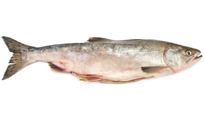
A cohort study performed on middle-aged and elderly Korean populations has suggested that a seafood and seaweed diet can help in sarcopenia prevention [5]. Preclinical animal research also suggests improvements in muscle functions following seaweed consumption, specifically Ishige okamurae.

Another study investigating  I. okamurae extract (IO) and DPHC (a marine polyphenol from I. okamurae) showed that both of them “were able to reverse the aging parameters and sex hormonal imbalance in 14-month-old female C57BL/6J mice” [6].

Animal and human research show promise 

In this study, the researchers followed 64 participants between 50 to 85 years. Participants received IO supplements or placebo for 12 weeks. The researchers assessed the difference between leg muscle strength at baseline and after 12 weeks of treatment. Leg strength was used as a marker because it is known to correlate with senescence progression [7].

As clinical biomarkers remained within normal values, IO was reported to be safe for the participants. The muscle testing revealed that “daily intake of IO helps prevent the decrease in muscle strength due to aging.”

To gain more understanding on the molecular level, the researchers turned to 12-month-old male mouse models. These mice received IO daily for 6 weeks.

The researchers started by testing their testosterone levels, as testosterone is known to promote muscle growth and protein synthesis [8]. Previous research in female mice has reported that IO alleviates age-related sexual hormone deficiencies [6].

IO supplementation improved testosterone serum levels and androgen receptor expression in the gastrocnemius muscle tissue compared to controls. In the control group, androgen receptor expression declined, but in the supplemented group, testosterone and androgen receptor levels were physiologically normal.

Most importantly, IO supplementation positively impacted the muscle mass of the treated mice. While bone mineral density and fat mass didn’t significantly differ between the placebo and IO-supplemented groups, the researchers noted that in the IO-supplemented group, mice had significantly increased lean mass compared to the age-matched placebo group. This increase was dose-dependent.

Muscle strength deteriorates as mice age. However, 6-week IO supplementation led to an increase in grip strength:

Seaweed sarcopenia

This increase in muscle growth and inhibition of muscle decomposition was also reflected in protein levels. IO treatment was found to downregulate expression of Atrogin-1, the protein implicated in protein degradation, and it significantly enhanced expression of myogenin, the primary myogenesis inducer [9,10].

IO supplementation also improved different markers of muscle health. One of them, Pax-7+, indicates “potential recovery ability against muscle damage” [11]. As expected, Pax-7+ levels were reduced in aged, control-fed mice compared to young ones. IO supplementation led to significant increases in Pax-7+ expression in a dose-dependent manner.

The proper functioning of muscles relies heavily on their mitochondria since they produce ATP, a molecule that serves as an energy source [12]. The authors used mitochondrial biogenesis processes, reflected by mitochondria complex protein expression, as a biomarker to evaluate muscle health. In general, mitochondrial complexes are sensitive to aging and are decreased in older mice compared to young ones. However, IO supplementation led to an increase of mitochondria complex protein expression.

Lack of mechanical understanding  

As of now, there is no effective way to stop or reverse sarcopenia. While exercise can help in delaying the age-related loss of muscle mass and function, elderly people generally have limited ability to exercise. That makes the need for other ways to alleviate muscle loss even more pressing.

The authors propose seaweed consumption as an easy-to-implement possible remedy. However, as of now, there is no established protocol or dosing or any other recommendations regarding such an intervention. The authors also recommend establishing additional safety evidence, and they stress the need to better understand the molecular pathways linking seaweed consumption to muscle functioning.

Regular intake of the brown alga I. okamurae can prevent age-related muscle weakness. A consistent seaweed diet may assist in the prevention of age-related diseases. However, large-scale studies are required to further elucidate the protective function of seaweeds against sarcopenia caused by aging.

We would like to ask you a small favor. We are a non-profit foundation, and unlike some other organizations, we have no shareholders and no products to sell you. All our news and educational content is free for everyone to read, but it does mean that we rely on the help of people like you. Every contribution, no matter if it’s big or small, supports independent journalism and sustains our future.

Literature

[1] Hyun, J., Lee, S. Y., Ryu, B., & Jeon, Y. J. (2023). A Combination Study of Pre- and Clinical Trial: Seaweed Consumption Reduces Aging-Associated Muscle Loss!. Aging and disease, 10.14336/AD.2023.0927. Advance online publication.

[2] Iso, H., Kubota, Y., & Japan Collaborative Cohort Study for Evaluation of Cancer (2007). Nutrition and disease in the Japan Collaborative Cohort Study for Evaluation of Cancer (JACC). Asian Pacific journal of cancer prevention : APJCP, 8 Suppl, 35–80.

[3] Murai, U., Yamagishi, K., Sata, M., Kokubo, Y., Saito, I., Yatsuya, H., Ishihara, J., Inoue, M., Sawada, N., Iso, H., Tsugane, S., & JPHC Study Group (2019). Seaweed intake and risk of cardiovascular disease: the Japan Public Health Center-based Prospective (JPHC) Study. The American journal of clinical nutrition, 110(6), 1449–1455.

[4] Cao, L., & Morley, J. E. (2016). Sarcopenia Is Recognized as an Independent Condition by an International Classification of Disease, Tenth Revision, Clinical Modification (ICD-10-CM) Code. Journal of the American Medical Directors Association, 17(8), 675–677.

[5] Kim, S. A., Ha, J., Lim, B., Kim, J. M., & Shin, S. (2020). The Association between Major Dietary Pattern and Low Muscle Mass in Korean Middle-Aged and Elderly Populations: Based on the Korea National Health and Nutrition Examination Survey. Nutrients, 12(11), 3543.

[6] Hyun, J., Ryu, B., Oh, S., Chung, D. M., Seo, M., Park, S. J., Byun, K., & Jeon, Y. J. (2022). Reversibility of sarcopenia by Ishige okamurae and its active derivative diphloroethohydroxycarmalol in female aging mice. Biomedicine & pharmacotherapy = Biomedecine & pharmacotherapie, 152, 113210.

[7] Marcell, T. J., Hawkins, S. A., & Wiswell, R. A. (2014). Leg strength declines with advancing age despite habitual endurance exercise in active older adults. Journal of strength and conditioning research, 28(2), 504–513.

[8] Vingren, J. L., Kraemer, W. J., Ratamess, N. A., Anderson, J. M., Volek, J. S., & Maresh, C. M. (2010). Testosterone physiology in resistance exercise and training: the up-stream regulatory elements. Sports medicine (Auckland, N.Z.), 40(12), 1037–1053.

[9] Bentzinger, C. F., Wang, Y. X., & Rudnicki, M. A. (2012). Building muscle: molecular regulation of myogenesis. Cold Spring Harbor perspectives in biology, 4(2), a008342.

[10] Gomes, M. D., Lecker, S. H., Jagoe, R. T., Navon, A., & Goldberg, A. L. (2001). Atrogin-1, a muscle-specific F-box protein highly expressed during muscle atrophy. Proceedings of the National Academy of Sciences of the United States of America, 98(25), 14440–14445.

[11] Chen, W., Datzkiw, D., & Rudnicki, M. A. (2020). Satellite cells in ageing: use it or lose it. Open biology, 10(5), 200048.

[12] Marzetti, E., Calvani, R., Cesari, M., Buford, T. W., Lorenzi, M., Behnke, B. J., & Leeuwenburgh, C. (2013). Mitochondrial dysfunction and sarcopenia of aging: from signaling pathways to clinical trials. The international journal of biochemistry & cell biology, 45(10), 2288–2301.

Glaucoma test

David Sinclair’s Group Reverses Glaucoma in Mice

In a new study, scientists have achieved significant improvement of glaucoma in a mouse model via partial cellular reprogramming of retinal cells. However, it might take more than that to reverse other aspects of age-related vision loss [1].

The vision of restored vision

The group that authored this paper, led by the prominent geroscientist Dr. David Sinclair of Harvard, has been bringing us exciting news for quite some time. Their chosen target is glaucoma, an age-related neurodegenerative disease and the leading cause of vision loss in the elderly. Glaucoma is usually triggered by increased intraocular pressure and characterized by the progressive loss of retinal ganglion cells (RGCs) and their axons. Today, the only clinically approved treatment is decreasing intraocular pressure via drugs or surgery. This can slow, but not halt, the disease’s progression, and it doesn’t work in all patients.

Sinclair’s group has already achieved impressive results in murine and non-human primate models by partially reprogramming RGCs using three of the four classic Yamanaka factors: OSK [2]. The fourth factor, M (c-Myc), is a known oncogene [3]. It was found that c-Myc is not essential for reprogramming, and foregoing it improves safety.

Sustained improvement in the continuous model

In this new study, the researchers induced glaucoma in three-month-old mice by artificially increasing intraocular pressure. After the animals’ vision deteriorated, they were treated with viral vectors carrying OSK.

The mice were divided into four groups. The two study groups received RGC-specific viral vectors either with continuous OSK expression or with one induced by doxycycline. The third group was given a sham treatment of viral vectors without subsequent doxycycline induction, and the fourth group was of healthy controls.

Development of glaucoma predictably led to a sharp decline in visual acuity. However, after eight weeks of OSK expression, visual acuity was not only restored but surpassed that recorded at baseline. After this peak, the mice’s vision began declining again, even in the model with continuous OSK expression. However, the decline was much faster in the induced OSK expression group, as OSK expression levels gradually fell following doxycycline withdrawal.

In the continuous expression group, the mice’s vision remained significantly restored and close to healthy levels for 11 months after the beginning of the treatment, although in one more month, it fell sharply. In the induced expression group, the second wave of doxycycline treatment, started nine months after the first one, led to a modest restoration of visual acuity that approached statistical significance versus baseline. Doxycycline withdrawal caused a new sharp drop in visual acuity.

OSK Glaucoma

Simply aging

The researchers hypothesize that the gradual decline in visual acuity in the continuous expression group and the attenuation of the treatment’s effect in the induced expression group were caused simply by aging. Normally, mice only live for a bit over two years, and by the end of the experiment, they were experiencing other vision problems besides the induced glaucoma. “It is highly possible” the researchers note, “that at this time, visual acuity is declining, not due to the loss of the OSK reprogramming effect on RGCs, but due to the age-related decline of other retinal layers that did not receive OSK treatment”. In their 2020 study, the group was able to rescue age-related vision loss by partial cellular reprogramming in 12-month-old mice, but not in mice six months older.

Tumorigenesis remains a concern for cellular reprogramming. The researchers closely monitored the mice and found no signs of increased prevalence of tumors or changes in retinal structure. Interestingly, just recently, a study found that continuous expression of OSKM leads to quick health deterioration and death in mice, but this effect was attenuated when OSKM expression was blocked in the liver and intestine [4]. Whether it was due to the absence of c-Myc or to the localization of OSK expression to retinal cells, or both, long-term partial reprogramming appeared to be safe in this case. No adverse effects were recorded.

We performed year-long functional tracking of mice with experimentally induced glaucoma that were treated with AAV2-OSK to induce epigenetic reprogramming in RGCs. Our results indicated that vision loss due to glaucoma can be safely reversed. In the AAV2-tTA; TRE-OSK mice (OSK continuously on), vision loss was restored to healthy levels after 2 months of treatment and remained close to healthy levels for 11 months… Mice that received DOX that triggered OSK expression restored vision to healthy levels by 8 weeks, suggesting that the beneficial effects of OSK treatment can be achieved using a DOX-inducible dual AAV system. When DOX was withdrawn and OSK turned off in these mice, although significance was compromised compared with continuous ON group, the absolute vision performance stays higher than its glaucoma baseline until 11 months post-AAV injection.

We would like to ask you a small favor. We are a non-profit foundation, and unlike some other organizations, we have no shareholders and no products to sell you. All our news and educational content is free for everyone to read, but it does mean that we rely on the help of people like you. Every contribution, no matter if it’s big or small, supports independent journalism and sustains our future.

Literature

[1] Karg, M. M., Lu, Y. R., Refaian, N., Cameron, J., Hoffmann, E., Hoppe, C., … & Ksander, B. R. (2023). Sustained Vision Recovery by OSK Gene Therapy in a Mouse Model of Glaucoma. Cellular Reprogramming.

[2] Lu, Y., Brommer, B., Tian, X., Krishnan, A., Meer, M., Wang, C., … & Sinclair, D. A. (2020). Reprogramming to recover youthful epigenetic information and restore vision. Nature, 588(7836), 124-129.

[3] Miller, D. M., Thomas, S. D., Islam, A., Muench, D., & Sedoris, K. (2012). c-Myc and cancer metabolism. Clinical cancer research, 18(20), 5546-5553.

[4] Parras, A., Vílchez-Acosta, A., Desdín-Micó, G., Picó, S., Mrabti, C., Montenegro-Borbolla, E., … & Ocampo, A. (2023). In vivo reprogramming leads to premature death linked to hepatic and intestinal failure. Nature Aging, 1-12.

Knee arthritis

Repairing DNA with Sirtuins for Arthritis

In a paper published on Saturday in Aging, researchers have described a method of activating the sirtuin SIRT6 to reduce DNA damage, reducing senescence and possibly treating arthritis.

Sirtuins, genetic damage, and senescence

Extensive previous research has found that SIRT6, a deacetylase that is dependent on NAD+, is instrumental in DNA repair [1] and naturally homes in on places showing signs of DNA damage [2]. Other work has found that it maintains cellular functions in chondrocytes [3], the cells responsible for cartilage and whose dysfunction is linked to osteoarthritis.

We have recently posted about potential disease-modifying treatments for osteoarthritis that use stem cell signals to encourage chondrocytes to return from senescence to youthful function. These researchers have approached the problem differently, beginning with prior work from their lab showing that chrondrocytes, as cells that do not frequently divide, accumulate significant DNA damage over time [4]. As other work has raised the possibility of DNA damage being a major source of senescence [5], these researchers have turned to SIRT6 as a potential treatment. Specifically, they have chosen to test MDL-800, a compound that was found  to increase SIRT6 activity by more than 20 times [6], against senescence in chondrocytes.

Restoring cellular DNA repair capability

This experiment began with an assessment of the ability of human chondrocytes to repair damage. Cells derived from 45-year-olds and younger, 50- to 65-year-olds, and people over 70 years old were subjected to intense radiation. This damage was far higher than what the cells had been subjected to during their donors’ lifespans.

As expected, the cells derived from younger people were able to repair damage considerably faster than their older counterparts. A closer look showed that some of the older cells were able to rapidly repair damage; however, many of the cells from the older group only got more damaged over time. This happened to a much lesser extent in the middle group and to very few of the cells in the young group.

MDL-800 mitigated this, reducing the number of heavily damaged cells in the middle-aged and younger groups while significantly increasing the rate of repair. EX-527, a compound that suppresses the activity of SIRT6, increased the damage.

The researchers then tested MDL-800 against older cells that had not been irradiated at all. In cells derived from older people and mice, MDL-800 significantly decreased DNA damage by more than half, as determined by a comet tail assay.

Reducing senescence

For this experiment, the researchers used cells derived from fluorescent-reporter mice to determine the amount of the senescence marker p16. These cells were cultured for three weeks in a substrate containing MDL-800. In two separate cohorts, the cells that received MDL-800 had considerably less p16 than a control group of cells that did not.

The researchers hold that their data provides significant support for the idea that DNA damage, which can potentially be mitigated by compounds that improve SIRT6, is a causative and possibly reversible element of senescence in chondrocytes. However, this study was performed only in cells, not humans or live animals. Considerably more experiments will have to be conducted before MDL-800 or another SIRT6-enhancing compound can be considered for use in human beings.

We would like to ask you a small favor. We are a non-profit foundation, and unlike some other organizations, we have no shareholders and no products to sell you. All our news and educational content is free for everyone to read, but it does mean that we rely on the help of people like you. Every contribution, no matter if it’s big or small, supports independent journalism and sustains our future.

Literature

[1] Roichman, A., Kanfi, Y., Glazz, R., Naiman, S., Amit, U., Landa, N., … & Cohen, H. Y. (2017). SIRT6 overexpression improves various aspects of mouse healthspan. Journals of Gerontology Series A: Biomedical Sciences and Medical Sciences, 72(5), 603-615.

[2] Mao, Z., Hine, C., Tian, X., Van Meter, M., Au, M., Vaidya, A., … & Gorbunova, V. (2011). SIRT6 promotes DNA repair under stress by activating PARP1. Science, 332(6036), 1443-1446.

[3] Collins, J. A., Kapustina, M., Bolduc, J. A., Pike, J. F., Diekman, B. O., Mix, K., … & Loeser, R. F. (2021). Sirtuin 6 (SIRT6) regulates redox homeostasis and signaling events in human articular chondrocytes. Free Radical Biology and Medicine, 166, 90-103.

[4] Copp, M. E., Chubinskaya, S., Bracey, D. N., Shine, J., Sessions, G., Loeser, R. F., & Diekman, B. O. (2022). Comet assay for quantification of the increased DNA damage burden in primary human chondrocytes with aging and osteoarthritis. Aging Cell, 21(9), e13698.

[5] Yousefzadeh, M., Henpita, C., Vyas, R., Soto-Palma, C., Robbins, P., & Niedernhofer, L. (2021). DNA damage—how and why we age?. Elife, 10, e62852.

[6] Huang, Z., Zhao, J., Deng, W., Chen, Y., Shang, J., Song, K., … & Zhang, J. (2018). Identification of a cellularly active SIRT6 allosteric activator. Nature chemical biology, 14(12), 1118-1126.

The Journal Club is a monthly livestream hosted by Dr. Oliver Medvedik which covers the latest aging research papers.

Journal Club December – Improving Mitochrondrial Function

The Journal Club is back for one final time this year! Dr. Oliver Medvedik will return on December 15th at 12:00 Eastern on the lifespan.io Facebook page to review the recent paper “A drug-like molecule engages nuclear hormone receptor DAF-12/FXR to regulate mitophagy and extend lifespan”. Join us and find out how researchers are boosting mitochondria and lifespan.

The mitochondria are vital for our cells to function, and their dysfunction is one of the reasons we age. The main function of the mitochondria is to produce energy via cellular respiration. Doing this converts nutrients from food into adenosine triphosphate (ATP), which our bodies use as energy. ATP is essentially a universal energy “currency” that all our cells can use, and it is essential for their survival, function, and growth.

Finding ways to combat mitochondrial dysfunction is a potential way to improve health and longevity in humans, and while this experiment was only in worms, it paves the way for testing this in people.

Abstract

Autophagy–lysosomal function is crucial for maintaining healthy lifespan and preventing age-related diseases. The transcription factor TFEB plays a key role in regulating this pathway. Decreased TFEB expression is associated with various age-related disorders, making it a promising therapeutic target. In this study, we screened a natural product library and discovered mitophagy-inducing coumarin (MIC), a benzocoumarin compound that enhances TFEB expression and lysosomal function. MIC robustly increases the lifespan of Caenorhabditis elegans in an HLH-30/TFEB-dependent and mitophagy-dependent manner involving DCT-1/BNIP3 while also preventing mitochondrial dysfunction in mammalian cells. Mechanistically, MIC acts by inhibiting ligand-induced activation of the nuclear hormone receptor DAF-12/FXR, which, in turn, induces mitophagy and extends lifespan. In conclusion, our study uncovers MIC as a promising drug-like molecule that enhances mitochondrial function and extends lifespan by targeting DAF-12/FXR. Furthermore, we discovered DAF-12/FXR as a previously unknown upstream regulator of HLH-30/TFEB and mitophagy.

Join the livestream

Join Zoom Meeting https://lifespan-io.zoom.us/j/82762680226?pwd=U2k0MDMxMk5XTlM0T2dOQlFyUlRidz09

Meeting ID: 827 6268 0226

Passcode: 662753

Literature

Chamoli, M., Rane, A., Foulger, A., Chinta, S. J., Shahmirzadi, A. A., Kumsta, C., … & Andersen, J. K. (2023). A drug-like molecule engages nuclear hormone receptor DAF-12/FXR to regulate mitophagy and extend lifespanNature Aging, 1-15.20

The Journal Club is a monthly livestream hosted by Dr. Oliver Medvedik which covers the latest aging research papers.

Boosting Mitochondrial Function

The Journal Club is back for one final time this year! Dr. Oliver Medvedik will return on December 15th at 12:00 Eastern on the lifespan.io Facebook page to review the recent paper “A drug-like molecule engages nuclear hormone receptor DAF-12/FXR to regulate mitophagy and extend lifespan”. Join us and find out how researchers are boosting mitochondria and lifespan.

The mitochondria are vital for our cells to function, and their dysfunction is one of the reasons we age. The main function of the mitochondria is to produce energy via cellular respiration. Doing this converts nutrients from food into adenosine triphosphate (ATP), which our bodies use as energy. ATP is essentially a universal energy “currency” that all our cells can use, and it is essential for their survival, function, and growth.

Finding ways to combat mitochondrial dysfunction is a potential way to improve health and longevity in humans, and while this experiment was only in worms, it paves the way for testing this in people.

Abstract

Autophagy–lysosomal function is crucial for maintaining healthy lifespan and preventing age-related diseases. The transcription factor TFEB plays a key role in regulating this pathway. Decreased TFEB expression is associated with various age-related disorders, making it a promising therapeutic target. In this study, we screened a natural product library and discovered mitophagy-inducing coumarin (MIC), a benzocoumarin compound that enhances TFEB expression and lysosomal function. MIC robustly increases the lifespan of Caenorhabditis elegans in an HLH-30/TFEB-dependent and mitophagy-dependent manner involving DCT-1/BNIP3 while also preventing mitochondrial dysfunction in mammalian cells. Mechanistically, MIC acts by inhibiting ligand-induced activation of the nuclear hormone receptor DAF-12/FXR, which, in turn, induces mitophagy and extends lifespan. In conclusion, our study uncovers MIC as a promising drug-like molecule that enhances mitochondrial function and extends lifespan by targeting DAF-12/FXR. Furthermore, we discovered DAF-12/FXR as a previously unknown upstream regulator of HLH-30/TFEB and mitophagy.

Literature

Chamoli, M., Rane, A., Foulger, A., Chinta, S. J., Shahmirzadi, A. A., Kumsta, C., … & Andersen, J. K. (2023). A drug-like molecule engages nuclear hormone receptor DAF-12/FXR to regulate mitophagy and extend lifespanNature Aging, 1-15.

Plant foods

Twin Study: Healthy Vegan Diet Beats Healthy Omnivorous Diet

In a randomized controlled trial, eight weeks of healthy vegan eating, but not of healthy omnivorous eating, led to weight loss and significant improvements in LDL cholesterol and insulin levels in a middle-aged twin study [1].

A tale of two diets

Most of our knowledge about the health effects of various diets comes from epidemiological studies. Notoriously noisy and hard to interpret, they still are the best tool we have for measuring long-term impacts. However, randomized controlled trials, limited in their duration but much more rigorous, can also provide valuable insights.

There has been considerable debate about the pros and cons of plant-based diets [2], especially compared to well-balanced and wholesome omnivorous diets. This new study tested 22 pairs of identical twins to investigate the effects of a healthy omnivorous diet versus a healthy vegan diet on several metabolic markers. Using identical twins is beneficial since it allows scientists to control for genetic differences and for many environmental factors, such as upbringing.

For each pair, one twin ate the vegan diet and the other ate the omnivorous diet for eight weeks. For the first four weeks, the meals were provided via a meal delivery service. For the second half of the experiment, after dietary coaching, the participants prepared their own diet-appropriate meals and snacks.

Better metabolic outcomes

The primary outcome was the change in low-density lipoprotein (LDL) cholesterol levels from the baseline. LDL is known as the “bad” cholesterol due to its association with cardiovascular disease [3]. Secondary outcomes included changes in cardiometabolic factors, such as plasma lipids, glucose, and insulin levels, as well as body weight.

The cohort was generally healthy, middle-aged, predominantly white and female, with a borderline normal average body mass index (BMI) of 25.9. The mean LDL level at baseline was in the “nearly optimal” range in both groups, which, according to the researchers, “left minimal room for participants to improve through diet alone.”

Yet, the plant-eaters, but not the omnivores, significantly improved their LDL levels by the end of the study period, with a mean decrease of 15.2 mg/dL (13.9 mg/dL more than the omnivores). The first group also lost about 2 kilograms of weight on average, beating the second group by a lot.

The plant-eating group also experienced a significant decrease in fasting insulin levels, along with larger but non-significant decreases in fasting high-density lipoprotein (HDL) cholesterol, vitamin B12, triglycerides, and glucose levels. HDL is considered the “good” cholesterol, but it is less strongly associated with health outcomes than LDL is. B12 is an essential vitamin that can be obtained almost solely from animal food sources. Many vegans supplement B12.

The possible causes

Digging deeper into the dietary data reveals that the vegan group consumed, on average, about 200 fewer calories a day than the omnivores, which might explain their weight loss. The vegans got a smaller percentage of their calories from protein and fat and a higher percentage from carbohydrates.

The two diets also had different lipid profiles: the vegans consumed much less saturated fats (generally considered unhealthy) and more mono- and polyunsaturated fats. This is what might have led to the significant decrease in LDL cholesterol among the vegans. The vegans also consumed much more fiber, a healthy dietary ingredient that promotes satiation.

Many dietary randomized controlled trials are conducted in metabolically unhealthy populations. This one suggests that healthy people can expect to glean quick metabolic benefits from a vegan diet, even compared to a balanced omnivorous one. If there was one downside to the vegan diet in this study, it’s that the vegan group reported lower food satisfaction. However, for most vegans, this gets better with time.

In this randomized clinical trial of healthy, adult identical twins, the 8-week change in LDL-C level—the primary outcome—was significantly lower for twins receiving the vegan diet compared with twins receiving the omnivorous diet. Insulin levels and weight were also significantly lower among the twins on the vegan diet from baseline to 8 weeks. Vegan-diet participants had total lower protein intake as a percentage of calories, lower dietary satisfaction, lower intake of dietary cholesterol, but higher intake of vegetable servings and intake of dietary iron. Vegans had lower intake of vitamin B12, yet serum vitamin B12 levels were not statistically different than omnivores at 8 weeks, likely because of preserved stores. Long-term vegans are typically encouraged to take a cyanocobalamin (vitamin B12) supplement.

We would like to ask you a small favor. We are a non-profit foundation, and unlike some other organizations, we have no shareholders and no products to sell you. All our news and educational content is free for everyone to read, but it does mean that we rely on the help of people like you. Every contribution, no matter if it’s big or small, supports independent journalism and sustains our future.

Literature

[1] Landry, M. J., Ward, C. P., Cunanan, K. M., Durand, L. R., Perelman, D., Robinson, J. L., … & Gardner, C. D. (2023). Cardiometabolic Effects of Omnivorous vs Vegan Diets in Identical Twins: A Randomized Clinical Trial. JAMA Network Open, 6(11), e2344457-e2344457.

[2] Fehér, A., Gazdecki, M., Véha, M., Szakály, M., & Szakály, Z. (2020). A Comprehensive Review of the Benefits of and the Barriers to the Switch to a Plant-Based Diet. Sustainability, 12(10).

[3] Abdullah, S. M., Defina, L. F., Leonard, D., Barlow, C. E., Radford, N. B., Willis, B. L., … & Khera, A. (2018). Long-term association of low-density lipoprotein cholesterol with cardiovascular mortality in individuals at low 10-year risk of atherosclerotic cardiovascular disease: results from the Cooper Center Longitudinal Study. Circulation, 138(21), 2315-2325.

Pink salmon

Astaxanthin and Meclizine Extend Lifespan in Mice

Scientists in the Intervention Testing Program have found that the antioxidant astaxanthin and the seasickness medication meclizine, both sold over the counter, significantly extend median lifespan in mice [1].

Astaxanthin and meclizine are in, but fisetin is out

The Intervention Testing Program (ITP) has existed for about 20 years, and it is considered the gold standard for testing longevity interventions in mice for good reasons. First, ITP uses genetically heterogeneous HET3 mice rather than the inbred strains that are usually used in studies. Second, it uses a lot of those mice, which ensures considerable statistical power. Finally, the studies are run in three research centers simultaneously, with conditions meticulously replicated in order to increase overall reliability.

The ITP’s first major success came in the late 2000s, when rapamycin was found to substantially extend lifespan in mice even if given later in life [2]. Since then, while a hundred molecules have been tested, only three of them have produced significant increases in lifespan: acarbose, canagliflozine, and 17α-estradiol. There have also been some notable failures, such as with resveratrol and metformin. You can learn more about the program’s workings from our interview with one of its leaders, Prof. Richard Miller.

Any new data coming out of the ITP is met with excitement and anticipation. Earlier this month, in a new paper, the researchers announced that two more compounds were found to significantly increase mouse lifespan: astaxanthin and meclizine. The former increased the median lifespan by 12%, and the latter increased it by 8%. Unfortunately, both seemed to work only in males. Five more molecules, fisetin, SG1002 (a hydrogen sulfide donor), dimethyl fumarate, mycophenolic acid, and 4-phenylbutyrate, failed to produce statistically significant results.

Who are the winners?

Excitingly, both astaxanthin and meclizine are over-the-counter compounds and the first ones to be found effective by the ITP. Astaxanthin is a naturally occurring antioxidant and inflammation modulator. It belongs to the carotenoid family of molecules and gives salmon its pink color. Algae, yeast, trout, krill, shrimp, and crayfish also contain it. Algae are the original source, and the astaxanthin they contain is passed up the food chain. Astaxanthin has an excellent safety profile and is widely available as a supplement.

Meclizine has been in use for decades against seasickness and vertigo and as an antihistamine. Recently, it was found to inhibit the protein complex mTORC1 [3]. Rapamycin, the best-known geroprotective molecule, is a potent inhibitor of mTORC1 as well. However, it also inhibits a closely related protein complex, mTORC2, which can be harmful and accounts for rapamycin’s questionable safety profile. Meclizine, on the other hand, seems to act exclusively on mTORC1.

However, as Richard Miller explained in his recent appearance on Peter Attia’s podcast, the researchers were unable to prove that the lifespan extension produced by meclizine was due to mTORC1 inhibition. Meclizine’s benefits for the central nervous system could be an alternative explanation.

Sex specificity and fisetin’s failure

Both molecules were given to mice in relatively high doses, which might pose a problem for translation into humans, but this remains to be seen. As to the sex specificity, there is nothing new about it: all four molecules that were successful in the ITP work either only in one sex or better in one sex than in the other.

One of the possible reasons is the observed sex-related differences in the drug’s blood concentration. For instance, rapamycin works better in females, and in the ITP experiments, females had higher concentrations of rapamycin in the blood. For astaxanthin and meclizine, the concentration was higher in males. Hence, it might be possible to achieve an effect in females by adjusting dosage.

Among the failed compounds, fisetin deserves a special mention. This naturally occurring polyphenol has been touted as a reasonably potent senolytic, a drug that removes senescent cells. Fisetin has shown some effectiveness in animal models, even causing lifespan and healthspan extension in wild-type mice [4], which led to its acceptance into the ITP. Interestingly, the drug was unable not only to extend lifespan but even to remove senescent cells, according to the popular senescence marker p16. Here, too, the problem might be with the dosage, meaning that fisetin’s ITP “career” is not necessarily over.

We would like to ask you a small favor. We are a non-profit foundation, and unlike some other organizations, we have no shareholders and no products to sell you. All our news and educational content is free for everyone to read, but it does mean that we rely on the help of people like you. Every contribution, no matter if it’s big or small, supports independent journalism and sustains our future.

Literature

[1] Harrison, D. E., Strong, R., Reifsnyder, P., Rosenthal, N., Korstanje, R., Fernandez, E., … & Miller, R. A. (2023). Astaxanthin and meclizine extend lifespan in UM-HET3 male mice; fisetin, SG1002 (hydrogen sulfide donor), dimethyl fumarate, mycophenolic acid, and 4-phenylbutyrate do not significantly affect lifespan in either sex at the doses and schedules used. GeroScience, 1-22.

[2] Harrison, D. E., Strong, R., Sharp, Z. D., Nelson, J. F., Astle, C. M., Flurkey, K., … & Miller, R. A. (2009). Rapamycin fed late in life extends lifespan in genetically heterogeneous mice. Nature, 460(7253), 392-395.

[3] Allen, S. A., Tomilov, A., & Cortopassi, G. A. (2018). Small molecules bind human mTOR protein and inhibit mTORC1 specifically. Biochemical pharmacology, 155, 298-304.

[4] Yousefzadeh, M. J., Zhu, Y. I., McGowan, S. J., Angelini, L., Fuhrmann-Stroissnigg, H., Xu, M., … & Niedernhofer, L. J. (2018). Fisetin is a senotherapeutic that extends health and lifespan. EBioMedicine, 36, 18-28.

Arthritis treatment

A Microvesicle Hydrogel to Treat Arthritis

In the Journal of Nanobiotechnology, researchers have found that embedding microvesicles in a slow-release hydrogel may be an effective treatment for osteoarthritis.

A line of inquiry begins to bear fruit

Researchers have repeatedly found that the extracellular vesicles (EVs) released by stem cells, particularly mesenchymal stem cells (MSCs), are the main drivers behind the effectiveness of many stem cell-related approaches [1]. These intercellular messengers contain molecules with clear biological effects, such as microRNAs, proteins, and cytokines [2]. EVs are also considerably smaller than cells, small enough to penetrate deeply into otherwise impermeable tissues, such as cartilage [3].

We have repeatedly reported on the potential of EV-based treatments to alleviate age-related conditions, including cellular senescence. In this paper, the researchers developed a suite of EVs specifically to counteract senescent cells, with the goal of curing osteoarthritis caused by their accumulation in joints.

Special EVs and a special substrate

Because interferon gamma (IFN-γ) is a critical part of both causing senescence and causing senescent cells to die, and its presence affects how MSCs produce EVs [4], the researchers chose to cultivate both regular EVs (specifically microvesicles, MVs) and ones that were created after exposure to IFN-γ (iMVs).

However, EVs, including iMVs, are vulnerable and don’t last long in the human body. Therefore, researchers have investigated hydrogels as potential long-term treatments, as these substances can slowly release their contents over time [5]. Some hydrogels can be triggered to release their contents only in the presence of another substance or class of substances, such as reactive oxygen species (ROS) [6], which is the approach that these researchers took, delving deep into the specifics of their new material. They report that their hydrogel can continue to release MVs in the presence of ROS for nearly three weeks.

Microvesicles for Osteoarthritis

Effectiveness in cells and model rats

The researchers tested their new hydrogel substrate, complete with the incorporation of their MVs and iMVs, in the presence of cartilage-making cells (chondrocytes). This co-culturing was found to have no significant negative effects on the chrondrocytes. Instead, the presence of three key senescence markers, p16, p21, and p53, were substantially downregulated compared to a hydrogel-only control group in the presence of hydrogen peroxide.

The effects on energy transport were also analyzed. The membranes of mitochondria were improved after treatment with MVs and iMVs, although these vesicle types affected different respiratory chains at different levels. Hydrogel containing MVs or iMVs was found to significantly decrease the ROS in chondrocytes exposed to it. Additionally, exposure to iMVs was found to trigger mitochondrial fusion significantly beyond the effect of MVs.

A rat model was then employed to test this approach in vivo. This experiment consisted of eight weeks of induced arthritis followed by five weeks of testing. Both the empty hydrogel and treatment with iMVs alone were found to have some therapeutic benefit in this model. However, combining them had an enormous benefit: their cartilage had been nearly restored to the level of rats that had never had induced arthritis at all.

Just as in the cellular testing, this approach had been found to substantially reduce senescence markers. It also encouraged mitochondrial fusion and discouraged mitochondrial fission.

While this study was only conducted in cells and animal models, its results are promising and potentially groundbreaking. Combating arthritis by targeting cellular senescence has not always been met with success. If these rat results can be confirmed in human clinical trials, a vesicle and hydrogel-based approach may be more effective than senolytics

In summary, Hydrogel@iMVs effectively inhibits cartilage cell wear and delays the progression of OA by enhancing mitochondrial function and breaking the vicious cycle of senescence.

We would like to ask you a small favor. We are a non-profit foundation, and unlike some other organizations, we have no shareholders and no products to sell you. All our news and educational content is free for everyone to read, but it does mean that we rely on the help of people like you. Every contribution, no matter if it’s big or small, supports independent journalism and sustains our future.

Literature

[1] Phinney, D. G., & Pittenger, M. F. (2017). Concise review: MSC-derived exosomes for cell-free therapy. Stem cells, 35(4), 851-858.

[2] Liu, X., Wei, Q., Sun, Z., Cui, S., Wan, X., Chu, Z., … & Wang, S. (2023). Small extracellular vesicles: Yields, functionalization and applications in diabetic wound management. Interdisciplinary Medicine, e20230019.

[3] Harrell, C. R., Jovicic, N., Djonov, V., Arsenijevic, N., & Volarevic, V. (2019). Mesenchymal stem cell-derived exosomes and other extracellular vesicles as new remedies in the therapy of inflammatory diseases. Cells, 8(12), 1605.

[4] BaoB, Y. Z., LvC, X. L., WangB, Y. Z., & WangA, G. F. (2017). IFN-? induces senescence-like characteristics in mouse bone marrow mesenchymal stem cells. Adv. Clin. Exp. Med, 26, 201-206.

[5] Wang, J., Zhu, M., Hu, Y., Chen, R., Hao, Z., Wang, Y., & Li, J. (2023). Exosome-Hydrogel System in Bone Tissue Engineering: A Promising Therapeutic Strategy. Macromolecular Bioscience, 23(4), 2200496.

[6] Wu, Y., Wang, Y., Long, L., Hu, C., Kong, Q., & Wang, Y. (2022). A spatiotemporal release platform based on pH/ROS stimuli-responsive hydrogel in wound repairing. Journal of Controlled Release, 341, 147-165.

Bottleneck

The Longevity Biotech Fellowship Bottleneck Survey

The LBF community is dedicated to maximally accelerating progress in longevity. While the field has seen substantial advances in the past decade, there are clear and systemic deficiencies that have received little attention but retard progress for many. To coordinate efforts and identify the highest leverage intervention points, it is necessary to first understand and map these deficiencies and bring them into the limelight.

The LBF aimed to establish an empirical, systematic, rigorous, and minimally biased methodology to map bottlenecks across the field. To do so, they surveyed 400 participants across various sectors of longevity, asking them for their biggest bottlenecks and most wanted solutions in structured and free-form questions. From the more than 1000 answers, they built a classification system of Bottlenecks and Solutions and tabulated the frequency of each. The result is a first-of-a-kind taxonomy of the biggest needs and biggest wants of the longevity space.

On their site, you’ll find the main takeaways of this work. For more detail, see their preprint paper, explore the answers in their interactive site, or access the full dataset for your own analysis.

Most importantly, if you want to make an impact and help solve or fund solving any of these bottlenecks, sign up to a working group.

We would like to ask you a small favor. We are a non-profit foundation, and unlike some other organizations, we have no shareholders and no products to sell you. All our news and educational content is free for everyone to read, but it does mean that we rely on the help of people like you. Every contribution, no matter if it’s big or small, supports independent journalism and sustains our future.

Thin person

Caloric Restriction Associated With Reduced Senescence

New research in Aging Cell has identified senescence-associated biomarkers whose levels are altered by caloric restriction in humans [1].

Slowing down senescence with fewer calories

Multiple animal studies have found that reducing caloric intake without causing malnutrition (caloric restriction) has great lifespan-extending potential and can delay the onset of several age-related diseases [2]. These studies suggest that caloric restriction can delay the Hallmarks of Aging.[3], one of which is cellular senescence.

While the influence of caloric restriction on cellular senescence has been broadly studied in animals, the effect of caloric restriction on cellular senescence in humans is less explored. This is why the Comprehensive Assessment of Long-term Effects of Reducing Intake of Energy (CALERIE) study was conducted. It was a 2-year long randomized controlled trial for assessing the influence of caloric restriction on healthy, non-obese, young to middle-aged volunteers [4].

So far, CALERIE is the most rigorous study that has been conducted in this area. Its participants were able to obtain ~12% caloric restriction.

Senescence markers and metabolic changes

The authors investigated clinical data and plasma samples of 199 CALERIE participants, 128 of which practiced caloric restriction and 71 of which ate normally.

The researchers investigated 28 candidate senescence biomarkers, which included cytokines, chemokines, matrix remodeling proteins, and growth factors. They were carefully selected based on scientific literature, senescence model screens, and appearing in human blood “in contexts associated with increased senescent cell burden” [5,6,7].

They compared the levels of the biomarkers at baseline, 12 months, and 24 months. After 12 months of caloric restriction, the researchers observed a reduction in nine senescence biomarkers. Four of those were still reduced following 24 months of caloric restriction, joined by reduced levels of two more.

Only one biomarker, sclerostin (SOST), was increased after 12 months. SOST negatively regulates bone formation, which might suggest changes in bone metabolism [8].

After 24 months, only one biomarker was increased, but this time it was the receptor for advanced glycation end-products (RAGE), a protein implicated in DNA repair.

Researchers then used the obtained data regarding changes in these senescence biomarkers to assess whether these changes are associated with the changes in the metabolic measurements in study participants who followed caloric restriction.

Those metabolic measurements included homeostatic model assessment of insulin resistance (HOMA-IR), insulin sensitivity index, and resting metabolic rate (RMR) residual. RMR is the amount of energy the body uses when it is in the resting state. RMR residual is the difference between a person’s actual RMR and the RMR predicted based on that person’s fat mass and lean mass.

Senescence is linked to metabolism

The researchers identified that changes in several senescence biomarkers played a role as predictors of changes in metabolism. Baseline measurements of HOMA-IR, insulin sensitivity index, and RMR residual were good predictors of where these biomarkers would be in 12 and 24 months. Incorporating five key senescence-related biomarkers into the model led to a great improvement in its ability to predict all three metabolic measurements after a year. However, this is only an association, and the authors could not conclude if those changes are causally connected or only parallel observations.

The researchers also analyzed a “recently defined gene set of 125 secreted factors, transmembrane proteins, and intracellular proteins centered on cellular senescence and the SASP, named SenMayo” [9]. Gene expression data was derived from the adipose tissue of eight study participants at baseline, 12 months, and 24 months. The authors observed a reduced SenMayo response following caloric restriction.

Overall, this study adds another layer of evidence that dietary interventions can counter biological aging.

High-quality data with some weaknesses

While this study’s strength is the high-quality and rigorously obtained data from the CALERIE clinical trial, the study also has some limitations. As the authors say, this selection of biomarkers doesn’t “unequivocally and universally detect senescent cells.” While they were chosen to the best of current knowledge, further studies are necessary to validate them.

Furthermore, the models used in the study suggest an association between changes in several biomarker levels and changes in HOMA-IR, insulin sensitivity index, and RMR residual. However, this relationship cannot imply causation.

Additionally, stringent inclusion criteria for CALERIE study participants might imply that the results cannot be generalized to the entire population.

This study does not also provide information on the cause of the reduced levels of senescence-associated biomarkers. Authors speculate that it could be reduced accumulation of senescent cells, increased clearance, or the inhibition of their SASP.

In conclusion, our results show that 2 years of moderate CR with adequate nutrient intake reduces biomarkers of cellular senescence in healthy young to middle-aged humans without obesity. Additional research is needed to fully elucidate the senotherapeutic implications of CR, validate the biomarkers signatures that emerged as predictive of metabolic health outcomes, and determine the generalizability and durability of the observed effects. Our data further highlight the impact of lifestyle factors on fundamental mechanisms of aging.

We would like to ask you a small favor. We are a non-profit foundation, and unlike some other organizations, we have no shareholders and no products to sell you. All our news and educational content is free for everyone to read, but it does mean that we rely on the help of people like you. Every contribution, no matter if it’s big or small, supports independent journalism and sustains our future.

Literature

[1] Aversa Z, White TA, Heeren AA, Hulshizer CA, Saul D, Zhang X, Molina AJA, Redman LM, Martin CK, Racette SB, Huffman KM, Bhapkar M, Khosla S, Das SK, Fielding RA, Atkinson EJ, LeBrasseur NK. Calorie restriction reduces biomarkers of cellular senescence in humans. Aging Cell. 2023 Nov 14:e14038. doi:10.1111/acel.14038. Epub ahead of print. PMID: 37961856.

[2] Balasubramanian, P., Howell, P. R., & Anderson, R. M. (2017). Aging and Caloric Restriction Research: A Biological Perspective With Translational Potential. EBioMedicine, 21, 37–44.

[3] Green, C. L., Lamming, D. W., & Fontana, L. (2022). Molecular mechanisms of dietary restriction promoting health and longevity. Nature reviews. Molecular cell biology, 23(1), 56–73.

[4] Ravussin, E., Redman, L. M., Rochon, J., Das, S. K., Fontana, L., Kraus, W. E., Romashkan, S., Williamson, D. A., Meydani, S. N., Villareal, D. T., Smith, S. R., Stein, R. I., Scott, T. M., Stewart, T. M., Saltzman, E., Klein, S., Bhapkar, M., Martin, C. K., Gilhooly, C. H., Holloszy, J. O., … CALERIE Study Group (2015). A 2-Year Randomized Controlled Trial of Human Caloric Restriction: Feasibility and Effects on Predictors of Health Span and Longevity. The journals of gerontology. Series A, Biological sciences and medical sciences, 70(9), 1097–1104.

[5] Aversa, Z., Atkinson, E. J., Carmona, E. M., White, T. A., Heeren, A. A., Jachim, S. K., Zhang, X., Cummings, S. R., Chiarella, S. E., Limper, A. H., & LeBrasseur, N. K. (2023). Biomarkers of cellular senescence in idiopathic pulmonary fibrosis. Respiratory research, 24(1), 101.

[6] Fielding, R. A., Atkinson, E. J., Aversa, Z., White, T. A., Heeren, A. A., Achenbach, S. J., Mielke, M. M., Cummings, S. R., Pahor, M., Leeuwenburgh, C., & LeBrasseur, N. K. (2022). Associations between biomarkers of cellular senescence and physical function in humans: observations from the lifestyle interventions for elders (LIFE) study. GeroScience, 44(6), 2757–2770.

[7] Schafer, M. J., Zhang, X., Kumar, A., Atkinson, E. J., Zhu, Y., Jachim, S., Mazula, D. L., Brown, A. K., Berning, M., Aversa, Z., Kotajarvi, B., Bruce, C. J., Greason, K. L., Suri, R. M., Tracy, R. P., Cummings, S. R., White, T. A., & LeBrasseur, N. K. (2020). The senescence-associated secretome as an indicator of age and medical risk. JCI insight, 5(12), e133668.

[8] Delgado-Calle, J., Sato, A. Y., & Bellido, T. (2017). Role and mechanism of action of sclerostin in bone. Bone, 96, 29–37.

[9] Saul, D., Kosinsky, R. L., Atkinson, E. J., Doolittle, M. L., Zhang, X., LeBrasseur, N. K., Pignolo, R. J., Robbins, P. D., Niedernhofer, L. J., Ikeno, Y., Jurk, D., Passos, J. F., Hickson, L. J., Xue, A., Monroe, D. G., Tchkonia, T., Kirkland, J. L., Farr, J. N., & Khosla, S. (2022). A new gene set identifies senescent cells and predicts senescence-associated pathways across tissues. Nature communications, 13(1), 4827.

Jellyfish

JellyfishDAO to Create Longevity Films and Television

A group of award-winning filmmakers, longevity experts, and blockchain developers have come together to launch JellyfishDAO, a new decentralized autonomous organization (DAO) focused on funding and distributing movies and TV shows related to extending health and lifespan.

“We created JellyfishDAO to shift the narrative around longevity science,” said Tim Maupin, co-founder of the DAO. “There are so many misconceptions about life extension, and we believe film and television are the perfect mediums to inspire people with optimistic visions of extremely long and healthy lifespans.”

The core team of twelve includes award-winning film producers Aimee Schoof, Isen Robbins (Tesla), and Daniel Sollinger (Immortality or Bust), longevity pioneers Keith Comito of lifespan.io and Adam Gries of Vitalism, and blockchain developers Laurence Ion of VitaDAO and Victor Forissier of Polyfire.

Rounding out the core team is Actress and Producer Madeline Bellariu (The Young Pope), director and actress Galia Barkol (Don’t Let the Night End), Actor and Producer Alessandro de Marco (Kingsman: The Golden Circle), and founder and investor Guy Bahat.

JellyfishDAO will function as a fan-driven funding platform for longevity-focused film and TV projects, allowing community members to provide input on projects and share in profits. The DAO is issuing limited NFTs that provide exclusive membership utility including voting rights, access to premieres and events, production credits, exclusive discussions with cast and crew, and more.

The first three films confirmed to participate in the DAO are “The Last Generation to Die” – a sci-fi feature film exploring a future where age reversal therapies are becoming reality; “AgeLess” – a feature-length documentary on cutting-edge longevity science; and a feature film thriller titled “Genethic”.

“Our goal is to ignite enthusiasm for the advancements in longevity science and to keep audiences informed about the latest breakthroughs in this exciting field.” said Producer Schoof.

For more information or to acquire JellyfishDAO membership NFTs, visit https://www.jellyfishdao.org/

We would like to ask you a small favor. We are a non-profit foundation, and unlike some other organizations, we have no shareholders and no products to sell you. All our news and educational content is free for everyone to read, but it does mean that we rely on the help of people like you. Every contribution, no matter if it’s big or small, supports independent journalism and sustains our future.
Conflict of interest disclosure: It should be noted in the interest of transparency that Keith Comito, President of lifespan.io, is a member of JellyfishDAO and has a financial interest in the company.
Dinosaur chasing mammal

Was Mammals’ Longevity Shaped by Living Among Dinosaurs?

A prominent geroscientist suggests that more than 100 million years of saurian domination might have deprived mammals of longevity-promoting traits that are found in today’s reptiles [1].

The longevity bottleneck

Today, mammals rule the world, but, sadly, we only have an average of 80 years to enjoy our superiority. That’s a lot by mammalian standards: humans are one of the longest-lived mammals, surpassed only by some cetaceans. However, animals that have remained largely unchanged for hundreds of millions of years, such as some reptiles, age far slower than we do.

According to the renowned geroscientist Dr. João Pedro de Magalhães, the roots of mammals’ generally underwhelming lifespans may lie in the roughly 100 million years when the roles were reversed and giant reptiles ruled the Earth. During that period, mammals were small, subdued, mostly nocturnal, and short-lived creatures. De Magalhães’ hypothesis, which he calls “the longevity bottleneck”, was published in the journal BioEssays.

Live fast, die young

It is a logical conjecture that evolution has shaped the aging patterns of species. For instance, if you have a lot of enemies, it pays off to reproduce quickly before they get to you. Genetic traits that confer longevity are not selected for, and even worse: evolution might select for traits that help an animal survive when it’s young but become detrimental as it gets older; this is known as antagonistic pleiotropy. This might be a major reason why maximum lifespan positively correlates with body size across species.

De Magalhães suggests that during that very long period of time when “live fast, die young” was mammals’ pre-eminent evolutionary strategy, many longevity traits that flourished in dinosaurs were lost in mammals. Those may include various regeneration abilities, such as constantly growing new sets of teeth. The inability to do this limits lifespan in today’s mammals, such as elephants who, in nature, often die of starvation after grinding down their last set of teeth.

Another interesting example is a photolyase DNA protection system, which was lost in placental mammals during the time of the dinosaurs. Photolyases are enzymes that repair the damage to DNA caused by exposure to ultraviolet light. Early mammals might have lost this specialized defense mechanism due to their nocturnal way of life, although this remains a speculation. Other longevity-promoting traits common to reptiles include oocyte regeneration, limb regeneration, and cancer resistance.

Longevity bottleneck

Not even close to birds and reptiles

Reptiles include some of the world’s champions in longevity, such as the Galapagos turtle, whose maximum lifespan is estimated to be close to 200 years. This and many other reptile species are thought to display negligible senescence [2]: their chances of dying do not increase with age (in humans, they double roughly every eight years). Some stay reproductive and keep growing during their entire life. In mammals, only the famous naked mole rat is suspected of being endowed with negligible senescence [3], although recent research has put this in doubt, as they do show some signs of aging and experience epigenetic aging.

One of the alternative explanations for mammals’ shorter lifespan might be that they are warm-blooded, as opposed to cold-blooded reptiles and amphibians. High body temperature might indeed accelerates several processes of aging. However, de Magalhães correctly mentions the fact that birds, who are also direct descendants of dinosaurs, are notoriously long-lived for their body sizes, despite being warm-blooded and leading energetically demanding lives.

After mammals were freed from the “yoke” of dinosaurs, the class exploded into a remarkable variety of body sizes and aging rates. However, the slowest-aging mammals still don’t get anywhere near the slowest-aging reptiles, birds, and amphibians:

Clade longevity

Making sense of aging

While de Magalhães’ hypothesis might not bear immediate implications for geroscience, it can help us make sense of the longevity landscape in the animal kingdom, including in humans. One of the cutting-edge directions in geroscience is adapting longevity-promoting mechanisms found in animals for humans.

We have covered some of the exciting advances in this field in our interviews with Vera Gorbunova and Ashley Zehnder. For example, the photolyase protection system that de Magalhães mentions was found to improve repair in transgenic mice [4]. De Magalhães, who is currently Professor of Molecular Biogerontology in the Institute of Inflammation and Ageing at the University of Birmingham, said about his idea:

The ‘longevity bottleneck hypothesis’ may shed light on evolutionary forces that have shaped the way that mammals have aged over millions of years. While we see humans among different species that are among the longest living animals, there are many reptiles and other animals that have a much slower ageing process and show minimal signs of senescence over their lives. Some of the earliest mammals were forced to live towards the bottom of the food chain, and have likely spent 100 million years during the age of the dinosaurs evolving to survive through rapid reproduction. That long period of evolutionary pressure has, I propose, an impact on the way that we humans age.

We would like to ask you a small favor. We are a non-profit foundation, and unlike some other organizations, we have no shareholders and no products to sell you. All our news and educational content is free for everyone to read, but it does mean that we rely on the help of people like you. Every contribution, no matter if it’s big or small, supports independent journalism and sustains our future.

Literature

[1] de Magalhães, J. P. (2023). The longevity bottleneck hypothesis: Could dinosaurs have shaped ageing in present-day mammals?. BioEssays, 2300098.

[2] da Silva, R., Conde, D. A., Baudisch, A., & Colchero, F. (2022). Slow and negligible senescence among testudines challenges evolutionary theories of senescence. Science, 376(6600), 1466-1470.

[3] Buffenstein, R. (2008). Negligible senescence in the longest living rodent, the naked mole-rat: insights from a successfully aging species. Journal of Comparative Physiology B, 178, 439-445.

[4] Schul, W., Jans, J., Rijksen, Y. M., Klemann, K. H., Eker, A. P., De Wit, J., … & Van der Horst, G. T. (2002). Enhanced repair of cyclobutane pyrimidine dimers and improved UV resistance in photolyase transgenic mice. The EMBO journal, 21(17), 4719-4729.

Future City

Vitalia: A “Los Alamos” for Longevity Starting in Roatán

Vitalia is a new venture that aims to redefine the longevity biotech landscape, fostering drug development at warp speed: 4 months to get to market, instead of 10+ years.

From January 6 to March 1, Vitalia will be hosting a two-month pop-up city experience similar to Vitalik Buterin’s Zuzalu. The event will take place on the Caribbean paradise island of Roatán, Honduras.

This venture will feature more than 200 people from all over the world, creating a community of people driven by an entrepreneurial spirit who love life and want to see the future, both experts and citizen scientists.

Visionary Mission

Vitalia 2024 is not just an event. It’s a movement with a bold mission: to accelerate progress in extending healthy human lifespan.

Vitalia’s first pop-up city is only setting the foundation for a permanent longevity district on Roatán. The community aims to build 3-5 such lasting hubs in the real world with tens of thousands of residents, locals and internationals, coordinated by a cloud community.

The strategy for Vitalia was inspired by Balaji Srinivasan’s idea of “The Network State.”

Vitalia is a moonshot project. The initiators see Vitalia as necessary to make longevity escape velocity a reality in our lifetime by creating an environment for thousands of biotech companies to accelerate.

Structured Innovation Weeks

This upcoming event is structured around four bi-weekly themes:

  • Longevity & Human Improvement: An introduction to longevity as a concept linked with all aspects of human improvement; exploring bioscience, business models, healthcare economics & regulation, and ethics to inspire life-saving innovations.
  • Crypto Cities & Network States: Exploring the societal impact of crypto technology by examining shifts in governance, economies, and communities in a way that is focused on real-world challenges and inspires participants to develop practical solutions.
  • AI & Technological Progress: A deep dive into the potential of AI solving problems of modern life and in nearly all areas of industry that focuses on the broader implications of technology. This week synthesizes discussions from previous weeks towards future-oriented solutions.
  • Pathways to Healthy Life Extension: Presenting solutions for healthy life extension by collaborating with the broader aging research and longevity biotech industry while showcasing the concrete outcomes and projects that emerged during Vitalia.

Community Engagement and Collaboration

Vitalia 2024 is not only an intellectual exploration; it’s a call to action. Residents are called “builders” and are encouraged to set ambitious goals and build new ventures. Regular pitch competitions, crowdfunding, and legal advice will help the builders get started building the next blockbuster drug, new insurance models, or healthcare practices.

Why Roatán?

Próspera, a Special Economic Zone on Roatán, was strategically chosen for its progressive legal framework, which allows for medical freedom and fosters an environment that is conducive to innovation with entrepreneurship.

The island of Roatan is known for its safety, world-class coral reef, and growing popularity with international tourism.

Biotech company Minicircle has conducted clinical studies on the island under an IRB by the GARM Clinic, a stem cell clinic founded by reputed physician Dr. Glenn Terry. Minicircle made waves by treating longevity super-influencer Bryan Johnson with follistatin gene therapy.

Open Invitation and Application Process

Vitalia 2024 extends an open invitation to individuals passionate about shaping the future of longevity. There are few spots left, so applicants are highly encouraged to apply here soon.

Additional Information

Visit the official Vitalia 2024 website for comprehensive details, FAQs, financing options, and travel guides (including visas and housing).

Note: This press release provides a high-level overview. Detailed information and updates are available on the official Vitalia 2024 website.

Young to old

Pre-Senescent Cells May Be Damaging and Treatable

Researchers have described an intermediary state between senescent and young cells, the inflammatory problems caused by these cells, and how young cells secrete a protein that may alleviate these problems.

What happens before senescence?

As these researchers note, the problems with senescent cells are very well-known and thoroughly documented. However, despite this, senescent cells are not the only older cells in the tissues of older people; many more cells are on their way to senescence. The researchers have termed these pre-senescent cells “mid-old” cells.

A difference in inflammation

This experiment began by culturing human fibroblasts in vitro and observed their chemical changes over time. They divided the cells into three groups: young cells (1-2 days old) that had almost none of the senescence marker SA-ß-gal, mid-old cells (5-7 days old) that still had very little SA-ß-gal, and fully senescent cells over two weeks old that were heavily infused with SA-ß-gal.

In most cases, mid-old fibroblasts behaved much like younger fibroblasts. They retained their ability to respond to external stimuli, which senescent fibroblasts largely cannot do. The researchers hold that this is because mid-old cells have only slight decreases in the necessary signaling proteins. Mid-old cells could also still proliferate, although with slightly decreased ability compared to younger cells.

Of the well-known senescence markers p16, p21, and p53, there was a clear distinction: like with SA-ß-gal, mid-old cels produced slightly more but fully senescent cells produced far more. Some, but not all, inflammatory and anti-inflammatory cytokines also followed a relatively smooth pattern; senescent cells produce some of both in greater and lesser quantities than younger cells, and in most cases, the mid-old cells’ production is only slightly deviated in the older cells’ direction.

Gene expression analysis confirmed this overall pattern. In general, mid-old cells have much more in common with young cells than with senescent cells.

This was not true, however, for the inflammatory cytokine IL-1ß, which has broad-spectrum effects, particularly relating to inflammatory diseases [1]. Senescent fibroblasts produce more than twice as much IL-1ß as young fibroblasts. However, mid-old cells were producing roughly twice as much as the senescent fibroblasts were. The researchers singled out the acute response protein SAA1, which is known to drive inflammation [2] and muscle atrophy [3], as the driving factor behind this change.

Tissue analysis shows a sharp difference

With these differences in hand, the researchers then examined donated human tissues. They found that elderly people had far more mid-old cells than young people did. While this was not true for epithelial tissues, whose cells are rapidly replaced, fibroblast-rich tissues, such as the colon and lung, were found to have far more IL-1ß and SAA1 – just as they were in mice. Smooth muscle tissues in humans also have these mid-old signifiers substantially upregulated.

The researchers posit that this increase, along with a decrease in anti-inflammatory cytokines such as CXCL12, is a substantial part of why inflammation is such a large problem for older people.

This increase in SAA1 was also hypothesized to harm the structures of organs. The extracellular matrix, particularly the basement membrane, is required for proper function, even in epithelial tissues. An increase in MMP9, which occurs in mid-old and old cells and is driven by SAA1 [4], harms this membrane [5]. An examination of older tissues, along with an in vitro experiment, confirmed this hypothesis: preventing mid-old cells from expressing MMP9 also prevented collagen degradation.

The anti-SASP

The researchers then attempted to rescue these mid-old cells by co-culturing them with young cells. This rescued many of the mid-old cells’ functions, including better proliferation. This experiment also showed a tendency towards reduced inflammation, particularly in the heavily upregulated IL-1ß. This, the researchers believe, demonstrates the existence of a secretory phenotype that is the functional opposite of the SASP: the Juvenile-Associated Secretory Phenotype (JASP). Further experiments revealed that the anti-inflammatory cytokine SLIT2, which is secreted by young cells, is a significant part of the JASP.

This line of inquiry was continued. The researchers injected 23-month-old mice, nearly at the end of their lifespans, with a recombinant murine variant of SLIT2. After a month of treatment, the SLIT2 group was found to have considerably larger muscles and could run notably faster than the control group.

SLIT2 muscle weight

SLIT2 running

As usual, these results were found in cultured cells and in mice. However, if SLIT2 can mitigate the damage that mid-old cells have been found to cause, it may be appropriate to conduct human clinical trials of this protein to test it against sarcopenia or other muscle- or tissue-related ailments.

We would like to ask you a small favor. We are a non-profit foundation, and unlike some other organizations, we have no shareholders and no products to sell you. All our news and educational content is free for everyone to read, but it does mean that we rely on the help of people like you. Every contribution, no matter if it’s big or small, supports independent journalism and sustains our future.

Literature

[1] Kaneko, N., Kurata, M., Yamamoto, T., Morikawa, S., & Masumoto, J. (2019). The role of interleukin-1 in general pathology. Inflammation and regeneration, 39, 1-16.

[2] Jensen, L. E., & WHITEHEAD, A. S. (1998). Regulation of serum amyloid A protein expression during the acute-phase response. Biochemical Journal, 334(3), 489-503.

[3] Langhans, C., Weber-Carstens, S., Schmidt, F., Hamati, J., Kny, M., Zhu, X., … & Fielitz, J. (2014). Inflammation-induced acute phase response in skeletal muscle and critical illness myopathy. PloS one, 9(3), e92048.

[4] Lee, H. Y., Kim, M. K., Park, K. S., Bae, Y. H., Yun, J., Park, J. I., … & Bae, Y. S. (2005). Serum amyloid A stimulates matrix-metalloproteinase-9 upregulation via formyl peptide receptor like-1-mediated signaling in human monocytic cells. Biochemical and biophysical research communications, 330(3), 989-998.

[5] Zeng, Z. S., Cohen, A. M., & Guillem, J. G. (1999). Loss of basement membrane type IV collagen is associated with increased expression of metalloproteinases 2 and 9 (MMP-2 and MMP-9) during human colorectal tumorigenesis. Carcinogenesis, 20(5), 749-755.

Skin up close

Mechanically Reprogrammed Fibroblasts for Skin Rejuvenation

Scientists have found a way to reprogram fibroblasts without chemicals by culturing them on a surface with specific mechanical properties. Transplanted into aged skin, those cells boosted rejuvenation in a human tissue model [1].

We’ve all got skin in the game

Skin aging might not be the deadliest part of organismal aging, but it certainly is one of the most conspicuous. For millennia, people have tried to slow down skin aging, and recently, various stem cell therapies have been proposed due to the fact that those cells drive rejuvenation of tissues [2].

However, harvesting stem cells is not easy. Stem cells also age, losing their “stemness” in the process, which research experiments have alleviated by using fully or partially reprogrammed cells. This cellular reprogramming allows the production of stem cells out of somatic cells and is accompanied by considerable rejuvenation. However, it is an intricate process, and reprogramming people’s cells with transcription factors might increase the risk of cancer.

More effective ECM remodeling

A while back, a group of scientists found that fibroblasts that are grown on a surface with specific mechanical properties undergo efficient non-chemical partial reprogramming, which leads to their rejuvenation. Those cells then redifferentiate back into functional fibroblasts that are better at producing elements of the extracellular matrix (ECM) and have less DNA damage than prior to reprogramming [3]. In this new paper published in Aging Cell, the same researchers report on the effects of implanting partially reprogrammed fibroblasts produced in this manner in an in vitro skin model.

Fibroblasts were obtained from a 75-year-old donor and an 11-year-old donor. Some of the cells from the old donor underwent partial reprogramming. Then, old cells, young cells, and old reprogrammed cells were transplanted into a skin model that structurally and physiologically mimicked aged human skin.

10 days after implantation, partially reprogrammed cells were producing as much collagen I as implanted young cells and much more than implanted old cells. Interestingly, with two other important ECM-remodeling proteins, elastin and fibronectin, the results were even more impressive: the partially reprogrammed cells far outperformed both young and old cells.

Mechanically reprogrammed cells

To be effective, fibroblasts must migrate upon transplantation, spreading out and ensuring homogenous skin rejuvenation. Partially reprogrammed cells spread uniformly into a larger area than old cells, thus ensuring more effective ECM remodeling.

Better wound healing

The researchers also tested partially reprogrammed cells in their skin wound model by making a deep cut. Cell implantation led to much more effective wound healing compared to no implantation at all. Interestingly, the transplanted fibroblasts produced a lot of αSMA, a protein that participates in wound closure. When transplanted into intact skin, partially reprogrammed fibroblasts did not produce αSMA. This shows that the transplanted cells were “aware” of their surroundings and could effectively perform various fibroblast functions.

In the wound healing model, partially reprogrammed cells exhibited a gene expression signature that significantly differed from old cells. Most differentially expressed genes (more than 200) were upregulated. Those included ECM-related genes, cytoskeleton-related genes, and wound response genes. The changes in gene expression closely resembled those happening during wound healing in vivo.

Finding ways to counteract skin aging is important for non-aesthetic reasons. Skin cancers are highly prevalent [4], even if less deadly than most other cancer types. Moreover, skin aging was found to drive inflammaging, the sterile age-related inflammation that has been implicated in various processes of aging. It would be interesting to see the results of an experiment similar to the one described here in vivo, which will probably be the next step.

In summary, our study demonstrates the potential of implanting mechanically reprogrammed aged fibroblasts for tissue regeneration and wound healing. We utilized an in vitro FT skin model that mimics physiologically relevant young and aged skin tissue. As expected, implanting only aged fibroblasts resulted in minimal ECM regeneration and remodeling. However, when the PR cells were implanted, they underwent redifferentiation, leading to a highly rejuvenated fibroblastic cell state. This rejuvenation was evident through the expression of fibroblast markers like vimentin and the deposition of increased ECM, including collagen I, elastin, and fibronectin. Furthermore, the rejuvenated cells demonstrated enhanced migratory capacity compared to aged fibroblasts, indicating their tissue regenerative potential over larger distances. Our results suggest that implanting PR cells in aged tissues could offer highly regenerative outcomes with both increased cell numbers and spatial distributions.

We would like to ask you a small favor. We are a non-profit foundation, and unlike some other organizations, we have no shareholders and no products to sell you. All our news and educational content is free for everyone to read, but it does mean that we rely on the help of people like you. Every contribution, no matter if it’s big or small, supports independent journalism and sustains our future.

Literature

[1] Roy, B., Pekec, T., Yuan, L., & Shivashankar, G. V. (2023). Implanting mechanically reprogrammed fibroblasts for aged tissue regeneration and wound healing. Aging Cell, e14032-e14032.

[2] Ojeh, N., Pastar, I., Tomic-Canic, M., & Stojadinovic, O. (2015). Stem cells in skin regeneration, wound healing, and their clinical applications. International journal of molecular sciences, 16(10), 25476-25501.

[3] Roy, B., Yuan, L., Lee, Y., Bharti, A., Mitra, A., & Shivashankar, G. V. (2020). Fibroblast rejuvenation by mechanical reprogramming and redifferentiation. Proceedings of the National Academy of Sciences, 117(19), 10131-10141.

[4] Garcovich, S., Colloca, G., Sollena, P., Andrea, B., Balducci, L., Cho, W. C., … & Peris, K. (2017). Skin cancer epidemics in the elderly as an emerging issue in geriatric oncology. Aging and disease, 8(5), 643.