The Blog

Building a Future Free of Age-Related Disease

David Barzilai

An Anonymous Longevity Enthusiast Unmasks Himself

A prolific anonymous poster who became popular in the longevity community has revealed his identity, and other longevity luminaries have chimed in.

The anonym

Anonymity is seldom practiced in the longevity community. Everyone knows everyone, and on social media, most people use their real names rather than cloak themselves behind handles. Of course, for every rule, there’s an exception.

Over a few years, an account on X (formerly Twitter), @agingdoc1, gained popularity in the community while remaining completely anonymous. The mysterious owner proudly displayed their degrees, which included MD and PhD, but not their name. The account posted plenty of  links to studies, old and new, related to aging (sometimes quite loosely), mostly without any commentary. However, when the anonym did choose to comment on a paper, it was usually succinct and insightful.

At some point, most of my Twitter notifications (admittedly, I’m not very active there) were coming from @agingdoc1. I tried to deduce the poster’s location and sleeping hours from their tweeting patterns but failed. Notifications just seemed to keep coming at all times.

There are several great accounts that post geroscience-related papers, but @agingdoc1 became my and many others’ personal favorite. There was something trustworthy about this minimal amount of personal touch on the otherwise impartial background. Even when studies were not top-quality, it did not reflect on the poster, as you assumed they were aware of it, too. If @agingdoc1 posted it, it somehow belonged in the mix. Importantly, @agingdoc1 never clashed with anyone or bashed anyone, never pushed their own opinion, and never promoted stuff. All in all, it was a very agreeable anonymous account.

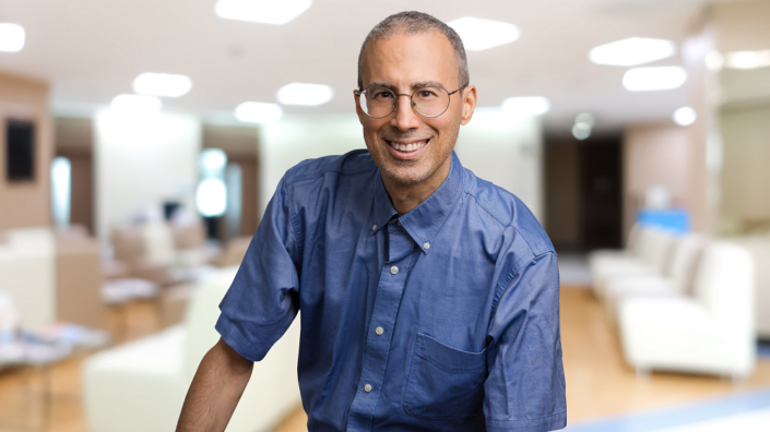
Somewhat unsurprisingly, it turned out that behind that handle, an equally agreeable human being had been hiding. A couple of weeks ago, the user @agingdoc1 revealed his true identity. His name is David Barzilai, he’s based in Boston, and he’s not connected to the star geroscientist Nir Barzilai. David came out of the shadows with a bang, announcing the launch of a podcast and of a longevity consulting service, so there was a lot for us to talk about when I asked him for a Zoom chat, and he kindly agreed.

Deep passion for everything longevity

First, David is not strictly a geroscientist in the sense of having a basic science lab, but he’s very enthusiastic about this scientific field. He says that his Ph.D. in health services (clinical) research and a strong background in evidence-based medicine offer him a unique perspective for evaluating and contributing to the field. However, he has degrees in abundance, and even calls himself “a degree hoarder.” He’s a practicing dermatologist, although he’s not doing it full time so that he can devote enough attention to his family and his passion, which includes longevity and making a personal impact in other ways.

I’ve always been very interested in my health since I understood that life naturally is finite. That sort of existential dread may have been part of what fueled a lifelong, extraordinarily deep passion. But I also had other things that led me to go into medicine. I’m fascinated by biology. I have a B.S. in cell and developmental biology. My B.A. in health and society is extremely diverse and includes things like community medicine, public health, medical anthropology, philosophy of medicine, medical sociology, history of medicine, and philosophy of health. That’s my background. To summarize, I have two bachelors, two masters, and two doctorates.

In addition to the two bachelor’s degrees and an M.D., David holds an M.Sc. in psychology, an MBA, a Ph.D. in health services research, and a couple of certificates, one of which, abbreviated as DipABLM, means “Diplomat of the American Board of Lifestyle Medicine.” This sparked my interest, and we made a brief detour into what lifestyle medicine is and how it differs from longevity medicine.

It’s a special exam that physicians can take regardless of specialty. As to how it differs from longevity medicine, lifestyle medicine relates to improving one’s health with a high emphasis on prevention through things in your lifestyle. This includes diet, sleep, your exercise, but also your relationships, your stress, your meaning, your circadian rhythm – a truly holistic approach.

Apparently, where longevity medicine turns to rapamycin, lifestyle medicine might choose keeping a healthy weight, staying physically active, pursuing a cleaner diet, getting better sleep, and generally “living healthy.”

The Twitter years

Several years ago, David decided to make the world better by sharing his excursions into longevity research literature.

I started off on Twitter because I was doing all these super-duper deep dives, healthy longevity pursuits for myself, my family, my friends, and I thought to myself, since I invest all that time in those literature searches, wouldn’t it be great to just put it out there for everyone’s benefit? And even if I disagree with some of what I find, this can be a foundation for a mutual discovery conversation. I would not have the time to discuss what I post at length, but I could be part of a larger ecosystem that self-corrects by inviting scientists and science communicators and, hopefully, changing minds and aligning individuals in the process, while also being a resource for geroscientists and longevity enthusiasts.

For me, it’s the adventure. It’s exploring the boundaries of this extremely deep hobby, passion, and, at some level, maybe obsession with learning everything that improves one’s healthspan, happiness, and even growing as a person, finding your one’s calling and life’s meaning. My Twitter activity was born from the healthspan aspect of it. I thought it would let people identify both good studies that are interesting and also bad studies. I’m not commenting much on the articles, except when I really feel it does good.

David is obviously aware of the existence of other accounts that post studies, but according to him, “none of them is as broad”. He lets himself go “off-topic” from time to time. This, he says, reflects his personality, interests, and attempt to make an impact in the lives of his readership. He’s also trying to “foster communication that’s free from all hype, nonsense, self-promotion, influence, etc.” “This can’t be perfect,” he admits, “anyone has their biases, including me. But maybe I can create a safe haven that’s friendly to everybody, where people can actually discuss topics.”

His success on Twitter came as a surprise even to himself.

My first followers were very small in number, but they had a lot of followers themselves. So somehow, even though I was mostly posting publications, I’d make a comment or two that probably made them think, ‘Oh, this guy seems to know what he’s talking about. He’s not just posting things, he’s not just a bot, he’s actually bringing an interesting perspective that I hadn’t considered’.

“I came up with something I thought would be novel in this crowded space,” he says about his ‘hybrid’ approach. “I broke every rule because most people either do listservs or they give their opinions. I don’t give opinions, but then I do a little bit,” he laughs. “I want a place where people can feel safe, no matter what they believe, and maybe they’re more likely to show up there, and maybe their own mind will be changed because they found a place where they can actually speak their mind.”

Like many people in the longevity space, David has a broad and inquisitive mind that often digresses into wider philosophical musings:

It’s also what democracy needs. Individuals to be able to discuss difficult problems, to really link minds and have the right focus, which is the focus on science, data, and treating each other with dignity, kindness, and respect, even while disagreeing. It’s OK to be critical about an analytical point but not saying that your opponent is a bad person or that they are motivated by greed. It’s OK to discuss matters of motivation, but we need to ask ourselves, in a given situation, what is most helpful for true progress? I believe most people are good at heart, and even if, in some situations, they are not, what good does it do to shame and mock people? It’s just not constructive. It ultimately is far more constructive to engage with people in a collegial manner, regardless of whether or not we agree with them, or even how we feel about their character in some instances.

But why stay anonymous for so long? Apparently, it’s not that simple to explain, and much thought went into this:

I thought to myself, who knows, maybe someone on Twitter, for instance, my patients, would recognize me. And I didn’t want them to think that if I’m talking about caloric restriction on Twitter, that’s also my advice to them, that’s what I think they should do. I’m not practicing longevity medicine on them, I’m wearing another hat when I’m on Twitter – this was very important to me.

There were other factors David had to consider. “All my decisions are multifactorial,” he says. “I’m constantly thinking about multiple variables, trying to optimize my behavior. So, that’s my gestalt of it. I decided to remain anonymous because I didn’t have to disclose my identity, and it seemed marginally better than the opposite option.”

“Project 49”

If remaining anonymous was the best thing to do for so long, why did he decide to emerge from the shadows? In part, it was acting on impulse. Usually, seeing the first digit of your age change is what might jolt you out of complacency and cause you to make resolutions you probably won’t keep. But being the thorough and forward-planning person he was, David felt an itch to shake things up when he turned 49.

On that birthday, I said to myself that I wanted to do more. I felt it was selfish for me to sit on my skill set, which is extremely deep, although you can’t tell that from my Twitter posts because of how rarely I comment on them. So, I thought, hey, if I coach and consult in this area, that would be so much fun and a phenomenal opportunity. And even though it came at some cost in terms of privacy, I owed it to myself and others.

This triggered my interest. I have interviewed many people from the budding realm of longevity medicine, but the concept of longevity consulting was new to me.

Basically, David wants to talk to people about longevity, lending them his expansive knowledge in the field, but also his skills as a life coach. It’s a concierge service where David offers Zoom appointments, starting with 10-minute-long introductory chats. These are strictly audio for his primary coaching service. “I’m not keen on video,” he says, “so I priced it so that very few would want to do it, but I would do a great job if somebody wanted video.”

Who would be his clients? Anyone, he says, starting with scientists and biohackers. Scientists might want to dip into his “deep command of literature,” running by him their thoughts and hypotheses. Biohackers might want to consult him on the intricacies of rapamycin regimens or on other potentially geroprotective drugs that can be taken off-label, such as canagliflozin.

A potential client might think: I’m reading this and that about a molecule or a treatment. They might want to bounce by me some things that they read and hear my thoughts, knowing I’m not going to say, ‘you must do this.’ I basically provide some expertise and an intelligent peer to discuss that. After all, this is a passion project for me. I want to emphasize that this coaching and consulting is not delivery of medicine, so no diagnosis or treatment such as medication is rendered. Rather, I support my clients in a collegial relationship with content expertise and as a sounding board on evidence and practices.

According to David, this will never become a big business, it’s just not scalable. And it’s not like David, with his MBA degree, doesn’t know how to turn this into a big business. He’s just not there for the money. It’s more about making a difference, not necessarily on a large scale, and applying his deep knowledge of the field he loves. He doesn’t intend to devote too much time to his practice. “I just thought it would be fun to do this for a few hours a week and exercise this part of me,” he says. Does he have a system? Not really.

Most other specialists have standardized algorithms for the highly defined populations they see. I do have my mental checklist and a strong framework, but all good coaches and consultants listen to the client, they want to know their objectives, personalizing the relationship around those values and needs. So, I try to get a sense of that and build the conversation accordingly. The sky is the limit. We could literally spend the whole time talking about their relationship with their kids or considering switching jobs – or about the finest minutiae of rapamycin in different protocols that have been tried in the literature, as long as we’re both having fun and it’s two intelligent people having an open discussion and deciding together how to spend our time.

In David’s view, various aspects of well-being and happiness come together to create a better and longer life:

On that same webpage, people can also choose the life coach track and I can go back and forth in between. Just to make it clear, I enjoy life coaching. I want every single person to walk out extremely happy.

While David says those who “gravitate towards him” tend to be M.D.s and Ph.D.s, novices are also welcome:

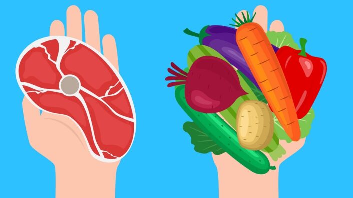
People can come to me with zero knowledge, and what I want to do is responsibly start with the basics for those individuals. With such people, I would start very heavily on low-hanging fruit, such as nutrition. I would try to understand what aspect of their healthspan is most important to them and what tradeoffs they are ready for. It’s just like when constructing a well-balanced portfolio. Everyone’s goals, their time horizon, their abilities are very individualized, just like everyone’s financial situation is different.

I enjoy working with individuals who are not experts in longevity, and just want guidance because it feels like I’m nurturing them, taking care of them, listening, discovering them. I’m very glad to be able to help them take those first steps and move their life in the right direction.

In fact, the average person that I encounter just wants to have a healthy, happy, well-balanced regular life. And that’s what I want for them. Health is a resource, one of many, just like economics and communities. They’re all aspects and facets to a well-balanced life that combines happiness, purpose, and meaning.

It’s also important for David to stay agile and responsive to new data. “I don’t sell products or invest in things that I peddle,” he says. “I want to be able to quickly change my opinions and recommendations whenever the evidence changes.”

A podcast is born

David did not want to abandon Twitter because it was “a way to reach everyone” but decided to spice things up. He launched the Agingdoc Podcast, immediately aiming at two big names in geroscience: Matt Kaeberlein and Aubrey de Grey.  Currently, it contains the first part of David’s interview with de Grey and two parts of his interview with Kaeberlein.

Just like with his Twitter persona, David thinks he’s found a winning combination of qualities for his newborn podcast:

I tried to go deeper into the science, issues, and controversies than introductory podcasts, while having a constructive conversation in a very friendly, mutually respecting way. It’s two intellectuals talking about how we think about this or that. I’m also trying to be very friendly and let the guest articulate their vision. This also relates to an extension of my role on Twitter. What makes it unique, in my view, is that I’m trying to have a place where it’s about intellect, respect, good data, and us being all on the same team. We all want to get healthy. We all want longevity.

If the description sounds a lot like Peter Attia’s podcast The Drive, it might not be a coincidence since David mentions The Drive as “the gold standard.” Others of David’s favorites are Translating Aging by BioAge Labs, Live Longer World by Aastha Jain Simes, and the podcast by VitaDAO, a decentralized geroscience organization and lifespan.io’s long-time ally. One of David’s first appearances after the “uncloaking” was on the popular Eleanor Sheekey’s podcast.

Dr. Kaeberlein was one of the first people in the longevity community to learn about David’s identity:

I first got to know @agingdoc1 as David Barzilai when he asked me if I would be willing to publicly confirm his credentials as an M.D. Ph.D. while keeping his identity private, which I agreed to do. He shared proof of his educational background with me, and I posted on Twitter that I’d confirmed his educational background at his request. During the course of our first conversation, I quickly perceived that David is a genuinely good and honest person who cares deeply for his patients and also has a passion for geroscience. After that, David and I kept in touch by email, and a few months later, he told me he was planning on ‘coming out’ and that he would be starting his own podcast. I happily agreed to be one of the first people he interviewed.

As is apparent from his posts and podcast, David has a breadth and depth of knowledge in the field that is quite impressive. He’s also a critical thinker, which is something I personally value highly, although he’s too nice to express critical opinions publicly very often. This is actually something we’ve talked about quite a bit, and I think still have somewhat different opinions on. I’ve encouraged David to be less hesitant to call out flawed science as such, instead of just presenting it without comment.  He argues that he can do more good as a consistently positive voice. Maybe he’s right. Either way, David Barzilai aka agingdoc1 is a good man, a friend, and a valuable member of our community.

Dr. de Grey, David’s second interlocutor, did not know him before appearing on his podcast, but he’s happy to welcome him to the party: “From the interview, I can say that I’m very pleased that someone who has been following the field for so many years has decided to become a full-fledged member of the community.”

With his eloquence, clout, and passion for science communication, would David consider becoming even more public, sort of an ambassador for the field? He stresses that, although he wants to make a difference, first and foremost, he’s committed to his own and his family’s health, longevity, and happiness. “Do I see myself as a sort of public figure? Sure, to the degree to which I can leverage my earned credibility to deliver hype-free, responsible education.”

Optimism and pessimism

Since David struck me as a thoroughly optimistic person, I wondered whether his optimism, combined with his deep knowledge of the field, extends to the current state of affairs in geroscience.

There are reasons for pessimism and reasons for optimism, and they coexist. I simultaneously marvel about how much insight we have compared to what we used to but also, at the same time, how little we understand. In relative terms, we know so much more compared to what we knew before, we understand things so much better than before. We already have to decentralize the knowledge we’ve acquired because it’s just unfathomable. But the fact that it’s unfathomable for the human mind doesn’t mean it’s not possible with technology.

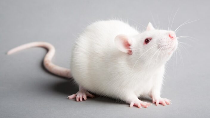
But there’s room for pessimism, too, from the standpoint of how much progress we’ve made in terms of maximum life extension. We’re slowly learning to stop all the more common things, and then we still all die at about the same time, as everything just peters out. I think this is the hard part: this simultaneous petering out of various systems. This is going to be the hardest thing for us to crack. The maximum we’ve achieved in model organisms is around 60% maximum lifespan extension with caloric restriction, and that’s in a mouse – a model that might be less optimized for aging than we are.

So, we haven’t really come up with anything that’s much better. Rapamycin is doing better than anything else, and it still doesn’t extend lifespan in those model organisms as much as caloric restriction does in some rodent strains. And nobody’s talking about calorically restricting humans or expecting humans to respond the same way.

That didn’t sound optimistic, but then David explained further:

There’s also a part of me that admires how far we’ve come in our fundamental understanding and in our basic tools. Some of these tools, I believe, are game changers – not just will they be game changers, they already are. I see publications coming out now, I see accelerating progress. Those tools are changing the game not directly, but by accelerating the rate of progress, and I mean things like single-cell sequencing, CRISPR, so many more and, importantly, those new much, much more sophisticated AI models.

So, according to David, while we still haven’t solved aging, we have acquired tools so powerful that they might soon drive an exponential increase in our knowledge and abilities.

I thought this was a great capstone for our interview, but David wasn’t done just yet. Spotting something in my tone of voice, he asked if I was feeling okay emotionally. A bit surprised, I said I wasn’t because I had a fight with my best friend the day before. I ended up receiving a bit of advice that was simple but effective. I don’t know whether following it prolonged my lifespan, but it felt good, so it might have.

We would like to ask you a small favor. We are a non-profit foundation, and unlike some other organizations, we have no shareholders and no products to sell you. All our news and educational content is free for everyone to read, but it does mean that we rely on the help of people like you. Every contribution, no matter if it’s big or small, supports independent journalism and sustains our future.
Chromatin

Pushing Forward on Understanding Cellular Decline

Researchers publishing in Nature Aging have investigated one of the core biological reasons behind the decline of stem cells’ ability to proliferate.

Heterochromatin and euchromatin

The genetic component of cells is comprised of two main parts: heterochromatin and euchromatin. Heterochromatin is largely silent, as it is a tightly packed mass that consists primarily of non-coding DNA. Euchromatin, on the other hand, is rich in actively transcribed genes and is considerably less packed.

However, heterochromatin can be methylated and demethylated just like euchromatin can. Age-related changes in methylation, the hallmark of aging known as epigenetic alterations, can happen to heterochromatin as well [1]. The regions to which this happens are known as partially methylated domains (PMDs), and they are associated with the lamina, the protective envelopes around nuclei that preserve genomic stability; laminal dysfunction is the key aspect of the rapid aging known as progeria. Changes to PMDs are associated with aging and cancer [2].

DNA methylation, these researchers explain, is mediated by the DNA methyltransferases (DNMTs), which produce 5mC, which promotes the stability of heterochromatin [3]. On the other hand, the TET family of enzymes oxidize 5mC to produce 5hmC, which is associated with the accessibility of euchromatin. The balance between DMNTs and TETs in regulating both forms of chromatin has not been fully explained [4].

Removing TET appears to help stem cells

These researchers used a mouse model that did not produce TET enzymes. The primary focus of this study was on hematopoietic stem and progenitor cells (HSPCs), which are responsible for creating blood. As expected, in 18- to 24-month-old wild-type mice, HSPCs were less able to divide and proliferate than in their young counterparts. The researchers state that “Tet2 depletion in HSPCs effectively rescued this age-related functional decline”.

Similarly, when cells from TET-knockout and wild-type mice were given to mice that had their natural cells’ proliferation destroyed by radiation, HSPCs from the TET-knockout mice proliferated far more readily. The stem cells of older wild-type mice spend more time in a non-dividing state, but this effect of aging was less pronounced in the TET-knockout mice. While rapid proliferation is often associated with cancer, the researchers found no evidence of these cells becoming cancerous.

There were also considerably fewer changes to gene expression. Wild-type mice have upregulated immune response and downregulated blood creation (hematopoiesis) as they age, but these effects of aging were also substantially blunted in TET-knockout mice.

A closer examination of the chromatin revealed interesting findings. Many epigenetic alterations were the same between the wild-type and TET-knockout groups. However, in the heterochromatin, 5hmC was substantially upregulated in older mice; the TET-knockout group, without the primary enzyme responsible for 5hmC, did not have this level of upregulation with age. The non-coding region has real effects after all.

Old viral DNA is expressed with age

Endogenous retroviruses (ERVs) are fragments of old viral DNA that have remained in the genome for millions of years. Normally, these genes are seldom expressed. However, as age-related changes to the location of heterochromatin occur, these genes become expressed more often, causing inflammation within cells. The HSPCs of TET-knockout mice did not have as many age-related location disturbances nor as much age-related upregulation of ERVs, and therefore they had less upregulation of inflammatory genes as well.

While this paper is very in-depth, and it certainly does not recommend TET knockout as a solution to human aging, it is clear that such complexities of biology need to be addressed if we are to get a handle on why our cells lose their fundamental abilities with age. Further work in this area may lead to better understanding and solutions to the problems of epigenetic alterations and genomic instability.

We would like to ask you a small favor. We are a non-profit foundation, and unlike some other organizations, we have no shareholders and no products to sell you. All our news and educational content is free for everyone to read, but it does mean that we rely on the help of people like you. Every contribution, no matter if it’s big or small, supports independent journalism and sustains our future.

Literature

[1] Hernando-Herraez, I., Evano, B., Stubbs, T., Commere, P. H., Jan Bonder, M., Clark, S., … & Reik, W. (2019). Ageing affects DNA methylation drift and transcriptional cell-to-cell variability in mouse muscle stem cells. Nature communications, 10(1), 4361.

[2] Brinkman, A. B., Nik-Zainal, S., Simmer, F., Rodríguez-González, F. G., Smid, M., Alexandrov, L. B., … & Stunnenberg, H. G. (2019). Partially methylated domains are hypervariable in breast cancer and fuel widespread CpG island hypermethylation. Nature communications, 10(1), 1749.

[3] Richards, E. J., & Elgin, S. C. (2002). Epigenetic codes for heterochromatin formation and silencing: rounding up the usual suspects. Cell, 108(4), 489-500.

[4] López-Moyado, I. F., Tsagaratou, A., Yuita, H., Seo, H., Delatte, B., Heinz, S., … & Rao, A. (2019). Paradoxical association of TET loss of function with genome-wide DNA hypomethylation. Proceedings of the National Academy of Sciences, 116(34), 16933-16942.

Meat versus plants

Plant-Based Alternatives Are Associated With Less Disease

A new meta-analysis has found that in many cases, switching from an animal product to a plant-based alternative is associated with less cardiovascular, diabetes, and all-cause mortality risk [1].

Supplant with plant?

Whether a completely or mostly plant-based diet is good for you has been the subject of a raging debate. On one hand, studies have consistently placed plant-based diets among the healthiest dietary options available [2]. On the other hand, complete exclusion of animal-based foods might not be ideal, especially for younger and older people [3]. Going vegan can make maintaining a balanced diet harder since some nutrients are scarce or even nonexistent in plants.

Apparently, a more nuanced analysis is needed into what happens when people substitute specific animal products with plant-based alternatives. This is the topic of a new review published in BMC. After sifting through hundreds of papers, the authors included 37 of them in their analysis. They then investigated associations between abandoning animal products in favor of plant-based alternatives and cardiovascular health, occurrence of type 2 diabetes, and all-cause mortality.

All of this review’s featured studies were prospective cohort studies, and no randomized, controlled trials were included. This is understandable, since detecting the occurrence of cardiovascular disease or diabetes, not to mention death, requires a long follow-up period: in this review, the mean follow-up duration was 19 years.

Switching to plants lowers risk

The researchers calculated shared hazard ratios (SHR): the difference in the prevalence of an outcome between two groups. For instance, substituting 50 grams of processed meat a day for 28-50 grams of nuts resulted in an SHR of 0.73 – that is, it lowered chances of getting any type of cardiovascular disease (CVD) by a hefty 27%. Substituting with 50 grams of legumes gave an SHR of 0.77. Whole grains scored the best, with an SHR of 0.64: a full 36% decrease in CVD incidence. This is consistent with previous research that has marked processed meat as highly unhealthy. The World Health Organization considers processed meat to be a carcinogen, but its detrimental effects are probably much broader.

Substituting one egg a day with nuts resulted in a 17% decrease in CVD incidence. Interestingly, substituting butter with olive oil only led to a slight 4% decrease in risk. The researchers also found that replacing red meat with nuts, unprocessed red meat with nuts or legumes, poultry with nuts, and eggs with legumes was associated with a lower risk of CVD, but for these associations, the certainty of evidence was low.

Butter, however, did not fare well in the analysis of type 2 diabetes incidence. The researchers calculated that replacing butter with olive oil was associated with a slight decline in risk. Interestingly, red meat also seemed to be more strongly associated with diabetes than with CVD: substituting red meat for nuts was associated with an 8% decrease in risk. The same parameter for replacing processed meat with nuts was 22%, replacing poultry with whole grains was 13%, and replacing eggs with nuts or whole grains was 8%.

Finally, replacing processed meat with nuts or whole grains was associated with a 21% decrease in all-cause mortality risk with a moderate certainty of evidence. Replacing eggs with nuts or legumes was a 15% decrease, replacing red meat with nuts or whole grains was a 7% decrease, and replacing butter with olive oil was a 6% decrease.

Importantly, switching to a plant-based alternative was never associated with an increase in risk, except for the slight uptick in CVD risk when replacing butter with margarine. However, the certainty of evidence for that particular association was very low.

Plant eaters tend to lead healthier lives in general

Populational studies can only indicate association, but not causation, so their results should be taken with a grain of salt. While meta-analyses generally improve the quality of evidence, they are still prone to the same problems as the individual studies they consist of.

According to the authors, the included studies were all adjusted for major lifestyle confounders, such as total energy intake, physical activity, alcohol intake, and smoking. However, completely eliminating confounding is impossible. One of the alternative explanations for the observed associations the authors provide is that people on plant-based diets tend to follow healthier lifestyles in general.

Our findings suggest that a shift in diet from a high consumption of animal-based foods, especially red and processed meat, to plant-based foods (e.g., nuts, legumes, and whole grains) is associated with a lower risk of all-cause mortality, CVD, and T2D. Thus, a change in dietary habits towards an increment of plant-based products appears to be important for cardiometabolic health. However, more research is needed in order to strengthen the existing evidence and to investigate new associations, especially with a focus on meat and dairy replacement products.

We would like to ask you a small favor. We are a non-profit foundation, and unlike some other organizations, we have no shareholders and no products to sell you. All our news and educational content is free for everyone to read, but it does mean that we rely on the help of people like you. Every contribution, no matter if it’s big or small, supports independent journalism and sustains our future.

Literature

[1] Neuenschwander, M., Stadelmaier, J., Eble, J. et al. (2023). Substitution of animal-based with plant-based foods on cardiometabolic health and all-cause mortality: a systematic review and meta-analysis of prospective studies. BMC Med 21, 404.

[2] Hodge, A. M., O’Dea, K., English, D. R., Giles, G. G., & Flicker, L. (2014). Dietary patterns as predictors of successful ageing. The journal of nutrition, health & aging, 18(3), 221–227.

[3] Domic, J., Grootswagers, P., van Loon, L. J., & de Groot, L. C. (2022). Perspective: vegan diets for older adults? A perspective on the potential impact on muscle mass and strength. Advances in nutrition, 13(3), 712-725.

Brain COVID

Senolytics May Work Against Long COVID in the Brain

A new publication in Nature Aging has used organoids and model mice to discover that senolytics may be effective treatments for neurodegeneration caused by long COVID.

A link between COVID-19 and cellular senescence

Post-acute infection syndromes (PAISes) are caused by a variety of viral and bacterial infections. The PAIS specific to the COVID-causing SARS-CoV-2, “long COVID”, is linked to multiple neurological problems, including fatigue and cognitive impairment [1]. Substantial evidence has found that SARS-CoV-2 invades the brain [2], and a study that used data from UK Biobank found that it is linked to changes in the brain’s structure [3].

The alterations linked to COVID are molecularly similar to brain aging [4]. As COVID-19 is also linked to cellular senescence in lung tissue [5], and senescent cells have been found to lead to neuropathology in the brain [6], these researchers developed a study to connect the two, attempting to determine the potential value of senolytics for treating long COVID’s effects on the brain.

Beginning with human organoids

The researchers began their investigation by using brain organoids, miniature organs grown for testing purposes from embryonic stem cells. After 8 months of natural aging, these organoids begin to develop significant cellular senescence. At this time, senolytic combinations were administered to various study groups.

In these organoids, astrocytes were more prone to senescence than neurons, but treating neurons was considerably more difficult. The well-known combination of dasatinib and quercetin was found to be the only senolytic that was effective at reducing the senescence marker p16 in neurons.

Interestingly, the various senolytics had different effects on the SASP-related mRNA expression of the cells in these organoids. For example, the laboratory senolytic ABT-737 reduced SERPINF1, while dasatinib and quercetin reduced SERPINE1 and IL1A instead. Overall, dasatinib and quercetin were found to be the best at reversing senescence-related gene expression.

After analyzing post-mortem brain tissue from both COVID-19 sufferers and age-matched controls that died of unrelated causes, the researchers found that the COVID group had substantially more markers of cellular senescence. They then infected brain organoids with various strains of SARS-CoV-2. Most of the variants significantly upregulated senescence markers along with the DNA damage biomarker γH2AX.

COVID variants senescence

Over 30% of the gene expressions of these organoids that were changed with natural aging were also changed with SARS-CoV-2 infection. Many of the affected genes were part of known senescence pathways. The Delta variant had a uniquely strong effect on senescence-related gene expression. Senolytic treatment was found to substantially reduce senescence markers in infected organoids.

Organoids senolytics

Mouse data agreed

As mice do not normally contract SARS-CoV-2, the researchers used a mouse model that expresses the human ACE2 protein that is a vector for the disease. One of the first and most startling findings was that senolytics reduced the mortality caused by this infection; after 10 days, all of the untreated mice were dead, but nearly half of the dasatinib and quercetin-treated mice were still alive after day 12. All of the tested senolytics also had substantial effects on the mice’s RNA gene expression, reducing inflammatory and senescence markers down nearly to the levels of uninfected controls.

Senolytics COVID RNA expression

Additionally, all of the senolytics also reduced viral RNA related to viral replication. The researchers hypothesize that SARS-CoV-2 preferentially proliferates through senescent cells.

While it is still unclear if senolytics could be useful as COVID-19 treatments in people, this research points in that direction. Clinical trials would need to be conducted to determine if senolytics can reduce the infectivity of COVID-19, are effective against long COVID or other PAISes, or can ameliorate the symptoms of ordinary brain aging.

We would like to ask you a small favor. We are a non-profit foundation, and unlike some other organizations, we have no shareholders and no products to sell you. All our news and educational content is free for everyone to read, but it does mean that we rely on the help of people like you. Every contribution, no matter if it’s big or small, supports independent journalism and sustains our future.

Literature

[1] Hartung, T. J., Neumann, C., Bahmer, T., Chaplinskaya-Sobol, I., Endres, M., Geritz, J., … & Finke, C. (2022). Fatigue and cognitive impairment after COVID-19: A prospective multicentre study. EClinicalMedicine, 53.

[2] Schwabenland, M., Salié, H., Tanevski, J., Killmer, S., Lago, M. S., Schlaak, A. E., … & Bengsch, B. (2021). Deep spatial profiling of human COVID-19 brains reveals neuroinflammation with distinct microanatomical microglia-T-cell interactions. Immunity, 54(7), 1594-1610.

[3] Douaud, G., Lee, S., Alfaro-Almagro, F., Arthofer, C., Wang, C., McCarthy, P., … & Smith, S. M. (2022). SARS-CoV-2 is associated with changes in brain structure in UK Biobank. Nature, 604(7907), 697-707.

[4] Mavrikaki, M., Lee, J. D., Solomon, I. H., & Slack, F. J. (2022). Severe COVID-19 is associated with molecular signatures of aging in the human brain. Nature Aging, 2(12), 1130-1137.

[5] Lee, S., Yu, Y., Trimpert, J., Benthani, F., Mairhofer, M., Richter-Pechanska, P., … & Schmitt, C. A. (2021). Virus-induced senescence is a driver and therapeutic target in COVID-19. Nature, 599(7884), 283-289.

[6] Ogrodnik, M., Evans, S. A., Fielder, E., Victorelli, S., Kruger, P., Salmonowicz, H., … & Jurk, D. (2021). Whole-body senescent cell clearance alleviates age-related brain inflammation and cognitive impairment in mice. Aging cell, 20(2), e13296.

Dietary choices

Promoting Health and Longevity Through Diet

A review published in the Journal of Internal Medicine summarized current knowledge on the impact of dietary factors on chronic diseases and longevity [1].

Restrict calories, live healthier and longer

The choice of what someone eats obviously has a profound impact on that person’s health, but the amount is also important. Caloric restriction, the practice of reducing caloric intake without causing malnutrition, has been shown in many laboratory organisms to increase lifespan and delay the onset of age-related diseases [2]. However, studying caloric restriction in humans is challenging.

In the longest caloric restriction trial, non-obese participants achieved 12% calorie reduction over 2 years. Researchers observed improvements in several biomarkers: blood lipids, blood pressure, insulin sensitivity, and pro-inflammatory cytokines [3]. However, due to the short duration of the study and the small sample, long-term chronic disease and mortality risk cannot be reliably assessed.

Studies assessing body weight and shape trajectories are used as a marker and substitute for detailed calorie intakes. Such studies show the health benefits of maintaining a stable-lean body shape, which include decreased risks of type 2 diabetes and cardiovascular diseases [4, 5, 6]. Results also show an association between an elevated risk of several diseases and weight gain, even in 5 kg (11 lb) increments, during young and middle adulthood [6].

5 kg weight gain

Quality and quantity of fats, protein, and carbs matters

Research spanning several decades provides a wealth of evidence supporting the idea that different types of fat are linked to different effects on health. Some studies have associated higher intake of unsaturated fats with lower mortality rates [7]. On the other hand, consumption of trans fats and saturated fats has been documented to have the opposite effect, and it is associated with increased mortality.

Fat and mortality

The food source of fat is also important, with plant sources, but not animal sources, lowering the risk of coronary artery disease [8]. Protein restriction, specifically restricting particular amino acids, such as methionine and tryptophan, extends the lifespan of laboratory model organisms [9, 10].

In humans, associations between protein intake and mortality are still being researched. Data from the National Health and Nutrition Examination Survey (NHANES) demonstrates that, for individuals between 50-65 years old, high animal protein, but not high plant protein, was associated with a 75% increase in overall mortality during 18 years of follow-up. However, for individuals over 65, higher protein intake was associated with lower mortality [11].

This hasn’t been observed in other cohort studies, where age was not a modifying factor. In those studies, higher animal protein intake was associated with cardiovascular mortality, and higher plant protein intake was inversely associated with all-cause and cardiovascular mortality [12].

Carbohydrate intake is also intensely studied. Animal studies on low-carbohydrate or ketogenic diets suggest that they can enhance longevity and healthspan [13]. Short-term randomized clinical trials show that restricting the consumption of carbohydrates can improve several biomarkers, such as by lowering blood glucose or improving insulin sensitivity [14].

However, adherence to a low-carb diet is challenging and can result in inadequate intake of fiber, vitamins, and minerals.

Current research suggests that the health impact of a low-carb diet depends on the type of fat and protein consumed [15]. An animal-based low-carb diet is associated with higher mortality. In comparison, a low-carb diet in which vegetables are mostly the sources of protein and fat is associated with lower mortality, especially mortality caused by cardiovascular diseases. Generally, studies agree that carbohydrate quality, more than quantity, plays a more important role in the development of chronic disease.

Polyphenol-rich plant foods

Polyphenols are a group of natural compounds with antioxidant, anti-inflammatory, and anticarcinogenic properties. They are found in many plant-based foods. Consumption of polyphenols is linked to cardiometabolic benefits, improved cognitive function, decreased neurodegenerative disease risk [16], and maintenance of healthy gut microbiota [17]. Some research has found that polyphenols have aging properties, and they influence many hallmarks of aging [18].

Bringing it all together into a healthy diet

The authors rightly notice that various healthy dietary components are not consumed in isolation but must be combined into a healthy dietary pattern. One of the diets, which is regarded as healthy, is the Mediterranean diet. The Mediterranean diet is abundant in plant foods, such as fruits, vegetables, whole grains, nuts, legumes, and olive oil.

Current research points out that adherence to the Mediterranean diet is associated with reduced risks of many conditions and diseases such as obesity, type 2 diabetes, hypertension, dyslipidemia, stroke, heart failure, neurodegenerative diseases, and mortality [19].

The Nordic diet has some similarities to the Mediterranean diet. It focuses on plant-based and locally sourced foods, with a major difference of using mainly rapeseed oil instead of olive oil. Available data, although scarce, suggests that following a Nordic diet lowers the risk of cardiovascular diseases and type 2 diabetes. However, no long-term studies have been conducted so far [20].

This paper also discusses the Okinawan diet. Okinawa Island is one of the Blue Zones, a place with a high number of centenarians. Diet is one of the components believed to be responsible for the increased lifespan of Okinawa’s residents. It puts “emphasis on root vegetables (mainly purple sweet potatoes), green and yellow vegetables, soybean-based foods, seaweeds and algae, tea, and a variety of medicinal plants (e.g. bitter melon) and spices such as turmeric” with limited meat consumption. Additionally, Okinawans practice Hara Hachi Bu, that is, eating until they are 80% full, which resembles caloric restriction.

The authors also discuss vegetarian diets. Small randomized clinical trials have shown improvements in different biomarkers for participants following a vegetarian diet, e.g., reduced blood pressure, total and LDL cholesterol levels, body weight, and other cardiometabolic risk factors. Additionally, large cohort studies of vegetarians suggest they have a reduced risk of obesity, type 2 diabetes, and lower rates of cancer than nonvegetarians [21].

It’s not only about food

The study authors conclude that while dietary patterns have a profound impact on health, other lifestyle factors are important to increase healthspan and lifespan.

We defined five low-risk lifestyle factors as fulfilling either: never smoking, maintaining normal weight (BMI 18.5–24.9 kg/m2), 30+ minutes/day moderate to vigorous physical activity, moderate alcohol intake (no more than one drink per day for women and no more than two for men), and a high-quality diet.

Diet graphical abstract
We would like to ask you a small favor. We are a non-profit foundation, and unlike some other organizations, we have no shareholders and no products to sell you. All our news and educational content is free for everyone to read, but it does mean that we rely on the help of people like you. Every contribution, no matter if it’s big or small, supports independent journalism and sustains our future.

Literature

[1] Hu F. B. (2023). Diet strategies for promoting healthy aging and longevity: An epidemiological perspective. Journal of internal medicine, 10.1111/joim.13728. Advance online publication.

[2] Fontana, L., & Partridge, L. (2015). Promoting health and longevity through diet: from model organisms to humans. Cell, 161(1), 106–118.

[3] Kraus, W. E., Bhapkar, M., Huffman, K. M., Pieper, C. F., Krupa Das, S., Redman, L. M., Villareal, D. T., Rochon, J., Roberts, S. B., Ravussin, E., Holloszy, J. O., Fontana, L., & CALERIE Investigators (2019). 2 years of calorie restriction and cardiometabolic risk (CALERIE): exploratory outcomes of a multicentre, phase 2, randomised controlled trial. The lancet. Diabetes & endocrinology, 7(9), 673–683.

[4] Song, M., Hu, F. B., Wu, K., Must, A., Chan, A. T., Willett, W. C., & Giovannucci, E. L. (2016). Trajectory of body shape in early and middle life and all cause and cause specific mortality: results from two prospective US cohort studies. BMJ (Clinical research ed.), 353, i2195.

[5] Zheng, Y., Song, M., Manson, J. E., Giovannucci, E. L., & Hu, F. B. (2017). Group-Based Trajectory of Body Shape From Ages 5 to 55 Years and Cardiometabolic Disease Risk in 2 US Cohorts. American journal of epidemiology, 186(11), 1246–1255.

[6] Zheng, Y., Manson, J. E., Yuan, C., Liang, M. H., Grodstein, F., Stampfer, M. J., Willett, W. C., & Hu, F. B. (2017). Associations of Weight Gain From Early to Middle Adulthood With Major Health Outcomes Later in Life. JAMA, 318(3), 255–269.

[7] Marklund, M., Wu, J. H. Y., Imamura, F., Del Gobbo, L. C., Fretts, A., de Goede, J., Shi, P., Tintle, N., Wennberg, M., Aslibekyan, S., Chen, T. A., de Oliveira Otto, M. C., Hirakawa, Y., Eriksen, H. H., Kröger, J., Laguzzi, F., Lankinen, M., Murphy, R. A., Prem, K., Samieri, C., … Cohorts for Heart and Aging Research in Genomic Epidemiology (CHARGE) Fatty Acids and Outcomes Research Consortium (FORCE) (2019). Biomarkers of Dietary Omega-6 Fatty Acids and Incident Cardiovascular Disease and Mortality. Circulation, 139(21), 2422–2436.

[8] Zong, G., Li, Y., Sampson, L., Dougherty, L. W., Willett, W. C., Wanders, A. J., Alssema, M., Zock, P. L., Hu, F. B., & Sun, Q. (2018). Monounsaturated fats from plant and animal sources in relation to risk of coronary heart disease among US men and women. The American journal of clinical nutrition, 107(3), 445–453.

[9] Solon-Biet, S. M., McMahon, A. C., Ballard, J. W., Ruohonen, K., Wu, L. E., Cogger, V. C., Warren, A., Huang, X., Pichaud, N., Melvin, R. G., Gokarn, R., Khalil, M., Turner, N., Cooney, G. J., Sinclair, D. A., Raubenheimer, D., Le Couteur, D. G., & Simpson, S. J. (2014). The ratio of macronutrients, not caloric intake, dictates cardiometabolic health, aging, and longevity in ad libitum-fed mice. Cell metabolism, 19(3), 418–430.

[10] Miller, R. A., Buehner, G., Chang, Y., Harper, J. M., Sigler, R., & Smith-Wheelock, M. (2005). Methionine-deficient diet extends mouse lifespan, slows immune and lens aging, alters glucose, T4, IGF-I and insulin levels, and increases hepatocyte MIF levels and stress resistance. Aging cell, 4(3), 119–125.

[11] Levine, M. E., Suarez, J. A., Brandhorst, S., Balasubramanian, P., Cheng, C. W., Madia, F., Fontana, L., Mirisola, M. G., Guevara-Aguirre, J., Wan, J., Passarino, G., Kennedy, B. K., Wei, M., Cohen, P., Crimmins, E. M., & Longo, V. D. (2014). Low protein intake is associated with a major reduction in IGF-1, cancer, and overall mortality in the 65 and younger but not older population. Cell metabolism, 19(3), 407–417.

[12] Song, M., Fung, T. T., Hu, F. B., Willett, W. C., Longo, V. D., Chan, A. T., & Giovannucci, E. L. (2016). Association of Animal and Plant Protein Intake With All-Cause and Cause-Specific Mortality. JAMA internal medicine, 176(10), 1453–1463.

[13] Roberts, M. N., Wallace, M. A., Tomilov, A. A., Zhou, Z., Marcotte, G. R., Tran, D., Perez, G., Gutierrez-Casado, E., Koike, S., Knotts, T. A., Imai, D. M., Griffey, S. M., Kim, K., Hagopian, K., McMackin, M. Z., Haj, F. G., Baar, K., Cortopassi, G. A., Ramsey, J. J., & Lopez-Dominguez, J. A. (2017). A Ketogenic Diet Extends Longevity and Healthspan in Adult Mice. Cell metabolism, 26(3), 539–546.e5.

[14]  Ludwig, D. S., Hu, F. B., Tappy, L., & Brand-Miller, J. (2018). Dietary carbohydrates: role of quality and quantity in chronic disease. BMJ (Clinical research ed.), 361, k2340.

[15] Fung, T. T., van Dam, R. M., Hankinson, S. E., Stampfer, M., Willett, W. C., & Hu, F. B. (2010). Low-carbohydrate diets and all-cause and cause-specific mortality: two cohort studies. Annals of internal medicine, 153(5), 289–298.

[16] Ammar, A., Trabelsi, K., Boukhris, O., Bouaziz, B., Müller, P., M Glenn, J., Bott, N. T., Müller, N., Chtourou, H., Driss, T., & Hökelmann, A. (2020). Effects of Polyphenol-Rich Interventions on Cognition and Brain Health in Healthy Young and Middle-Aged Adults: Systematic Review and Meta-Analysis. Journal of clinical medicine, 9(5), 1598.

[17] Rana, A., Samtiya, M., Dhewa, T., Mishra, V., & Aluko, R. E. (2022). Health benefits of polyphenols: A concise review. Journal of food biochemistry, 46(10), e14264.

[18] Leri, M., Scuto, M., Ontario, M. L., Calabrese, V., Calabrese, E. J., Bucciantini, M., & Stefani, M. (2020). Healthy Effects of Plant Polyphenols: Molecular Mechanisms. International journal of molecular sciences, 21(4), 1250.

[19] Guasch-Ferré, M., & Willett, W. C. (2021). The Mediterranean diet and health: a comprehensive overview. Journal of internal medicine, 290(3), 549–566.

[20] Massara, P., Zurbau, A., Glenn, A. J., Chiavaroli, L., Khan, T. A., Viguiliouk, E., Mejia, S. B., Comelli, E. M., Chen, V., Schwab, U., Risérus, U., Uusitupa, M., Aas, A. M., Hermansen, K., Thorsdottir, I., Rahelic, D., Kahleová, H., Salas-Salvadó, J., Kendall, C. W. C., & Sievenpiper, J. L. (2022). Nordic dietary patterns and cardiometabolic outcomes: a systematic review and meta-analysis of prospective cohort studies and randomised controlled trials. Diabetologia, 65(12), 2011–2031.

[21] Wang, T., Masedunskas, A., Willett, W. C., & Fontana, L. (2023). Vegetarian and vegan diets: benefits and drawbacks. European heart journal, 44(36), 3423–3439.

Fang feng

Saposhnikovia Root Extract Improves Tendon Repair

A new study has identified a molecule found in Saposhnikovia root as a potent mediator of tendon regeneration in rats. The results were also replicated in human cells [1].

Aging does not spare tendons

Like with many tissues, the health of tendons, which are central to mobility and agility, hinges on the function of local stem cells. Tendon stem/progenitor cells (TSPCs) are hard at work doing maintenance, regeneration, and repair, but they experience loss of function with age. As a result, tendon ruptures, one of the leading causes of mobility impairment, heal much slower in older organisms [2].

In this new study published in Nature Bone Research, the scientists used a deep learning-based efficacy prediction system (DLEPS), which determined that prim-O-glucosylcimifugin (POG), which is found in Saposhnikovia root extract, is a molecule that can potentially promote stemness and inhibit senescence in tendon stem/progenitor cells (TSPC). Previous research has already found that POG has anticonvulsant, anti-inflammatory, and even anti-cancer effects [3], but it has not been investigated in the context of aging.

POG rescues senescence and improves healing in vitro

First, the researchers conducted in vitro experiments in a model of replicative senescence. By the twelfth passage (division), rat TSPCs showed numerous signs of senescence, and their stemness and differentiation potential were disrupted. Adding POG to the culture from the beginning significantly rescued the cells’ self-renewal and proliferative potential, allowing them to form many more colonies.

In the treated cells, several markers of senescence and aging were reduced, including β-galactosidase and p21, the apoptosis marker p53, and mRNA levels of the inflammatory cytokines Il-6, Il-1β, and TNF-α. When transferred to a medium that induces the formation of tendons (tenogenesis), POG-treated TSPCs secreted much more tenogenesis-associated markers and generated denser tendon collagen fibers when transplanted back into rats.

Works in naturally aged cells and animals

The researchers then wanted to experiment with ordinary aging rather than cellular senescence induced in a dish. As expected, tendons from 18-month-old rats showed signs of collagen degradation and increased senescence, and tendon injuries took longer to heal. TSPCs from naturally aged rats also showed many similarities to cells exhausted by replication in culture. POG proved to be almost as effective in countering natural aging in cells as it was against replicative senescence.

The researchers then administered POG to old rats for two months. Two days after the treatment stopped, the rats had their tendons injured. Seven days later, the animals from the study group showed basically the same levels of healing as young controls, and much higher than old untreated rats, with better collagen formation and lower inflammation. In uninjured old rats, POG treatment was able to alleviate collagen deterioration and cellular senescence.

As a replacement for multi-day oral administration, the researchers used nanoparticles for sustained POG release. Once injected into the site of the future injury, POG was gradually released from the nanoparticles over the course of two weeks. “Both the systemic administration of POG and the local delivery of POG nanoparticles”, the researchers wrote, “effectively enhanced the healing capacity of partial transection in aged rat tendons by enhancing rTSPC stemness, rejuvenating senescent phenotypes, and suppressing inflammatory progression.”

Increase in autophagy via mTOR downregulation

Finally, the researchers ran a series of experiments to investigate POG’s mechanism of action. RNA analysis revealed that the treatment downregulated several aging-related pathways, most notably NF-κB and mTOR. Since mTOR negatively regulates autophagy, which is also impaired in senescent cells, the researchers investigated the effect of POG on autophagy and found that POG increased autophagic flux and improved aberrant protein clearing. When the researchers knocked out a crucial autophagy-related gene, this abrogated POG’s effect, showing that POG indeed works via improvement of autophagy.

Since rodents are less than ideal models for human aging, the researchers also experimented in vitro with human TSPCs. The results were largely the same, including improvement in stemness and regeneration capacity, downregulation of mTOR, and increased autophagy. This improves the chances that POG therapy can be translated to humans.

In summary, this study employed a DLEPS to uncover a potential stemness-promoting drug, POG, which rescues aging-impaired TSPC stemness and functions by dual targeted inhibition of mTOR and nuclear factor-κB and activation of autophagy. The systemic administration of POG or the local delivery of POG nanoparticles restored structural and functional tendon regeneration in aged rats without transplanted cells (Fig. 8). These findings possess great translational potential for clinical tendon therapies.

We would like to ask you a small favor. We are a non-profit foundation, and unlike some other organizations, we have no shareholders and no products to sell you. All our news and educational content is free for everyone to read, but it does mean that we rely on the help of people like you. Every contribution, no matter if it’s big or small, supports independent journalism and sustains our future.

Literature

[1] Wang, Y., Jin, S., Luo, D., He, D., Yu, M., Zhu, L., … & Liu, Y. (2023). Prim-O-glucosylcimifugin ameliorates aging-impaired endogenous tendon regeneration by rejuvenating senescent tendon stem/progenitor cells. Bone Research, 11(1), 54.

[2] Ackerman, J. E., Bah, I., Jonason, J. H., Buckley, M. R., & Loiselle, A. E. (2017). Aging does not alter tendon mechanical properties during homeostasis, but does impair flexor tendon healing. Journal of Orthopaedic Research, 35(12), 2716-2724.

[3] Gao, W., Zhang, X., Yang, W., Dou, D., Zhang, H., Tang, Y., … & Sun, T. (2019). Prim-O-glucosylcimifugin enhances the antitumour effect of PD-1 inhibition by targeting myeloid-derived suppressor cells. Journal for immunotherapy of cancer, 7, 1-15.

Rat looking at camera

Targeting Metabolism to Protect Against Stroke

Japanese researchers publishing in Aging have described how improving the AMPK metabolic pathway improves outcomes in a rat model of stroke.

Targeting metabolism directly

This paper begins with a look back at previous research on the focus of this study, 5′-adenosine monophosphate-activated protein kinase (AMPK). This signaling molecule is described here as ‘evolutionarily conserved’, meaning that it fulfills the same function across a wide range of distantly related species. It regulates, and is regulated by, multiple aspects of metabolism and physiology. This molecule has been reported to be responsible for metformin’s effects on diabetes [1] and lowers blood pressure in animal models [2].

AMPK holds particular value in the brain. Dysregulation of AMPK is associated with Alzheimer’s and Parkinson’s diseases [3], and brain aging and AMPK dysregulation appear to be strongly linked [4]. AMPK is linked to the cellular maintenance process of autophagy in brain cells [5], and other research has suggested that it could be a target for stroke treatment [6].

While metformin and exercise have been reported to affect AMPK, these researchers instead chose 1,5-anhydro-D-fructose (1,5-AF), which is naturally formed by decomposition in the gut [7]. These researchers have previously reported that 1,5-AF helps mitochondria to form in neurons, providing a protective effect [8]. However, that study was done in cells, not living animals, and so for this study, these researchers have moved on to rodents.

Three models of aging show positive results

As their name suggests, stroke-prone spontaneously hypertensive rats (SHRSPs) have high blood pressure, nearly double that of ordinary rats, and are often used to develop therapies for stroke [9]. Another model used in this study is a senescence-prone mouse model, SAMP8, which is uniquely prone to brain aging [10]. The researchers also used a conventional model of acute ischemic stroke (AIS), which uses unmodified rats subjected to a stroke-inducing procedure.

In the AIS model, 1,5-AF administration drastically reduced the extent of the damage caused by stroke. Infarcts were far smaller, neurology was significantly less impacted, and mortality rate was considerably lower. 1,5-AF was found, as expected, to significantly upregulate a pathway related to AMPK. It was also found to reduce TNF-α, which encourages inflammation.

These findings continued to the SHRSP model. Rats of this breed that were given water high in salt had reduced blood pressure when also given 1,5-AF, and their mortality rate was substantially lower. AMPK was upregulated, and TNF-α was downregulated, in these animals as well. The 1,5-AF group also had additional muscle mass.

In the SAMP8 mice, the findings were positive although more spotty and less significant. While the total locomotion distance seemed unaffected, frailty in the mice given 1,5-AF was somewhat delayed from the norm of this breed, and the 1,5-AF mice ran slightly faster. The Morris water maze test did not return positive results, but the 1,5-AF group had better object recognition. Although the AMPK pathway was affected, TNF-α was not significantly affected in this mouse model.

A future for treatments

From the totality of these results, the researchers conclude that 1,5-AF causes neurovascular improvement in a way that protects against stroke. However, all of these are animal models, two of which are specifically engineered for the purpose. Further trials, particularly human clinical trials, will be required to determine if 1,5-AF or other AMPK-related therapies are valuable for preventing or treating strokes in people.

We would like to ask you a small favor. We are a non-profit foundation, and unlike some other organizations, we have no shareholders and no products to sell you. All our news and educational content is free for everyone to read, but it does mean that we rely on the help of people like you. Every contribution, no matter if it’s big or small, supports independent journalism and sustains our future.

Literature

[1] Musi, N. (2006). AMP-activated protein kinase and type 2 diabetes. Current Medicinal Chemistry, 13(5), 583-589.

[2] Ford, R. J., Teschke, S. R., Reid, E. B., Durham, K. K., Kroetsch, J. T., & Rush, J. W. (2012). AMP-activated protein kinase activator AICAR acutely lowers blood pressure and relaxes isolated resistance arteries of hypertensive rats. Journal of hypertension, 30(4), 725-733.

[3] Liu, Y. J., & Chern, Y. (2015). AMPK-mediated regulation of neuronal metabolism and function in brain diseases. Journal of neurogenetics, 29(2-3), 50-58.

[4] Mattson, M. P., & Arumugam, T. V. (2018). Hallmarks of brain aging: adaptive and pathological modification by metabolic states. Cell metabolism, 27(6), 1176-1199.

[5] Shi, Q., Cheng, Q., & Chen, C. (2021). The role of autophagy in the pathogenesis of ischemic stroke. Current neuropharmacology, 19(5), 629-640.

[6] Jiang, S., Li, T., Ji, T., Yi, W., Yang, Z., Wang, S., … & Gu, C. (2018). AMPK: potential therapeutic target for ischemic stroke. Theranostics, 8(16), 4535.

[7] Ito, T., Totoki, T., Takada, S., Otsuka, S., & Maruyama, I. (2021). Potential roles of 1, 5-anhydro-d-fructose in modulating gut microbiome in mice. Scientific reports, 11(1), 19648.

[8] Kasamo, Y., Kikuchi, K., Yamakuchi, M., Otsuka, S., Takada, S., Kambe, Y., … & Maruyama, I. (2021). 1, 5-anhydro-D-fructose protects against rotenone-induced neuronal damage in vitro through mitochondrial biogenesis. International journal of molecular sciences, 22(18), 9941.

[9] Yamagata, K. (2020). Astrocytic nutritional dysfunction associated with hypoxia-induced neuronal vulnerability in stroke-prone spontaneously hypertensive rats. Neurochemistry International, 138, 104786.

[10] Sanada, Y., Ikuta, Y., Ding, C., Shinohara, M., Yimiti, D., Ishitobi, H., … & Miyaki, S. (2022). Senescence-accelerated mice prone 8 (SAMP8) in male as a spontaneous osteoarthritis model. Arthritis Research & Therapy, 24(1), 235.

Food choices

Mediterranean, Keto, and Plant-Based Diets Vs. Cancer

A new review summarizes what we know about the Mediterranean, keto, and plant-based diets and their effects on cancer risk and progression [1].

Diet matters

Since the War on Cancer began in the early 1970s, science has made giant strides in understanding and treating cancer. However, the war is far from over. Cancer remains a deadly age-related disease, its advanced stages are still mostly untreatable, the response to therapies is not nearly universal, and those therapies themselves can be harmful and accelerate aging.

The upshot is clear: it’s better not to get cancer at all. In recent decades, studies have shown that a variety of lifestyle factors affect your chances of getting cancer and sometimes its progression. Smoking is the most obvious case, but other behaviors, such as diet, can also affect your fate. Processed meat is recognized by the World Health Organization as carcinogenic and red meat as probably carcinogenic. Bad dietary choices can lead to numerous problems associated with cancer, such as type 2 diabetes, which has been shown to substantially increase the risk of getting some cancers [2].

This new review summarizes the research into the relationships of three popular diets with cancer. The authors scrutinized literature on the Mediterranean diet (MD), ketogenic diets, and plant-based diets. Of the 23 papers included in the review, 15 were related to MD, the best-studied diet of the three, four to keto diets, and four to plant-based diets.

Breast cancer

One study followed 114 women with breast cancer. Higher adherence to MD was linked to smaller tumor size, absence of nodular metastases, and recurrence-free survival [3]. Here and elsewhere, this review does not report how the original study treated possible confounding variables.

Another study followed a cohort of about 10,000 women and found that the occurrence of breast cancer was significantly lower in those with high adherence to MD versus those who followed a Western diet (defined as higher in whole-fat dairy, processed meals, fast foods, and red meat while being lower in fruits, vegetables, whole grains, and fish) [4].

Yet another breast cancer study, which followed 1,017 patients versus a control cohort, found that participants who followed MD for five years prior to the diagnosis were less prone to getting breast cancer and had smaller tumor size on average. The researchers concluded that shifting to MD could be protective against the development and the progression of breast cancer [5].

The authors mention two connected keto diet studies by the same team in which keto diets were initiated in breast cancer patients. The treatment led to a significant decrease in tumor size and in overall survival rates compared to a control group. It also improved markers of inflammation [6].

Two studies evaluated the impact of plant-based diets on breast cancer. One study that was run in 412 patients and 456 controls found that adherence to a healthy plant-based diet dependently and highly significantly decreased the risk of developing the disease [7]. Another study in an even larger cohort reached similar conclusions. However, unhealthy plant-based diets were associated with a considerably higher risk.

Gastric and colorectal cancer

Overall, MD was associated with significantly lower risk of gastric and colorectal cancer. One study found a 68% reduction in chances of developing gastric cancer for people who adhered to a well-balanced and strict MD [8].

One study concluded that keto had no effect on gastric cancer risk [9]. However, according to another, interventional, study, keto diets as a treatment (in addition to chemotherapy) resulted in longer survival periods, although not in higher overall survival rates [10].

Conversely, a large study of 1,404 survivors of colorectal cancer showed that plant-based diets caused a significant reduction in mortality rate seven years into remission. As with some other studies, the quality of the plant-based diet was important, although this particular finding did not quite reach statistical significance [11].

Other cancers

The review only mentions other cancers in relation to MD, with the exception of a single study of plant-based diet in hepatocellular carcinoma patients. This large longitudinal study of 89,000 women and 48,000 men with 32 years of follow-up showed reduced risk for those who adhered to a plant-based diet. Substituting carbohydrates and refined grains for plant fats reduced the risk even more [12].

One study of 4000+ current or recent smokers, of whom 178 developed lung cancer, found that high consumption of vegetables, fruits, olive oil, and fish was associated with a significantly lower risk of getting the disease, while red meat consumption was associated with an even more substantial increase [13].

In a study that recruited a treatment group of 690 people and a control group of 665, high adherence to MD reduced the risk of bladder cancer development by 35% compared to the low-adherence group [14]. Finally, in a study of thyroid cancer patients, low adherence to MD was associated with more thyroid nodules and higher malignancy [15].

We would like to ask you a small favor. We are a non-profit foundation, and unlike some other organizations, we have no shareholders and no products to sell you. All our news and educational content is free for everyone to read, but it does mean that we rely on the help of people like you. Every contribution, no matter if it’s big or small, supports independent journalism and sustains our future.

Literature

[1] Nagy, S., Petrosky, S. N., Beckler, M. D., & Kesselman, M. M. (2023). The Impact of Modern Dietary Practices on Cancer Risk and Progression: A Systematic Review. Cureus, 15(10).

[2] Shahid, R. K., Ahmed, S., Le, D., & Yadav, S. (2021). Diabetes and cancer: risk, challenges, management and outcomes. Cancers, 13(22), 5735.

[3] Mantzorou, M., Tolia, M., Poultsidi, A., Vasios, G. K., Papandreou, D., Theocharis, S., … & Giaginis, C. (2022). Adherence to Mediterranean Diet and Nutritional Status in Women with Breast Cancer: What Is Their Impact on Disease Progression and Recurrence-Free Patients’ Survival?. Current Oncology, 29(10), 7482-7497.

[4] Gardeazabal, I., Romanos-Nanclares, A., Martínez-González, M. Á., Castello, A., Sánchez-Bayona, R., Perez-Gomez, B., … & Toledo, E. (2020). Mediterranean dietary pattern is associated with lower incidence of premenopausal breast cancer in the Seguimiento Universidad de Navarra (SUN) Project. Public health nutrition, 23(17), 3148-3159.

[5] Castelló, A., Pollán, M., Buijsse, B., Ruiz, A., Casas, A. M., Baena-Cañada, J. M., … & Martín, M. (2014). Spanish Mediterranean diet and other dietary patterns and breast cancer risk: case–control EpiGEICAM study. British journal of cancer, 111(7), 1454-1462.

[6] Khodabakhshi, A., Akbari, M. E., Mirzaei, H. R., Seyfried, T. N., Kalamian, M., & Davoodi, S. H. (2021). Effects of Ketogenic metabolic therapy on patients with breast cancer: A randomized controlled clinical trial. Clinical Nutrition, 40(3), 751-758.

[7] Sasanfar, B., Toorang, F., Booyani, Z., Vassalami, F., Mohebbi, E., Azadbakht, L., & Zendehdel, K. (2021). Adherence to plant-based dietary pattern and risk of breast cancer among Iranian women. European Journal of Clinical Nutrition, 75(11), 1578-1587.

[8] Álvarez-Álvarez, L., Vitelli-Storelli, F., Rubín-García, M., Aragonés, N., Ardanaz, E., Castaño-Vinyals, G., … & Martín, V. (2021). Relationship between the Risk of Gastric Cancer and Adherence to the Mediterranean Diet According to Different Estimators. MCC—Spain Study. Cancers, 13(21), 5281.

[9] Toorang, F., Zendehdel, K., Sasanfar, B., Hadji, M., & Esmaillzade, A. (2021). Adherence to low-carbohydrate diet in relation to gastric cancer: findings from a case-control study in Iran. European Journal of Cancer Prevention, 30(4), 297-303.

[10] Furukawa, K., Shigematsu, K., Katsuragawa, H., Tezuka, T., & Hataji, K. (2019). Investigating the effect of chemotherapy combined with ketogenic diet on stage IV colon cancer.

[11] Ratjen, I., Enderle, J., Burmeister, G., Koch, M., Nöthlings, U., Hampe, J., & Lieb, W. (2021). Post-diagnostic reliance on plant-compared with animal-based foods and all-cause mortality in omnivorous long-term colorectal cancer survivors. The American Journal of Clinical Nutrition, 114(2), 441-449.

[12] Liu, Y., Yang, W., VoPham, T., Ma, Y., Simon, T. G., Gao, X., … & Zhang, X. (2021). Plant-Based and Animal-Based Low-Carbohydrate Diets and Risk of Hepatocellular Carcinoma Among US Men and Women. Hepatology, 73(1), 175-185.

[13] Gnagnarella, P., Maisonneuve, P., Bellomi, M., Rampinelli, C., Bertolotti, R., Spaggiari, L., … & Veronesi, G. (2013). Red meat, Mediterranean diet and lung cancer risk among heavy smokers in the COSMOS screening study. Annals of oncology, 24(10), 2606-2611.

[14] Bravi, F., Spei, M. E., Polesel, J., Di Maso, M., Montella, M., Ferraroni, M., … & Turati, F. (2018). Mediterranean diet and bladder cancer risk in Italy. Nutrients, 10(8), 1061.

[15] Barrea, L., Muscogiuri, G., Alteriis, G. D., Porcelli, T., Vetrani, C., Verde, L., … & Savastano, S. (2022). Adherence to the Mediterranean diet as a modifiable risk factor for thyroid nodular disease and thyroid cancer: Results from a pilot study. Frontiers in Nutrition, 9, 944200.

Gift giving

Cells Regularly Donate Mitochondria to One Another

In a review published in Nature, researchers have explained how and why cells transfer mitochondria to one another, proposing that this transfer allows organisms to be more resilient against energy deficiencies.

A relatively recent discovery

It was not until 2006 when a team of researchers realized that mitochondria were not restricted to their cellular environments, as cells that had no mitochondria at all were rescued by culturing them alongside other cells [1]. This landmark study set up a large number of other studies showing that mitochondrial transfer is a significant part of human biology, in both normal function and disease.

Three methods of transfer

When cells come into contact, they can transfer mitochondria directly. There are two methods: transient nanotubes (TNTs) [2] and gap junctional channels (GJCs) that are mediated by connexin 43, although this protein is also involved in TNTs [3]. These direct connections have been extensively documented in the literature, spanning a wide variety of tissues and even cancer cells. It remains unknown just how much control cells have over this process; for example, it is unclear if there are circumstances under which mitochondria or other cellular components are selected for transfer.

Extracellular vesicles can contain mitochondria. Sometimes, these are components that have been damaged through oxidative stress and are simply being excreted for disposal rather than destroyed within the cell; this release is not always a bad thing, as it can prepare organs such as the heart to repair damage [4]. Healthy mitochondria can also be released to help alleviate or repair damage; for example, in the brain following a stroke, astrocytes will release mitochondria to neurons to help protect against hypoxia [5].

Mitochondria can also be released without vesicles. Free mitochondria can be generated by platelets [6] and fat cells [7]. This mechanism is the least well-understood of the three, in large part because cells normally consume freely floating particles. However, research has noted that freely floating mitochondria have some mechanism of being incorporated into cells rather than simply digested [8].

Sharing generation and mediating inflammation

The reviewers posit their own ‘generator hypothesis’ for mitochondrial transfer. Living organisms have evolved to handle energy problems by allowing cells to borrow generation until it is no longer necessary. This hypothesis is supported by research suggesting that certain cells do not use freely floating, purified mitochondria unless the cells are metabolically stressed [7].

Research has found that mitochondrial transfer modulates inflammation in both directions. Platelets releasing mitochondria to neutrophils encourages inflammation as part of wound healing [9]. This inflammation can be immediately harmful: mitochondria play a significant role in organ rejection as well [10]. On the other hand, mitochondria from mesenchymal stem cells to T cells have been shown to encourage them to play an immunomodulatory role, restricting systemic inflammation [11].

Furthermore, osteoblasts, the cells responsible for building bone, have been found to donate their own mitochondria to osteoblasts, encouraging them to differentiate into more osteoblasts [12].

An avenue for therapies

The researchers note that a mitochondrial therapy is already in the clinical trial process. Children who had problems with mitochondrial DNA have had samples of their own stem cells removed in order to give those cells healthy mitochondria. Those stem cells were then cultured and released back into their donors, with positive results [13].

Other research might be done to block, rather than promote, mitochondrial transfer, as it can be harmful in more contexts than organ rejection. A recent study found that cancer cells steal mitochondria from T cells [14].

As healthy cells gradually lose their mitochondria to dysfunction, it may be a viable strategy to use extracellular vesicles in order to replenish their depleted stocks. Ideally, if every cell in the body can be gifted with healthy and functional mitochondria, then mitochondrial dysfunction can be effectively crossed off as an aspect of aging.

We would like to ask you a small favor. We are a non-profit foundation, and unlike some other organizations, we have no shareholders and no products to sell you. All our news and educational content is free for everyone to read, but it does mean that we rely on the help of people like you. Every contribution, no matter if it’s big or small, supports independent journalism and sustains our future.

Literature

[1] Spees, J. L., Olson, S. D., Whitney, M. J., & Prockop, D. J. (2006). Mitochondrial transfer between cells can rescue aerobic respiration. Proceedings of the National Academy of Sciences, 103(5), 1283-1288.

[2] Lou, E., Fujisawa, S., Morozov, A., Barlas, A., Romin, Y., Dogan, Y., … & Moore, M. A. (2012). Tunneling nanotubes provide a unique conduit for intercellular transfer of cellular contents in human malignant pleural mesothelioma. PloS one, 7(3), e33093.

[3] Yao, Y., Fan, X. L., Jiang, D., Zhang, Y., Li, X., Xu, Z. B., … & Fu, Q. L. (2018). Connexin 43-mediated mitochondrial transfer of iPSC-MSCs alleviates asthma inflammation. Stem cell reports, 11(5), 1120-1135.

[4] Crewe, C., Funcke, J. B., Li, S., Joffin, N., Gliniak, C. M., Ghaben, A. L., … & Scherer, P. E. (2021). Extracellular vesicle-based interorgan transport of mitochondria from energetically stressed adipocytes. Cell Metabolism, 33(9), 1853-1868.

[5] Hayakawa, K., Esposito, E., Wang, X., Terasaki, Y., Liu, Y., Xing, C., … & Lo, E. H. (2016). Transfer of mitochondria from astrocytes to neurons after stroke. Nature, 535(7613), 551-555.

[6] Boudreau, L. H., Duchez, A. C., Cloutier, N., Soulet, D., Martin, N., Bollinger, J., … & Boilard, E. (2014). Platelets release mitochondria serving as substrate for bactericidal group IIA-secreted phospholipase A2 to promote inflammation. Blood, The Journal of the American Society of Hematology, 124(14), 2173-2183.

[7] Borcherding, N., Jia, W., Giwa, R., Field, R. L., Moley, J. R., Kopecky, B. J., … & Brestoff, J. R. (2022). Dietary lipids inhibit mitochondria transfer to macrophages to divert adipocyte-derived mitochondria into the blood. Cell metabolism, 34(10), 1499-1513.

[8] Cowan, D. B., Yao, R., Thedsanamoorthy, J. K., Zurakowski, D., Del Nido, P. J., & McCully, J. D. (2017). Transit and integration of extracellular mitochondria in human heart cells. Scientific reports, 7(1), 17450.

[9] Boudreau, L. H., Duchez, A. C., Cloutier, N., Soulet, D., Martin, N., Bollinger, J., … & Boilard, E. (2014). Platelets release mitochondria serving as substrate for bactericidal group IIA-secreted phospholipase A2 to promote inflammation. Blood, The Journal of the American Society of Hematology, 124(14), 2173-2183.

[10] Scozzi, D., Ibrahim, M., Liao, F., Lin, X., Hsiao, H. M., Hachem, R., … & Gelman, A. E. (2019). Mitochondrial damage–associated molecular patterns released by lung transplants are associated with primary graft dysfunction. American Journal of Transplantation, 19(5), 1464-1477.

[11] Court, A. C., Le-Gatt, A., Luz-Crawford, P., Parra, E., Aliaga-Tobar, V., Bátiz, L. F., … & Khoury, M. (2020). Mitochondrial transfer from MSCs to T cells induces Treg differentiation and restricts inflammatory response. EMBO reports, 21(2), e48052.

[12] Suh, J., Kim, N. K., Shim, W., Lee, S. H., Kim, H. J., Moon, E., … & Lee, Y. S. (2023). Mitochondrial fragmentation and donut formation enhance mitochondrial secretion to promote osteogenesis. Cell metabolism, 35(2), 345-360.

[13] Jacoby, E., Bar-Yosef, O., Gruber, N., Lahav, E., Varda-Bloom, N., Bolkier, Y., … & Toren, A. (2022). Mitochondrial augmentation of hematopoietic stem cells in children with single large-scale mitochondrial DNA deletion syndromes. Science Translational Medicine, 14(676), eabo3724.

[14] Zhang, H., Yu, X., Ye, J., Li, H., Hu, J., Tan, Y., … & Li, B. (2023). Systematic investigation of mitochondrial transfer between cancer cells and T cells at single-cell resolution. Cancer Cell, 41(10), 1788-1802.

Extra Virgin Olive Oil

Olive Oil Fights Cognitive Decline in Clinical Trials

In a recent review published in Frontiers in Nutrition, the researchers reviewed studies linking olive oil consumption to cognitive performance [1].

Dietary interventions to improve cognition

As life expectancy rises, there is an increasing number of elderly people, many of whom struggle with cognitive decline. However, current treatments for age-related cognitive decline are limited, and so there has been ongoing research into preventing or delaying those processes. Dietary interventions seem to be a promising and easy-to-implement avenue [2].

The authors of this review have proposed that the Mediterranean diet may have the potential to improve cognition [3], as it has been reported to reduce the likelihood of suffering from neurodegenerative diseases [4].

One of the key components of the Mediterranean diet is extra-virgin olive oil. Previous work has linked frequent olive oil consumption and improvements in cognitive function. It is also suggested that olive oil consumption can help with dementia prevention [5].

In this review, the researchers analyzed clinical trials, cohort studies, and cross-sectional studies to determine the role of olive oil consumption on cognitive functions in people over 55.

Clinical trials were largely positive

In a randomized, controlled trial conducted in Greece, participants who received high-phenolic, early-harvest, extra-virgin olive oil outperformed people who received moderate-phenolic, extra-virgin olive oil or participants who only received instructions for the Mediterranean diet in almost all cognitive tests [6].

Similar results were obtained in a clinical trial performed in Spain [7]. In this study, the group that consumed the Mediterranean diet and extra-virgin olive oil “exhibited superior performance on all cognitive domains measured” compared to people who consumed the Mediterranean diet plus nuts or an ordinary low-fat diet.

At the end of the 6.5-year follow-up period, a mild cognitive impairment diagnosis was confirmed among 7.8% of people who ate the Mediterranean diet with extra-virgin olive oil. For the groups eating Mediterranean diet with nuts or a low-fat die, it was 11.8% and 19.3%, respectively, suggesting a protective role of olive oil against mild cognitive impairment.

A third randomized, controlled trial was performed in Italy [8]. Study groups were given a plain Mediterranean diet or a Mediterranean diet enriched with olive oil for one year. The researchers found improvements in the Alzheimer’s Disease Assessment Scale’s cognitive subscale, which measures several cognitive abilities, such as memory and language, in both study groups. However, they didn’t report significant differences in other tests.

Cross-sectional studies showed different results

Cross-sectional studies did not always agree with the results of the randomized, controlled trials. In one from Greece, the authors used the Food Frequency Questionnaire to evaluate the participants’ diets. This analysis didn’t find a significant relationship between the consumption of olive oil and cognitive health [9].

A Spanish cross-sectional study used the Rey Auditory Verbal Learning Test (RAVLT), “a well-recognized measure of a person’s ability to encode, combine, store and recover verbal information in different stages of immediate memory” [10]. The results of this study suggested an association between total olive oil and virgin olive oil consumption and improved cognitive functions [11].

Similar results were obtained in a Polish study, which used different tests to measure cognitive functions [12], and in a study conducted in Morocco, where olive oil was the only dietary component of the Mediterranean diet that has shown protection against cognitive impairment [13].

Cohort studies were mixed

In a study conducted in France, participants were divided into three groups based on olive oil consumption as reported on the Food Frequency Questionnaire: none, moderate (used for either dressing or cooking), and intensive (used for dressing and cooking). Cognitive tests indicated that, compared to people who didn’t consume olive oil, moderate and intensive olive oil consumption lowers the odds of cognitive impairment in verbal fluency and visual memory. However, the researchers didn’t observe any relationship regarding global cognitive functioning [14].

The Food Frequency Questionnaire also assessed olive oil intake among participants in a Greek cohort study. Cognitive tests performed after 6 to 13?years of follow-up indicated a non-significant association between the consumption of olive oil and cognitive functioning [15].

A study of Spanish adults that assesses diet and cognition with six and eight-year follow-up periods concluded that “Participants with low or moderate olive oil consumption demonstrated a larger cognitive decline over the measurement period than those with higher consumption” [16].

On the other hand, a study conducted in Germany that followed participants for over 10 years didn’t find an association between higher olive oil consumption and lower risk of Alzheimer’s disease or memory decline [17].

Limitations of heterogeneous data

The studies included in this review were heterogeneous in their designs, ways of reporting olive oil intake, and reported outcomes. This prevented the review authors from conducting meta-analyses or sub-group analyses.

Additionally, some reported studies lacked data regarding participants and the intake of different food groups. The authors also mention that the food intake data in the included cross-sectional and cohort studies relied on self-reported intake reported in Food Frequency Questionnaires. The authors question the reliability of this data. This applies especially to elderly people with cognitive impairment, who might not precisely remember their food intake.

The authors were also concerned about the lack of study protocol registration, which might result in bias. Additionally, their bias analysis indicated a “considerable risk of bias” in half of the randomized, controlled trials. Considering this, the authors call for high-quality clinical trials to strengthen the data about olive oil consumption and cognitive impairments.

Despite the limitations, the authors concluded that:

“Despite some heterogeneity in the findings, the results of the 11 studies were reasonably consistent. The findings from the RCTs indicated that the consumption of olive oil could increase cognitive performance in almost all cognitive domains measured.“

Literature

[1] Fazlollahi, A., Motlagh Asghari, K., Aslan, C., Noori, M., Nejadghaderi, S. A., Araj-Khodaei, M., Sullman, M. J. M., Karamzad, N., Kolahi, A. A., & Safiri, S. (2023). The effects of olive oil consumption on cognitive performance: a systematic review. Frontiers in nutrition, 10, 1218538.

[2] Dominguez, L. J., & Barbagallo, M. (2018). Nutritional prevention of cognitive decline and dementia. Acta bio-medica : Atenei Parmensis, 89(2), 276–290.

[3] Wesselman, L. M. P.,  van Lent, D. M., Schröder, A., van de Rest, O., Peters, O., Menne, F., Fuentes, M., Priller, J., Spruth, E. J., Altenstein, S., Schneider, A., Fließbach, K., Roeske, S., Wolfsgruber, S., Kleineidam, L., Spottke, A., Pross, V., Wiltfang, J., Vukovich, R., Schild, A. K., … Wagner, M. (2021). Dietary patterns are related to cognitive functioning in elderly enriched with individuals at increased risk for Alzheimer’s disease. European journal of nutrition, 60(2), 849–860.

[4] Sofi, F., Cesari, F., Abbate, R., Gensini, G. F., & Casini, A. (2008). Adherence to Mediterranean diet and health status: meta-analysis. BMJ (Clinical research ed.), 337, a1344.

[5] Klimova, B., Novotný, M., Kuca, K., & Valis, M. (2019). Effect Of An Extra-Virgin Olive Oil Intake On The Delay Of Cognitive Decline: Role Of Secoiridoid Oleuropein?. Neuropsychiatric disease and treatment, 15, 3033–3040.

[6] Tsolaki, M., Lazarou, E., Kozori, M., Petridou, N., Tabakis, I., Lazarou, I., Karakota, M., Saoulidis, I., Melliou, E., & Magiatis, P. (2020). A Randomized Clinical Trial of Greek High Phenolic Early Harvest Extra Virgin Olive Oil in Mild Cognitive Impairment: The MICOIL Pilot Study. Journal of Alzheimer’s disease : JAD, 78(2), 801–817.

[7] Martínez-Lapiscina, E. H., Clavero, P., Toledo, E., San Julián, B., Sanchez-Tainta, A., Corella, D., Lamuela-Raventós, R. M., Martínez, J. A., & Martínez-Gonzalez, M. Á. (2013). Virgin olive oil supplementation and long-term cognition: the PREDIMED-NAVARRA randomized, trial. The journal of nutrition, health & aging, 17(6), 544–552.

[8] Mazza, E., Fava, A., Ferro, Y., Rotundo, S., Romeo, S., Bosco, D., Pujia, A., & Montalcini, T. (2018). Effect of the replacement of dietary vegetable oils with a low dose of extravirgin olive oil in the Mediterranean Diet on cognitive functions in the elderly. Journal of translational medicine, 16(1), 10.

[9] Anastasiou, C. A., Yannakoulia, M., Kosmidis, M. H., Dardiotis, E., Hadjigeorgiou, G. M., Sakka, P., Arampatzi, X., Bougea, A., Labropoulos, I., & Scarmeas, N. (2017). Mediterranean diet and cognitive health: Initial results from the Hellenic Longitudinal Investigation of Ageing and Diet. PloS one, 12(8), e0182048.

[10] Khosravi Fard, E., L Keelor, J., Akbarzadeh Bagheban, A., & W Keith, R. (2016). Comparison of the Rey Auditory Verbal Learning Test (RAVLT) and Digit Test among Typically Achieving and Gifted Students. Iranian journal of child neurology, 10(2), 26–37.

[11] Valls-Pedret, C., Lamuela-Raventós, R. M., Medina-Remón, A., Quintana, M., Corella, D., Pintó, X., Martínez-González, M. Á., Estruch, R., & Ros, E. (2012). Polyphenol-rich foods in the Mediterranean diet are associated with better cognitive function in elderly subjects at high cardiovascular risk. Journal of Alzheimer’s disease : JAD, 29(4), 773–782.

[12] Bajerska, J., Wozniewicz, M., Suwalska, A., & Jeszka, J. (2014). Eating patterns are associated with cognitive function in the elderly at risk of metabolic syndrome from rural areas. European review for medical and pharmacological sciences, 18(21), 3234–3245.

[13] Talhaoui, A., Aboussaleh, Y., Bikri, S., Rouim, F. Z., Ahami, A. (2023) The relationship between adherence to a Mediterranean diet and cognitive impairment among the elderly in Morocco. Acta Neuropsychologica 2023; 21 (2): 125-138

[14] Berr, C., Portet, F., Carriere, I., Akbaraly, T. N., Feart, C., Gourlet, V., Combe, N., Barberger-Gateau, P., & Ritchie, K. (2009). Olive oil and cognition: results from the three-city study. Dementia and geriatric cognitive disorders, 28(4), 357–364.

[15] Psaltopoulou, T., Kyrozis, A., Stathopoulos, P., Trichopoulos, D., Vassilopoulos, D., & Trichopoulou, A. (2008). Diet, physical activity and cognitive impairment among elders: the EPIC-Greece cohort (European Prospective Investigation into Cancer and Nutrition). Public health nutrition, 11(10), 1054–1062.

[16] Galbete, C., Toledo, E., Toledo, J. B., Bes-Rastrollo, M., Buil-Cosiales, P., Marti, A., Guillén-Grima, F., & Martínez-González, M. A. (2015). Mediterranean diet and cognitive function: the SUN project. The journal of nutrition, health & aging, 19(3), 305–312.

[17] Fischer, K., Melo van Lent, D., Wolfsgruber, S., Weinhold, L., Kleineidam, L., Bickel, H., Scherer, M., Eisele, M., van den Bussche, H., Wiese, B., König, H. H., Weyerer, S., Pentzek, M., Röhr, S., Maier, W., Jessen, F., Schmid, M., Riedel-Heller, S. G., & Wagner, M. (2018). Prospective Associations between Single Foods, Alzheimer’s Dementia and Memory Decline in the Elderly. Nutrients, 10(7), 852.

Older man exercising

Lifestyle Interventions Against Cellular Senescence

A new review summarizes our knowledge about lifestyle interventions, such as exercise, healthy diet, and good sleep, that can possibly reduce cellular senescence [1].

What do we do with a senescent cell?

Senescent cells play a complex role. In some contexts, such as early organismal development and wound healing, senescence plays a beneficial role. However, with age, the excessive accumulation of senescent cells has been reported to cause harm by reducing regenerative capacity and increasing inflammation. Cellular senescence is also highly heterogeneous across cell and tissue types, which makes studying it harder.

Targeting senescent cells in an aging organism has shown many benefits in preclinical studies. Numerous clinical trials are currently underway, featuring compounds that clear senescent cells out (senolytics) or make them less harmful (senomorphics). Some phytochemicals supposedly have senolytic activity, such as fisetin, which is available as a supplement. The budding field of senolytic cosmetics is also worth mentioning, although the actual evidence behind such products might be thin.

Run from senescence (but not too fast)

Another option is lifestyle interventions. Preclinical and clinical studies have shown promising results, which now have been summarized in a new review published in the Biomedical Journal. The authors start with exercise, which is a powerful anti-aging intervention when done right. In numerous rodent studies, various types of exercise have shown senolytic effects in multiple organs and tissues, including heart, liver, muscle, kidney, and fat [2]. Many times, those effects were beneficial for the animals’ health [3].

However, the authors note that “the relationship between exercise and senescence is not straightforward. For instance, exercise-induced senescence of fibro-adipogenic progenitor cells is beneficial for inducing regeneration of muscle cells following exercise” [4]. It is also known that too much exercise can be harmful, including in the context of cellular senescence. One study found that extremely intensive swimming caused senescence in the hippocampus and impaired memory in rats [5].

There are human studies showing that exercise lowers markers of senescence. For example, endurance running was found to effectively block the age-related increase in senescence [6]. Another study suggests that resistance training increases the clearance of senescent cells, even in young healthy adults [7].

Have another helping of senolytics

Diet can also be important in the context of cellular senescence. Obesity is a major driver of inflammation and cellular senescence, as is hyperglycemia [8]. High-glycemic diets lead to increased production of advanced glycation end-products (AGEs), which have been shown to induce cellular senescence in rats.

Conversely, caloric restriction, another powerful anti-aging intervention, can reduce senescent cell burden [9]. As for intermittent fasting, which is becoming increasingly popular, the researchers mention a study that found a trend towards lower cellular senescence in humans, although those results did not quite reach statistical significance [10].

Dietary protein has always been a matter of debate. Many studies suggest that high protein consumption, especially from animal sources, can be detrimental, while others have found that at least older people should consume more protein to slow their muscle mass loss. In the context of cellular senescence, one study found that in mice, high dietary protein and fat exacerbate hepatic senescence and the SASP [11]. High-fat diets are more clearly linked to cellular senescence [12], although this can be due to excess caloric intake resulting in obesity.

Other than fisetin, the phytochemicals that have shown promise against cellular senescence include quercetin, piperlongumine, berberine, and epigallocatechin gallate (EGCG), which is abundant in green tea.

Probiotics can apparently slow the accumulation of senescence cells [13]. Some known senolytics, such as quercetin, have been found to alter gut microbiota [14]. This, according to the authors, might explain part of their senolytic effects.

Sleep on it

The authors also discuss sleep quality, which is increasingly recognized as an important pillar of healthy lifespan. While the research on the relationship between sleep and cellular senescence is scarce, it mostly points in one direction. In a mouse model, chronic sleep deprivation led to the accumulation of the senescence marker p16 [15]. Participants in the Women’s Health Initiative study who reported sleep fragmentation and insomnia had higher levels of late-differentiated T cells, which might be senescent or near senescent [16]. Sleep deprivation was also shown to upregulate genes related to SASP and p16 in older adults [17].

This succinct review suggests that while we wait for powerful senolytics to be developed, well-known lifestyle interventions and some phytomolecules that are generally considered safe might slow harmful senescent cell accumulation. However, more research is needed, especially in understudied fields such as senescence and sleep quality.

We would like to ask you a small favor. We are a non-profit foundation, and unlike some other organizations, we have no shareholders and no products to sell you. All our news and educational content is free for everyone to read, but it does mean that we rely on the help of people like you. Every contribution, no matter if it’s big or small, supports independent journalism and sustains our future.

Literature

[1] Martel, J., Ojcius, D. M., & Young, J. D. (2023). Lifestyle interventions to delay senescence. Biomedical Journal, 100676.

[2] Zhang, X., Englund, D. A., Aversa, Z., Jachim, S. K., White, T. A., & LeBrasseur, N. K. (2022). Exercise counters the age-related accumulation of senescent cells. Exercise and sport sciences reviews, 50(4), 213.

[3] Bao, C., Yang, Z., Li, Q., Cai, Q., Li, H., & Shu, B. (2020). Aerobic endurance exercise ameliorates renal vascular sclerosis in aged mice by regulating PI3K/AKT/mTOR signaling pathway. DNA and cell biology, 39(2), 310-320.

[4] Saito, Y., Chikenji, T. S., Matsumura, T., Nakano, M., & Fujimiya, M. (2020). Exercise enhances skeletal muscle regeneration by promoting senescence in fibro-adipogenic progenitors. Nature communications, 11(1), 889.Chicago

[5] Liu, B., Liu, W., Liu, P., Liu, X., Song, X., Hayashi, T., … & Ikejima, T. (2019). Silibinin alleviates the learning and memory defects in overtrained rats accompanying reduced neuronal apoptosis and senescence. Neurochemical research, 44, 1818-1829.

[6] Demaria, M., Bertozzi, B., Veronese, N., Spelta, F., Cava, E., Tosti, V., … & Fontana, L. (2023). Long-term intensive endurance exercise training is associated to reduced markers of cellular senescence in the colon mucosa of older adults. npj Aging, 9(1), 3.

[7] Yang, C., Jiao, Y., Wei, B., Yang, Z., Wu, J. F., Jensen, J., … & Kuo, C. H. (2018). Aged cells in human skeletal muscle after resistance exercise. Aging (Albany NY), 10(6), 1356.

[8] Narasimhan, A., Flores, R. R., Camell, C. D., Bernlohr, D. A., Robbins, P. D., & Niedernhofer, L. J. (2022). Cellular Senescence in Obesity and Associated Complications: a New Therapeutic Target. Current diabetes reports, 22(11), 537-548.

[9] Fontana, L., Mitchell, S. E., Wang, B., Tosti, V., van Vliet, T., Veronese, N., … & Demaria, M. (2018). The effects of graded caloric restriction: XII. Comparison of mouse to human impact on cellular senescence in the colon. Aging Cell, 17(3), e12746.

[10] Erlangga, Z., Ghashang, S. K., Hamdan, I., Melk, A., Gutenbrunner, C., & Nugraha, B. (2023). The effect of prolonged intermittent fasting on autophagy, inflammasome and senescence genes expressions: An exploratory study in healthy young males. Human Nutrition & Metabolism, 32, 200189.Chicago

[11] Nehme, J., Yang, D., Altulea, A., Varela-Eirin, M., Wang, L., Hu, S., … & Demaria, M. (2023). High dietary protein and fat contents exacerbate hepatic senescence and SASP in mice. The FEBS Journal, 290(5), 1340-1347.

[12] Sone, H., & Kagawa, Y. (2005). Pancreatic beta cell senescence contributes to the pathogenesis of type 2 diabetes in high-fat diet-induced diabetic mice. Diabetologia, 48, 58-67.

[13] Boyajian, J. L., Ghebretatios, M., Schaly, S., Islam, P., & Prakash, S. (2021). Microbiome and human aging: probiotic and prebiotic potentials in longevity, skin health and cellular senescence. Nutrients, 13(12), 4550.

[14] Tamura, M., Hoshi, C., Kobori, M., Takahashi, S., Tomita, J., Nishimura, M., & Nishihira, J. (2017). Quercetin metabolism by fecal microbiota from healthy elderly human subjects. PLoS One, 12(11), e0188271.

[15] Carreras, A., Zhang, S. X., Peris, E., Qiao, Z., Gileles-Hillel, A., Li, R. C., … & Gozal, D. (2014). Chronic sleep fragmentation induces endothelial dysfunction and structural vascular changes in mice. Sleep, 37(11), 1817-1824.

[16] Carroll, J. E., Irwin, M. R., Levine, M., Seeman, T. E., Absher, D., Assimes, T., & Horvath, S. (2017). Epigenetic aging and immune senescence in women with insomnia symptoms: findings from the Women’s Health Initiative Study. Biological psychiatry, 81(2), 136-144.

[17] Carroll, J. E., Cole, S. W., Seeman, T. E., Breen, E. C., Witarama, T., Arevalo, J. M., … & Irwin, M. R. (2016). Partial sleep deprivation activates the DNA damage response (DDR) and the senescence-associated secretory phenotype (SASP) in aged adult humans. Brain, behavior, and immunity, 51, 223-229.

Tweaking the Growth of Stem Cells for Better Therapies

Researchers publishing in ACS Nano have described how culturing stem cells on nanogratings instead of flat substrates changes the effects of the extracellular vesicles (EVs) they send, potentially paving the way to a new system of therapies.

When signaling is critical

Many of the effects of stem cells can be traced back to the EVs, such as exosomes, that are sent by these cells, rather than the proliferation of the stem cells themselves [1]. Previous work has found that EVs have beneficial effects on muscle regeneration [2]. Therefore, a substantial amount of recent research has focused on cultivating and isolating these intercellular messengers for potential therapeutic use.

However, like any message, the contents of an EV are affected by both the sender and the circumstances under which it was sent. Stem cells send different EVs depending on what they are doing, and differentiating, rather than proliferating, stem cells had improved muscle regeneration in even young mice [3].

Being able to control the behavior of cells is a challenge, but it appears to have a relatively simple solution: topology. Previous work has found it to be possible to change the behavior of stem cells by changing the shape of the substrate on which they are grown [4]. These researchers put these two concepts together, controlling EVs by controlling the behavior of stem cells.

When shape affects biochemistry

Pulling from their previous work, the researchers chose nanogratings used in this study that aligned the stem cells in a way that simulated living tissue [5]. As expected, these cells were considerably more likely to differentiate into functional muscle tissue than their flat-substrate counterparts, and the resulting tissue showed substantially more muscle fibers and tubes. Using nanogratings also significantly upregulated p38 MAPK, a compound responsible for muscular differentiation.

EVs were found to be a large part of these effects. Blocking EVs with two different compounds removed the effects of the nanogratings almost entirely, whether the EVs were blocked on the way in or on the way out.

The EVs of cells cultured on flat surfaces or nanogratings were of similar concentration and size. However, their contents were different: the nanograting-cultured cells had fewer fats and sugars in their EVs (nEVs) than the flat-cultured ones (fEVs). The specific contents of the EVs were not examined, however; unpacking and analyzing these contents will be a problem for future work.

The researchers then drew stem cells from the muscle tissue of aged mice and exposed them to EVs of both kinds. Interestingly, fEVs did improve the formation of muscle tissue somewhat, but not to the extent of nEVs. On the other hand, fEVs were more effective at encouraging these aged cells to proliferate.

Each, in sequence, is needed to work

Using 3D-printed tissue generated from the stem cells of aged mice, the researchers performed many combinations of EV treatment. Neither fEVs nor nEVs by themselves were particularly beneficial. fEVs and nEVs given in combination were also of little help. However, the researchers discovered that, if fEVs were given early on, and then nEVs were delivered later, significant muscle regeneration could be induced in this construct.

The researchers found that this was also true for living animals. Injured mice given fEVs, nEVs, or both at once showed no physical benefit. However, introducing fEVs to induce proliferation, and then nEVs to promote differentiation, substantially improved the muscle regeneration of these animals, bringing them nearly to the levels of young mice.

EV effects on aged mice

This concept of sequential EV therapy is entirely novel and represents a possible new avenue of treatment for the treatment of the muscle wasting of sarcopenia along with regular muscle injury and sports medicine. However, these are still just mouse experiments so far, and trials will be required to bring this approach to the clinic.

We would like to ask you a small favor. We are a non-profit foundation, and unlike some other organizations, we have no shareholders and no products to sell you. All our news and educational content is free for everyone to read, but it does mean that we rely on the help of people like you. Every contribution, no matter if it’s big or small, supports independent journalism and sustains our future.

Literature

[1] Wiklander, O. P., Brennan, M. Á., Lötvall, J., Breakefield, X. O., & El Andaloussi, S. (2019). Advances in therapeutic applications of extracellular vesicles. Science translational medicine, 11(492), eaav8521.

[2] Casado-Díaz, A., Quesada-Gómez, J. M., & Dorado, G. (2020). Extracellular vesicles derived from mesenchymal stem cells (MSC) in regenerative medicine: applications in skin wound healing. Frontiers in Bioengineering and Biotechnology, 8, 146.

[3] Choi, J. S., Yoon, H. I., Lee, K. S., Choi, Y. C., Yang, S. H., Kim, I. S., & Cho, Y. W. (2016). Exosomes from differentiating human skeletal muscle cells trigger myogenesis of stem cells and provide biochemical cues for skeletal muscle regeneration. Journal of Controlled Release, 222, 107-115.

[4] Xu, B., Magli, A., Anugrah, Y., Koester, S. J., Perlingeiro, R. C., & Shen, W. (2018). Nanotopography-responsive myotube alignment and orientation as a sensitive phenotypic biomarker for Duchenne Muscular Dystrophy. Biomaterials, 183, 54-66.

[5] Wang, K., Man, K., Liu, J., Meckes, B., & Yang, Y. (2023). Dissecting Physical and Biochemical Effects in Nanotopographical Regulation of Cell Behavior. ACS nano, 17(3), 2124-2133.

Elderly Alcohol

Epigenetic Age Accelerated By Drinking Alcohol

A new study has found that higher levels of long-term alcohol consumption are associated with a larger gap between the person’s biological and chronological age, but not in the young [1].

Don’t drink and age

It is common knowledge that excessive drinking can be quite harmful [2], but previous research has suggested a horseshoe-like relationship, with moderate drinking being associated with better health than the two extremes [3]. Newer studies, however, put this conclusion in doubt [4], so even more research has become needed to settle this debate.

Epigenetic clocks appeared around a decade ago and have earned a reputation as a good metric for biological age. In this new study published in Aging, the researchers used two robust second-generation epigenetic clocks, GrimAge and PhenoAge, to assess the impact of drinking on biological age acceleration: the difference between a person’s biological and chronological ages.

Older people drink more

The researchers analyzed drinking patterns of 3823 participants of the Framingham Heart Study. The cohort was stratified by age (young: 24-44 years, middle-aged: 45-64 years, and older: 65-92 years) and average self-reported levels of alcohol consumption over a long period of time. Light drinkers consumed less than 1 drink per day for women and less than 2 drinks per day for men, at-risk drinkers consumed 1-2 drinks per day for women and 2-3 drinks per day for men, and heavy drinkers consumed more than 2 drinks per day for women and more than 3 drinks per day for men.

Interestingly, older people seemed to be more into booze. Only 5.6% of this group were non-drinkers, about twice as few as in the two other groups. Conversely, at-risk drinkers and heavy drinkers were much more prevalent in the older cohort, especially compared to the young cohort (11.8% and 2.3% at-risk drinkers and 5.6% and 0.7% heavy drinkers, respectively).

Hard liquor is the worst

Long-term average alcohol consumption was significantly associated with increased biological age acceleration in middle-aged and old cohorts but not in the young cohort. According to PhenoAge, one additional drink a day increased the discrepancy between biological and chronological age by 0.71 years in middle-aged people and by 0.6 years in older people. GrimAge showed slightly lesser, but still highly statistically significant effects (0.43 and 0.37, years respectively). The model was adjusted for sex, physical activity, educational level, body mass index, smoking, and chronological age.

When the researchers stratified by different types of alcohol, hard liquor emerged as the most harmful type by far, while the second place was hotly contested by beer and wine. According to PhenoAge (here, PAA), wine was more strongly associated with age acceleration in middle-aged participants than beer. For GrimAge (GAA), it was the other way around.

Alcohol effect sizes

Binge drinking and biological age

White blood cell composition emerged as a potential confounding variable. When adjusted for it, several associations became more robust. In another interesting finding, recent binge drinking showed significant associations with epigenetic age acceleration. Middle-aged participants who reported recent binge drinking (defined as women consuming over 4 drinks per day, men consuming over 5 drinks per day, or less than 2 alcohol-free days per week) had a 0.56-year increase and a 0.93-year increase in epigenetic age acceleration according to GrimAge and PhenoAge, respectively. A recent study showed that stress can cause a transient increase in epigenetic age [5].

This study had several limitations: for instance, it only included European-Americans, so its findings may not be generalizable to other ethnic groups. Participants’ educational level was higher than the national average. Since higher educational levels are associated with better health, this might have mitigated the effect of alcoholic consumption, especially in the young cohort. The researchers note that larger studies might be needed to reveal a true relationship between drinking and epigenetic age acceleration in this age group.

In this study, we explored the association of long-term average alcohol consumption with EAA across the whole adulthood in the 3823 FHS participants. We observed long-term average alcohol consumption (i.e., total alcohol, wine, beer, and liquor consumption) were associated with the increase in GAA and PAA in middle-aged and older participants, while no association were observed among young adults. Additionally, the association between long-term average alcohol consumption and EAA appeared to be linear by comparing associations of non-drinkers, at-risk drinkers, and heavy drinkers with light drinkers as the reference. Furthermore, we found that up to 28% of the association between alcohol consumption (total, beer, or liquor) and hypertension was potentially mediated by GAA and PAA in the pooled sample.

We would like to ask you a small favor. We are a non-profit foundation, and unlike some other organizations, we have no shareholders and no products to sell you. All our news and educational content is free for everyone to read, but it does mean that we rely on the help of people like you. Every contribution, no matter if it’s big or small, supports independent journalism and sustains our future.

Literature

[1] Wang, M., Li, Y., Lai, M., Nannini, D. R., Hou, L., Joehanes, R., … & Liu, C. (2023). Alcohol consumption and epigenetic age acceleration across human adulthood. Aging, 15.

[2] Room, R., Babor, T., & Rehm, J. (2005). Alcohol and public health. The lancet, 365(9458), 519-530.

[3] Djoussé, L., Biggs, M. L., Mukamal, K. J., & Siscovick, D. S. (2007). Alcohol consumption and type 2 diabetes among older adults: the Cardiovascular Health Study. Obesity, 15(7), 1758-1765.

[4] Zhao, J., Stockwell, T., Naimi, T., Churchill, S., Clay, J., & Sherk, A. (2023). Association Between Daily Alcohol Intake and Risk of All-Cause Mortality: A Systematic Review and Meta-analyses. JAMA Network Open, 6(3), e236185-e236185.

[5] Poganik, J. R., Zhang, B., Baht, G. S., Tyshkovskiy, A., Deik, A., Kerepesi, C., … & Gladyshev, V. N. (2023). Biological age is increased by stress and restored upon recovery. Cell Metabolism, 35(5), 807-820.

Ancient clock

Predicting Future Frailty with an Epigenetic Clock

An accepted manuscript in the Journals of Gerontology has described how a third-generation epigenetic clock is a predictor  of future frailty.

Are epigenetic and physiological metrics connected?

This paper begins with an examination of two different methods of measuring aging processes. The first is the well-known epigenetic clocks, and this paper lists several, including the first-generation Horvath and Hannum clocks, the second-generation PhenoAge and GrimAge, and the third-generation DunedinPACE, which is built to estimate how rapidly someone is biologically aging.

On the other hand, frailty refers to physiological changes, and these researchers define it as “a state of increased vulnerability to stressors due to multisystem decline”. The frailty index is used to measure the total weakness of systems and organs [1], providing a physical biomarker for overall aging.

Previous studies have reported mixed results on their correlation. Cross-sectional studies involving first- and second-generation clocks have found that there is one [2], which has been corroborated by some longitudinal studies [3]. However, other longitudinal studies have found that there is no correlation between epigenetic age and a greater risk of frailty later in the study [4].

To shed further light on the subject, these researchers decided to use a twin study based on the Swedish Adoption/Twin Study of Aging (SATSA), which uses data from Swedish twins gathered every three years from 1984 to 2014 [5]. Data from a total of 524 people was used for this analysis.

Longevity Literacy NOVOS Novos Labs uses DunedinPACE for its epigenetic tests.

Only one clock predicted future frailty

For reliability, this study used the principal components of several first- and second-generation clocks along with DunedinPACE. The frailty index was already calculated as part of SATSA, which had pioneered its construction with 42 physical parameters. This study used data from 50- to 90-year-olds, with the appropriate data being placed in two-year bins for modeling.

Importantly, while epigenetic clocks, including the acceleration-measuring DunedinPACE, have largely linear correlations with chronological age, the frailty index does not. It has a slight upward curve instead: the older a person is, the faster that person is likely to become more frail. However, it is highly variable, and some frailty is temporary.

The four first-generation clocks and the frailty index appeared to be directly correlated at the 50-year baseline: at that chronological age, people with older epigenetic ages had more frailty. However, the researchers did not observe any causal or temporal connections between the two: those epigenetic clocks could not predict future frailty, nor vice versa.

On the other hand, DunedinPACE was slightly predictive of future frailty, although not the other way around. The researchers note that DunedinPACE is built from biomarkers that are associated with frailty [6]. Therefore, they surmise that this clock may be acting as an early warning system: the same biological changes causing a rise in DunedinPACE appear to be causing accelerated frailty as well.

However, they also note that this study had limited power and was not built with a particularly large dataset. The precise epigenetic components that could more strongly suggest a causal relationship were also not investigated. Further work will need to be done to see if systemic frailty can be more precisely predicted at the epigenetic level.

We would like to ask you a small favor. We are a non-profit foundation, and unlike some other organizations, we have no shareholders and no products to sell you. All our news and educational content is free for everyone to read, but it does mean that we rely on the help of people like you. Every contribution, no matter if it’s big or small, supports independent journalism and sustains our future.

Literature

[1] Howlett, S. E., Rutenberg, A. D., & Rockwood, K. (2021). The degree of frailty as a translational measure of health in aging. Nature Aging, 1(8), 651-665.

[2] Gale, C. R., Marioni, R. E., Harris, S. E., Starr, J. M., & Deary, I. J. (2018). DNA methylation and the epigenetic clock in relation to physical frailty in older people: the Lothian Birth Cohort 1936. Clinical epigenetics, 10(1), 1-8.

[3] Verschoor, C. P., Lin, D. T., Kobor, M. S., Mian, O., Ma, J., Pare, G., & Ybazeta, G. (2021). Epigenetic age is associated with baseline and 3-year change in frailty in the Canadian Longitudinal Study on Aging. Clinical epigenetics, 13, 1-10.

[4] Vetter, V. M., Kalies, C. H., Sommerer, Y., Spira, D., Drewelies, J., Regitz-Zagrosek, V., … & Demuth, I. (2022). Relationship between 5 epigenetic clocks, telomere length, and functional capacity assessed in older adults: cross-sectional and longitudinal analyses. The Journals of Gerontology: Series A, 77(9), 1724-1733.

[5] Finkel, D., & Pedersen, N. L. (2004). Processing speed and longitudinal trajectories of change for cognitive abilities: The Swedish Adoption/Twin Study of Aging. Aging Neuropsychology and Cognition, 11(2-3), 325-345.

[6] Mak, J. K., Kananen, L., Qin, C., Kuja-Halkola, R., Tang, B., Lin, J., … & Jylhävä, J. (2023). Unraveling the metabolic underpinnings of frailty using multicohort observational and Mendelian randomization analyses. Aging Cell, e13868.

Rejuve.ai logo

Rejuve’s Longevity App Welcomes Peptide Bioregulator

Longevity’, an app designed to empower everyone to live longer, healthier lives, welcomes its latest partner, Peptide Bioregulator, a high quality supplement company. The app, featuring top-tier partners in supplements, wearables, testing, DNA sequencing, and epigenetics, will leverage AI, cutting-edge research, and blockchain technology to democratise access to longevity treatments and advice, making longevity a universal right, not an exclusive privilege.

The partnership marks an exciting year of research, development and outreach for the Longevity app creator, Rejuve.AI. Since it was founded, the app now has 11 partners and has raised approximately $3.5 million in funding in 2023 alone.

Peptide Bioregulator specialises in peptide supplements, which are drugs designed to optimise cellular metabolism and increase the body’s resistance to disease, while also helping to restore damaged organs and improve the immune system. The company also provides DNA testing, to help people discover genetic health risks. Thanks to this partnership, users of the Longevity app will be able to spend the tokens earned from sharing their biological, lifestyle or medical data on treatments and supplements from Peptide Bioregulator.

The Longevity app’s main goal is to give its users control over their own data and make sure they get a fair share of the compensation from its use. Users can safely share their medical, biological, and lifestyle info with a database that gives them personalised health advice. As a thanks for sharing, users will get Rejuve (RJV) tokens, which they can spend on things like supplements, services at longevity clinics and spas, wearable devices, and medical and DNA tests. The app will keep this data safe and private, and researchers will use it to find new ways to help people live longer.

Jasmine Smith, CEO of Rejuve said: “Groundbreaking science and pioneering individuals will only get us so far. To meaningfully fast-track radical healthspan extension, we need improved ways to streamline and govern interactions among biomedical players and stakeholders.

“Technology holds the answer. Combine cutting edge technology with world-leading longevity research, and we can make a huge difference to the human lifestyle, bringing access to longevity knowledge and treatments to anyone who wants to live a healthier, longer life. This is Rejuve’s reason for existence, and why we’re so excited for our app to launch. ”

The app is anticipated to launch later this year, both on Android and iOS. The app will be free to download, with premium services available by subscription, which is set to arrive later next year.

We would like to ask you a small favor. We are a non-profit foundation, and unlike some other organizations, we have no shareholders and no products to sell you. All our news and educational content is free for everyone to read, but it does mean that we rely on the help of people like you. Every contribution, no matter if it’s big or small, supports independent journalism and sustains our future.