Fat Cells Can Secrete a Senescence Promoter
- Asprosin encourages mitochondria to fall apart.

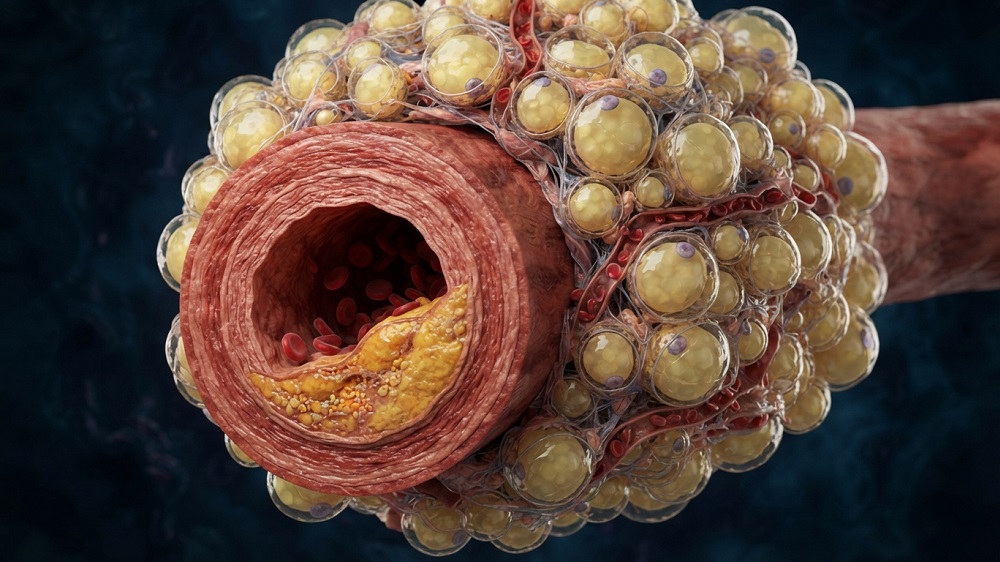
- With aging, asprosin is increasingly secreted by fat cells.
- Cells cultured in the presence of asprosin have their mitochondria damaged, leading to STING activation and senescence.
- Asprosin was found to accelerate vascular aging in mice.
- In a mouse model of accelerated senescence, mice that were given an anti-asprosin antibody or that did not express asprosin had reduced senescence effects.
A team of researchers has linked asprosin, which is produced by aging fat cells, to mitochondrial dysfunction and cellular senescence in the cells that line blood vessels.
Vulnerable vessels
Cellular senescence and vascular aging are closely connected; the senescence-associated secretory phenotype (SASP) is emitted into the bloodstream, where it is easily taken up by the endothelial cells that line the walls of blood vessels. Unfortunately, there are no interventions that are known to directly affect this process or shield these cells from the SASP.
Of particular interest to these researchers is the cGAS-STING pathway, which we have reported on extensively, as it has been implicated in several inflammatory and age-related diseases [1]. Mitochondrial DNA (mtDNA) is particularly vulnerable to negative changes with aging, leading to mitochondrial dysfunction; this work touches upon its relationship to STING, which has been confirmed in previous studies [2].
This study focuses on a particular molecule that these researchers have extensively worked on before: asprosin, a metabolic regulator that is secreted by fat cells [3]. These researchers had found that it damages epithelial cells in a mouse model of diabetic nepropathy [4] and that it is detrimental in many other disorders, including vascular inflammation [5]. Therefore, they conducted another study to determine the extent of its effects in this area and whether targeting it would affect mtDNA and STING.
Senescence-related effects
Unsurprisingly, asprosin was found to be significantly upregulated with senescence in human endothelial cells and with aging in wild-type mice. The researchers then rapidly aged a population of mice using D-galactose, driving their cells to senescence, and then injected some of them with an anti-asprosin antibody (AASP). As expected, AASP ameliorated some of the negative effects, reducing the levels of various senescence biomarkers and inflammatory cytokines. Another population of mice was injected with asprosin; these mice had roughly the same levels of inflammatory and senescence biomarkers as the mice exposed to D-galactose.
Both D-galactose and asprosin had significant negative effects on vascular structure, including thickening blood vessel walls and increasing systolic blood pressure. Not every effect of D-galactose and asprosin was the same, however. Inducing senescence through D-galactose caused dysfunctions in vascular dilation that AASP did not change.
The researchers also generated a population of mice that were incapable of natively producing asprosin, then subjected them to D-galactose. Like the D-galactose + AASP group, the inflammatory and senescence biomarkers of this group were reduced compared to the mice that only received D-galactose. Senescence-associated vascular alterations, including thickening and collagen accumulation, were also significantly reduced in these mice compared to the D-galactose group.
An in vitro experiment using conditioned media from adipocytes explained these results. Adipocytes that could not produce asprosin yielded conditioned media that was much healthier for human endothelial cells, including blunting the senescence-inducing effects of hydrogen peroxide while improving both proliferation and migration in these cells.
When mitochondria fall apart
The researchers examined the effects of asprosin on mitochondria and STING. Treating cells with asprosin yielded similar effects to treating them with hydrogen peroxide, damaging mitochondrial membranes and causing mitochondrial fragmentation. Combining hydrogen perofixe with asprosin worsened the effect. Asprosin was found to cause mtDNA to leak into the cytosol, a known trigger of STING; knocking down STING in these cells prevented the resulting senescence.
This increase in mtDNA was connected to an increase in mitochondrial reactive oxygen species (mtROS). Treating cells with mitoTEMPO, which specifically scavenges mtROS, mitigated many of the effects of asprosin, reducing mtDNA leakage and senescence while restoring proliferation.
Asprosin is one potential fate of profibrillin-1, which is encoded by the gene FBN1, which itself increases in expression with senescence. However, the researchers note that although they have established a link between asprosin, mitochondrial dysfunction, and senescence, they have not established the full mechanistic relationship between the drivers of aging and asprosin’s increase. They also note that they are unsure if targeting asprosin would provide benefits beyond current vascular treatments and suggest that, if any such therapies are developed, they should be tested alongside current standard-of-care options.
Literature
[1] Oduro, P. K., Zheng, X., Wei, J., Yang, Y., Wang, Y., Zhang, H., … & Wang, Q. (2022). The cGAS–STING signaling in cardiovascular and metabolic diseases: future novel target option for pharmacotherapy. Acta Pharmaceutica Sinica B, 12(1), 50-75.
[2] Kim, J., Kim, H. S., & Chung, J. H. (2023). Molecular mechanisms of mitochondrial DNA release and activation of the cGAS-STING pathway. Experimental & molecular medicine, 55(3), 510-519.
[3] Farrag, M., Ait Eldjoudi, D., González-Rodríguez, M., Cordero-Barreal, A., Ruiz-Fernandez, C., Capuozzo, M., … & Gualillo, O. (2022). Asprosin in health and disease, a new glucose sensor with central and peripheral metabolic effects. Front Endocrinol (Lausanne) 13: 1101091.
[4] Huang, Q., Xiong, X., Chen, S., Wang, Y., Wang, L., Liu, W., … & Huang, Q. (2025). Asprosin aggravates tubular epithelial cell injury and phenotypic transformation via mitochondrial dynamics disorder mediated by excessive Drp1 SUMOylation in diabetic nephropathy mice. Advanced Science, 12(39), e03259.
[5] Huang, Q., Chen, S., Xiong, X., Yin, T., Zhang, Y., Zeng, G., & Huang, Q. (2023). Asprosin Exacerbates Endothelium Inflammation Induced by Hyperlipidemia Through Activating IKKβ-NF-κBp65 Pathway: Huang, Chen, Xiong, Yin, Zhang, Zeng, and Huang. Inflammation, 46(2), 623-638.
About the author

Josh Conway