
Researchers have established how a protein that is nearly absent in the literature, PTCHD4, is linked to cellular senescence.
Very little previous work
In most papers, after a customary discussion of the mechanics of cellular senescence and current methods of handling it and its repercussions, the researchers go on to discuss previous work relating to the particular approach at hand. However, that does not pertain to this paper; PTCHD4 is only known as being structurally related to PTCH1, which affects the Hedgehog signaling pathway [1]. While some work has linked dysregulation of Hedgehog pathways to accelerated neurodegeneration [2], the specific role of PTCHD4 was not clear.
The researchers found that, when exposed to the genotoxin bleomycin, PTCHD4 increases alongside inflammation markers in a variety of cells, including human fetal lung diploid fibroblasts, mouse embryonic fibroblasts (MEFs), alveolar epithelial type II cells (AEC2s), human umbilical vein endothelial cells (HUVECs) and adult retinal pigment epithelial cells. Aging Atlas data confirmed this upregulation in other types of cells as well, and NCBI data listed this molecule as being disease-associated: its presence is negligible under ordinary conditions.
The team then examined young mice, old mice, and progeric mice, staining for PTCHD4 alongside the senescence biomarker p16. The two biomarkers were expressed in similar quantities among each group in multiple tissues. Similar results were found in human tissue samples; idiopathic pulmonary fibrosis (IPF) patients expressed more PTCHD4 in their lungs than people without the disease, and human lung cells expressing PTCHD4 were more likely to also express established senescence markers.
Significant effects on senescence
The next experiment involved creating MEFs that do not express PTCHD4 and then driving them senescent through replication. PTCHD4 was found to be a critical driver of senescence in this group; after an average of eight passages, ordinary MEFs exhibited signs of senescence, such as the key biomarker SA-β-gal, while this did not occur to the PTCHD4-deficient MEFs until an average of passage 14.
This line of experimentation continued with PTCHD4-overexpressing MEFs, which began exhibiting senescence-related features such as SA-β-gal early in passage 5. Similar results were found in AEC2s; the inflammatory biomarkers IL6 and IL8, along with SA-β-gal, were upregulated alongside PTCHD4 overexpression.
This testing continued with live mice. The researchers utilized mice that do not express PTCHD4 along with wild-type mice, and they exposed both groups to D-galactose, which mimics many of the symptoms of natural aging. The mice that did not express PTCHD4 were much more resistant to D-galactose than wild-type mice, and there were no detectable physical or biochemical problems caused by this absence.
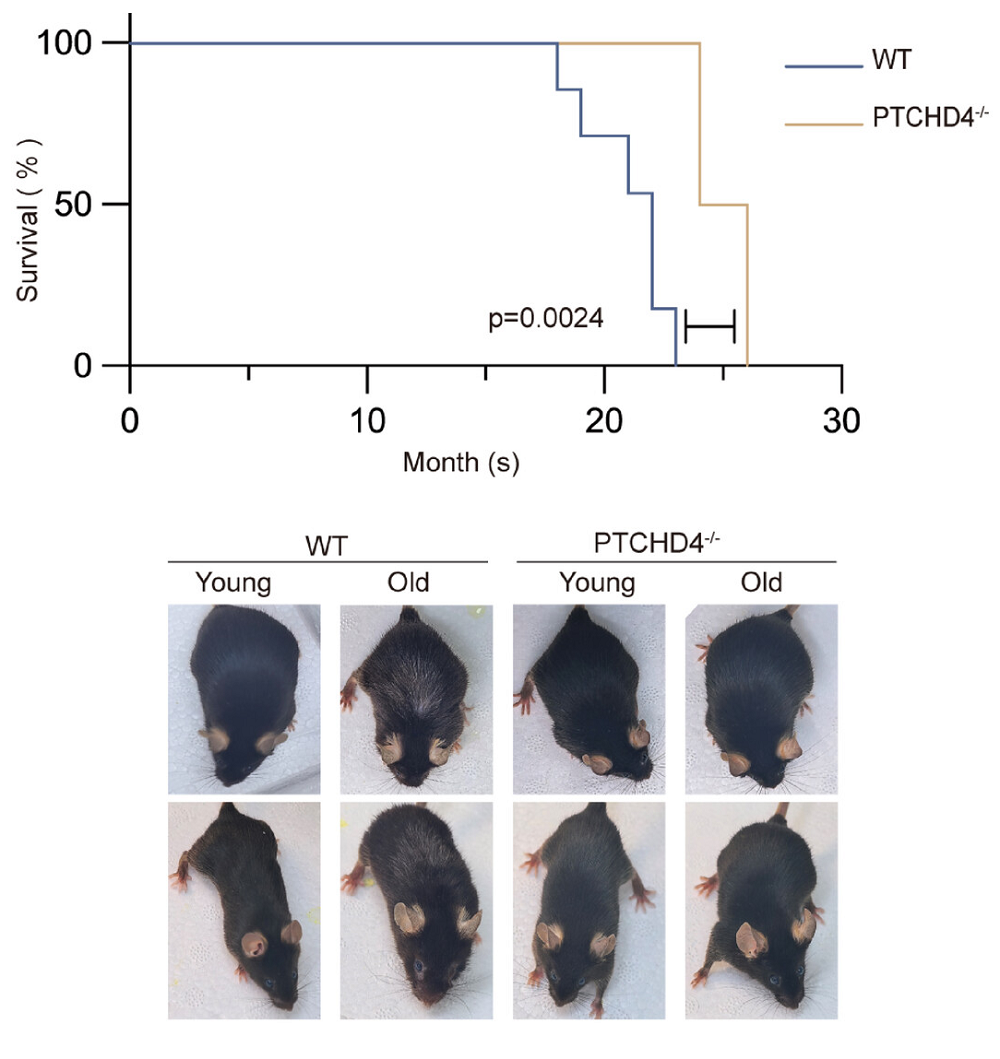
The team then continued with naturally aging animals. They found that mice that do not express PTCHD4 naturally live months longer than wild-type mice, and mice without it apparenly do not develop white hair the way that wild-type animals do.
A further experiment involved subjecting mice to bleomycin, which harms lung function in a way that mimics IPF in people. As expected, the mice exhibited features of lung fibrosis within 21 days. However, the PTCHD4-less mice suffered from fewer symptoms of the disease, retaining more lung capacity, less immune infiltration, less destruction of alveolar tissues, and less fibrosis than wild-type mice.
A well-known signaling pathway
In this study, PTCHD4 was found to have no effects on the Hedgehog pathway or related genes. Instead, its effects were found to be due to AKT signaling. AKT activation normally increases with aging, but a lack of PTCHD4 diminished this. The team created MEFs that do not express PTCHD4 but express AKT through a different construct, and these MEFs were not protected by their lack of PTCHD4. Similar experiments involving the direct expression of AKT in other cell types confirmed these results, recapitulating the effects of PTCHD4.
The researchers believe that “PTCHD4 may represent a candidate target for senescence-associated interventions” and that there is “a potential role for PTCHD4 in age-related fibrotic disease.” However, this is an initial study. Further work will need to be done to confirm these results, elucidate the relationship of PTCHD4 and AKT, ascertain any side effects of eliminating or suppressing PTCHD4, and determine if PTCHD4 can be targeted by interventions.
Literature
[1] Harvey, M. C., Fleet, A., Okolowsky, N., & Hamel, P. A. (2014). Distinct effects of the mesenchymal dysplasia gene variant of murine Patched-1 protein on canonical and non-canonical Hedgehog signaling pathways. Journal of Biological Chemistry, 289(15), 10939-10949.
[2] Esmaeli, M., Dehabadi, M. D., Ghanbari, A., & Yancheshmeh, F. S. A. (2025). The role of Sonic Hedgehog (SHH) in the formation of motor neurons and neurodegenerative diseases. Discover Medicine, 2(1), 231.