The Blog

Building a Future Free of Age-Related Disease

Supplements

What is Arginine? A Summary of L-Arginine

Arginine is a popular sports supplement, but could it also be useful in the context of healthy longevity?

What is arginine?

Arginine, also known as L-arginine, is an amino acid, a building block used to make proteins, that plays an important role in regulating blood flow. Arginine was first isolated from the extract of lupin seedlings by the German chemist Ernst Schultze in 1886.

Arginine is classed as a semi-essential amino acid, as it is normally needed during child development, but deficiency is rare in adults. Preterm infants are unable to produce arginine themselves, so for them, it is essential; however, it is nonessential for healthy adults, who can produce it internally.

Arginine-rich foods

Turkey is a good source of protein, and one cooked breast can have as much as 16 grams of arginine. Turkey also contains B vitamins and omega-3 fatty acids.

Pork loin is another good source of protein, with an arginine content of up to 14 grams per rib. It is also one of the less fatty cuts of pork.

Chicken is also a good source of arginine. One breast can have up to 9 grams of arginine and as much as 70% of the recommended amount of daily protein.

Pumpkin seeds are also a great source of arginine, with a cup yielding up to 7 grams. These seeds are also a good source of iron and zinc. Another option is soybeans, which can have up to 4.6 grams of arginine per cup. They are also a good way to get potassium and magnesium.

Another good way to get arginine is from peanuts, which can contain up to 4.6 grams per cup. They also contain niacin, folate, and vitamin E.

Spirulina is a kind of blue-green algae harvested from the ocean. A cup of spirulina can have up to 4.6 grams of arginine, though the typical tablespoon amount used in smoothies would equate to 0.28 grams. The algae is also an excellent source of calcium, iron, potassium, and niacin.

Chickpeas are a source of arginine, and a cup of cooked chickpeas can contain up to 1.3 grams. Chickpeas also have up to 14.5 grams of protein and as much as 12.5 grams of dietary fiber, making them ideal for people who do not eat meat but want to get enough arginine, protein, and fiber.

Finally, lentils are another source of arginine, yielding up to 1.3 grams per cup, and can provide up to 63 percent of recommended dietary fiber.

What does arginine do?

Arginine is one of three compounds that create creatine, a molecule involved in energy metabolism and produces adenosine triphosphate (ATP), the molecule that provides the energy used by our cells.

In both humans and mammals, approximately 80% of the nitrogen excreted is in the form of urea, which is produced via a series of reactions occurring in the liver. These reactions are collectively called the urea cycle or the Krebs-Henseleit cycle.

Arginine is an intermediary in the urea cycle with L-ornithine, L-citrulline, and argininosuccinate [1]. Ammonia is produced as part of nitrogen metabolism and is toxic, so it is removed from our bodies. The urea cycle converts ammonia into urea in the liver; it then enters the bloodstream, is filtered by the kidneys, and then is excreted as urine. Arginine plays an essential role in this chain of events, facilitating the removal of ammonia from the body.

Arginine is popular with sportsmen and women

Arginine is also a popular sports supplement used by athletes, as it directly produces nitric oxide via the nitric oxide synthase enzymes. Nitric oxide dilates arteries and blood vessels, increasing blood flow, and is a reason why athletes often take arginine supplements.

However, this effect seems to be inconsistent; while there have indeed been studies that have shown an increase of nitric oxide, other studies have shown no effect whatsoever [2-3]. This inconsistency may result from the poor absorption of arginine by the intestines. L-citrulline is a possible alternative that has superior absorption and increases levels of arginine at a higher level and a longer duration than L-arginine supplementation.

However, despite the unreliable results, arginine persists as a favorite for sports and athletics enthusiasts.

Potential arginine benefits

Arginine helps to produce the byproduct agmatine, a signaling molecule shown to regulate multiple molecular targets, including neurotransmitters and ion channels. Agmatine also plays a role in nitric oxide (NO) synthesis in combination with ornithine and argininosuccinate, which widens and relaxes the blood vessels, improving blood flow.

In a research study, a combination of arginine and citrulline in patients with hypertension reduced systolic and diastolic blood pressure in both ankle and brachial measurements [4]. Another study looked at patients with peripheral artery disease, and it showed that an infusion of arginine was able to double blood flow to extremities [5].

Impressively, it performed on par with prostaglandin E1, a commercial drug designed to induce vasodilation (widening of the blood vessels). A significant increase of nitric oxide was also noted during the study. In a follow-up study, these benefits were observed with oral supplementation of arginine as well [6].

There is some suggestion that nitric oxide signaling might also have a role in aging, mainly due to inflammation. Inflammation increases levels of inducible nitric oxide synthase (iNOS), resulting in increased oxidative byproducts, such as peroxynitrite, which causes neurotoxicity [7]. However, more research is needed in this area before we have a complete picture of its role in aging, but it is likely a downstream effect of the primary aging processes.

Arginine side effects

Taking arginine as a supplement can have possible side effects, including abdominal pain or bloating, diarrhea, and gout. It may also make breathing in people with asthma more difficult. Arginine can also potentially interact with medications that lower blood pressure. As always when taking supplements, cease taking them immediately and consult your doctor should you feel any adverse effects.

Disclaimer

This article is only a very brief summary, is not intended as an exhaustive guide, and is based on the interpretation of research data, which is speculative by nature. This article is not a substitute for consulting your physician about which supplements may or may not be right for you. We do not endorse supplement use or any product or supplement vendor, and all discussion here is for scientific interest.

We would like to ask you a small favor. We are a non-profit foundation, and unlike some other organizations, we have no shareholders and no products to sell you. All our news and educational content is free for everyone to read, but it does mean that we rely on the help of people like you. Every contribution, no matter if it’s big or small, supports independent journalism and sustains our future.

Literature

[1] Guoyao, W. U., & Morris, S. M. (1998). Arginine metabolism: nitric oxide and beyond. Biochemical Journal, 336(1), 1-17. [2] Liu, T. H., Wu, C. L., Chiang, C. W., Lo, Y. W., Tseng, H. F., & Chang, C. K. (2009). No effect of short-term arginine supplementation on nitric oxide production, metabolism and performance in intermittent exercise in athletes. The Journal of nutritional biochemistry, 20(6), 462-468. [3] Bailey, S. J., Winyard, P. G., Vanhatalo, A., Blackwell, J. R., DiMenna, F. J., Wilkerson, D. P., & Jones, A. M. (2010). Acute L-arginine supplementation reduces the O2 cost of moderate-intensity exercise and enhances high-intensity exercise tolerance. Journal of Applied Physiology, 109(5), 1394-1403. [4] Figueroa, A., Sanchez-Gonzalez, M. A., Wong, A., & Arjmandi, B. H. (2012). Watermelon extract supplementation reduces ankle blood pressure and carotid augmentation index in obese adults with prehypertension or hypertension. American journal of hypertension, 25(6), 640-643. [5] Bode-Böger, S. M., Böger, R. H., Alfke, H., Heinzel, D., Tsikas, D., Creutzig, A., … & Frölich, J. C. (1996). L-Arginine induces nitric oxide–dependent vasodilation in patients with critical limb ischemia. Circulation, 93(1), 85-90. [6] Böger, R. H., Bode-Böger, S. M., Thiele, W., Creutzig, A., Alexander, K., & Frölich, J. C. (1998). Restoring vascular nitric oxide formation by L-arginine improves the symptoms of intermittent claudication in patients with peripheral arterial occlusive disease. Journal of the American College of Cardiology, 32(5), 1336-1344. [7] Calabrese, V., Mancuso, C., Calvani, M., Rizzarelli, E., Butterfield, D. A., & Stella, A. M. G. (2007). Nitric oxide in the central nervous system: neuroprotection versus neurotoxicity. Nature Reviews Neuroscience, 8(10), 766-775.
Saunas can activate heat shock proteins and may be a potential way to improve health and potentially, longevity.

Saunas for a Personal Health and Longevity Strategy

There are a few reasons to consider including sauna use as part of your personal health and longevity strategy.
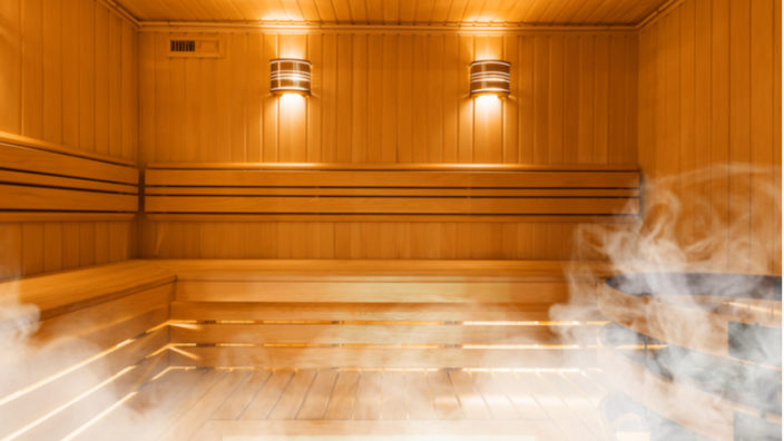
What is a sauna?

The word sauna comes from the Finnish language, and it means an earth or snow pit. While Finnish saunas today are typically log-walled, wooden paneled, or even tiled structures, these modern forms evolved from the more ancient pits. Traditionally, a sauna has wooden benches, often made from spruce, aspen, or other types of wood common to the region.

While saunas can be found around the world, a significant amount of health research on sauna use has been done in Finland due to their popularity there. Sauna design does vary, as does the heat source, humidity, and typical usage time.

Sauna use can be described as a short-duration passive exposure to high levels of heat. The result of this exposure is essentially a mild form of hyperthermia, in which the core temperature of the body rises, triggering a thermoregulatory response that attempts to restore homeostasis (balance) to the body. This process also acclimates the body to better deal with future events involving heated environments.

How are saunas heated?

The historical heat source for saunas was wood, something that continues to this day in rural areas of Finland and other countries. However, modern saunas are normally heated using electric or infrared heaters.

An electric heater will typically warm the air to between 70 – 100°C, or 158°F – 212°F. The heat then transfers from the warm air into the body.

In the case of infrared heaters, these give out thermal radiation, which not only heats the body directly but also warms the surrounding air. Infrared saunas are cooler than their electric and wood-burning counterparts, generally reaching 45°C to 60°C (113°F to 140°F).

There are two kinds of infrared sauna heaters – near- and far-infrared heaters. Near-infrared heaters use incandescent bulbs to generate thermal radiation, and they mostly produce near infrared and a little middle-wavelength infrared. Far-infrared heaters employ metallic or ceramic heating elements that mostly produce energy in the far-infrared range, which is closer to that produced by natural sunlight.

There are essentially two kinds of saunas: wet and dry. A wet sauna is really a steam sauna; it has very high humidity, often exceeding 50%, which prevents sweat from evaporating. This is why a wet sauna can often feel hotter than a dry sauna. The heart also has to work harder in a wet sauna because of the absence of evaporative cooling provided by sweat.

Dry saunas have a fairly low humidity in the 10 to 20% range, though in saunas that include heater rocks, water can be poured on them to increase that humidity somewhat.

How long do you use a sauna for?

The traditional Finnish method is to have between 1-3 individual sessions in the sauna, which typically last about 20 minutes each. These sessions are divided by a cooling period with the aim of triggering the thermoregulatory response.

This cooling period can take some well-known extreme forms, such as immediately leaving the sauna to roll around in the snow or drenching with cold water. A comparison might also be made with the ancient Romans and their practice of taking heated baths and then diving into a cold plunge pool afterwards, again aiming to activate the thermoregulatory response.

This rapid cooling taxes the cardiovascular system more so than regular cooling, forcing the heart to work harder [1]. However, doing so holds little risk of a heart attack or similar cardiac event in a healthy person.

A 2015 study discovered that there was a dose-dependent reduction of cardiovascular associated mortality, all-cause mortality, and occurrence of Alzheimer’s disease associated with sauna use [2]. This specifically related to saunas of at least 78.9°C (174°F) that were used for 20 minutes or longer.

What happens when our bodies are exposed to heat stress?

High temperature causes stress to the body, which triggers a thermoregulatory response that seeks to restore homeostasis. For example, during warm weather or in a hot environment, the hypothalamus reacts to the increased heat and sends signals to the blood vessels, instructing them to dilate.

This allows the movement of warm blood, salts, and other fluids to the skin. The blood is cooled, and the other fluids evaporate. In fact, between 50-70% of blood flow is rerouted away from the body core to the skin surface to optimize cooling via sweating.

How hard the heart works also increases as a response to heat stress. When exposed to high temperatures, cardiac output can increase by 60% or more to supply the body with oxygen. The heart rate also increases, though the amount of blood pumped with each stroke remains the same.

Finally, during severe exposure to heat, there is an increase in total plasma volume to compensate for the decreased volume of blood in the body core. The increased plasma volume acts as a backup source of fluid for use in sweat production. It also helps the body to cool and keeps the core temperature from increasing too quickly, which aids heat acclimation.

Regular sauna use triggers hormesis

Regular sauna use allows the body to acclimate to the heat and helps the body optimize its thermoregulatory response. This results in the body being better prepared for handling heat stress in the future and is likely due to hormesis.

Hormesis is a biological phenomenon in which a harmful stress-causing stimulus conveys a beneficial effect when given in small doses. Various sources of stimuli, such as exercise, exposure to low doses of toxic substances, dietary modifications, and environmental stressors such as heat or cold, can trigger hormesis.

The body responds to hormetic stressors such as heat with a compensatory defense response. Hormesis causes a wide range of protective systems to kick in, which boosts cell repair and starts the development of acclimation. This is in anticipation of future stress from the same source and may even help against an increased level of the same stressor.

Essentially, hormesis causes our cells to enter a defensive mode, a bit like how a spaceship in a sci-fi movie might raise its shields. Each time the cells encounter the same source of stress, such as heat in the case of sauna use, it gets better at protecting itself and is more resilient. Some researchers in our field are studying the effects of these hormetic stress responses and how they may influence aging and longevity.

This is also why sauna use is often recommended to people who cannot exercise due to a disability or have a medical condition that prevents it. Indeed, sauna use is similar to exercise in how it produces some of the same physiological responses [3].

Is it OK to sauna everyday?

Sauna use may lower blood pressure, so people with low blood pressure should talk to their doctor prior to using one. People who have recently had a heart attack should also consult their doctor first. That being said, if you are healthy with no underlying medical issues, it is generally considered safe to use a sauna every day.

The heat shock response protects our cells

Heat stress responses include the activation of heat shock proteins, the production of various transcription factors, and the production of both inflammatory and anti-inflammatory factors such as certain interleukins.

The heat shock proteins

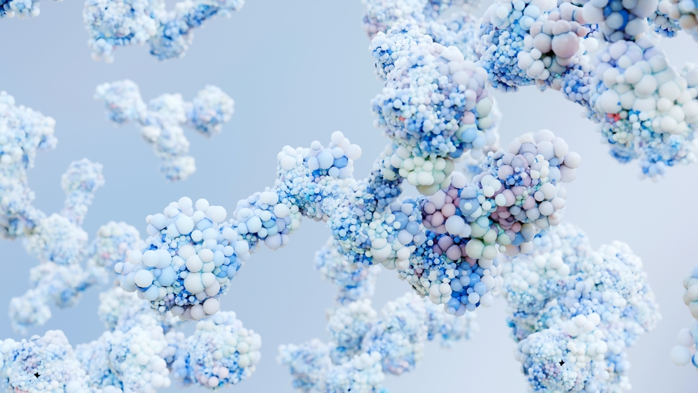
Heat shock proteins are a family of proteins that are produced by our cells as a response to stressful conditions, such as excessive heat. They are important in a number of cellular processes, such as regulation of the cell cycle, cellular signaling, and functioning of the immune system.

Healthy cells always have a standing or basal level of heat shock proteins to support the day-to-day operation of protein production, export, and regulation. Unfortunately, even during healthy functioning, cells produce harmful waste and byproducts. Chief among them are the reactive oxygen and nitrogen species produced by the mitochondria as a byproduct of energy metabolism [4].

These harmful species then bounce around the inside of the cell and can damage proteins and alter their structure as well as damaging mitochondrial DNA [5]. This damage to the mitochondrial DNA is believed to be one of the nine reasons we age.

The presence of large amounts of misfolded proteins is also a feature of neurodegenerative diseases. These damaged proteins can aggregate together to form clumps and are associated with Alzheimer’s and Parkinson’s disease [6]. Misfolded proteins are also believed to be another reason we age.

Increasing our heat shock proteins may potentially protect us from neurodegenerative diseases. Animal studies suggest that additional heat shock proteins help to repair damaged proteins and may protect us from diseases such as Alzheimer’s and Parkinson’s [7].

The heat shock proteins in action

When our cells are exposed to environmental stressors, it can cause new proteins to misfold or existing ones to unfold, which impairs their function. During exposure to extreme heat and other sources of stress, our cells increase the production of heat shock proteins in an attempt to repair the damaged proteins. This damage control process is known as the heat shock response and heat shock proteins are the first line of defence.

Multiple studies have shown that heat shock proteins increase in response to heat exposure in people as they do in animals. A 2012 study showed that people who stayed in a heat chamber at 73°C (163°F) for thirty minutes saw a 49% increase of heat shock protein HSP72 levels [8].

A 2018 study using deep tissue heat therapy over a period of six days also appeared to increase the levels of heat shock proteins [9]. The researchers found that the levels of the heat shock proteins HSP70 and HSP90 rose by 45 and 38%, respectively.

They also noted that mitochondrial function also improved around 28%, and they observed an increase in mitochondrial biogenesis, a process by which cells increase mitochondrial mass and can produce more energy.

The heat shock proteins play an important role in responding to stressors such as high temperatures and are a key part of the defences our cells use. Regular sauna use is going to activate the heat shock proteins more often making our cells more robust and resistant to damage and stress.

The interleukins

In transient and tightly controlled amounts, inflammation can help to facilitate wound healing and rouse the immune system to attack invading pathogens. However, chronic or uncontrolled inflammation is a known contributing factor in many age-related diseases.

To remain healthy, it is essential that the body maintains a balance between inflammatory and anti-inflammatory factors. Too much inflammation, and cellular dysfunction and age-related diseases can occur; too little inflammation, and healing processes and immune responses are impaired. Therefore, balance is the key for the body to effectively trigger inflammatory responses, regulate them appropriately, and to resolve them when no longer required.

Interleukins are a group of naturally occurring proteins that were first discovered being expressed by white blood cells. There are over fifty interleukins in the human genome, and they can be divided into four major groups based on their structural features [10].

The immune system relies greatly on the presence and activity of interleukins, and deficiencies in some of them are associated with autoimmune diseases or immune deficiency. Most interleukins are produced by immune cells, chiefly T helper cells (helper CD4 T lymphocytes), though monocytes, macrophages and endothelial cells also produce them.

Interleukins also encourage the development and differentiation, a process by which a more generic cell changes to become specialized in form and function, of T and B lymphocytes and hematopoietic cells.

While there are many interleukins, we are concerned with interleukin 6 (IL-6) and interleukin 10 (IL-10) in the context of sauna use and heat stress.

Interleukin-6 and Interleukin-10

Sauna use increases the expression of IL-6, a pro-inflammatory cytokine that has a key role in regulating immunological functions as well as helping the body to maintain homeostasis. It has a critical role in the final differentiation of B cells maturing into immunoglobulin-secreting cells as well as in supporting nerve cell differentiation.

The presence of IL-6 is greatly increased by muscles during exercise, is pro-inflammatory, and appears to be required for the insulin-sensitizing benefits of exercise

However, while IL-6 itself is pro-inflammatory in nature, it also serves to dampen the inflammatory response by triggering IL-10, which is a powerful anti-inflammatory cytokine [11]. IL-10 plays an important role in limiting the host immune response to pathogens. This limiting function prevents damage to the host due to excessive immune responses and helps to maintain homeostasis.

FOXO3

The gene FOXO3 (Forkhead Box O3), which encodes the transcription factor FOXO3, is one of the genes associated with longevity in humans [12]. FOXO3 proteins regulate a wide range of genes that protect us against damage to DNA, proteins, and lipids along with genes that aid efficient stem cell function [13].

FOXO3 increases the expression of genes that facilitate tumor suppression to help keep us safe from cancer [14]. It also plays a role in healthy immune function [15] and is a key regulator or cellular quality control, regulating both proteostasis and autophagy [16]. FOXO3 is also a regulator of the controlled cell death known as apoptosis.

After heat stress is experienced, FOXO3 proteins make a complex with sirtuin 1 (SIRT1), an enzyme associated with longevity due to its influence with various pathways associated with aging. The sirtuins have long been referred to as “longevity genes”, and their proteins regulate cellular health via controlling the release of insulin, ensuring that lipids are working correctly, responding to stressful stimuli, and modulating lifespan in multiple species.

The presence of SIRT1 boosts FOXO3’s ability to resist oxidative stress and shifts its focus from apoptosis to resisting stress instead. This means when it combines with SIRT1 as a complex, it helps our cells to better resist heat stress and become more resilient [17].

Nrf2

NF-E2–related factor 2 (Nrf2) is an essential transcription factor that regulates the expression of more than 1000 genes in the cell, both during stressful conditions and normal operation. Nrf2 is typically triggered by oxidative stress, electrophilic stress, or chronic inflammation.

Once activated, it travels from the cytoplasm to the cell nucleus, where it regulates an array of genes with antioxidant, anti-inflammatory, and cytoprotective properties. Heat stress from a sauna is enough to activate Nrf2, which then increases the production of the heat shock protein, heme oxygenase-1 (HO-1).

This HO-1 then goes on to break down heme, a pro-oxidant, into carbon monoxide and bilirubin, which are anti-inflammatory and antioxidant, respectively [18]. The increased presence of HO-1 also suppresses the expression of a number of inflammatory molecules associated with cardiovascular disease [19].

Health benefits of using a sauna

There are multiple potential health benefits associated with regular sauna use.

Cardiovascular health

Exposure to heat triggers a number of protective responses; some of these may help protect the cardiovascular system and protect us from heart disease. Exercise is an excellent way to support a healthy cardiovascular system, and again, sauna use appears to trigger many of the same responses that exercise does.

Like exercise, sauna use can increase heart rate to around 100 beats per minute in moderate heat or at higher temperatures to as much as 180 beats per minute, which is similar to what is seen during moderate to more vigorous exercise [20-21].

Research suggests that regular sauna use may be useful in improving blood pressure and circulation while reducing inflammation. Sauna use may be particularly useful for people who cannot engage in regular exercise perhaps due to injury, frailty, or another medical condition. As always, if in doubt, consult your doctor before using a sauna.

Cardiovascular disease

The World Health Organization (WHO) suggests that around 18 million people a year die as the result of cardiovascular disease. This makes it the number one killer worldwide, and sauna use combined with lifestyle changes may help to reduce your risk.

A Finnish study compared people who used the sauna only once a week, people who used it 2-3 times a week, and people who used it 4-7 times a week. The moderate 2-3 per week people were 22% less likely to have a heart attack, and the frequent users at 4-7 times a week were an impressive 63% less at risk [2]. The frequent sauna users also saw a 37% reduction in premature death.
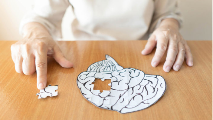
Cognitive Decline

Healthy brain function requires proper blood flow to deliver oxygen and nutrients to the brain. Therefore, it is no surprise that during aging, the progression of cardiovascular disease is often accompanied by cognitive decline and even neurodegenerative diseases.

High blood pressure can damage brain structure and reduce the blood flow to the brain, causing loss of cognition. This reduction in blood flow may also impair the ability of our brain to remove accumulated misfolded proteins and could also speed up the onset and progression of Alzheimer’s, Parkinson’s, and other dementias.

The Finnish study showed that men who used a sauna 4-7 times a week saw a 65% reduced risk of developing Alzheimer’s disease compared to people who use a sauna only once a week [2].

Conclusion

There are many reasons to consider using a sauna regularly, and while we have focused mostly on the cardiovascular and cognitive benefits, there are many other studies that suggest plenty of other benefits.

Regular heat stress via a sauna to activate the heat shock proteins and other hormetic response could be a useful way to increase your resilience and condition your cells to become more robust in the face of oxidative stress and other stressors that drive aging. Regular use of a sauna, therefore, may offer a viable approach to delaying aging.

We would like to ask you a small favor. We are a non-profit foundation, and unlike some other organizations, we have no shareholders and no products to sell you. All our news and educational content is free for everyone to read, but it does mean that we rely on the help of people like you. Every contribution, no matter if it’s big or small, supports independent journalism and sustains our future.

Literature

[1] Pilch, W., Szygula, Z., Palka, T., Pilch, P., Cison, T., Wiecha, S., & Tota, L. (2014). Comparison of physiological reactions and physiological strain in healthy men under heat stress in dry and steam heat saunas. Biology of sport, 31(2), 145–149.

[2] Laukkanen, T., Khan, H., Zaccardi, F., & Laukkanen, J. A. (2015). Association between sauna bathing and fatal cardiovascular and all-cause mortality events. JAMA internal medicine, 175(4), 542-548.

[3] McCarty, M. F., Barroso-Aranda, J., & Contreras, F. (2009). Regular thermal therapy may promote insulin sensitivity while boosting expression of endothelial nitric oxide synthase–effects comparable to those of exercise training. Medical hypotheses, 73(1), 103-105.

[4] Lacza, Z., Kozlov, A. V., Pankotai, E., Csordás, A., Wolf, G., Redl, H., Kollai, M., Szabó, C., Busija, D. W., & Horn, T. F. (2006). Mitochondria produce reactive nitrogen species via an arginine-independent pathway. Free radical research, 40(4), 369–378.

[5] Squier, T. C. (2001). Oxidative stress and protein aggregation during biological aging. Experimental gerontology, 36(9), 1539-1550.

[6] Soto, C., & Estrada, L. D. (2008). Protein misfolding and neurodegeneration. Archives of neurology, 65(2), 184-189.

[7] Leak, R. K. (2014). Heat shock proteins in neurodegenerative disorders and aging. Journal of cell communication and signaling, 8(4), 293-310.

[8] Iguchi, M., Littmann, A. E., Chang, S. H., Wester, L. A., Knipper, J. S., & Shields, R. K. (2012). Heat stress and cardiovascular, hormonal, and heat shock proteins in humans. Journal of athletic training, 47(2), 184–190.

[9] Hafen, P. S., Preece, C. N., Sorensen, J. R., Hancock, C. R., & Hyldahl, R. D. (2018). Repeated exposure to heat stress induces mitochondrial adaptation in human skeletal muscle. Journal of applied physiology, 125(5), 1447-1455.

[10] Brocker, C., Thompson, D., Matsumoto, A., Nebert, D. W., & Vasiliou, V. (2010). Evolutionary divergence and functions of the human interleukin (IL) gene family. Human genomics, 5(1), 1-26.

[11] Ahmed, S. T., & Ivashkiv, L. B. (2000). Inhibition of IL-6 and IL-10 signaling and Stat activation by inflammatory and stress pathways. The Journal of Immunology, 165(9), 5227-5237.

[12] Morris, B. J., Willcox, D. C., Donlon, T. A., & Willcox, B. J. (2015). FOXO3: a major gene for human longevity-a mini-review. Gerontology, 61(6), 515-525.

[13] Murtaza, G., Khan, A. K., Rashid, R., Muneer, S., Hasan, S. M. F., & Chen, J. (2017). FOXO transcriptional factors and long-term living. Oxidative medicine and cellular longevity, 2017.

[14] Renault, V. M., Thekkat, P. U., Hoang, K. L., White, J. L., Brady, C. A., Broz, D. K., … & Brunet, A. (2011). The pro-longevity gene FoxO3 is a direct target of the p53 tumor suppressor. Oncogene, 30(29), 3207-3221.

[15] Peng, S. L. (2008). Foxo in the immune system. Oncogene, 27(16), 2337-2344.

[16] Webb, A. E., & Brunet, A. (2014). FOXO transcription factors: key regulators of cellular quality control. Trends in biochemical sciences, 39(4), 159-169.

[17] Brunet, A., Sweeney, L. B., Sturgill, J. F., Chua, K. F., Greer, P. L., Lin, Y., … & Greenberg, M. E. (2004). Stress-dependent regulation of FOXO transcription factors by the SIRT1 deacetylase. science, 303(5666), 2011-2015.

[18] Yet, S. F., Melo, L. G., Layne, M. D., & Perrella, M. A. (2002). Heme oxygenase 1 in regulation of inflammation and oxidative damage. Methods in enzymology, 353, 163-176.

[19] Lin, C. C., & Yang, W. C. (2009). Prognostic factors influencing the patency of hemodialysis vascular access: literature review and novel therapeutic modality by far infrared therapy. Journal of the Chinese Medical Association, 72(3), 109-116.

[20] Taggart, P., Parkinson, P., & Carruthers, M. (1972). Cardiac responses to thermal, physical, and emotional stress. Br Med J, 3(5818), 71-76.

[21] Kukkonen-Harjula, K., Oja, P., Laustiola, K., Vuori, I., Jolkkonen, J., Siitonen, S., & Vapaatalo, H. (1989). Haemodynamic and hormonal responses to heat exposure in a Finnish sauna bath. European journal of applied physiology and occupational physiology, 58(5), 543-550.

Elderly Person Coughing

Teasing Apart the Effects of Aging and COVID-19

Researchers have used a big data approach to disentangle cellular and physiological changes that are specific to COVID-19 from those caused by aging [1]. Identifying these improves our understanding of the disease and may help explain why COVID-19 has a more severe effect on older people.

A tangled skein

The enormous impact of COVID-19 around the world has led to a massive research effort to characterize the disease, including how our immune system responds to the challenge it poses. Many studies have investigated the differences in the immune cell population of COVID-19 patients and healthy individuals, but they did not distinguish between changes caused by COVID-19 and age-related changes.

There is good reason to expect significant age-related immune changes to color these analyses. COVID-19 has disproportionately affected older people. People over 65 have the highest fatality rate and make up nearly 80% of COVID-19 deaths in the U.S., and COVID-19 infections are significantly more likely to become severe in older people. The disproportionate representation of the elderly among COVID-19 patients means that analyses of the changes in patients might capture not only its effect on the immune system but also age-related immune changes.

Separating threads

To distinguish age-related changes from COVID-19-driven changes, an international team of researchers analyzed data from healthy individuals and people with respiratory diseases, including COVID-19. The data included clinical blood tests, immune cell populations, and plasma proteomic profiles. The inclusion of patients with other respiratory conditions enabled the team to identify changes specific to COVID-19 (rather than pulmonary distress more generally), and comparing people of different age groups with and without COVID-19 distinguished between the COVID-driven changes and changes caused by aging.

The team found an increase in the proportion of B cells in patients with COVID-19 and other respiratory illnesses. Changes in certain T cell populations were specific to COVID-19 patients, and a unique population of T cells was seen in patients with moderate COVID-19. A proteomics analysis showed that the pathways that were the most upregulated in COVID-19 patients – matrisome proteins and extracellular matrix proteins – were, in fact, strongly upregulated with age. Without this analysis, the changes in these pathways would be considered a consequence of the disease, clouding our understanding of its effects.

Further analysis identified pathways that are specifically upregulated in COVID-19 patients independently of age. These include interferon response, interleukins, complement, and lysosome proteins. These pathways merit further investigation for potential biomarkers or perhaps even interventions aimed at COVID-19.

We examine the cellular and soluble determinants of coronavirus disease 2019 (COVID-19) relative to aging by performing mass cytometry in parallel with clinical blood testing and plasma proteomic profiling of ~4,700 proteins from 71 individuals with pulmonary disease and 148 healthy donors (25–80 years old). Distinct cell populations were associated with age (GZMK+CD8+ T cells and CD25low CD4+ T cells) and with COVID-19 (TBET-EOMES- CD4+ T cells, HLA-DR+CD38+ CD8+ T cells and CD27+CD38+ B cells). A unique population of TBET+EOMES+ CD4+ T cells was associated with individuals with COVID-19 who experienced moderate, rather than severe or lethal, disease. Disease severity correlated with blood creatinine and urea nitrogen levels. Proteomics revealed a major impact of age on the disease-associated plasma signatures and highlighted the divergent contribution of hepatocyte and muscle secretomes to COVID-19 plasma proteins. Aging plasma was enriched in matrisome proteins and heart/aorta smooth muscle cell-specific proteins. These findings reveal age-specific and disease-specific changes associated with COVID-19, and potential soluble mediators of the physiological impact of COVID-19.

Conclusion

This study isn’t about longevity in any way, but it demonstrates the importance of geroscience in general. Age is not only a risk factor for many conditions but also a confounding factor of many diseases. This is strikingly clear in the case of COVID-19, making it critical to separate how age and COVID-19 affect individuals and to understand how their interaction increases risk.

We would like to ask you a small favor. We are a non-profit foundation, and unlike some other organizations, we have no shareholders and no products to sell you. All our news and educational content is free for everyone to read, but it does mean that we rely on the help of people like you. Every contribution, no matter if it’s big or small, supports independent journalism and sustains our future.

Literature

[1] Arthur, L., et al. Cellular and plasma proteomic determinants of COVID-19 and non-COVID-19 pulmonary diseases relative to healthy aging. Nature Aging (2021), doi: 10.1038/s43587-021-00067-x

Glioblastoma

Senescent Cells, p65, and a Possible Treatment for Cancer

A report in the Journal of Cell Science has outlined different stages of cellular senescence alongside a mechanism that reverses it, along with a potential new treatment for glioblastoma.

Old paradigms in cellular senescence

Cellular senescence contributes to aging and age-related diseases, likely primarily through the inflammatory SASP. Senescence is often discussed in black-and-white terms: a cell is either senescent or it isn’t. It’s also widely accepted that once a cell enters a senescent state, its cell cycle arrest is permanent. However, more research is coming to light that paints a more complicated picture, including a recent study from the Homi Bhabha National Institute that challenges both of these assertions. [1]

On first consideration, the reversal of senescence may seem like a good thing. However, given the anti-cancer effects of senescence, the greater result of its reversal may be tumorigenesis rather than an anti-aging effect. In fact, previous research by the same lab has presented evidence of various cancer cells becoming senescent after treatment with anti-cancer therapies and then later resuming their proliferation.

Ciprofloxacin induces senescence in glioblastoma cells

Ciprofloxacin is an antibiotic that has also shown anti-tumor properties in some old, largely overlooked studies. However, these studies did not investigate a mechanism of action for these effects. Here, the researchers first showed it inhibited growth in four tumor cell lines in vitro primarily by inducing senescence. Ciprofloxacin caused an increase in SA ß-gal staining, SASP factors, reactive oxygen species (ROS), DNA damage, and senescence gene signatures via RNAseq. Many of these measures became positive in upwards of 80% or 90% of cells.

When ciprofloxacin was removed after 15 days, cells did not begin to proliferate, indicating that their senescence was not reversed. However, cells in which the treatment was removed after 5 or 10 days resumed proliferation. These cells were sorted by size (senescent cells typically show an enlarged morphology) to test whether senescent cells were returning to proliferation or if the proliferation observed was simply from the non-senescent cells within this treated population. The larger cells showed extensive SA ß-gal staining and, after a short delay, proliferated to equal numbers as the smaller, SA ß-gal negative cells.

p65 moves into the cell nucleus during senescence reversal

Whole-transcriptome analysis identified the p65 gene network as highly differential in ciprofloxacin-treated cells. Diving deeper, the researchers found that p65 was localized primarily in the cytoplasm of senescent cells and in the nucleus after removal of the treatment. Preliminary experiments suggested that SMAD7 may mediate this process. However, this did not occur in the cells that were treated for 15 days, which did not resume proliferation.

To test whether movement of p65 to the nucleus was involved in reversing senescence in the 5- or 10-day treated cells, the researchers also treated the cells with drugs that prevented this movement of p65. These cells showed increased levels of senescence, especially with respect to ROS-related measures. Rather than return to proliferation after ciprofloxacin removal, these cells began to die. When implanted into mice, cells treated for 10 days with ciprofloxacin (both the senescent and non-senescent subpopulations) formed tumors while cells treated for 15 days or for 10 days plus a p65 nuclear localization inhibitor did not.

Here, we demonstrate that ciprofloxacin-induced senescence in glioma-derived cell lines and primary glioma cultures is defined by SA-ß-gal positivity, a senescence-associated secretory phenotype (SASP), a giant cell (GC) phenotype, increased levels of reactive oxygen species (ROS), ?-H2AX and a senescence-associated gene expression signature, and has three stages of senescence –initiation, pseudo-senescence and permanent senescence. Ciprofloxacin withdrawal during initiation and pseudo-senescence reinitiated proliferation in vitro and tumor formation in vivo. Importantly, prolonged treatment with ciprofloxacin induced permanent senescence that failed to reverse following ciprofloxacin withdrawal. RNA-seq revealed downregulation of the p65 (RELA) transcription network, as well as incremental expression of SMAD pathway genes from initiation to permanent senescence. Ciprofloxacin withdrawal during initiation and pseudo-senescence, but not permanent senescence, increased the nuclear localization of p65 and escape from ciprofloxacin-induced senescence. By contrast, permanently senescent cells showed loss of nuclear p65 and increased apoptosis. Pharmacological inhibition or genetic knockdown of p65 upheld senescence in vitro and inhibited tumor formation in vivo. Our study demonstrates that levels of nuclear p65 define the window of reversibility of therapy-induced senescence and that permanent senescence can be induced in GBM cells when the use of senotherapeutics is coupled with p65 inhibitors.

Conclusion

The framework presented by this study suggests three stages of senescence: initiation, pseudo-senescence, and permanent senescence. During pseudo-senescence, many of the typical characteristics of cellular senescence can be seen, but this state is reversible. Stopping a senescence-inducing, anti-cancer therapy too early may result in partially senescent cells returning to proliferation. However, this study also opens the door to combination therapies that block this reversal in order to prevent cancer recurrence.

In addition to the study’s relevance to the age-related disease of cancer, it also has important implications for the broader longevity field. More and more, scientists are recognizing the heterogeneity of senescent cells based on original cell type, senescence inducer, and other factors. So too, must they now grapple with the added complication of senescence as a spectrum, rather than a binary switch that is turned on or off.

The question remains, what should ultimately become of these pseudo-senescent cells when designing a longevity treatment? Should they be cleared by senolytics or left alone? Is there a safe way to reverse senescence in these cell populations without triggering cancerous proliferation? Furthermore, what do these cells look like in non-cancerous cell lines? Armed with this knowledge, researchers can build more targeted therapeutics to help minimize side effects and maximize benefits.

We would like to ask you a small favor. We are a non-profit foundation, and unlike some other organizations, we have no shareholders and no products to sell you. All our news and educational content is free for everyone to read, but it does mean that we rely on the help of people like you. Every contribution, no matter if it’s big or small, supports independent journalism and sustains our future.

Literature

[1] Salunkhe, S., et al. Nuclear localization of p65 reverses therapy-induced senescence. Journal of Cell Science (2021). https://doi.org/10.1242/jcs.253203

Mice playing

Enriched Environments Youthen the Brains of Mice

A study published in Nature Communications has shown that an environment rich in stimuli changes the epigenetics of the mouse hippocampus, making the brains of aged mice more like those of young mice [1].

What is an enriched environment?

For mice, enriched environments (ENRs) are “large cages equipped with frequently rearranged toys, providing physical, cognitive, sensory, and social stimulation”. In this experiment, the researchers took an enclosure roughly 3/4 of a square meter in size and filled it with tunnels and plastic toys. Ten mice were placed into this enclosure. The control group was put into standard polycarbonate cages, five mice per cage. Food and water were provided ad libitum to both groups, and there was no difference in the food quality.

The researchers cite many previous studies showing the positive effects of environmental enrichment, including but not limited to neurogenesis in the hippocampus [2], behavioral and cognitive improvements [3], resistance to neurodegenerative diseases [4], and improved learning and brain plasticity [5]. The fact that mental stimulation is good for the brains of mammals is backed by substantial amounts of research.

While previous studies have examined the effects of ENRs on methylation [6], this study is the first to examine the relationship between age-related epigenetic alterations and epigenetic alterations caused by an enriched environment.

The relationship between environment and aging

To begin, the researchers first examined the effects of ENRs on the epigenetics of cells in the dentate gyrus, a part of the brain next to the hippocampus that is responsible for forming memories. Compared to the control mice, ENR-exposed mice had altered gene expression related to synaptic plasticity, synaptic organization, axon guidance, receptor use, signaling, and metabolism. The critical neuronal protein Npas4 was upregulated as well.

The researchers compared the methylation of both young and aged mice in both control groups. The results were striking. Overall methylation is drastically decreased with aging in these cells, but ENR mitigated much of this. Approximately 40% of the specific methylation sites affected by aging were countered by ENR. In multiple visualizations, young control group mice, young ENR mice, and older ENR mice look similar in their methylation profiles; the aged control group clearly stands out.

Methylation profiles

The researchers conducted further testing on aged mice, and they showed that late-life ENR provides similar benefits to lifelong ENR after three months. 17-month-old mice were expressing multiple genes critical to neurogenesis in the same way as their three-month-old counterparts; the control group was expressing these genes little if at all.

Finally, the researchers studied how many of the affected genes were found in human beings, and they found that there was a significant overlap between the genes positively affected by ENR in mice and the genes that decline in expression alongside human cognitive decline, including Alzheimer’s disease.

Abstract

The decline of brain function during aging is associated with epigenetic changes, including DNA methylation. Lifestyle interventions can improve brain function during aging, but their influence on age-related epigenetic changes is unknown. Using genome-wide DNA methylation sequencing, we here show that experiencing a stimulus-rich environment counteracts age-related DNA methylation changes in the hippocampal dentate gyrus of mice. Specifically, environmental enrichment prevented the aging-induced CpG hypomethylation at target sites of the methyl-CpG-binding protein Mecp2, which is critical to neuronal function. The genes at which environmental enrichment counteracted aging effects have described roles in neuronal plasticity, neuronal cell communication and adult hippocampal neurogenesis and are dysregulated with age-related cognitive decline in the human brain. Our results highlight the stimulating effects of environmental enrichment on hippocampal plasticity at the level of DNA methylation and give molecular insights into the specific aspects of brain aging that can be counteracted by lifestyle interventions.

Conclusion

This research makes it extremely clear that lifestyle interventions relating to the brain have identifiable biological effects. Mental stimulation and a changing environment, at least in mice, is shown to drastically improve not only mental abilities but the fundamental gene expression that makes these abilities possible, and it has been shown to have positive effects on the human brain as well [7].

Further studies may allow us to identify the exact sorts of stimulation that promote neurogenesis in humans and the genes that they affect. It may be possible to one day develop a treatment that stimulates expression of these genes, boosting human intelligence and directly counteracting epigenetic age-related cognitive decline.

We would like to ask you a small favor. We are a non-profit foundation, and unlike some other organizations, we have no shareholders and no products to sell you. All our news and educational content is free for everyone to read, but it does mean that we rely on the help of people like you. Every contribution, no matter if it’s big or small, supports independent journalism and sustains our future.

Literature

[1] Zocher, S., Overall, R. W., Lesche, M., Dahl, A., & Kempermann, G. (2021). Environmental enrichment preserves a young DNA methylation landscape in the aged mouse hippocampus. Nature Communications,12(1), 1-15.

[2] Kempermann, G., Kuhn, H. G., & Gage, F. H. (1997). More hippocampal neurons in adult mice living in an enriched environment. Nature, 386(6624), 493-495.

[3] Sampedro-Piquero, P., & Begega, A. (2017). Environmental enrichment as a positive behavioral intervention across the lifespan. Current neuropharmacology, 15(4), 459-470.

[4] Fischer, A. (2016). Environmental enrichment as a method to improve cognitive function. What can we learn from animal models?. Neuroimage, 131, 42-47.

[5] Speisman, R. B., Kumar, A., Rani, A., Pastoriza, J. M., Severance, J. E., Foster, T. C., & Ormerod, B. K. (2013). Environmental enrichment restores neurogenesis and rapid acquisition in aged rats. Neurobiology of aging, 34(1), 263-274.

[6] Zhang, T. Y., Keown, C. L., Wen, X., Li, J., Vousden, D. A., Anacker, C., … & Meaney, M. J. (2018). Environmental enrichment increases transcriptional and epigenetic differentiation between mouse dorsal and ventral dentate gyrus. Nature communications, 9(1), 1-11.

[7] Hertzog, C., Kramer, A. F., Wilson, R. S., & Lindenberger, U. (2008). Enrichment effects on adult cognitive development: can the functional capacity of older adults be preserved and enhanced?. Psychological science in the public interest, 9(1), 1-65.

Bad news

HDAC Inhibitors Linked to Cellular Senescence

A study published in Aging has shown that inhibiting histone deacetylases (HDACs), an approach used for treating cancer and other diseases, can lead to cellular senescence.

What is a histone, anyway?

In biology, a histone is a protein that binds to DNA in order to control its access. Histone acetyl-transferases (HATs) add acetyl groups to histones. These extra groups take away the histones’ positive charge, thus reducing their binding to DNA and opening the door to transcription: the accessible DNA can be used to make proteins. On the other hand, histone deacetylases (HDACs) remove these acetyl groups, causing histones to tightly bind to DNA once more and closing off access.

A histone deacetylase inhibitor, therefore, holds the door open; genes that would otherwise be prevented from making proteins are allowed to continue doing so. HDAC inhibitors have seen use in treating cancer, cardiovascular diseases, and inflammation-related disorders [1].

This new research has demonstrated a negative aspect of this approach, showing that HDAC expression is decreased in cellular senescence and that inhibiting HDACs can cause cells to become senescent.

Which HDACs are important?

In order to test the generalized inhibition of HDACs, the researchers first used SAHA, which affects all HDACs, in dermal fibroblasts. The results were starkly negative, as four components of the SASP skyrocketed: the inflammatory cytokines IL-6 and IL-8 along with the matrix metalloproteinases MMP-1 and MMP-3. Lamin B1, a part of the nuclear lamin that protects DNA from damage, was dramatically decreased. The proportion of cells that express SA beta-galactose (SA ß-gal), another well-known marker of cellular senescence, was also increased.

Using targeted silencing RNA, researchers then inhibited the two HDACs most reduced in dermal fibroblasts, HDAC2 and HDAC7. While HDAC2 inhibition caused the cells to become senescent, halting growth, only the HDAC7 silencer RNA caused the cells to consistently and heavily express the SASP proteins. The researchers showed that they were able to partially offset this effect by blocking the NF-kB pathway.

Causing HDAC expression

Given the results of inhibiting HDACs, the researchers then set out to discover what would happen if they were to cause the expression of HDACs in dermal fibroblasts rather than inhibit it. For this purpose, they infected pre-senescent cells with HDAC2 and HDAC7 lentiviruses. While HDAC2 had little effect, cells given the HDAC7 lentivirus were able to resume proliferation, and the number of cells expressing SA ß-gal was decreased.

There was just one problem: the SASP. Far from decreasing SASP expression, stimulating cells to produce HDAC7 also caused the SASP factors IL-6, IL-8, MMP-1, and MMP-3 to increase, just as inhibiting HDAC7 had. Apparently, directly targeting this HDAC, in either direction, triggers dermal fibroblasts to express the SASP.

Abstract

Originally simply reported to be in a stable and irreversible growth arrest in vitro, senescent cells are now clearly associated with normal and pathological ageing in vivo. They are characterized by several biomarkers and changes in gene expression that may depend on epigenetic factors, such as histone acetylation, involving a balance between histone acetyltransferases (HATs) and histone deacetylases (HDACs). In this study, we investigate the expression and the role of HDACs on the senescent phenotype of dermal fibroblasts. We report that during replicative senescence, most canonical HDACs are less expressed. Moreover, treatment with SAHA, a histone deacetylase inhibitor (HDACi) also known as Vorinostat, or the specific downregulation of HDAC2 or HDAC7 by siRNA, induces the appearance of senescence biomarkers of dermal fibroblasts. Conversely, the ectopic re-expression of HDAC7 by lentiviral transduction in pre-senescent dermal fibroblasts extends their proliferative lifespan. These results demonstrate that HDACs expression can modulate the senescent phenotype, highlighting their pharmaceutical interest in the context of healthy ageing.

Conclusion

While illuminating for people interested in the relationship between histones and senescence. this research shows bad news for people who intend to develop HDAC inhibitors as treatments for age-related diseases. While HDAC inhibitors have been used as treatments for some diseases, more research needs to be done into their long-term effects, whether or not other cells act the same way as dermal fibroblasts when exposed to HDAC inhibitors, and a more complete picture of the role that HATs and HDACs have in cellular senescence as a whole.

We would like to ask you a small favor. We are a non-profit foundation, and unlike some other organizations, we have no shareholders and no products to sell you. All our news and educational content is free for everyone to read, but it does mean that we rely on the help of people like you. Every contribution, no matter if it’s big or small, supports independent journalism and sustains our future.

Literature

[1] Yoon, S., & Eom, G. H. (2016). HDAC and HDAC inhibitor: from cancer to cardiovascular diseases. Chonnam medical journal, 52(1), 1-11.

Blockchain looks set to play a key role in funding aging research.

PulseChain Airdrop Raises $20m for SENS

The SENS Research Foundation has apparently already raised four times its annual income thanks to the PulseChain Airdrop.

The PulseChain airdrop supporting aging research

Richard Heart, the founder of HEX, is about to launch a new cryptocurrency called PulseChain. As part of that launch, he has also arranged an airdrop to give away some of the new cryptocurrency in order to support the SENS Research Foundation (SRF).

The SRF is one of the most important players in the context of organizations conducting aging research. Its goal is to combat age-related diseases through the development of therapies that can reverse the damage that aging does, thus keeping us biologically younger and healthier.

At the time of writing, the PulseChain airdrop has raised an amazing 20 million dollars for aging research at SRF. To put this into perspective, this is four times the annual revenue that the SRF normally receives and around half of the Buck Institute’s revenue for a year. If that was not incredible enough, this money was raised in just 2 days!

This kind of money will be transformative for the SRF, and with it, we could likely see an escalation of its key projects, such as MitoSENS to create backup copies of mitochondrial genes in the nucleus. The funds could allow them to increase the scope and pace of research at the foundation and potentially help to bring therapies to the clinical trial stage sooner.

The airdrop works by people making a “sacrifice”, or making a donation to SRF during this sacrifice phase. People who donate during this time will be placed into the airdrop and will get the chance to win some of the new cryptocurrency once it launches. SRF has asked people to read the instructions for donating through the airdrop before doing anything.

Growing support for life extension from the cryptocurrency community 

The PulseChain airdrop is another example of how the worlds of cryptocurrency and healthy life extension technology have been moving closer together in the past year or two. There is a real intersection between cryptocurrency and aging research, and the interest and activity in this area has really started picking up pace recently.

Earlier this year, cryptocurrency mogul Vitalik Buterin donated significant funds to the Methuselah Foundation and has also donated to SRF and other aging research initiatives. Indeed, he is such an avid supporter of healthy life extension that he said that The Best Thing to Donate Money to Is the Fight Against Aging during a 2018 interview with us.

Under the guidance of Tyler Golato, VitaDAO also launched this year. The last few years have seen the rise of decentralized autonomous organizations, called DAOs, which is where the name VitaDAO is derived from. VitaDAO is built on blockchain technology, the same technology that Bitcoin uses. However, Bitcoin’s blockchain serves as a ledger to record transactions of Bitcoin between users. DAOs use other blockchains, most often Ethereum, to record things like voting records and membership, which are usually represented by a token specific to the DAO. DAOs have already enjoyed some success in the cryptocurrency space, with many DAOs currently valued at a billion dollars or more each.

PulseChain may be a great opportunity for the SRF

Cryptocurrency-based funding offers a great alternative to traditional sources of funding such as grants, which come with a host of issues, including risk aversion to fund moonshot research goals like healthy life extension. It isn’t hard to see how progress could be greatly sped up thanks to cryptocurrency, which is free from these restraints and so allows science to explore the directions it truly wishes to instead of having to appease grant givers or pander to conservative views within the establishment.

PulseChain is one such initiative that has the potential to help drive our field forward and make healthy life extension one step closer. If you do decide to donate make sure that you read these instructions for donating first.

Solving brain puzzle

Loss of Peptide Not Amyloid Plaques a New Alzheimer’s Target

A recent study, led by the University of Cincinnati in collaboration with the Karolinska Institute in Sweden, challenges the popular amyloid hypothesis that has dominated Alzheimer’s research for decades.

Amyloid accumulation may be the consequence, not the cause

Over 100 years ago, Alois Alzheimer originally identified amyloid-beta plaques in the brains of people with the disease, and since then, a lot of focus has been on targeting those plaques. This has also meant that the greater share of funding has gone into developing drugs that target the plaques rather than exploring other possible avenues of approach.

Going against the established amyloid hypothesis, the researchers suggest that the formation of amyloid plaques is a consequence but not the cause of Alzheimer’s disease. 

Results from the Alzheimer’s Disease Neuroimaging Initiative study suggests that the successful treatment of Alzheimer’s disease might not be in targeting amyloid plaques [1]. Instead, normalizing the levels of amyloid-beta peptide, a protein present in the brain, may be the key to combating this disease.

Amyloid-beta peptide is an important protein for brain health while in its soluble form. However, it can begin to misfold and form clumps known as amyloid plaques. The accumulation of amyloids is linked to loss of proteostasis and is believed to be one of the reasons we age.

Testing their new hypothesis

The researchers believe that the cognitive impairment caused by Alzheimer’s could be due to a decline of the soluble amyloid-beta peptide and not the buildup of amyloid-beta.

In order to test this hypothesis, the team analyzed brain scans and spinal fluid samples from 600 participants enrolled in their study. The participants all had amyloid plaques present in their brains. 

Using the data from this, they then compared the presence of plaques and levels of soluble amyloid-beta peptide against people with healthy cognitive function. They discovered that regardless of the presence of amyloid plaques, people with high levels of soluble amyloid-beta peptide had normal cognitive function.

In addition, they also reported that there was an association with a high level of soluble amyloid-beta peptide and a larger hippocampus, a part of the brain responsible for memory.

The researchers noted that as we grow older, most people will have some level of amyloid plaques. They suggest that by the age of 85, around 60% of people have some level of amyloid accumulation, and yet only 10% will develop Alzheimer’s.

The next step for the researchers is to test their findings in animal models of Alzheimer’s disease. This will also include testing replenishing soluble amyloid-beta peptide levels in order to restore healthy brain function. 

Another approach that could work in combination is to prevent the peptide from clumping and forming plaques in the first place.

Abstract

Brain amyloidosis does not invariably predict dementia. We hypothesized that high soluble 42-amino acid β amyloid (Aβ42) peptide levels are associated with normal cognition and hippocampal volume despite increasing brain amyloidosis.

This cross-sectional study of 598 amyloid-positive participants in the Alzheimer’s Disease Neuroimaging Initiative cohort examined whether levels of soluble Aβ42 are higher in amyloid-positive normal cognition (NC) individuals compared to mild cognitive impairment (MCI) and Alzheimer’s disease (AD) and whether this relationship applies to neuropsychological assessments and hippocampal volume measured within the same year. All subjects were evaluated between June 2010 and February 2019. Brain amyloid positivity was defined as positron emission tomography-based standard uptake value ratio (SUVR) ≥1.08 for [18] F-florbetaben or 1.11 for [18]F-florbetapir, with higher SUVR indicating more brain amyloidosis. Analyses were adjusted for age, sex, education, APOE4, p-tau, t-tau, and centiloids levels.

Higher soluble Aβ42 levels were observed in NC (864.00 pg/ml) than in MCI (768.60 pg/ml) or AD (617.46 pg/ml), with the relationship between NC, MCI, and AD maintained across all amyloid tertiles. In adjusted analysis, there was a larger absolute effect size of soluble Aβ42 than SUVR for NC (0.82 vs. 0.40) and MCI (0.60 vs. 0.26) versus AD. Each standard deviation increase in Aβ42 was associated with greater odds of NC than AD (adjusted odds ratio, 6.26; p < 0.001) or MCI (1.42; p = 0.006). Higher soluble Aβ42 levels were also associated with better neuropsychological function and larger hippocampal volume.

Conclusion

The data appears to suggest that the development of Alzheimer’s may be more dependent on the decline of soluble amyloid-beta peptide than the accumulation of amyloid plaques. 

As the old saying goes, never put all your eggs in one basket. In biology, there are often multiple possible routes to take to tackle a problem, and we should be open to exploring all of those with scientific merit. 

Let us hope that this direction of research yields positive results and breaks the stalemate that amyloid-targeting drugs appear to have reached. Speaking as someone who has watched a close relative develop Alzheimer’s and have their memories and who they were slowly stolen from them, the day we find a cure could not come soon enough!

We would like to ask you a small favor. We are a non-profit foundation, and unlike some other organizations, we have no shareholders and no products to sell you. All our news and educational content is free for everyone to read, but it does mean that we rely on the help of people like you. Every contribution, no matter if it’s big or small, supports independent journalism and sustains our future.

Literature

[1] Sturchio, A., Dwivedi, A. K., Young, C. B., Malm, T., Marsili, L., Sharma, J. S., … & Espay, A. J. (2021). High cerebrospinal amyloid-β 42 is associated with normal cognition in individuals with brain amyloidosis. EClinicalMedicine, 100988.

Invisibility

Engineering Cells to Avoid Immune Detection in Transplants

Gene editing can make stem cells invisible to the immune system, making it possible to carry out cell therapy transplants without suppressing the patients’ immune response [1].

Help me help you

Cellular therapies involve transplanting cells into a patient to replace the activity of malfunctioning or damaged cells. For example, cardiomycetes could be transplanted to repair or restore heart function, and endothelial cell transplants could be used to treat vascular diseases. While these therapies hold exciting potential, rejection of the transplanted cells is a significant hurdle to their widespread use. Rejection can be overcome by immunosuppression or by growing custom cells using a sample from the patient, but both approaches have drawbacks.

Instead, scientists in the US and Germany are using immune engineering to develop universal cell products that could be used in all patients. The idea is to create stem cells that evade the immune system; these hypoimmune stem cells are then used to generate cells of the desired type that can be transplanted into any patient without the need for immunosuppression, since the cells won’t elicit an immune response.

Hidden helpers

To accomplish this, the researchers used CRISPR-Cas9 to knock out two genes involved in the major histocompatibility complex, which is used for self/non-self discrimination. They also increased the expression of a protein that acts as a “don’t eat me” signal to protect cells from macrophages. Together, these changes made the stem cells look less foreign and avoid clearance by macrophages.

The team then differentiated endothelial cells and cardiomycetes from the engineered stem cells, and they used these to treat three different diseases in mice. Cell therapy treatments using the hypoimmune cells were effective in rescuing hindlimbs from vascular blockage, preventing lung damage in an engineered mouse model, and maintaining heart function following a myocardial infarction. In all three cases, transplanted cells derived from ordinary stem cells, which hadn’t been engineered to evade the immune system, were not effective.

“We showed that immune-engineered hypoimmune cells reliably evade immune rejection in mice with different tissue types, a situation similar to the transplantation between unrelated human individuals,” said Tobias Deuse of the University of California, San Francisco, in a press release.

Abstract

The emerging field of regenerative cell therapy is still limited by the few cell types that can reliably be differentiated from pluripotent stem cells and by the immune hurdle of commercially scalable allogeneic cell therapeutics. Here, we show that gene-edited, immune-evasive cell grafts can survive and successfully treat diseases in immunocompetent, fully allogeneic recipients. Transplanted endothelial cells improved perfusion and increased the likelihood of limb preservation in mice with critical limb ischemia. Endothelial cell grafts transduced to express a transgene for alpha1-antitrypsin (A1AT) successfully restored physiologic A1AT serum levels in mice with genetic A1AT deficiency. This cell therapy prevented both structural and functional changes of emphysematous lung disease. A mixture of endothelial cells and cardiomyocytes was injected into infarcted mouse hearts, and both cell types orthotopically engrafted in the ischemic areas. Cell therapy led to an improvement in invasive hemodynamic heart failure parameters. Our study supports the development of hypoimmune, universal regenerative cell therapeutics for cost-effective treatments of major diseases.

Conclusion

This technology is still far from clinical application, but this is nevertheless an exciting first step. Immunosuppression poses obvious risks to a patient, and generating custom cells for transplant therapy is expensive, often prohibitively so. The development of universal donor cells that can be used as therapeutics could bring the cost down significantly, making cellular therapeutics available to many more patients in a much safer way.

We would like to ask you a small favor. We are a non-profit foundation, and unlike some other organizations, we have no shareholders and no products to sell you. All our news and educational content is free for everyone to read, but it does mean that we rely on the help of people like you. Every contribution, no matter if it’s big or small, supports independent journalism and sustains our future.

Literature

[1] Deuse, T. Hypoimmune induced pluripotent stem cell–derived cell therapeutics treat cardiovascular and pulmonary diseases in immunocompetent allogeneic mice. PNAS (2021), doi: 10.1073/pnas.2022091118

Aging timeline

The Enormous Economic Benefits of Targeting Aging

Using Peter Pan, Wolverine, and a powerful economic model, a trio of prominent scientists argues that slowing aging can bring gargantuan economic benefits – an order of magnitude bigger than previously thought [1].

The marriage of economics and geroscience

The paper, published in Nature Aging, looks rather unusual. It is co-authored by one of the leaders in geroscience, David Sinclair, and two prominent economists, Andrew J. Scott of the London Business School and Martin Ellison of the University of Oxford. It is full of economic jargon, and it deals with the economic value of fighting aging. While this question had been raised before by the proponents of the Longevity Dividend (LD) theory, such as Jay Olshansky [2], the new paper employs a different methodology and arrives at different results, albeit reinforcing LD’s point.

Why should we even consider the economic value of extending human lifespan and healthspan? Isn’t it a moral imperative to invest massive efforts in this enterprise? In an ideal world, maybe. In real life, though, decision makers must take the financial aspects into account. Today, increased longevity is associated with the growing mountain of expenses incurred by fighting the swarm of age-related diseases. An average person’s HLE (healthy life expectancy) is around 10 years shorter than their total lifespan, which means this last decade is spent in bad health and frailty, with the society sponsoring the increasingly expensive healthcare.

While modern society does invest considerable efforts in keeping the elderly alive, the returns on investment quickly diminish with age. According to the theory of competing risks, even if an elderly person has successfully evaded or fought off cancer, other age-related diseases will catch up with that person quite soon. This is the reason why even full eradication of cancer or any other age-related disease would increase average life expectancy only slightly. As Olshanski notes in his commentary on the paper, chronic fatal conditions are not independent of each other because they are underpinned by the same processes of biological aging [3].

Philanthropic and govenmental resources are both limited and distributed among many different social and economic programs. Causes compete for finances and attention, and this important paper aims to place fighting aging high on that list by arguing that it brings along enormous economic benefits.

To get decisionmakers’ attention, the authors base their research on the model called VSL – Value of Statistical Life. The obvious advantage of this approach is that this model is already widely used for making decisions that affect citizens’ lifespan and healthspan. To influence politicians, it is important to speak their language.

Is Peter Pan stronger than Wolverine?

The authors offer four scenarios corresponding to four different hypothetical ways in which life expectancy can be increased. They wittily chose to name those scenarios after famous fictional characters. The first one is named after Struldbruggs, one of the many strange peoples Jonathan Swift’s Gulliver encounters during his travels. Struldbruggs have discovered the secret of eternal life but not of eternal health. Therefore, Struldbruggs’ health diminishes indefinitely as they age, making them miserable and longing for death. This first scenario is about extending the maximal lifespan without a similar effect on healthspan. The authors then analyze the economic value of this particular type of life extension according to VSL.

The second is the “Dorian Grey scenario”, after the Oscar Wilde’s character who retained good health and youthful looks until his timely death, with his portrait soaking up all the damages of aging in his stead. This scenario corresponds to increasing healthspan without noticeably affecting lifespan.

The third scenario is named after Peter Pan – a character who ages, albeit very slowly. In this scenario, both healthspan and lifespan are increased, which corresponds to slowing aging. This scenario, the preferred one in terms of VSL, is precisely what geroscience is all about.

The fourth scenario, called Wolverine, is worth mentioning as well, though the authors admit that it is more hypothetical than the rest. This scenario imagines treatments that reverse aging. Hypothetical, but not outlandish. Some novel approaches currently in development, such as parabiosis, senolytics, and thymic rejuvenation, aim specifically at repairing the damages of aging.

In real world, healthspan and lifespan are, of course, interconnected, and one cannot be extended without affecting the other to some degree. Yet, those simplified scenarios are enough to prove that targeting aging itself is a superior strategy. According to the paper, not only is it superior to other types of life extension but also to eradication of any single age-related disease.

A real-life example

In a forceful move, the authors apply their model to an actual intervention that most probably works according to the Peter Pan scenario: metformin. Long before becoming a promising anti-aging drug, metformin had been prescribed to people with Type 2 diabetes. Yet, according to Nir Barzilai, the leading scientist behind the upcoming TAME (Targeting Aging with Metformin) study, diabetic people who receive metformin tend to live longer than diabetics on other medications and even longer than people without diabetes.

As Barlizai notes in his recent lifespan.io interview, we cannot definitely say whether metformin also increases average human lifespan (it does moderately increase lifespan in model organisms), since such research would require decades to complete, but we can reasonably suggest that metformin works that way, since it seems to successfully slow the advance of numerous age-related diseases at once. When all the competing risks are addressed simultaneously, the result should be an extension of both healthspan and lifespan.

According to the paper, if metformin, a cheap, safe, and readily available drug, lives up to current estimates, its use as an anti-aging drug can bring enormous economic benefits.

Getting to the bottom line

The authors have calculated that just one year of average lifespan extension done the “Peter Pan way” – i.e., by slowing aging – will bring the US economy a benefit of 38 trillion dollars, and a 10-year gain would be a staggering 370 trillion. This is an order of magnitude larger than the previous estimates done by LD theorists. The main reason is that, according to the paper, there is a positive feedback loop at play: when we slow aging, older people stay healthier for longer, which means they value the next extension of their healthy lifespan even more. On top of that, the proportion of older people in the population grows, causing the overall value of life extension (as a sum of individual values) to increase.

The optimistic projection of this is that as soon as society makes a bold step that slows aging (such as approving wide use of metformin), the demand for further gains and their economic impact will both skyrocket.

We asked one of the co-authors, Andrew J. Scott, a few questions about the paper.

Could you explain in a few words the main takeaways and the importance of the paper?

We consider the economic value of various improvements in health and show that whilst longer lives are valuable, most valuable of all is making sure that both healthspan and lifespan increase – i.e., improvements that delay or reverse aging. Given how long people live and the rising incidence of age-related diseases, a focus on delaying aging has to be central to modern health systems. Treatments that delay aging bring forth multi-trillion-dollar benefits, as they reduce the incidence of numerous diseases and thus have a bigger impact on improving the quality of life. Further, the better we age and the more older people there are, the bigger the gains to delaying aging.

Who is the paper aimed at? Is it aimed primarily at decision makers?

We wanted to use sophisticated but standard tools of health economics to evaluate economic gains from treatments focused on aging. Too often, the longevity debate slips into discussions of science fiction and metaphysics, but we set the issue up as a health debate. Converting it into trillions of dollars seems the way to get greater focus and to shift the debate more into the worlds of decision makers sitting in finance ministries etc. That said, as an economist, I would say that there isn’t much study on the impacts of healthy longevity at later ages on the economy, and so I also want this paper to start a research theme, paying greater attention to the importance, opportunities, and problems that it could bring.

What do you expect the effect of the paper to be? What is your preferred outcome?

We would like to raise awareness of the practical value of tackling aging as a source of age-related diseases, increasing funding around biological pathways of aging and potential treatments arising from it, starting an economic debate about how we achieve the economic and health gains that healthy longevity can bring.

The COVID pandemic highlighted the case for adding infectious diseases to the list of age-related diseases. How does that impact your conclusions?

We calibrated our model using US health and mortality data but didn’t have any data which showed the influence of COVID. Allowing for COVID would obviously make the number even larger, although it’s hard to guess how much larger, but it would clearly be sizeable.

How do you see the relationship between the economic argument for life extension and the moral one?

In this paper, we treat anti-aging therapies as a health issue and like any other health intervention that leads to improvements in mortality and health. That means it’s separate from any moral issues. There always are, of course, moral issues involved in health treatments, and in arriving at a calculation of economic value, these are not taken into account. However, that is standard when applying this approach to any health intervention. I would hope that the modelling we do here might be useful to help frame moral debate in more concrete ways.

What were your reasons for illustrating your point with metformin?

Our paper is based on simulations and ‘what ifs’. There was a published study that presented some estimates of the impact of metformin on various age-related diseases, and that helped us calibrate our simulations in a more detailed way than just making assumptions.

Discussing metformin, you mention education as something that impacts health outcomes and, as such, can be considered an anti-aging intervention. Does the same logic apply to physical activity and healthy diet?

Absolutely. The methodology is very general. You evaluate the gains post and pre-treatment, and a broad range of things can be considered as treatments.

Your model does not account for income inequality. You mention this in the paper and add that a wide access to anti-aging therapies is needed in order to maximize the gains. Could you elaborate on this?

Healthy longevity is a function of many factors, including behavior, environment, healthcare systems, and available treatments. Whilst there is great interest in treatments that could target aging, the fact that large health inequalities exist today even in the absence of such treatments points to a range of behavioral and environmental factors that could be addressed. Life expectancy in the bottom 10% of the US income distribution is 14 years less than the top 10%. That speaks to the importance of socioeconomic determinants of health and longevity. When widespread gains in infant health were achieved, it was done through concerted public health efforts. Something similar needs to be done today at older ages.

Literature

[1] Scott, A.J., Ellison, M. & Sinclair, D.A. The economic value of targeting aging. Nat Aging (2021).

[2] Olshansky, S. J., Perry, D., Miller, R. A., & Butler, R. N. (2007). Pursuing the longevity dividend.

[3] Olshansky, S.J. Aging Like Struldbruggs, Dorian Gray or Peter Pan. Nat Aging (2021).

Runner

NMN Enhances Aerobic Capacity in Amateur Runners

A clinical trial of endurance runners recently published in the Journal of the International Society of Sports Nutrition found nicotinamide mononucleotide (NMN) to increase several measures of aerobic capacity.

Could NMN and exercise be synergistic?

NMN is a NAD+ precursor well known to longevity researchers for its role in various aging pathways, particularly ones related to energy usage and metabolism. NMN supplementation has been shown in both rodents and humans to trigger some of the same molecular pathways as exercise. However, there’s also reason to believe that NMN and exercise may work together for even greater effects.

The NAD+ salvage pathway helps replenish NAD+ levels by generating it from NAD+ precursors. These precursors come from both our diets and recycling NAD+ that has been used up and broken down into other molecules. Exercise increases the rate-limiting enzyme in this process (NAMPT), potentially allowing more NMN in the body to be converted to NAD+. Furthermore, studies in both young and aged rodents have shown NMN plus exercise to increase cardiovascular fitness more than by either intervention alone.

Researchers at Guangzhou University recently conducted the first clinical trial studying the combined effects of NMN and exercise in humans. [1]

NMN supplementation routines and exercise regimens

40 men and 8 women between the ages of 27 and 50 were recruited for the study. Each subject was a regular endurance runner from the same running club in China and abstained from caffeine for the duration of the study. For six weeks, participants took either a placebo or 150 mg, 300 mg, or 600 mg of powdered NMN twice per day, once in the morning and once at lunch or in the afternoon prior to a training run.

For training, all subjects exercised 5-6 sessions per week, with 2 sessions of cycling and 3-4 of running. Each session lasted between 40-60 minutes. Target heart rates were given individually for all subjects based on their aerobic fitness at the start of the study.

Did NMN help improve aerobic fitness? 

No statistically significant differences were found with NMN supplementation in measures of weight loss and body composition. The researchers also conducted a multitude of aerobic fitness measurements before and after the interventions. In general, at peak effort, most measures were unchanged with NMN supplementation, such as VO2 max, maximum heart rate, and peak power. However, subjects showed a dose-dependent improvement of fitness at more moderate effort of the first ventilatory threshold (VT1).

For example, at VT1, subjects who took 600 mg NMN twice per day had higher VO2 and lower heart rates than the lower doses and placebo. The optimal dose of NMN in humans is not clear, but several experiments in rodents have found better results with low doses of NMN compared to high doses, including lower doses than the human equivalents used in this study. However, these previous studies were looking at pathological conditions rather than exercise. This suggests that the optimal NMN dose in humans is likely context-specific.

The authors attributed these dose-dependent improvements to an increased utilization of O2 by skeletal muscle. An improvement in cardiac function is another typical cause of improved aerobic fitness. However, this would also have likely led to higher peak performances, which were not seen in this study. NMN is also known to disproportionately impact skeletal muscle relative to other tissues. Therefore, NMN likely acted by improving muscular efficiency, although the researchers did not take any muscle biopsies or conduct any biochemical analyses.

No adverse events were reported during the intervention, consistent with previous studies in rodents and humans.

Here, we administrated three dosages (300, 600, and 1200 mg/d) of NMN supplementation to healthy amateur runners during a 6-week exercise training program. The main finding of this study is that NMN supplementation during exercise improved first ventilatory threshold (VT1) and power@VT2 without changing the VO2 max and that this improvement was dose-dependent.

Conclusion

NMN, a drug known for its effects on longevity, has had mixed results in human clinical trials despite robust data in rodents. A decline in physical fitness is one of the most noticeable effects of aging, and in this study, NMN further improved fitness above and beyond regular exercise.

Although there was no NMN-only group to compare to, it seems likely from previous studies that such a group would have been outperformed by the exercise groups. With this assumption, it can be concluded that the two interventions have a synergistic effect. The double-blind, placebo-controlled study design, as well as the dose-dependent relationship borne out in the results (i.e. placebo < 300 mg < 600 < 1200 mg/day), provide a lot of confidence that NMN truly was responsible for the improvements seen in these subjects.

However, as always, questions remain. Subjects were exclusively Chinese, young or middle-aged, healthy, and regular but amateur runners. NMN has shown sex-dependent effects in previous studies, but there were not enough female participants to do a separate statistical analysis. Additionally, the effects were only investigated at 6 weeks, so more long-term results remain unknown. Only physiological measures were taken from subjects, so little can be concluded as to why these effects were seen. This study hopefully will inspire more investigation regarding the applicability of these results to other populations and the molecular pathways involved.

We would like to ask you a small favor. We are a non-profit foundation, and unlike some other organizations, we have no shareholders and no products to sell you. All our news and educational content is free for everyone to read, but it does mean that we rely on the help of people like you. Every contribution, no matter if it’s big or small, supports independent journalism and sustains our future.

Literature

[1] Liao, B., et al. Nicotinamide mononucleotide supplementation enhances aerobic capacity in amateur runners: a randomized, double-blind study. Journal of the International Society of Sports Nutrition (2021). https://dx.doi.org/10.1186%2Fs12970-021-00442-4

Fibrotic liver

Telomere Loss And The Development Of Fibrosis

A new study published in Aging has shown a link between a reduction in telomerase reverse transcriptase (TERT), the gene that allows for telomere maintenance, and the development of myofibroblasts, cells that are a part of organ fibrosis. It also documents the link between telomere attrition and cellular senescence, two of the hallmarks of aging.

What’s wrong with myofibroblasts?

Like many other human diseases, fibrosis is caused by a normal process going out of control. Under normal conditions, myofibroblasts play a role in wound healing, and their ability to contract makes them crucial in repairing damaged tissues. Unfortunately, they are also strongly linked to organ fibrosis, as they accumulate scar tissue [1]. As these cells are found in nearly every organ in the body, preventing their overabundance is key in preventing a host of fibrotic diseases.

A straightforward link between telomeres and senescence

Telomerase, which is produced through the TERT gene, is required for stem cells to continue to divide. The expression of telomerase is one of the main differences between stem cells and the somatic (ordinary) cells into which they differentiate.

This cellular research shows that the expression of TERT is directly linked to the repression of two separate genes. The first is the gene CDKN2A, which expresses the senescence marker p16. This comes as no surprise, as telomere attrition has long been known as an upstream cause of cellular senescence.

The researchers found that this relationship was direct and easy to understand. TERT directly binds to the CDKN2A gene promoter, preventing it from being expressed. Therefore, under normal circumstances, cells that are extending their telomeres through TERT do not express the senescence marker p16.

The link between telomeres and fibrosis is less straightforward

The researchers also found that TERT expression also represses the gene ACTA2, which encodes a-smooth muscle actin (aSMA), which is responsible for cells differentiating into myofibroblasts. It does not do this by binding directly to its gene promoter.

Instead, the researchers found that another transcription factor, YB-1, is positively affected by the presence of TERT and negatively affects the expression of ACTA2. However, the researchers also found that knocking down the YB-1 gene did not fully stop the effects of TERT on ACTA2.

Unfortunately, despite their efforts, the researchers did not discern the precise relationship between YB-1, TERT, and ACTA2. Further studies are needed to understand how and why TERT prevents cells from differentiating into myofibroblasts on the biochemical level.

Abstract

Many aging related diseases such as cancer implicate the myofibroblast in disease progression. Furthermore genesis of the myofibroblast is associated with manifestation of cellular senescence of unclear significance. In this study we investigated the role of a common regulator, namely telomerase reverse transcriptase (TERT), in order to evaluate the potential significance of this association between both processes. We analyzed the effects of TERT overexpression or deficiency on expression of CDKN2A and ACTA2 as indicators of senescence and differentiation, respectively. We assess binding of TERT or YB-1, a repressor of both genes, to their promoters. TERT repressed both CDKN2A and ACTA2 expression, and abolished stress-induced expression of both genes. Conversely, TERT deficiency enhanced their expression. Altering CDKN2A expression had no effect on ACTA2 expression. Both TERT and YB-1 were shown to bind the CDKN2A promoter but only YB-1 was shown to bind the ACTA2 promoter. TERT overexpression inhibited CDKN2A promoter activity while stimulating YB-1 expression and activation to repress ACTA2 gene. TERT repressed myofibroblast differentiation and senescence via distinct mechanisms. The latter was associated with TERT binding to the CDKN2A promoter, but not to the ACTA2 promoter, which may require interaction with co-factors such as YB-1.

Conclusion

While this research is clearly in its infancy and we are only beginning to understand the biochemical relationships involved, it already has significant implications for aging research and the development of future therapies. We have known that cells falter due to a lack of telomerase, telomere attrition is linked to cellular senescence, and cancer-resistant mice that overexpress TERT live longer than their unmodified counterparts [3].

However, a link between TERT and myofibroblasts means that any future treatment that affects TERT as part of an anti-senescence strategy may affect organ fibrosis as well. Researchers and biotech companies seeking treatments for organ fibrosis may choose to examine TERT as a potential target, as it is worth studying if a TERT-related gene therapy can possibly be used to effectively treat both fibrosis and diseases related to cellular senescence.

Literature

[1] Pakshir, P., Noskovicova, N., Lodyga, M., Son, D. O., Schuster, R., Goodwin, A., … & Hinz, B. (2020). The myofibroblast at a glance. Journal of Cell Science, 133(13), jcs227900.

[2] Sun, K. H., Chang, Y., Reed, N. I., & Sheppard, D. (2016). a-Smooth muscle actin is an inconsistent marker of fibroblasts responsible for force-dependent TGFß activation or collagen production across multiple models of organ fibrosis. American Journal of Physiology-Lung Cellular and Molecular Physiology, 310(9), L824-L836.

[3] Tomás-Loba, A., Flores, I., Fernández-Marcos, P. J., Cayuela, M. L., Maraver, A., Tejera, A., … & Blasco, M. A. (2008). Telomerase reverse transcriptase delays aging in cancer-resistant mice. Cell, 135(4), 609-622.

Mouse back

Senolytics Improve Spinal Cord Recovery in Mice

A new study published in Cell Reports has shown that the senolytic navitoclax (ABT-263) helps mice heal from spinal cord injuries [1].

The difference between mice and zebrafish

The zebrafish is a well-known species studied for its regenerative abilities, so the researchers began their investigation by examining what happens to the spines of mice and zebrafish after a spinal cord injury. The difference was stark, neatly illustrating the difference between what happens in injured mammals and what we would like to see happen instead.

There are basic similarities. Imaging and co-location analysis have shown that these senescent cells, as measured by their expression of SA-ß-gal, are predominantly neurons. Other markers are largely the same between mice and zebrafish.

When their spinal cords are injured, zebrafish exhibit a temporary increase in senescence, as measured by the senescence biomarker senescence-associated ß-galactosidase (SA-ß-gal). This increase peaks after 15 days and returns back to normal 60 days afterwards, as if they had never been injured at all.

Mice, on the other hand, show a completely different reaction. Their senescent cell levels continue a sharp rise from the time of injury onward; 60 days past injury, a full 35% of the neurons at the site of injury have become senescent.

Enter the senolytic

The researchers set out to see how much senescent cells contributed to continued spinal dysfunction after injury. To this end, they chose navitoclax, a drug that was originally developed to fight cancer but has shown significant promise as a senolytic. Because the blood-spinal cord barrier, which is similar to the blood-brain barrier, is leaky during the first 14 days after injury, the researchers chose that time window to administer the drug.

The results were significant. Mice given navitoclax showed substantial improvements in walking ability. Walking, body stability, and forelimb-hindlimb coordination were improved in ways “that were never achieved in vehicle-treated mice” – that is, mice given a sham injection. The treatment also benefited bladder function and cold sensitivity.

Myelination, a critical factor in the ability of sensory and motor neurons, was better preserved in mice given navitoclax. Neuroplasticity, the critical ability of neurons to reorganize themselves, was better maintained, and the size of the fibrotic scar left behind after injury was reduced.

Many improvements were also seen on the biochemical level, beyond just a reduction in cellular senescence. Mice that were injured but not treated with navitoclax had many inflammatory markers substantially increased, even doubled; in treated animals, this inflammation was reduced to the level of uninjured mice. Compounds related to fibrosis and immune cell adhesion were also similarly reduced.

In sum, the researchers show that senolytic treatment after spinal cord injury reduces scarring, promotes healthy regrowth, and provides tangible benefits in restoring the abilities of mice.

Summary

Persistent senescent cells (SCs) are known to underlie aging-related chronic disorders, but it is now recognized that SCs may be at the center of tissue remodeling events, namely during development or organ repair. In this study, we show that two distinct senescence profiles are induced in the context of a spinal cord injury between the regenerative zebrafish and the scarring mouse. Whereas induced SCs in zebrafish are progressively cleared out, they accumulate over time in mice. Depletion of SCs in spinal-cord-injured mice, with different senolytic drugs, improves locomotor, sensory, and bladder functions. This functional recovery is associated with improved myelin sparing, reduced fibrotic scar, and attenuated inflammation, which correlate with a decreased secretion of pro-fibrotic and pro-inflammatory factors. Targeting SCs is a promising therapeutic strategy not only for spinal cord injuries but potentially for other organs that lack regenerative competence.

Conclusion

Spinal injuries are crippling. If an injury occurs in the lower spine, it can result in the well-known symptom of paraplegia: the inability to control the lower body. Upper spine injuries can be even worse, as they can render patients unable to move their arms or legs or even breathe independently. It goes without saying that treatments for spinal cord injuries are in high demand.

Obviously, navitoclax does not represent a complete cure, as mice given this drug were able to regain only some of their abilities and it is doubtful that humans would fare any better. However, this research highlights the need for a human clinical trial of navitoclax for spinal injuries, illustrates the role that senescent cells play in the inability of spinal injuries to heal properly, and makes it clear that senolytic treatment is likely to be part of a complete future therapy, one that simultaneously restores healthy cell populations while removing harmful senescent cells.

Literature

[1] Paramos-de-Carvalho, D., Martins, I., Cristóvão, A. M., Dias, A. F., Neves-Silva, D., Pereira, T., … & Saúde, L. (2021). Targeting senescent cells improves functional recovery after spinal cord injury. Cell Reports36(1), 109334.
Scientific Announcement

Senolytics Show Positive Results in Phase 1 Clinical Trial

Leading the pack is never easy, but today, UNITY Biotechnology is back with some positive news for senolytics.

Being first is not always easy

UNITY Biotechnology is a company that has been developing senolytics for the removal of senescent cells, which accumulate with age, drive chronic inflammation, and spur various age-related diseases.

Unity was the first company focused on clearing senescent cells to appear and led the pack from its founding some years ago. Unfortunately, being the first is not always easy, and UNITY has had setbacks such as the disappointing news in August 2020, which saw its lead candidate UBX0101 essentially fail to work as intended.

UBX0101 is an MDM2-p53 inhibitor designed to treat osteoarthritis of the knee, but, unfortunately, it failed in its trial. There are a number of potential reasons why: it could be that the target tissue just did not contain enough senescent cells using that particular pro-survival pathway for their removal to achieve much, that senescent cell accumulation does not play a key role in osteoarthritis, that the drug simply did not remove senescent cells, or even that senescent cell clearance in humans is not as beneficial as it is in other animals. The reason remains unclear.

Many people were ready to write UNITY off as a failure at this point, but there was always the potential for it to succeed with one of its other drug candidates, as we pointed out earlier this year.

A second shot on goal

Recently, UNITY announced positive results from its phase 1 study of UBX1325 in patients with advanced DME or wet AMD. The study participants had also reached the point where anti–vascular endothelial growth factor therapy (anti-VEGF therapy) had ceased to be useful.

UBX1325 is different from UBX0101 and targets a different pro-survival pathway that senescent cells use to evade apoptosis, a form of programmed self-destruction that cells should initiate when damaged. UBX1325 works by inhibiting Bcl-xL and is the first senolytic drug that has been used in an ophthalmological context.

The study found that UBX1325 is well tolerated, no adverse effects reported, and no dose-limiting toxicities were observed. While phase 1 studies are conducted in order to determine safety, the majority of participants who were administered UBX1325 showed significant improvement in best-corrected visual acuity, central subfield thickness, and subretinal and intraretinal fluid. This is excellent news, as these are all indicative markers of disease progression.

UNITY has announced that it will now be moving to a phase 2a study to further assess the safety and efficacy of UBX1325 in a larger group of participants with DME. The data for this is anticipated for the first part of 2022.

Also, it is going to be enrolling additional patients with advanced wet AMD in the phase 1 study in order to seek data to support a phase 2a study.

Quotes from the press release

“The imaging data demonstrating structural improvements in the retina are compelling at this stage of clinical development and represent defined endpoints for disease improvement,” said Jeffrey Heier, M.D., Director of the Vitreoretinal Service and Retina Research at Ophthalmic Consultants of Boston. “Importantly, UBX1325 is an entirely new treatment modality for eye disease and is particularly exciting for this patient population for whom new therapeutic options could provide significant additional benefits alone or in combination with anti-VEGF agents.” – Source.

Jamie Dananberg, M.D., chief medical officer of UNITY, added, “The patients enrolled in this study had advanced disease for whom anti-VEGF therapies, the standard of care for DME and wet AMD, were no longer thought to be of benefit. Seeing treatment-related improvement in these difficult-to-treat patients is very promising and supports the investigation of UBX1325’s potential as a differentiated, disease-modifying treatment option for a broad patient population. We look forward to further exploring the efficacy of this novel mechanism to alter the course of disease progression in DME patients in the recently initiated Phase 2a study.” – Source.

Conclusion

Tackling the complexity of aging and developing interventions is arguably the greatest challenge humanity has ever faced. Along the way, there will likely be hundreds of failed experiments and studies before the problem is solved. UNITY was the first company to step up and develop senolytic drugs to solve one aspect of aging, and while its first shot on goal missed, this second shot could hit the mark.

The road we have ahead of us is a long one; there will be more failures, and there will be more setbacks, but, importantly, there will also be successes too. We can learn not only through success but also through failure, and as our knowledge and understanding of what aging is and how it works grows, the successes will come more often. This is a good step forward.

We would like to ask you a small favor. We are a non-profit foundation, and unlike some other organizations, we have no shareholders and no products to sell you. All our news and educational content is free for everyone to read, but it does mean that we rely on the help of people like you. Every contribution, no matter if it’s big or small, supports independent journalism and sustains our future.
Sheep

Castration Lengthens Lifespan of Sheep by Altering Androgens

Researchers have shown that castration makes male sheep live longer, and they’ve also used genomic data to get a basic understanding of the mechanism behind this longevity [1]. While castration is unlikely to be part of a life extension therapy, unraveling its effect on lifespan will help flesh out the link between sex hormones and lifespan.

A poorly understood relationship

It’s well established that there is a link between sex and lifespan in mammals, with females of many species – including humans – outliving males, but the mechanism behind this difference isn’t yet understood. Scientists expect the link to be mediated by sex hormones, and this notion is supported by various experiments. Castration increases the lifespan of lab rodents and domestic cats, and ovariectomies have been shown to accelerate the epigenetic clock.

Now, an international team of researchers have developed an epigenetic clock for sheep, and in the process of this research, they developed a better understanding of the link between sex and lifespan. Because the populations from which they collected tissue samples included castrated sheep grown for wool production, the researchers were able to compare the readings of their epigenetic clock in castrated males, intact males, and females.

“Both farmers and scientists have known for some time that castrated male sheep live on average much longer than their intact counterparts; however, this is the first time anyone has looked at DNA to see if it also ages slower,” said the first author of the study, University of Otago PhD student Victoria Sugrue in a press release.

Analyzing the epigenetics

There was no difference in epigenetic age between juvenile intact and castrated males, but in sheep older than 18 months, the castrated males had a significantly lower epigenetic age than intact males. According to the epigenetic clock, mature castrated males were an average of roughly 3 months younger than their chronological ages, while there was no difference in the epigenetic and chronological ages of intact males or of females.

The team built on these findings to investigate which factors contribute to the truncated lifespan of intact males. They identified roughly 4700 methylation probes that were expressed differently in castrated and intact males. Many of these become progressively less methylated over the life of intact males but retain methylation in castrates; of the top 50 differentially expressed sites, only two had a gain of methylation in the castrates.

Further examination revealed that more than half of the top 50 sites are bound to by androgen receptor, including the 14 most significantly differing loci. The most prominent differentially methylated sites also had a negative correlation with sheep mass in intact males and a positive correlation in castrates, consistent with the observation that intact males tended to be heavier than castrates. Taken together, these results demonstrate a link between castration, androgens, and DNA methylation patterns that act together to influence the lifespan of male sheep.

“We found that males and females have very different patterns of DNA aging in sheep; and that despite being male, the castrates (wethers) had very feminine characteristics at specific DNA sites. Interestingly, those sites most affected by castration also bind to receptors of male hormones in humans at a much greater rate than we would expect by chance. This provides a clear link between castration, male hormones and sex-specific differences in DNA aging,” said Tim Hore of the University of Otago, the study’s lead author, in a press release.

Abstract

In mammals, females generally live longer than males. Nevertheless, the mechanisms underpinning sex-dependent longevity are currently unclear. Epigenetic clocks are powerful biological biomarkers capable of precisely estimating chronological age and identifying novel factors influencing the aging rate using only DNA methylation data. In this study, we developed the first epigenetic clock for domesticated sheep (Ovis aries), which can predict chronological age with a median absolute error of 5.1 months. We have discovered that castrated male sheep have a decelerated aging rate compared to intact males, mediated at least in part by the removal of androgens. Furthermore, we identified several androgen-sensitive CpG dinucleotides that become progressively hypomethylated with age in intact males, but remain stable in castrated males and females. Comparable sex-specific methylation differences in MKLN1 also exist in bat skin and a range of mouse tissues that have high androgen receptor expression, indicating that it may drive androgen-dependent hypomethylation in divergent mammalian species. In characterizing these sites, we identify biologically plausible mechanisms explaining how androgens drive male-accelerated aging.

Conclusion

The basic research in this paper is the construction of a pair of epigenetic clocks for sheep as well as a dual human-sheep clock. However, by taking advantage of the fact that their sample population included castrated sheep, the researchers were able to probe the link between sex hormones and longevity. Building on this research will help us understand how male sex hormones affect aging, which could lead to less drastic interventions that might yield similar benefits.

We would like to ask you a small favor. We are a non-profit foundation, and unlike some other organizations, we have no shareholders and no products to sell you. All our news and educational content is free for everyone to read, but it does mean that we rely on the help of people like you. Every contribution, no matter if it’s big or small, supports independent journalism and sustains our future.

Literature

[1] Sugrue, VJ et al. Castration delays epigenetic aging and feminizes DNA methylation at androgen-regulated loci. Elife (2021), doi: 10.7554/eLife.64932