The Blog

Building a Future Free of Age-Related Disease

Quantum computer

Can Quantum Computing Help Discover New Drugs?

With a new announcement from IBM and Germany’s Fraunhofer Institute, the era of quantum computing has finally arrived in Europe. Can its advance speed up the discovery of new drugs?

Europe joins the race

Despite being a research and manufacturing powerhouse, Germany, like Europe as a whole, has been lagging behind in what is probably the next tech revolution: quantum computing. Now, IBM, one of the world leaders in quantum computing, has announced a collaboration with the revered Fraunhofer Institute, which includes operating what the company says is Europe’s most powerful quantum computer to date.

The new computer belongs to the IBM Q System One class – the first-ever line of commercially available quantum computers. Despite its relatively modest 20 quantum bit (qubit) capacity, it may still greatly benefit German industry, according to some potential clients. BMW, for instance, touted the computer’s ability to help industrial robots find the most efficient way to seal seams on a car.

While we all would love to see even more solidly built German cars, what does all this have to do with longevity? It turns out that quantum computers can greatly speed up drug discovery, including new geroprotective drugs.

Superposition, interference, and entanglement

What makes quantum computing special? In a nutshell, quantum computers utilize the unique characteristics of quantum states. Contrary to regular bits, which can only assume the states 0 or 1, a qubit can be in an indeterminate superposition, with certain probabilities of ending up as 0 or 1 when measured.

Other quantum properties that are used in quantum computing are interference (the ability of two superpositions to cancel or amplify each other in a wave-like fashion) and entanglement (the ability of quantum systems to be correlated without being connected). A quantum computing algorithm utilizes all these qualities to suppress the probability that the system will arrive at a wrong answer and to enhance the probability of the correct answer.

Quantum computers are especially suitable for solving certain kinds of problems, such as optimization problems. On the other hand, it is extremely hard to maintain the qubits in the pristine state that they need to operate properly, which is why today’s quantum computers are bulky and weird-looking.

IBM's quantum computer

IBM Q at CES 2020 (Credit: IBM)

Quantum chemistry calls for quantum computing

What is important for our field, though, is that quantum computing is built on the principles of quantum mechanics: the same ones that form the basis of physics and chemistry. When looking for new drugs, researchers must be able to reliably predict the way in which a candidate molecule will interact with its target, usually a protein, and with other proteins. On such a tiny scale, quantum effects come into play.

According to a paper published by IBM researchers, “quantum computers can perform certain important quantum chemistry tasks in ways that are beyond the ability of classical computers” [1]. Another paper notes that “computing the full electronic wavefunction of an average drug molecule numerically is expected to take longer than the age of the universe on any current supercomputer, while even a modestly sized quantum computer may be able to solve this in a timescale of days” [2]. To summarize, quantum computers can both figure out the properties of a molecule and shuffle through large libraries of candidate molecules much faster than regular computers can [3].

Researchers have been working to incorporate quantum mechanics into drug discovery for years, with dozens of startups operating in the field. Many of these companies design algorithms that take quantum effects into account but can still run on regular computers. While such an approach has already proved its worth, to kick off the quantum revolution in drug discovery, quantum algorithms may need to be run on quantum computers with 300-1000 qubits.

Are we there yet?

How far are we from hitting this target? Google’s newest quantum computer boasts 72 qubits, while IBM is lagging slightly behind with 65 but promises to launch a 127-qubit processor by the end of this year and a 433-qubit processor by the end of 2022. By the end of 2023, IBM expects to reach a 1,121-qubit milestone, so data scientists better be ready with their quantum algorithms.

Though China and the US hold the lion’s share of patents in quantum computing, Europe can soon emerge as a worthy contender, with many startups and research institutions having been doing important work under the radar. Let us not forget that Angela Merkel, Germany’s soon-to-retire chancellor, has a PhD in quantum chemistry, though she probably does not do research anymore.

Quantum computing is one of the few technologies that has a potential to revolutionize the field of geroscience in the near future, so we should watch its advance closely. As the physicist Richard Feynman famously said: “Nature isn’t classical, dammit, and if you want to make a simulation of nature, you better make it quantum mechanical, and by golly, it’s a wonderful problem because it doesn’t look so easy”.

We would like to ask you a small favor. We are a non-profit foundation, and unlike some other organizations, we have no shareholders and no products to sell you. All our news and educational content is free for everyone to read, but it does mean that we rely on the help of people like you. Every contribution, no matter if it’s big or small, supports independent journalism and sustains our future.

Literature

[1] Cao, Y., Romero, J., & Aspuru-Guzik, A. (2018). Potential of quantum computing for drug discovery. IBM Journal of Research and Development, 62(6), 6-1.

[2] Outeiral, C., Strahm, M., Shi, J., Morris, G. M., Benjamin, S. C., & Deane, C. M. (2021). The prospects of quantum computing in computational molecular biology. Wiley Interdisciplinary Reviews: Computational Molecular Science, 11(1), e1481.

[3] De Wolf, R. (2017). The potential impact of quantum computers on society. Ethics and Information Technology, 19(4), 271-276.

What is Pterostilbene? A Summary of Pterostilbene

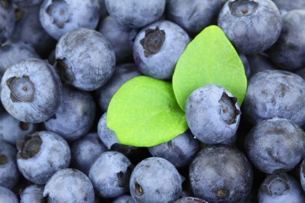
There are few things as tasty as a good blueberry, but science is showing that there could be more health benefits to this delicious fruit than people might think.

What is Pterostilbene?

Pterostilbene (trans-3,5-dimethoxy-4-hydroxystilbene) is a naturally occurring polyphenol, a type of molecule that occurs in plants. It is part of the stilbene group of compounds and the main antioxidant component of blueberries. In plants, it serves a defensive antimicrobial and often antioxidative role.

It was first discovered in 1977 by Langcake and Pryce and has been studied for its antioxidant properties and potential health benefits [1].

Pterostilbene is chemically related to resveratrol, another popular dietary supplement; some studies suggest that it may be more useful for health than its close relation, sometimes giving rise to the suggestion that it may be a “better resveratrol”.

What foods are high in pterostilbene?

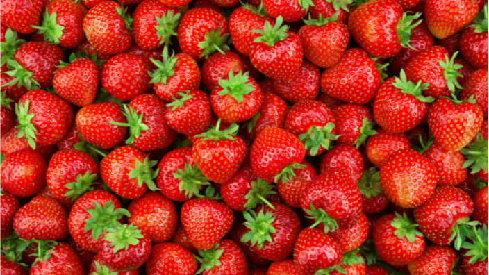
Pterostilbene is found in blueberries, with the estimated content per blueberry varying between 99 ng to 520 ng, depending on the type of blueberry [2-3]. To put this into perspective, an average blueberry punnet weighs around 340 grams.

If you ate the entire punnet, the total amount of pterostilbene you would get is only 0.03 to 0.18 mg, and based on the dose used in limited human studies of 100mg a day, that would be a huge amount of blueberries a day!

However, if you are looking to naturally obtain the kind of levels of pterostilbene that dietary supplements offer, then you would really, really, need to like blueberries, not to mention the high cost of buying that much fruit. In realistic terms, this is impractical. Supplement dosages range from 50 mg to 1,000 mg per capsule.

Pterostilbene is also found in almonds, grape leaves and vines, cranberries and related Vaccinium berries, such as lingonberries, bilberries, and huckleberries.

Which is better resveratrol or pterostilbene?

Pterostilbene has more bioavailability than resveratrol and other stilbenes due to two methoxy groups, which cause it to exhibit increased lipophilic and oral absorption [4]. In animal studies, pterostilbene was shown to have 80% bioavailability compared to 20% for resveratrol, suggesting that it is a superior choice [5].

So far, the data for pterostilbene looks promising, but it should be noted that it has significantly less research than resveratrol.

Potential pterostilbene benefits

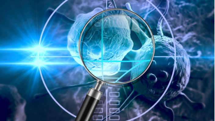
Various studies have demonstrated the antioxidant, anti-inflammatory [6], and anticarcinogenic properties of pterostilbene, which has led to the improved function of healthy cells and the inhibition of malignant cells [7-9].

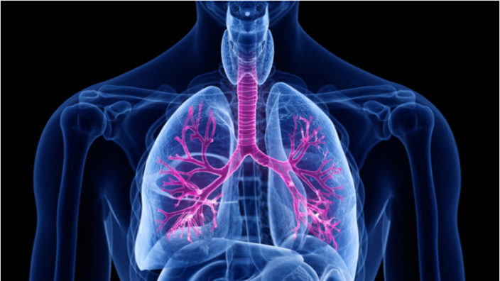
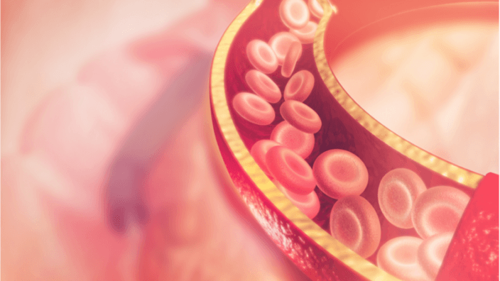
Pterostilbene has been implicated in cardiovascular health. with one study showing that it has a protective effect against atherosclerosis [10] and another showing that it improves aspects of autophagy and helps to counteract the pro-atherosclerosis effect of oxidized low-density lipoprotein on vascular endothelial cells [11-12]. It has also demonstrated potential utility in treating ischemia-reperfusion injury [13].

Studies have also shown the potential of pterostilbene in relation to neurodegenerative conditions such as Alzheimer’s disease. A study on mice with accelerated aging showed that pterostilbene, even in low doses, has a significant effect on improving cognitive ability [14].

Another study suggests that pterostilbene is involved in neural plasticity and its associated cognitive and motor functions and that rats given pterostilbene perform better in cognitive tests [15].

Pterostilbene is a powerful anti-inflammatory and can suppress NF-Kb, a protein complex that regulates the transcription of DNA, cytokine production and cell survival [16]. A recent study showed that pterostilbene can potentially treat severe acute pancreatitis by reducing serum levels of inflammatory TNF-a, IL-1b, and NF-kB and that it reduces the generation of reactive oxygen species.

Pterostilbene also has some data that suggests it might be useful for treating arthritis, and given its anti-inflammatory properties, this is hardly a surprise. While research is limited to date regarding arthritis, a rat study did suggest some potential for treating this condition [17].

Pterostilbene influences energy metabolism

While not unique to pterostilbene but important to mention, pterostilbene activates AMP-activated protein kinase (AMPK), a master regulator of cellular energy homeostasis. AMPK is one of the two catabolic signaling systems, the other being sirtuins; these two work together in a feedback loop and signal nutrient scarcity [18]. These systems are part of our energy metabolism, and deregulation of that system is thought to be one of the reasons we age.

Increasing these two pathways favors health and longevity (on the other hand, the IIS and mTOR pathways reduce lifespan when their levels are increased). In combination, AMPK and sirtuins sense low energy states by detecting high AMP levels and high NAD+ levels, respectively.

AMPK activation has a number of positive effects on metabolism as well as autophagy [19]. It has been shown to influence lifespan in mice that were given metformin. Even better, AMPK even inhibits the mTOR signaling pathway, which increases autophagy [20-21].

Finally, pterostilbene also increases sirtuins, which are best known for their pro-longevity effects in dietary restriction experiments in a variety of animal species [22-23]. Sirtuins adjust cellular metabolism based on nutrient availability and regulate many metabolic functions, including DNA repair, genomic stability, inflammatory response, apoptosis, cell cycle, and mitochondrial functions.

Pterostilbene side effects

Pterostilbene is considered safe, and no adverse effects have been reported up to a dose of 250 mg per day. However, some people may experience increased LDL cholesterol when using it. Given this compound is commonly found in food, dietary levels of pterostilbene should be safe.

That said, pterostilbene has few human studies compared to resveratrol and could potentially interact with other medications in ways not yet known. Caution is advised at the higher doses provided by supplements, as these greatly exceed typical dietary intake. If you do decide to take it and experience any adverse effects, cease taking it immediately and consult your doctor.

Disclaimer

This article is only a very brief summary. It is not intended as an exhaustive guide and is based on the interpretation of research data, which is speculative by nature. This article is not a substitute for consulting your physician about which supplements may or may not be right for you. We do not endorse supplement use or any product or supplement vendor, and all discussion here is for scientific interest.

Literature

[1]  Langcake, P., & Pryce, R. J. (1977). A new class of phytoalexins from grapevines. Cellular and Molecular Life Sciences, 33(2), 151-152. [2]  Rimando, A. M., Kalt, W., Magee, J. B., Dewey, J., & Ballington, J. R. (2004). Resveratrol, pterostilbene, and piceatannol in vaccinium berries. Journal of agricultural and food chemistry, 52(15), 4713-4719. [3]  Rodríguez-Bonilla, P., López-Nicolás, J. M., Méndez-Cazorla, L., & García-Carmona, F. (2011). Development of a reversed phase high performance liquid chromatography method based on the use of cyclodextrins as mobile phase additives to determine pterostilbene in blueberries. Journal of Chromatography B, 879(15), 1091-1097. [4]  Perečko, T., Drábiková, K., Račková, L., Číž, M., Podborská, M., Lojek, A., … & Jančinová, V. (2010). Molecular targets of the natural antioxidant pterostilbene: effect on protein kinase C, caspase-3 and apoptosis in human neutrophils in vitro. Neuroendocrinology Letters, 31(2), 84. [5]  Kapetanovic, I. M., Muzzio, M., Huang, Z., Thompson, T. N., & McCormick, D. L. (2011). Pharmacokinetics, oral bioavailability, and metabolic profile of resveratrol and its dimethylether analog, pterostilbene, in rats. Cancer chemotherapy and pharmacology, 68(3), 593-601. [6]  Choo, Q. Y., Yeo, S. C. M., Ho, P. C., Tanaka, Y., & Lin, H. S. (2014). Pterostilbene surpassed resveratrol for anti-inflammatory application: Potency consideration and pharmacokinetics perspective. Journal of Functional Foods, 11, 352-362. [7]  Rimando, A. M., Cuendet, M., Desmarchelier, C., Mehta, R. G., Pezzuto, J. M., & Duke, S. O. (2002). Cancer chemopreventive and antioxidant activities of pterostilbene, a naturally occurring analogue of resveratrol. Journal of agricultural and food chemistry, 50(12), [8]  Remsberg, C. M., Yáñez, J. A., Ohgami, Y., Vega‐Villa, K. R., Rimando, A. M., & Davies, N. M. (2008). Pharmacometrics of pterostilbene: preclinical pharmacokinetics and metabolism, anticancer, antiinflammatory, antioxidant and analgesic activity. Phytotherapy Research, 22(2), 169-179. [9]  Kong, Y., Chen, G., Xu, Z., Yang, G., Li, B., Wu, X., … & Chang, G. (2016). Pterostilbene induces apoptosis and cell cycle arrest in diffuse large B-cell lymphoma cells. Scientific Reports, 6. [10] Ekshyyan, V. P., Hebert, V. Y., Khandelwal, A., & Dugas, T. R. (2007). Resveratrol inhibits rat aortic vascular smooth muscle cell proliferation via estrogen receptor dependent nitric oxide production. Journal of cardiovascular pharmacology, 50(1), 83-93. [11] Zhang, L., Zhou, G., Song, W., Tan, X., Guo, Y., Zhou, B., … & Chen, L. (2012). Pterostilbene protects vascular endothelial cells against oxidized low-density lipoprotein-induced apoptosis in vitro and in vivo. Apoptosis, 17(1), 25-36. [12]  Zhang, L., Cui, L., Zhou, G., Jing, H., Guo, Y., & Sun, W. (2013). Pterostilbene, a natural small-molecular compound, promotes cytoprotective macroautophagy in vascular endothelial cells. The Journal of nutritional biochemistry, 24(5), 903-911. [13] Guo, Y., Zhang, L., Li, F., Hu, C. P., & Zhang, Z. (2016). Restoration of sirt1 function by pterostilbene attenuates hypoxia-reoxygenation injury in cardiomyocytes. European journal of pharmacology, 776, 26-33. [14] Chang, J., Rimando, A., Pallas, M., Camins, A., Porquet, D., Reeves, J., … & Casadesus, G. (2012). Low-dose pterostilbene, but not resveratrol, is a potent neuromodulator in aging and Alzheimer’s disease. Neurobiology of aging, 33(9), 2062-2071. [15] Joseph, J. A., Fisher, D. R., Cheng, V., Rimando, A. M., & Shukitt-Hale, B. (2008). Cellular and behavioral effects of stilbene resveratrol analogues: implications for reducing the deleterious effects of aging. Journal of agricultural and food chemistry, 56(22), 10544-10551. [16]  Pan, M. H., Chang, Y. H., Tsai, M. L., Lai, C. S., Ho, S. Y., Badmaev, V., & Ho, C. T. (2008). Pterostilbene suppressed lipopolysaccharide-induced up-expression of iNOS and COX-2 in murine macrophages. Journal of agricultural and food chemistry, 56(16), 7502-7509. [17] Macickova, T., Drábiková, K., Nosal, R., Bauerová, K., Mihalova, D., Harmatha, J., & Pecivova, J. (2009). In vivo effect of pinosylvin and pterostilbene in the animal model of adjuvant arthritis. Neuro endocrinology letters, 31, 91-95. [18] Price, N. L., Gomes, A. P., Ling, A. J., Duarte, F. V., Martin-Montalvo, A., North, B. J., … & Hubbard, B. P. (2012). SIRT1 is required for AMPK activation and the beneficial effects of resveratrol on mitochondrial function. Cell metabolism, 15(5), 675-690. [19]  Salminen, A., & Kaarniranta, K. (2012). AMP-activated protein kinase (AMPK) controls the aging process via an integrated signaling network. Ageing research reviews, 11(2), 230-241. [20] Mihaylova, M. M., & Shaw, R. J. (2011). The AMPK signalling pathway coordinates cell growth, autophagy and metabolism. Nature cell biology, 13(9), 1016-1023. [21]  Kim, J., Kundu, M., Viollet, B., & Guan, K. L. (2011). AMPK and mTOR regulate autophagy through direct phosphorylation of Ulk1. Nature cell biology, 13(2), 132-141. [22] Guo, Y., Zhang, L., Li, F., Hu, C. P., & Zhang, Z. (2016). Restoration of sirt1 function by pterostilbene attenuates hypoxia-reoxygenation injury in cardiomyocytes. European journal of pharmacology, 776, 26-33. [23] Cheng, Y., Di, S., Fan, C., Cai, L., Gao, C., Jiang, P., … & Li, T. (2016). SIRT1 activation by pterostilbene attenuates the skeletal muscle oxidative stress injury and mitochondrial dysfunction induced by ischemia reperfusion injury. Apoptosis, 21(8), 905-916.
Lung branches

A Look at Idiopathic Pulmonary Fibrosis

A new review published in the American Journal of Respiratory Cell and Molecular Biology discusses the causes of, and potential treatments for, idiopathic pulmonary fibrosis (IPF).

Fibrosis, but not focused on fibroblasts

In the lungs, the trachea (airway) breaks out into a tree-like system of nodes. Each branch ends in alveoli, which are responsible for gas exchange between the air and the bloodstream. This is where the distal epithelium resides, and it has two types of cells: AEC1 and AEC2. AEC1 cells are involved in the gas exchange itself, as their thin membranes allow the entry of oxygen and the release of carbon dioxide. AEC2 cells are cubic in shape, and they secrete a surfactant that prevents the alveoli from collapsing. Furthermore, they are able to self-renew and differentiate into AEC1 cells, so they function like stem cells [1].

The direct cause of the fibrosis in IPF is fibroblasts. Previous research has shown that cellular senescence is linked to IPF, and one paper has shown that the senescence-associated secretory phenotype (SASP) is linked to aberrantly activated fibroblasts [2].

However, this review cites research showing that IPF begins with the distal epithelium, not the fibroblasts, and that targeting the disease here may be sufficient to slow or reverse it [3]. The researchers portray this as wound healing gone wrong, and in addition to genetic predispositions for the disease, they cite multiple aspects of aging that may contribute to IPF.

Extracellular matrix dysfunction

The accumulation of cross-links in the extracellular matrix has been associated with multiple aspects of aging, including arterial stiffness. Here, the reviewers cite research showing that the extracellular matrix of IPF patients affects fibroblasts more strongly than where the fibroblasts themselves originated [4]. The lungs of IPF patients are less elastic than the lungs of people without this disease, and one experiment showed that stretching IPF tissue causes the release of TGF-beta 1 [5], a compound that has positive effects in normal concentrations but is known to cause fibrosis in excess.

Cellular senescence and apoptosis

Research has shown that fibroblasts are not the only cells whose senescence implicates IPF; senescent distal epithelial cells are involved as well. In particular, the senescence marker and transcription factor p53 is directly linked to the progression of the disease, as it drives aberrant cellular behavior in AEC2 cells [6].

The reviewers also suggest that premature or aberrant induction of apoptosis (cell death) pathways might also be partially responsible for the disease. One mouse experiment reduced GSTP, a chemical known to activate apoptosis, and the effects on IPF were positive [7].

Loss of proteostasis and mitochondrial dysfunction

One cause of IPF may be unfolded or misfolded proteins, which is one of the hallmarks of aging: the loss of proteostasis. In the context of IPF, the loss of proteostasis places stress on the endoplasmic reticulum (ER), the part of the cell that is responsible for protein maintenance. In one mouse experiment, the unfolded protein response was triggered in the ER of AEC2 cells, which led directly to fibrosis [8]. The reviewers also cite multiple research papers in which mutations to the genes responsible for the surfactant proteins of AEC2 cells are shown to drive the development of IPF.

Mitochondrial dysfunction, another hallmark of aging, was also cited as a cause of ER stress and, ultimately, IPF. Specifically, the reviewers highlight the lack of autophagy. In autophagy, cells consume their own organelles, eating parts of themselves as part of survival and maintenance. While too much autophagy can lead to apoptosis, not enough can cause dysfunctional mitochondria to proliferate, and these dysfunctional mitochondria are linked to IPF [9].

Potential treatments

The reviewers first point out that there is a strategy that has never been shown to work: directly targeting the fibroblasts themselves. Attempting to treat IPF with anti-inflammatories has also returned generally negative results.

They then go on to show the difficulties inherent in targeting signaling pathways, particularly in the case of IPF, as the number of interlinked pathways makes it difficult to create a targeted drug. There are a great number of clinical trials aimed at these signaling pathways in IPF, although none of them have yet surpassed Phase 2.

The reviewers hold that, on top of a lack of drugs that target the distal epithelium, there is an unmet need for more advanced approaches to IPF, including stem cell therapies, gene therapies, and other treatments that go beyond simple pharmacology, such as nanoparticles and nanotechnology-based antibodies.

Abstract

Idiopathic pulmonary fibrosis is a fatal interstitial lung disease with limited therapeutic options. Current evidence suggests that IPF may be initiated by repeated epithelial injury in the distal lung followed by abnormal wound healing responses which occur due to intrinsic and extrinsic factors. Mechanisms contributing to chronic damage of the alveolar epithelium in IPF include dysregulated cellular processes such as apoptosis, senescence, abnormal activation of developmental pathways, aging, as well as genetic mutations. Therefore, targeting the regenerative capacity of the lung epithelium is an attractive approach in the development of novel therapies for IPF. Endogenous lung regeneration is a complex process involving coordinated cross-talk between multiple cell types and re-establishment of a normal extracellular matrix environment. This review will describe the current knowledge of reparative epithelial progenitor cells in the alveolar region of the lung and discuss potential novel therapeutic approaches for IPF focusing on endogenous alveolar repair. This article is open access and distributed under the terms of the Creative Commons Attribution Non-Commercial No Derivatives License 4.0 (https://creativecommons.org/licenses/by-nc-nd/4.0/).

Conclusion

This review is extremely in-depth, and it cites multiple avenues of research that have discovered many different biological problems that are linked to IPF. This raises the concern that IPF may be fundamentally caused by multiple upstream factors, so there cannot be a single treatment to cure it, just as there is no one cure for all cancers. However, as research progresses and we learn more about how to deal with this frustrating and deadly disease, we may be able to restore AEC2 populations, decrease fibrosis, and, ultimately, allow people to breathe easier.

We would like to ask you a small favor. We are a non-profit foundation, and unlike some other organizations, we have no shareholders and no products to sell you. All our news and educational content is free for everyone to read, but it does mean that we rely on the help of people like you. Every contribution, no matter if it’s big or small, supports independent journalism and sustains our future.

Literature

[1] Barkauskas CE, Cronce MJ, Rackley CR, Bowie EJ, Keene DR, Stripp BR, Randell SH, Noble PW, Hogan BLM. Type 2 alveolar cells are stem cells in adult lung. The Journal of clinical investigation 2013; 123: 3025-3036.

[2] Álvarez, D., Cárdenes, N., Sellarés, J., Bueno, M., Corey, C., Hanumanthu, V. S., … & Rojas, M. (2017). IPF lung fibroblasts have a senescent phenotype. American Journal of Physiology-Lung Cellular and Molecular Physiology, 313(6), L1164-L1173.

[3] Yuan T, Volckaert T, Redente EF, Hopkins S, Klinkhammer K, Wasnick R, Chao C-M, Yuan J, Zhang J-S, Yao C, Majka S, Stripp BR, Günther A, Riches DWH, Bellusci S, Thannickal VJ, De Langhe SP. FGF10-FGFR2B Signaling Generates Basal Cells and Drives Alveolar Epithelial Regeneration by Bronchial Epithelial Stem Cells after Lung Injury. Stem Cell Reports 2019; 12: 1041-1055.

[4] Parker MW, Rossi D, Peterson M, Smith K, Sikström K, White ES, Connett JE, Henke CA, Larsson O, Bitterman PB. Fibrotic extracellular matrix activates a profibrotic positive feedback loop. The Journal of clinical investigation 2014; 124: 1622-1635.

[5] Froese AR, Shimbori C, Bellaye P-S, Inman M, Obex S, Fatima S, Jenkins G, Gauldie J, Ask K, Kolb M. Stretch-induced Activation of Transforming Growth Factor-ß1 in Pulmonary Fibrosis. American Journal of Respiratory and Critical Care Medicine 2016; 194: 84-96.

[6] Xu Y, Mizuno T, Sridharan A, Du Y, Guo M, Tang J, Wikenheiser-Brokamp KA, Perl AKT, Funari VA, Gokey JJ, Stripp BR, Whitsett JA. Single-cell RNA sequencing identifies diverse roles of epithelial cells in idiopathic pulmonary fibrosis. JCI Insight 2016; 1: e90558-e90558.

[7] Roach KM, Sutcliffe A, Matthews L, Elliott G, Newby C, Amrani Y, Bradding P. A model of human lung fibrogenesis for the assessment of anti-fibrotic strategies in idiopathic pulmonary fibrosis. Sci Rep 2018; 8: 342.

[8] Borok Z, Horie M, Flodby P, Wang H, Liu Y, Ganesh S, Firth AL, Minoo P, Li C, Beers MF, Lee AS, Zhou B. Grp78 Loss in Epithelial Progenitors Reveals an Age-linked Role for Endoplasmic Reticulum Stress in Pulmonary Fibrosis. 2020; 201: 198-211.

[9]  Hill C, Li J, Liu D, Conforti F, Brereton CJ, Yao L, Zhou Y, Alzetani A, Chee SJ, Marshall BG, Fletcher SV, Hancock D, Ottensmeier CH, Steele AJ, Downward J, Richeldi L, Lu X, Davies DE, Jones MG, Wang Y. Autophagy inhibition-mediated epithelialmesenchymal transition augments local myofibroblast differentiation in pulmonary fibrosis. Cell Death Dis 2019; 10: 591-591.

Developing healthy practices could help you live a longer life.

Starting Your Personal Longevity Strategy

Every day, researchers get together in their labs and try to find ways of bringing aging under medical control. Despite the ongoing global effort, it is estimated that a relatively complete system of controlling biological aging is at least 20 years away.

That means there is likely going to be considerable time before life extension technologies are available. In order to live long enough to benefit from them, developing a personal longevity strategy is a wise idea.

The goal therefore, would be to live at least another 20 years in relatively good health. The difficulty of this depends on how old you are now, but regardless of your age, you can develop a personal longevity strategy today. To help you on this path, we have created some practical health tips to help you take care of yourself and your family.

Know yourself

The first and most important step towards developing a personal longevity strategy is proper diagnostics.

Many diseases and conditions start silently. It’s possible to feel changes in the body without being able to attribute them to deficiencies or abnormal biomarkers. When we get sick, we simply feel bad or in pain. It is important to find out what is wrong – preferably, before it becomes a real problem.

To receive valuable data on your health, you can use three types of diagnostic tests. All of them are valuable, but some of them might be more appropriate than others in different cases.

  • Genetic tests spot any genetic predisposition to a disease. Many companies now offer all sorts of genetic tests, from individual diseases to a full exome (the coding part of the genome) test or a full DNA test. In most cases, you only need to take such a broad test once in order to use its information to improve decision making and help your medical advisor understand your condition.
  • Regular complex check-ups to see what is going on in your body. Complex checkups test a large number of biomarkers, such as lipid profile, thyroid hormones, glucose and insulin, biomarkers of the liver and kidney, inflammation biomarkers such as C-reactive protein and interleukin-6, and levels of certain vitamins and minerals that are vital for health and well-being. Biomarker composition normally changes with age, and older people may benefit from checking more things than younger people.
  • Regular check-ups specific to your diseases or conditions that will show you how well treatments are working. If you are taking drugs for age-related diseases regularly check if each drug is working well and does not need replacement or dose adjustment. This way, you make sure that each treatment is beneficial and keeping your diseases under control.

Talk to your medical advisor

Most people don’t have a medical degree. While you can find lots of useful information on the internet, a visit to a doctor remains the most reliable way to get a proper assessment of your physical condition.

Properly interpreting data is vital for developing a good strategy, and a doctor may advise you on how to combine your treatments in a safe way if you need to address several issues at once. Some drugs may be incompatible, or your genetic layout may predispose you to a weak response to certain treatments. A doctor can also prescribe additional tests if the picture is unclear and there is no reliable diagnostic hypothesis.

Working together with medical advisors, we can achieve the best results possible. Here are some tips that may make your visit more effective:

  • Describe your sensations in enough detail.
  • Try to remember what happened before or after you felt that something was wrong, including social situations, sports, or overwork that may have served as stress factors
  • List the medications that you are already taking and the reasons why you are taking them; this helps to exclude the influence of those medications on the diagnosis.
  • Ask about the suspected diagnosis and how to confirm it.
  • Ask about what can be done in your case, including the best strategy and the second-best strategy.
  • Don’t forget to set another appointment to check how your treatment is going.

New supplements should be introduced one at a time

The range of dietary supplements is already overwhelmingly large and it is important to develop a science-based approach when considering taking them. Before taking anything, carefully check the information about each supplement and see if it can indeed be beneficial.

There is a lot of misleading marketing and overpriced supplements with little to no supporting data that they will do anything for longevity, so due diligence is a must before considering taking anything. Some supplements contain vitamins that might already be abundant in the body, some can interact with medical treatments in the wrong way, and some may be incompatible with you personally.

In any case, if you choose to take supplements, it makes sense to introduce only one supplement at a time into your regime and carefully check how you feel after it. If you use several new supplements or drugs at once and feel bad, you may never understand what exactly has happened nor which substance is causing issues.

Supplements should supplement a healthy lifestyle, not replace it

It is very tempting to stop working on maintaining a healthy lifestyle if you can just take a drug or a supplement that may prolong healthy life. However, the increase in healthy years that often come from a good diet and physical activity can easily compete with the effects of existing therapies that slow down age-related diseases.

This is largely explained by deeply ingrained genetic mechanisms that make mild hunger, physical activity, good sleep, and a diet with a low glycemic index beneficial for health. For thousands of years, humans have adjusted to harsh living conditions, such as hard work and long walks just to gather food, until the body learned how to use those survival efforts to postpone age-related diseases, reduce their risks, and stay healthy for longer. This legacy is not to be underestimated.

Some simple things you can do now

While we are waiting for powerful longevity treatments to arrive, here are some tips.

  • Try to find out how many calories your body needs to be healthy and eat as close to this as is practical. A calorie counter app may help you to track your intake.
  • Limit the amount of fast carbs in your diet. WHO recommends only 5% of your calorie intake be from free sugars, such as cakes and sweet drinks.
  • Eat plenty of raw vegetables and fruits. According to WHO, over 400 grams of vegetables every day should provide health benefits. And fiber is good for beneficial gut bacteria.
  • Try to walk more than 10000 steps a day. If you have health issues, adjust this number to your actual ability, but still move as much as you can
  • If you are a healthy adult, do some intense physical activity 2-3 times a week. If you have some health issues, adjust this amount of exercise to your actual ability but still try to make your muscles work.
  • Sleep in a room that is dark, ventilated, and cool enough. 7-8 hours of sleep seems to be a healthy amount of sleep. If your sleep is much shorter, you might need to discuss it with your medical advisor – as you age, the production of the sleep hormone melatonin decreases, and your advisor might recommend taking it as a supplement.
  • Practice some self-regulation techniques – meditation or relaxation can contribute to better sleep and better control over your health behaviors.
  • Saunas have a number of health benefits that relate to health and longevity, try to use them as often as possible. The mild heat stress causes your cells to produce protective proteins and increase the activity of cellular repair systems.
  • Get rid of bad habits such as heavy drinking, smoking, and harmful drugs. These may reduce your healthy period of life because they accelerate aging.

Get some good company

It has been shown in numerous studies that social interaction can be beneficial for your health. Even though the pandemic currently requires many of us to limit in-person meetings and wear protective masks, having friends and communicating with relatives on a regular basis is important.

Apart from the enjoyment from human companionship, your relatives or friends may notice if something is going wrong and help you in case of a crisis. Helping other people may also have positive effects on your health.

People who volunteer often feel happier and more fulfilled in life than those who don’t. Centenarians are known to contribute to their social circles, which might be a factor why they live so long.

We would like to ask you a small favor. We are a non-profit foundation, and unlike some other organizations, we have no shareholders and no products to sell you. All our news and educational content is free for everyone to read, but it does mean that we rely on the help of people like you. Every contribution, no matter if it’s big or small, supports independent journalism and sustains our future.

PEARL Rapamycin Campaign

PEARL Is Funded, Rapamycin Longevity Clinical Trials Begin

Today is a doubly important day: it marks the final day of the PEARL campaign and it is a celebration of another victory for the life extension community.

PEARL smashed its initial fundraising goal and sailed through its two stretch goals, raising just under $183k thanks to the generous support of the community.

What is PEARL?

The Participatory Evaluation (of) Aging (with) Rapamycin (for) Longevity Study, or PEARL, will launch the first large-scale placebo-controlled clinical trial to determine the effects of rapamycin on human aging. The principal investigator is Dr. James P. Watson based at UCLA.

What is rapamycin?

Rapamycin is a naturally occurring antifungal antibiotic produced by soil bacteria originally discovered on Easter Island, also known as Rapa Nui.

While most people know it as a drug used in high doses for organ rejection during transplants, it has potential as a drug to slow down aging in far lower doses. It was the first drug proven to extend the lifespan of mice, even when given late in life, and has been shown to reliably increase the lifespan of yeast, worms, and rats.

Rapamycin works through the mTOR signaling pathway, a master regulator of cellular and energy metabolism, and a potent trigger of autophagy, the ultimate cellular recycling mechanism.

Recent research shows that it also improves how our DNA is stored which helps combat genomic instability resulting from DNA damage and harmful epigenetic alterations to gene expression which drive the aging process.

Until now, there has not been a proper clinical trial to evaluate if rapamycin can slow down aging in humans. PEARL is going to change that thanks to our supporters.

Why PEARL is an important milestone for life extension

This large-scale clinical trial will allow the researchers to find out if the effects of rapamycin will translate to people. If that were to happen, then the potential impact could be massive. Having a drug that demonstrably slows down human aging confirmed by large-scale clinical trial data would really open doors. The data could help garner wider public support for life extension therapies as well as support dialogue with policymakers and healthcare regulatory bodies.

Also, rapamycin is a relatively cheap, off-patent, generic drug and widely available. It has been a registered drug for decades with a well-known safety profile and characteristics. If longevity benefits are confirmed, then it could potentially start being used in healthcare to address aging almost immediately.

The community should be proud of what it has achieved today

This has been the most successful crowdfunding campaign we have ever hosted on lifespan.io, and it is all thanks to you, our amazing life extension community.

Thank you to everyone who has supported PEARL!

Over 409 of you have donated just under $183k to the campaign since its launch on May 17th, helping to make this one of the largest, if not the largest, crowdfunded aging focused biology projects in the world.

This successful fundraising really showcases the growing power of the life extension community, and part of this power comes from the cryptocurrency community. In the last few years, thanks to people like Vitalik Buterin, there has been an increasing level of support from the cryptocurrency community, which is greatly appreciated.

Special thanks

The PEARL team and lifespan.io are incredibly grateful to everyone who donated to PEARL and would like to give special thanks to Vitalik Buterin, Tom Moya Schau, Micah Zoltu, and Brad Armstrong, who contributed to the campaign with large donations. Thank you so much for your wonderful support; you have really helped to drive this project to success!

What is next?

Due to the huge success of the crowdfunding campaign, the PEARL team wanted to let you all know how the additional funds would be used. The extra money will allow the researchers to measure additional important biomarkers, which will further strengthen the data.

The original planned biomarkers:

  • IGF-1 (fasting)
  • Lipid Panel, Standard
  • TNFa
  • IL-6
  • VLDL
  • Comprehensive Metabolic Panel*
  • HgAC1
  • Uric Acid
  • CBC & DIFF
  • CD4/CD8
  • CMV IgG titers
  • hs-CRP
  • Vitamin D, 25-OH Total  (VIT D,25-OH,TOTAL,IA)
  • sirolimus level

The extra funds will be used to measure these additional biomarkers:

  • Cystatin C
  • GDF 15
  • Adiponectin
  • Leptin

Once again from the PEARL team and lifespan.io, thank you to everyone who has helped make this project possible! For people in the U.S. interested in participating, please get in touch with Agelessrx and a trial administrator should get back to you.

We would like to ask you a small favor. We are a non-profit foundation, and unlike some other organizations, we have no shareholders and no products to sell you. All our news and educational content is free for everyone to read, but it does mean that we rely on the help of people like you. Every contribution, no matter if it’s big or small, supports independent journalism and sustains our future.
Elderly muscle

The Aged Microenvironment Linked to Sarcopenia

Muscular degradation with age isn’t the result of a decline in the intrinsic regenerative ability of muscles, according to new research [1].  Instead, sarcopenia is likely due to changes in the muscle microenvironment that reduce repair and regeneration.

Declining muscles

Age-related muscle loss can begin as early as a person’s fourth decade, and sarcopenia can eventually lead to a cumulative loss of 30-50% of skeletal muscle mass. The complicated process involves a lot of interacting factors, including the gut microbiome and changes in the muscle microenvironment. Accumulating damage to muscle tissue doubtlessly plays a part, but it’s unclear why this damage doesn’t get repaired.

One suggested possibility has been that the mechanisms responsible for muscle repair might become exhausted with age; therefore, the quiescent muscle precursor cells that normally produce and regenerate muscle lose their ability to do so. However, it’s also possible that there is no change in the intrinsic competence of these cells and that muscle damage goes unrepaired because of changes in the muscle microenvironment.

To determine which of these possibilities is correct, a team of U.S. researchers developed and used a system to graft human muscles into mice. They collected muscle tissue from human cadavers that had undergone autopsies and transplanted it into mice. By using tissue from cadavers of people from different ages, they were able to evaluate the intrinsic regenerative ability of muscle cells at these ages.

Undaunted stem cells

The transplants were equally successful regardless of whether the tissue came from the body of a 36-year old or a 78-year old. Donor tissue from cadavers of various ages integrated into the mouse host and began producing muscle within three weeks and continued for at least six weeks. The researchers also carried out a transcriptomic analysis and detected the expression of human transcription factors involved in muscle differentiation.

These findings make it clear that precursor cells in elderly people retain the ability to generate muscle tissue. They do exactly that when placed in an appropriate environment, such as in healthy mice. The fact that the damage accumulates in elderly muscle tissue thus cannot be attributed to a decrease in the regenerative competence of these cells. The researchers argue that the most likely explanation is that changes in the muscle microenvironment somehow reduce regenerative competence.

The team also tested transplants of tissue after different post-mortem intervals to find out how long the cells retained their regenerative capacity. They found that the transplants were still successful after 11 days, the longest interval they tested. Not only were the transplants successful but there was no change in the success rate from shorter intervals. This suggests that the muscle results are highly resistant to the anoxic conditions of post-mortem tissues.

Age-related loss of muscle mass and strength is widely attributed to limitation in the capacity of muscle resident satellite cells to perform their myogenic function. This idea contains two notions that have not been comprehensively evaluated by experiment. First, it entails the idea that we damage and lose substantial amounts of muscle in the course of our normal daily activities. Second, it suggests that mechanisms of muscle repair are in some way exhausted, thus limiting muscle regeneration. A third potential option is that the aged environment becomes inimical to the conduct of muscle regeneration. In the present study, we used our established model of human muscle xenografting to test whether muscle samples taken from cadavers, of a range of ages, maintained their myogenic potential after being transplanted into immunodeficient mice. We find no measurable difference in regeneration across the range of ages investigated up to 78 years of age. Moreover, we report that satellite cells maintained their myogenic capacity even when muscles were grafted 11 days postmortem in our model. We conclude that the loss of muscle mass with increasing age is not attributable to any intrinsic loss of myogenicity and is most likely a reflection of progressive and detrimental changes in the muscle microenvironment such as to disfavor the myogenic function of these cells.

Conclusion

Figuring out how and why sarcopenia happens is obviously important to being able to address it, and this study brings us one step closer to that. The xenografting transplant approach, together with the demonstration that muscle tissue can be harvested from cadavers of different ages at varying times after death, will also likely prove useful tools in our efforts to understand and control sarcopenia.

We would like to ask you a small favor. We are a non-profit foundation, and unlike some other organizations, we have no shareholders and no products to sell you. All our news and educational content is free for everyone to read, but it does mean that we rely on the help of people like you. Every contribution, no matter if it’s big or small, supports independent journalism and sustains our future.

Literature

[1] Novak, JS, et al. Human muscle stem cells are refractory to aging. Aging Cell (2021), doi: 10.1111/acel.13411
Blood cells in an artery

Brain Blood Flow Restriction Resembles Brain Aging in Mice

A mouse study published in Aging Cell has outlined the similarities between artificially restricted cerebral blood flow and the reduced blood flow associated with aging.

Aiming to understand vascular dementia

Prior research has shown that reduced cerebral blood flow, known as hypoperfusion, results in vascular dementia and brain atrophy through multiple mechanisms, including neuroinflammation and mitochondrial dysfunction [1]. The researchers wanted to determine which genes and signaling pathways are involved in the destructive processes of hypoperfusion and how they relate to normal aging. They focused their study on the hippocampus, a critical part of memory, learning, and executive function that is strongly affected by vascular dementia.

Restricting blood flow to match aging

In this experiment, mice were subjected to bilateral common cortical artery stenosis (BCAS), a procedure that uses microcoils to artificially induce hypoperfusion, reducing the blood available to the brain. This procedure reduced the cerebral blood flow of young mice nearly exactly to that of old mice immediately afterwards, taking away roughly 30% of their cerebral blood flow and thus brain oxygen.

After 7 days, the blood flow of young mice had slightly increased, but this procedure resulted in substantial changes to their gene expression, particularly after 30 days. These genes included many critical functions of neurons along with fundamental metabolic processes, as their brains struggled to perform their function in a low-oxygen environment.

Interestingly, while the blood flow of older mice was further reduced by this procedure, they changed their gene expression substantially less than young mice did, and their blood flow returned closer to their norm. It is logical to conclude that, as their blood flow was already being restricted, the procedure caused few effects that had not already occurred.

The mitochondrial connection

Genes relating to fundamental aspects of the ribosome, the cellular machinery that engages in protein synthesis, as well as many genes related to mitochondrial integrity, were still different between young mice subjected to BCAS and normal old mice.

However, the researchers noted that young mice subjected to BCAS and normal old mice had similar gene expressions related to mitochondrial oxidation and, partially, the production of NAD+ from NADH and the basic synthesis of ATP, the energy our cells use to function. The mitochondria, the powerhouses of the cell, were prohibited from producing as much power as they normally would, both in BCAS and aging. Other similarly downregulated genes included heat shock proteins that serve neuroprotective functions.

This begs the question of how much of aging is caused by macro-scale physical processes that are caused by other aspects of aging. These results suggest this sort of causal chain: as some processes of aging cause ischemia, this limits critical oxygen flow to the brain, thus resulting in fewer neuroprotective proteins and increased mitochondrial dysfunction. More research is needed to see if this holds true in other tissues and in human beings.

Abstract

Vascular dementia (VaD) is a progressive cognitive impairment of vascular etiology. VaD is characterized by cerebral hypoperfusion, increased blood-brain barrier permeability and white matter lesions. An increased burden of VaD is expected in rapidly aging populations. The hippocampus is particularly susceptible to hypoperfusion, and the resulting memory impairment may play a crucial role in VaD. Here we have investigated the hippocampal gene expression profile of young and old mice subjected to cerebral hypoperfusion by bilateral common carotid artery stenosis (BCAS). Our data in sham-operated young and aged mice reveal an age-associated decline in cerebral blood flow and differential gene expression. In fact, BCAS and aging caused broadly similar effects. However, BCAS-induced changes in hippocampal gene expression differed between young and aged mice. Specifically, transcriptomic analysis indicated that in comparison to young sham mice, many pathways altered by BCAS in young mice resembled those already present in sham aged mice. Over 30 days, BCAS in aged mice had minimal effect on either cerebral blood flow or hippocampal gene expression. Immunoblot analyses confirmed these findings. Finally, relative to young sham mice the cell type-specific profile of genes in both young BCAS and old sham animals further revealed common cell-specific genes. Our data provide a genetic-based molecular framework for hypoperfusion-induced hippocampal damage and reveal common cellular signaling pathways likely to be important in the pathophysiology of VaD.

Conclusion

While this study builds on our knowledge of the effects of long-term hypoperfusion on gene expression and makes strides in uncovering the mechanisms behind vascular dementia, it also pointedly illustrates just how much damage aging actually causes. To cause a young mouse to suffer the same amount as an old mouse in just one way, researchers must squeeze the arteries going to its brain. The old mouse still has many more medical problems that lead to dysfunction and death; to cause a young mouse to suffer all the problems of an old mouse through surgery would be both inhumane and impossible.

Meanwhile, ischemic stroke, vascular dementia, and many more crippling and fatal age-related diseases are occurring, right now, in hundreds of millions of human beings around the world.

We would like to ask you a small favor. We are a non-profit foundation, and unlike some other organizations, we have no shareholders and no products to sell you. All our news and educational content is free for everyone to read, but it does mean that we rely on the help of people like you. Every contribution, no matter if it’s big or small, supports independent journalism and sustains our future.

Literature

[1] Aliev, G., Obrenovich, M. E., Smith, M. A., & Perry, G. (2003). Hypoperfusion, mitochondria failure, oxidative stress, and Alzheimer disease. Journal of Biomedicine and Biotechnology, 2003(3), 162-163.

Creatine helps to build muscle and may slow down aging.

What is Creatine? A Summary of Methylguanidine Acetic Acid

Meat is rich in creatine, an important energy buffer in muscle cells, and the main constituent of a popular sports supplement used by athletes. However, we’re now finding out that there’s more to it than ‘meats’ the eye.

What is creatine?

Creatine (α-methyl guanidine-acetic acid) is a substance that is present in our muscle cells. Functionally, it helps our muscles to produce energy for lifting weights, high-intensity exercise, and other strenuous activities. It has enjoyed great popularity as a supplement with athletes and bodybuilders in order to gain muscle, boost strength and increase exercise performance.

It was first identified by French chemist Michel Eugène Chevreul in 1832, who isolated it from skeletal muscle. Due to its presence in living tissue, Chevreul named it after the Greek word κρέας (kreas), meaning meat.

In 1912, Otto Folin and Wiley Glover Denis of Harvard University found that ingesting creatine led to a significant increase in intramuscular stores [1], sparking an interest in its potential as an oral supplement.

By the 1930s, German scientists began to study the relationship between creatine levels and muscle contraction, concluding that the more creatine present in muscle cells, the longer they could contract before producing lactic acid [2-3], allowing for extended training times.

During the 1960s, an interest surged in the possible uses of creatine to treat muscle diseases. In 1964, Fitch et al. were able to show that the skeletal muscles of muscular dystrophy patients have lower concentrations than their normal counterparts, which they attributed to a defect in creatine binding in muscle cells [4-5].

The potential of creatine as a performance-enhancing supplement came into public scrutiny after the 1992 Barcelona Olympics, in which several gold medallists admitted to taking it during training. Today, supplements exist in a wide variety of forms and are one of the most widely used nutritional supplements worldwide [6].

What foods contain creatine?

It is produced inside the body from the amino acids glycine and arginine, and it is widely distributed to tissues with high energy demands, such as the brain and muscles. About 95% of the body’s creatine is stored in skeletal muscle, but it is also found in small amounts in the liver, kidneys, and testes.

On average, the body produces approximately 1 gram of creatine per day in young adults [7-8], while the rest is obtained through diet. 

Creatine is naturally occurring in many foods, particularly animal protein, such as meat and fish. One pound of raw beef contains approximately 2.3g of creatine, while one pound of raw salmon contains up to 2g.

Cooking denatures creatine, so unless you like your steak extra bloody or are a big fan of sashimi, it will be difficult to get enough of it in your diet to benefit from its health properties. Red meat is also high in saturated fat and may increase your risk of all-cause mortality [9-10], so consuming that much meat to begin with might not be the wisest choice.

Luckily, it is widely available as a health supplement, and is extremely affordable. This is particularly relevant for vegans and vegetarians, whose intake is greatly diminished or absent altogether. These supplements have been found to have increased potency in vegetarians and confer other beneficial effects, such as increased cognitive capacity and performance, compared to omnivores [11-12]. Recently, some studies have suggested that this might be due to an underlying creatine deficiency [13-14].

Creatine benefits

It is an important molecule in the maintenance of cellular adenosine triphosphate (ATP) homeostasis, the cell’s balancing act. ATP is essential for the upkeep of physiological processes and is the main transporter of energy for use in metabolism. During exercise, ATP levels in muscle cells deplete very quickly, leading to the accumulation of lactic acid and the onset of cramps.

In order to be able to replenish ATP quickly, muscle cells contain stores of phosphocreatine (PCr), a high-energy phosphate compound which can donate a phosphate group to ADP to quickly form ATP. This reaction is reversible, and during periods of low energy demands, ATP can be used to convert creatine back to phosphocreatine for later use [15-16]. This important “energy reservoir” is what allows it to improve exercise performance.

The use of creatine supplements in combination with strength training has been found to increase muscle fiber size [17-18] and improve performance in high-intensity repetitive exercise in several studies [19-21]. Other studies have found no beneficial effects on performance, however [22-25]. This inconsistency has recently been attributed to conflicting experimental designs, making the literature on the effects of creatine in humans difficult to interpret.

Creatine is an energy buffer

Due to its ability to act as an energy buffer, creatine has also been shown to be neuroprotective against low oxygen levels, preventing neuronal death by regulating NMDA receptor function – a critical channel for the development of the central nervous system – and reducing oxidative stress [26-27].

There is evidence that impairments in energy production may play a role in the development of neurodegenerative diseases such as Huntington’s [28-29], and a study exploring the effect of oral administration of creatine on brain lesions found that feeding animals a mixture containing 1% creatine lead to an 83% reduction in lesion volume after two weeks [30]. Other studies have found that it might protect the brain from damage after stroke [31-33], and increase overall cognitive performance in the elderly [34] but not in young adults [35].

Phosphocreatine has also been found to be cardioprotective in several studies, particularly during heart failure, where it becomes the primary source of energy for cardiac tissue [36-37]. During periods of low oxygen, the creatine kinase system plays an important role in cardiac recovery by providing high-energy phosphate to the heart muscles [38].

Last but not least, creatine may restore skin elasticity and reduce wrinkles by replenishing collagen stores [39-40] and protecting against UV-induced DNA damage [41-42]. One study using creatine as a topical skin cream (compounded with glycerol and guarana) found a significant skin-tightening effect and reduction of wrinkles over 6 weeks [43], while another study, which used topical creatine and folic acid, also found notable improvements in skin regeneration and elasticity [44].

Disclaimer

This article is only a very brief summary, is not intended as an exhaustive guide, and is based on the interpretation of research data, which is speculative by nature. This article is not a substitute for consulting your physician about which supplements may or may not be right for you. We do not endorse supplement use or any product or supplement vendor, and all discussion here is for scientific interest.

References

[1] Folin, Otto; Denis, W. (1912). Protein metabolism from the standpoint of blood and tissue analysis. Journal of Biological Chemistry, 12 (1): 141–61.

[2] Lohmann, K. (1934). Ober die enzymatische Aufspaltung der Kreatinphosphore; zugleich ein Beitrag zum Chemismus der Muskelkontratraktion. Biochem. Z. 271, 264.

[3] Lundsgaard, E. (1930). Weitere Untersuchungen iiber Muskelkontraktionen ohne Milchsiiurebildung. Biochem. Z. 227, 5,1.

[4] Fitch, C. D., & Sinton, D. W. (1964). A Study of Creatine Metabolism in Diseases Causing Muscle Wasting. Journal of Clinical Investigation, 43(3): 444–452.

[5] Vignos, P.J.JR & Warner, J.L. (1963). Glycogen, creatine, and high energy phosphate in human muscle disease. Journal of Laboratory and Clinical Medicine 62: 579.

[6] Williams M.H., Kreider R.B., Branch J.D. (1999). Creatine: The Power Supplement. Human Kinetics, Champaign, IL.

[7] Brosnan J.T., da Silva R.P., Brosnan M.E. (2011). The metabolic burden of creatine synthesis. Amino Acids 40 (5): 1325–31.

[8] Cooper R., Naclerio F., Allgrove J., Jimenez A. (2012). Creatine supplementation with specific view to exercise/sports performance: an update. Journal of the International Society of Sports Nutrition 9 (1): 33.

[9] Sinha R., Cross A.J., Graubard B.I., et al. (2009). Meat intake and mortality: a prospective study of over half a million people. Archive of Internal Medicine 169(6):562–71.

[10] Larsson S.C. & Orsini N. (2014). Red meat and processed meat consumption and all-cause mortality: a meta-analysis. American Journal of Epidemiology 179: 282–289.

[11] Lukaszuk J.M., et al. (2005). Effect of a defined lacto-ovo-vegetarian diet and oral creatine monohydrate supplementation on plasma creatine concentration. Journal of Strength and Conditioning Research 19(4):735-40.

[12] Maccormick V.M., et al. (2004). Elevation of creatine in red blood cells in vegetarians and nonvegetarians after creatine supplementation. Canadian Journal of Applied Physiology 29(6):704-13.

[12] Benton D., Donohoe R. (2011). The influence of creatine supplementation on the cognitive functioning of vegetarians and omnivores. British Journal of Nutrition 105(7):1100-5.

[13] Rae C., et al. (2003). Oral creatine monohydrate supplementation improves brain performance: a double-blind, placebo-controlled, cross-over trial. Proceedings. Biological Sciences 270(1529): 2147-50.

[14] Meyer RA, et al. (1984). A simple analysis of the “phosphocreatine shuttle”. American Journal of Physiology 246:C365–C377

[15] Bessman S.P., Carpenter C.L. (1985). “The creatine-creatine phosphate energy shuttle.” Annual Review of Biochemistry 54:831–862

[16] Volek et al. (2004). The effects of creatine supplementation on muscular performance and body composition responses to short-term resistance training overreaching. European Journal of Applied Physiology 91(5-6):628-37.

[17] Olsen et al. (2006). Creatine supplementation augments the increase in satellite cell and myonuclei number in human skeletal muscle induced by strength training. Journal of Physiology 573(Pt 2): 525-34.

[18] Dawson B., Cutler M., Moody A., Lawerence S., Goodman C., Randall N. (1995). Effects of oral creatine loading on single and repeated maximal short sprints. Australian Journal of Science and Medicine in Sports 27, 56-61

[19] Meir R. (1995) Practical application of oral creatine supplementation in professional rugby league: A case study. Australian Strength and Conditioning Coach 3, 6-10.

[20] Jacobs I., Bleue S., Goodman J. (1997) Creatine ingestion increases anaerobic capacity and maximum accumulated oxygen deficit. Canadian Journal of Applied Physiology 22, 231-243

[21] Barnett C., Hinds M., Jenkins D.G. (1995) Effects of oral creatine loading on multiple sprint cycle performance. Australian Journal of Science and Medicine in Sports 28, 35-39

[22] Snow R.J., McKenna M.J., Selig S.E., Kemp J., Stathis C.G., Zhao S. (1998) Effect of creatine supplementation on sprint exercise performance and muscle metabolism. Journal of Applied Physiology 84, 1667-1673

[23] Deutekom M.J., Beltman G.M., De Ruiter C.J., De Koning J.J., De Haan A. (2000) No acute effects of short-term creatine supplementation on muscle properties and sprint performance. European Journal of Applied Physiology 82, 23-229

[24] Biwer C.J., Jensen R.L., Schmidt W.D., Watts P.B. (2003) The effect of creatine on treadmill running with high-intensity intervals. Journal of Strength and Conditioning Research 17, 439-445

[25] Bird, S. P. (2003). Creatine Supplementation and Exercise Performance: A Brief Review. Journal of Sports Science & Medicine, 2(4), 123–132.

[26] Genius J., et al. (2012). Creatine protects against excitoxicity in an in vitro model of neurodegeneration . PLoS One 7(2).

[27] Matthews R.T., et al. (1999). Creatine and cyclocreatine attenuate MPTP neurotoxicity. Exp Neurol.

[28] Tabrizi S.J., et al. (2005). High-dose creatine therapy for Huntington disease: a 2-year clinical and MRS study. Neurology 64(9):1655–1656

[29] Matthews et al. (1998). Neuroprotective effects of creatine and cyclocreatine in animal models of Huntington’s disease. Journal of Neuroscience 18:156–163.

[30] Balestrino et al. (1999). Exogenous creatine delays anoxic depolarization and protects from hypoxic damage: dose-effect relationship. Brain Research, 816, 124–130.

[31] Dechent et al., (1999). Increase of total creatine in human brain after oral supplementation of creatine-monohydrate. American Journal of Physiology 277(3 Pt 2):R698-704.

[32] Balestrino et al. (2002). Role of Creatine and Phosphocreatine in Neuronal Protection From Anoxic and Ischemic Damage. Amino Acids 23 (1-3), 221-229.

[33] McMorris T., et al. (2007). Creatine supplementation and cognitive performance in elderly individuals. Neuropsychology, Development, and Cognition, Section B: Aging Neuropsychology and Cognition 14(5):517-28.

[34] Rawson E.S., et al. (2008). Creatine supplementation does not improve cognitive function in young adults. Physiological Behaviour 95(1-2):130-4

[35] Akki A., et al. (2012). Creatine kinase overexpression improves ATP kinetics and contractile function in postischemic myocardium. American Journal of Physiology: Heart and Circulatory Physiology 303(7).

[36] Rodriguez P., et al. (2003). Importance of creatine kinase activity for functional recovery of myocardium after ischemia-reperfusion challenge. Journal of Cardiovascular Pharmacology 41(1):97-104.

[37] Bittl J.A., Balschi J.A., Ingwall J.S. (1987). Contractile failure and high-energy phosphate turnover during hypoxia: 31P-NMR surface coil studies in living rat. Circulation Research 60(6):871-8.

[38] Neubauer S, et al. (1988). Velocity of the creatine kinase reaction decreases in postischemic myocardium: a 31P-NMR magnetization transfer study of the isolated ferret heart. Circulatory Research 63(1):1-15.

[39] Blatt T., et al. (2005). Stimulation of skin’s energy metabolism provides multiple benefits for mature human skin. Biofactors 25(1-4):179-85.

[40] El-Domyati M., et al. (2002). Intrinsic aging vs. photoaging: a comparative histopathological, immunohistochemical, and ultrastructural study of skin. Experimental Dermatology 11(5):398-405.

[41] Scharffetter-Kochanek K., et al. (1997). UV-induced reactive oxygen species in photocarcinogenesis and photoaging. Biological Chemistry 378(11):1247-57.

[42] Lenz H., et al. (2005). The creatine kinase system in human skin: protective effects of creatine against oxidative and UV damage in vitro and in vivo. Journal of Investigative Dermatology 124(2):443-52.

[43] Peirano R.I., et al. (2011). Dermal penetration of creatine from a face-care formulation containing creatine, guarana and glycerol is linked to effective antiwrinkle and antisagging efficacy in male subjects. Journal of Cosmetic Dermatology 10(4):273-81

[44] Knott A., et al. (2008). A novel treatment option for photoaged skin. Journal of Cosmetic Dermatology 7(1):15-22

Magnifying glass on cancer

First Multi-Cancer Blood Test Available Now

Biotech company GRAIL made history earlier this month when it introduced the first commercially available multi-cancer blood test. The test is based on analyzing tumor DNA in circulation and could be a game-changer in the field of cancer diagnostics, especially if the company continues to improve it.

Discovering cancer early

Medicine has made great strides in fighting cancer, but it remains one of the most lethal diseases in the world. Even when therapy is successful, cancer can have a devastating effect on the patient’s quality of life and remaining lifespan. While many novel therapeutical approaches are being explored, including immunotherapy, the next major success could lie in better diagnostics.

Cancer is much more treatable at early stages (the average 5-year survival rate is 91% for early-stage cancer and only 26% for late-stage cancer) [1] but also much harder to detect. Only a handful of cancers have an approved early detection method, which can be invasive or uncomfortable, such as colonoscopy. This makes people apprehensive about taking the test and results in fewer early detections. One of the rare exceptions is prostate cancer, which can be detected by the PSA (prostate-specific antigen) blood marker, but this does not change the overall picture: most cancer types currently lack a recommended early screening option, invasive or otherwise.

A multi-cancer blood test addresses two major problems. First, it is no more invasive than a regular blood test, so people have fewer reasons to avoid it. Second, a single test can detect dozens of types of cancer, rather than one specific type.

It’s in the blood

GRAIL’s test, Galleri, is based on analyzing the methylation patterns of circulating cell-free DNA (cfDNA), which consists of degraded DNA molecules. These remnants, which have escaped from living or dead cells into the bloodstream, have a length of 50 to 200 base pairs. Cancer cells also shed cfDNA, which can be recognized by specific methylation patterns using machine learning. Yet, even with cutting-edge technologies, it is not easy to locate trace amounts of cfDNA from cancer cells among other free-floating DNA, which makes GRAIL’s achievement even more impressive.

In trials, Galleri’s sensitivity was highly dependent on the clinical stage of the disease, which is to be expected [2]. Across all cancer types, sensitivity (the probability that a test will correctly indicate that someone has the disease) was 18% at stage I, 43% at stage II, 81% at stage III, and 93% at stage IV. In many cancer types, including highly prevalent cancers and cancers that lack early detection methods, sensitivity was higher.

Given the lack of alternatives, GRAIL’s test is highly valuable and will undoubtedly save many lives. Galleri also excels in predicting the tissue of origin (TOO) of the cancer signal: in trials, the TOO was correctly predicted in 96% of samples. Overall, Galleri can detect more than 50 types of cancer, a majority of which lack recommended screening tests in the U.S., with a false positive rate of less than 1%.

Support, not replacement

GRAIL admits that Galleri “should complement, not replace, existing single-cancer screening tests”. In a recent paper, GRAIL researchers note that of the 89% cases of prostate cancer that had been diagnosed through a conventional screening by PSA, only 6% were detected by Galleri [3]. For this specific cancer type, Galleri will probably find limited use, but it can do a lot of good in others.

This study also shows that Galleri preferentially detects more aggressive cancers, even when controlled for the stage of the disease. For instance, the test excels at detecting small-cell lung cancer and hormone-receptor negative breast cancer, which are both considered especially deadly. That means that false negatives, the cancer cases that the Galleri test misses, are probably less deadly than the cancers it detects.

In today’s medicine, it rarely happens that a certain field of study is dominated by just one team. Researchers race against each other and build on each other’s advancements. Multi-cancer blood tests are not an exception, with several teams currently working on perfecting the technology [4]. Nevertheless, beating others to the shelves is a major achievement, considering how hard this last leg of the journey is.

Conclusion

A multi-cancer blood test is another sharp arrow in the quiver of oncology. Although it is easier to obtain regulatory approval for a test than for a treatment, a product hitting the market is still a rare event. Improvements in early diagnostics can have a drastic effect on cancer outcomes, especially for cancers that currently lack alternative methods of early detection. It is important to remember that Galleri is the first generation of multi-cancer blood tests, and it will inevitably be improved upon.

We would like to ask you a small favor. We are a non-profit foundation, and unlike some other organizations, we have no shareholders and no products to sell you. All our news and educational content is free for everyone to read, but it does mean that we rely on the help of people like you. Every contribution, no matter if it’s big or small, supports independent journalism and sustains our future.

Literature

[1] Siegel, R. L., Miller, K. D., & Jemal, A. (2016). Cancer statistics, 2016. CA: a cancer journal for clinicians, 66(1), 7-30.

[2] Liu, M. C., Oxnard, G. R., Klein, E. A., Swanton, C., Seiden, M. V., Liu, M. C., … & Youngren, J. (2020). Sensitive and specific multi-cancer detection and localization using methylation signatures in cell-free DNA. Annals of Oncology, 31(6), 745-759.

[3] Chen, X., Dong, Z., Hubbell, E., Kurtzman, K. N., Oxnard, G. R., Venn, O., … & Liu, M. C. (2021). Prognostic Significance of Blood-Based Multi-cancer Detection in Plasma Cell-Free DNA. Clinical Cancer Research.

[4] Chen, X., Gole, J., Gore, A., He, Q., Lu, M., Min, J., … & Jin, L. (2020). Non-invasive early detection of cancer four years before conventional diagnosis using a blood test. Nature communications, 11(1), 1-10.

Strawberries are a source of fisetin.

What is Fisetin? A Summary of Fisetin

We take a look at the popular supplement fisetin and see how it stacks up as a potential anti-aging supplement.

What is Fisetin?

Fisetin is a plant polyphenol and part of the flavonoid group in the flavonol sub-category.  It is present in many trees and plants, including Eudicotyledons, Acacia greggii, Quebracho colorado, Rhus cotinus, and Butea frondosa. In fact, the earliest record of isolated fisetin dates back to 1833 taken from the smoke bush (Rhus cotinus), so it has been around for a long time.

Its basic chemical characteristics were later defined by J. Schmidt in 1886, but it was not until the 1890s when S. Kostanecki defined its chemical structure and confirmed it via synthesis. Kostanecki launched a study of plant pigments during this period and coined group names for sub-categories, including flavones, flavonol, chromones, and chalcones.

The traditional yellow/ochre dye known as young fustic uses fisetin extracted from the wood of the smoke bush and was a popular way to color fabrics and clothes until synthetic dyes replaced it.

What foods contain Fisetin?

It can be found in many common fruits and vegetables, although the amounts can vary considerably.

Fruit/Vegetable Amount in µg/g
Strawberry 160
Apple 26.9
Persimmon 10.6
Lotus Root 5.8
Onion 4.8
Grape 3.9
Kiwi 2.0
Peach 0.6
Cucumber 0.1

Clearly, strawberries have a much higher concentration of this flavonol than other fruits and vegetables, which may be why people associate it with this fruit in particular. Typical supplement pills are in the 100-mg range, which is significantly higher than dietary sources would typically provide. However, without proper clinical trials, there is no way to know if this 100-mg dose is beneficial or harmful in humans.

Of course, the supplement industry offers a fisetin supplement for those wishing to increase their intake beyond that achieved from food.

What is Fisetin good for?

Like many plant polyphenols, it is known to have antioxidant properties and demonstrates the specific biological activity of protecting functional macromolecules against stress, resulting in a benefit to cellular cytoprotection.

It is also known to have anti-inflammatory, chemopreventive, and chemotherapeutic properties.

Fisetin and its senolytic potential

It has been in the spotlight most recently for its potential as a senolytic therapeutic, which can destroy harmful senescent cells that linger in the body and cause inflammation. Senescent cell accumulation is thought to be a reason we age, so fisetin may, in a very real sense, be targeting aging directly and has potential as an anti-aging therapeutic.

This interest in its senolytic properties was initiated in 2018, when researchers from the University of Minnesota Medical School and Mayo Clinic published “Fisetin is a senotherapeutic that extends health and lifespan” in the journal EBioMedicine [1]. The study showed that fisetin given to aged mice could destroy senescent cells and improved both their healthspan and lifespan. Importantly, no adverse side effects were noted, even when the mice were given very high doses.

The researchers also compared it against other compounds, including resveratrol, luteolin, rutin, epigallocatechin gallate, curcumin, pirfenidone, myricetin, apigenin, and catechin. This study showed that it was the most effective of these compounds.

Since fisetin has a good safety profile, Mayo Clinic followed these mouse studies by launching three trials to see if the compound is effective for humans.

Start End Participants
November 15, 2018 April 28, 2020 Alleviation by Fisetin of Frailty, Inflammation, and Related Measures in Older Adults
February 6, 2018 June 30, 2020 Alleviation by Fisetin of Frailty, Inflammation, and Related Measures in Older Women
January 2, 2018 April 2022 Inflammation and Stem Cells in Diabetic and Chronic Kidney Disease

Further fisetin studies

Fisetin has been studied prior to the interest in senolytics and, like many polyphenols, it has anti-inflammatory activity. While not all of the following studies are directly about aging per se, they do explore inflammation, which is a critical component of aging.

A 2018 study tested fisetin, curcumin, and three modified variants of these two compounds and discovered that they all reduced some biomarkers of aging, increased median lifespan in mice and flies, and reduced the signs of dementia [2]. While the modified compounds worked somewhat better, the unmodified fisetin also worked.

It also appears to have a potent effect on inflammation by blocking the activity of lipoxygenases, thus reducing the level of pro-inflammatory factors [3-4].

There also appears to be some potential for fisetin to address high blood sugar in diabetics. In a 2014 study, researchers discovered that it was able to block the inflammatory response to prevent damage to blood vessels and tissue in mice [5]. Similar results were found for human cell lines tested during the same study.

A 2014 study found that it was an effective treatment for eczema in mice, as it was able to reduce the presence of immune cells such as T cells, mast cells, and eosinophils, which are commonly encountered in the skin lesions that eczema causes [6].

There are a number of additional studies are interest:

Condition PMID
Diabetes PMID: 21738623
Diabetes PMID: 24939606
Diabetes PMID: 23791753
Diabetes PMID: 21816145
Diabetes PMID: 25064342
Hypertension PMID: 26741654
Hypertension PMID: 26759702
Obesity PMID: 23517912

Fisetin side effects

No adverse effects in humans has been reported. However, clinical trial data is still quite limited in particular regarding long-term use. As always, if you do decide to take a fisetin supplement and experience any adverse effects, you should cease taking it immediately and consult your doctor.

Does fisetin work?

Currently, the lack of clinical trial data makes it impossible to say if fisetin is a geroprotector in humans, though the signs are positive for animal studies. With Mayo Clinic and other groups conducting human trials, we likely will not have to wait too long before we have the answers.

It has an good safety profile and is well tolerated and cheap, so if human trials confirm its effectiveness as a geroprotector, it would be a real low-hanging fruit for people with an interest in combating the effects of aging. We will have to wait and see what such trials say before we jump on the bandwagon.

Disclaimer

This article is only a very brief summary. It is not intended as an exhaustive guide and is based on the interpretation of research data, which is speculative by nature. This article is not a substitute for consulting your physician about which supplements may or may not be right for you. We do not endorse supplement use or any product or supplement vendor, and all discussion here is for scientific interest.

Literature

[1] Yousefzadeh, M. J., Zhu, Y., McGowan, S. J., Angelini, L., Fuhrmann-Stroissnigg, H., Xu, M., … & McGuckian, C. (2018). Fisetin is a senotherapeutic that extends health and lifespan. EBioMedicine, 36, 18-28.

[2] Schubert, D., Currais, A., Goldberg, J., Finley, K., Petrascheck, M., & Maher, P. (2018). Geroneuroprotectors: Effective Geroprotectors for the Brain. Trends in pharmacological sciences, 39(12), 1004-1007.

[3] Sadik, C. D., Sies, H., & Schewe, T. (2003). Inhibition of 15-lipoxygenases by flavonoids: structure–activity relations and mode of action. Biochemical pharmacology, 65(5), 773-781.

[4] Maher, P. (2015). Fisetin Acts on Multiple Pathways to Reduce the Impact of Age and Disease on CNS Function. Frontiers in bioscience (Scholar edition), 7, 58.

[5] Kwak, S., Ku, S. K., & Bae, J. S. (2014). Fisetin inhibits high-glucose-induced vascular inflammation in vitro and in vivo. Inflammation Research, 63(9), 779-787.

[6] Kim, G. D., Lee, S. E., Park, Y. S., Shin, D. H., Park, G. G., & Park, C. S. (2014). Immunosuppressive effects of fisetin against dinitrofluorobenzene-induced atopic dermatitis-like symptoms in NC/Nga mice. Food and Chemical Toxicology, 66, 341-349.

Bosu exercise

Exercise Decreases Circulating Senescence Proteins

A new study published in Aging Cell shows that a 12-week program of structured exercise lowers the activity of the inflammatory SASP in people in their mid-60s.

The expected results

To begin the study, participants were measured, weighed, tested on physical abilities, and asked about their quality of life. They then spent a week wearing accelerometers before engaging in a 12-week program of both strength and endurance training, after which they were measured once more and retained their accelerometers for another week.

To little surprise, this exercise program improved the health of the people who partook in it. Participants enjoyed a roughly 1-kilogram loss in fat mass and an almost 2-centimeter decrease in waist circumference, and both physical and mental composite scores increased slightly. Lean mass was also shown to increase very slightly, although this result was not statistically signfiicant.

Interestingly, the baseline activity of the participants did not change after these 12 weeks. People who were sedentary before the intervention remained sedentary afterwards, as measured by their accelerometers.

The decrease of the SASP

Multiple biomarkers associated with the SASP were shown to decrease after this intervention. CD3(+) T cells expressed less of multiple senescence-related genes, including the well-known p16, p21, and TNF-alpha. Ten different proteins related to cellular senescence were also shown to decrease. While the effect was modest, it was statistically significant and broad in scope.

Responders and non-responders

Unfortunately, this intervention did not work on everyone, and the timed up-and-go (TUG) test, which measures how quickly someone can get up, walk, and sit back down again, was used to differentiate people who responded from people who did not. Several SASP biomarkers, some more than others, were correlated with the TUG results. People who were shown to have physically benefited from this intervention were also shown to have reduced their circulating SASP; people who were generally unaffected also had their SASP generally unaffected.

While it is inconclusive as to cause and effect, this result strongly links the decrease of the SASP with the benefits of exercise in older people.

Abstract

Cellular senescence has emerged as a significant and potentially tractable mechanism of aging and multiple aging-related conditions. Biomarkers of senescent cell burden, including molecular signals in circulating immune cells and the abundance of circulating senescence-related proteins, have been associated with chronological age and clinical parameters of biological age in humans. The extent to which senescence biomarkers are affected by interventions that enhance health and function has not yet been examined. Here, we report that a 12-week structured exercise program drives significant improvements in several performance-based and self-reported measures of physical function in older adults. Impressively, the expression of key markers of the senescence program, including p16, p21, cGAS, and TNFa, were significantly lowered in CD3+ T cells in response to the intervention, as were the circulating concentrations of multiple senescence-related proteins. Moreover, partial least squares discriminant analysis showed levels of senescence-related proteins at baseline were predictive of changes in physical function in response to the exercise intervention. Our study provides first-in-human evidence that biomarkers of senescent cell burden are significantly lowered by a structured exercise program and predictive of the adaptive response to exercise.

Conclusion

While this study went in-depth in its analyses, it only had 34 participants and did not have a control group. For an exercise study, the lack of a control group is less important than in many other studies, as it is impossible to engage in vigorous placebo exercise. While it is possible that the self-reported results were brought about by the placebo effect, this cannot meaningfully explain the weight loss, physical improvements, and SASP decrease experienced by the people who benefited from this regimen.

The SASP and other biomarkers of aging do not significantly, spontaneously decrease in aged individuals. Therefore, people looking to achieve longevity should seek safe and proven interventions that decrease these biomarkers, reduce the effects of biological aging, and improve their health.

In this case, the intervention is something that is very inexpensive and available to nearly everyone. While few people can afford personal trainers and many seniors have their exercise options limited due to other effects of aging (such as osteoporosis), regular exercise has repeatedly been shown to improve human health, even in the elderly. This study simply sheds light on some of the reasons why this happens and a potential biomarker-based method for determining whether or not any given exercise program is working as intended.

The researchers state the most crucial fact as follows:

Exercise remains the most promising intervention to improve physical function in older adults.

We would like to ask you a small favor. We are a non-profit foundation, and unlike some other organizations, we have no shareholders and no products to sell you. All our news and educational content is free for everyone to read, but it does mean that we rely on the help of people like you. Every contribution, no matter if it’s big or small, supports independent journalism and sustains our future.

Chromatin and histones

Rapamycin Improves How Our DNA Is Stored

Researchers have demonstrated that rapamycin, a drug that has long been believed to slow down aging, changes the way DNA is stored inside cells to support gut health and longevity [1]. This improvement to DNA storage has been observed in both fruit flies and mice in the lab, and the researchers believe that those benefits could translate to humans.

Our DNA is stored inside our cells and is so tightly and efficiently packed that each two-meter-long molecule fits into the tiny cell nucleus. Our cells achieve this by winding the DNA a number of times around the histones, a family of proteins associated with nuclear DNA that helps to compact it into chromatin. Once the DNA is compacted and wrapped, it can then start forming chromosomes.

How tightly wound the DNA is around the histones also decides which genes can be expressed. Interestingly, the number of histones is also known to decline with advancing age in various species. This would, by its nature, lead to less tightly packed DNA and, presumably, more genes being expressed, which is not good news if some of those genes support aging.

Until now it was not clear if these changes in histone numbers might be a point of intervention in slowing aging.

A new target of rapamycin 

Rapamycin is best known for its targeting of the TOR pathway, so much so that TOR stands for target of rapamycin. This suppression of TOR is why researchers have long believed it could potentially slow down aging.

TOR is one of the four key metabolic pathways that regulate aging. Studies have shown that turning down the activity of this pathway reliably increases lifespan in yeast, worms, and mice. Rapamycin reduces the activity of TOR, which regulates energy metabolism and nutrient sensing.

It also influences an array of cellular functions, including stress status and protein assembly. Finally, it has been shown to be a potent inducer of autophagy in a wide range of cells, including yeast and mammalian cells. Autophagy is an important recycling system in cells that allows them to break down defective or unwanted proteins and cellular components in order to assemble new ones.

Essentially, TOR is a master regulator of cellular and energy metabolism, and drugs that can reduce its activity may help to slow down human aging.

The link between TOR and histones

Researchers already knew that histone levels decline with age and influence aging. However, it was unclear whether there was a link between the TOR pathway and those histone levels along with whether such a link could be used as a drug target to potentially slow down aging.

To determine this, the researchers administered rapamycin to fruit flies and examined the organs and tissues both before and after. They observed that histone levels increased following rapamycin treatment. However, perhaps the most intriguing thing was that this only happened in gut cells of the flies and not in other tissues.

Next, the team showed that an increased level of histones in gut cells known as enterocytes could reduce the incidence of tumors and their growth. This also improved gut health and led to an increase in lifespan of the animals. The lifespan was not really a surprise given that rapamycin can reliably increase healthy lifespan in many species.

They also repeated this in mice, and similar results were observed following administration of rapamycin.

Abstract

Age-related changes to histone levels are seen in many species. However, it is unclear whether changes to histone expression could be exploited to ameliorate the effects of ageing in multicellular organisms. Here we show that inhibition of mTORC1 by the lifespan-extending drug rapamycin increases expression of histones H3 and H4 post-transcriptionally through eIF3-mediated translation. Elevated expression of H3/H4 in intestinal enterocytes in Drosophila alters chromatin organisation, induces intestinal autophagy through transcriptional regulation, and prevents age-related decline in the intestine. Importantly, it also mediates rapamycin-induced longevity and intestinal health. Histones H3/H4 regulate expression of an autophagy cargo adaptor Bchs (WDFY3 in mammals), increased expression of which in enterocytes mediates increased H3/H4-dependent healthy longevity. In mice, rapamycin treatment increases expression of histone proteins and Wdfy3 transcription, and alters chromatin organisation in the small intestine, suggesting that the mTORC1-histone axis is at least partially conserved in mammals and may offer new targets for anti-ageing interventions.

Conclusion

This study shows for the first time that there is a direct link between the TOR pathway and histone levels. Further, it also shows that this link is a regulator of longevity and health. The link establishes not only the metabolic targets of rapamycin but also that it has an influence on genomic instability and epigenetic alterations, both of which are primary causes of aging.

These results’ replication in mice also suggests the potential that this could translate to humans and that this is a common mechanism. This finding builds upon our knowledge of why we age and how each of these processes interact to determine longevity.

Rapamycin has the potential to slow down human aging and should be a focus of translational research right now. The initial patent on rapamycin expired in 1992, so it is a generic drug and could be manufactured cheaply and at scale, making it a great candidate for the first potential anti-aging drug.

We would like to ask you a small favor. We are a non-profit foundation, and unlike some other organizations, we have no shareholders and no products to sell you. All our news and educational content is free for everyone to read, but it does mean that we rely on the help of people like you. Every contribution, no matter if it’s big or small, supports independent journalism and sustains our future.

Literature

[1] Lu, Y. X., Regan, J. C., Eßer, J., Drews, L. F., Weinseis, T., Stinn, J., … & Partridge, L. (2021). A TORC1-histone axis regulates chromatin organisation and non-canonical induction of autophagy to ameliorate ageing. Elife, 10, e62233.

computational biology

Calico Scientists Develop Safer Cellular Reprogramming

In a preprint paper, scientists from Calico, Google’s longevity research behemoth, suggest that contrary to our previous understanding, transient reprogramming of cells using Yamanaka factors involves suppressing cellular identity, which may open the door to carcinogenic mutations. They also propose a milder reprogramming method inspired by limb regeneration in amphibians [1].

Rejuvenation that can give you cancer

In 2006, a group of scientists led by Shinya Yamanaka developed a technique for reprogramming somatic cells back into pluripotent stem cells by transfusing them with a cocktail of transcription factors [2]. These four pluripotency-associated genes, Oct4, Sox2, Klf4, and c-Myc (OSKM), became known as the Yamanaka factors. This breakthrough made it possible to produce patient-specific stem cells from their own somatic cells.

On the other hand, induced pluripotent stem cells (iPSCs), which are the product of cellular reprogramming, are known to acquire carcinogenic mutations. This hurdle has been limiting their use, with scientists all over the world trying to overcome it in order to fully utilize iPSCs’ immense potential [3].

When somatic cells revert to the pluripotent state, they also shed many features of cellular aging, effectively becoming young again. iPSCs from young and aged donors are almost indistinguishable, and this similarity remains even after the cells differentiate again into various cell types.

This led scientists to attempt cellular rejuvenation with Yamanaka factors but without reprogramming the cells back to a pluripotent state. Such “transient reprogramming”, in which the factors are introduced for a short period of time, stops before the cells reach the Point of No Return (PNR) on the road back to pluripotency – or so it was thought. Transient reprogramming has been shown to improve multiple physiological functions in aged animals and extend lifespan in progeroid mice [4].

To pluripotency and back

This new paper was published by scientists from Calico, a secretive and well-funded Alphabet (Google) subsidiary in the field of longevity research. Since its inception several years ago, expectations from Calico have been high, but we have only seen a slow trickle of papers. This study is one of the most important to ever come out of the company.

Utilizing their almost unlimited resources, Calico researchers were able to study the effects of transient reprogramming by performing single-cell RNA sequencing for tens of thousands of individual cells. They found that transient reprogramming restored youthful gene expression in adipogenic cells and mesenchymal stem cells, but, at the same time, temporarily suppressed their cell identity programs. These results stand in contrast with the previous notion that transient reprogramming rejuvenates cells without making them revert to a pluripotent state. By analyzing transcription levels of several pluripotency-associated genes on a single-cell level, the researchers showed that such reversion does occur, even if briefly and/or partially, with the cells subsequently reacquiring their cellular identities. These subtle back-and-forth transitions might not have been picked up by previous studies that used less precise bulk analysis.

If cellular identity is indeed suppressed by transient reprogramming, this brings back the specter of oncogenic mutations. The whole idea of transient reprogramming is to rejuvenate cells in vivo, where such mutations cannot be controlled or weeded out.

Can we do with fewer factors?

Since some Yamanaka factors are known to be more oncogenic than others, the researchers analyzed the effects of various combinations of factors to determine whether any of them could be left out. Surprisingly, they found that none of the factors were indispensable – probably because of the way they interact with each other. Apparently, when a combination of factors is introduced to the cell, it activates endogenic transcription of the missing factors. As a result, leaving out any single factor, or even two, only weakens the reprogramming effect, sometimes moderately. As an example, the SO cocktail (half of OSKM), while still effective in transient reprogramming and rejuvenation, was found to suppress cellular identity considerably less than the full array of Yamanaka factors.

The researchers applied the factors both to young and aged cells. While the aged cells were significantly rejuvenated by the treatment, on the transcriptomic map created by the researchers, these aged cells clustered differently from the young reprogrammed cells. This remaining difference probably means that certain features of aging might not be affected by transient reprogramming, but additional research is needed.

Among the gene sets that showed the biggest amplitude of change following the reprogramming was the set that regulates cellular inflammatory response. The genes in this set were upregulated in aged cells and significantly downregulated by reprogramming. As aging is linked to excessive inflammation, a situation known as inflammaging, the ability of transient reprogramming to downregulate these genes is great news.

The amphibian connection

Finally, the researchers attempted an unorthodox approach to transient reprogramming using factors that are associated with multipotency. As opposed to pluripotent cells which can differentiate to almost any cell type, multipotent cells can only differentiate into a small subset of types.

The researchers treated aged murine myocytes (smooth muscle cells) with the multipotency factor Msx1, which also facilitates limb regeneration in some amphibians. This multipotency cellular reprogramming successfully restored youthful gene expression in aged myogenic cells. People probably will not be growing back limbs any time soon, but induced rejuvenation of muscle cells is an important result.

Conclusion

While holding great promise, transient reprogramming apparently is not risk-free. This important paper showcases what can be done with proper funding – such as tens of thousands of single-cell RNA profiles. It expands our understanding of the intricate processes of acquiring and losing cellular identity and of possible ways of using reprogramming techniques to develop therapies.

We would like to ask you a small favor. We are a non-profit foundation, and unlike some other organizations, we have no shareholders and no products to sell you. All our news and educational content is free for everyone to read, but it does mean that we rely on the help of people like you. Every contribution, no matter if it’s big or small, supports independent journalism and sustains our future.

Literature

[1] Roux, A., Zhang, C., Paw, J., Zavala-Solorio, J., Vijay, T., Kolumam, G., … & Kimmel, J. C. (2021). Partial reprogramming restores youthful gene expression through transient suppression of cell identity. bioRxiv.

[2] Takahashi, K., & Yamanaka, S. (2006). Induction of pluripotent stem cells from mouse embryonic and adult fibroblast cultures by defined factors. cell, 126(4), 663-676.

[3] Abad, M., Mosteiro, L., Pantoja, C., Cañamero, M., Rayon, T., Ors, I., … & Serrano, M. (2013). Reprogramming in vivo produces teratomas and iPS cells with totipotency features. Nature, 502(7471), 340-345.

[4] Ocampo, A., Reddy, P., Martinez-Redondo, P., Platero-Luengo, A., Hatanaka, F., Hishida, T., … & Belmonte, J. C. I. (2016). In vivo amelioration of age-associated hallmarks by partial reprogramming. Cell, 167(7), 1719-1733.

Chamomile is a great source of Apigenin.

What is Apigenin? A Summary of Apigenin

This active ingredient in chamomile tea has traditionally been used to ease anxiety and reduce stress. We investigate if the scientific studies support this traditional use and how it relates to aging.

What is apigenin?

Apigenin is a very common and widely distributed flavonoid in the plant kingdom. Flavonoids are a class of naturally occurring phytochemicals that are found in plant tissues. Plants use flavonoids to protect themselves from pathogens and radiation from the sun. Some of them even serve a role in attracting pollinating insects, such as bees, butterflies, and moths. Flavonoids are also used by plants to regulate their metabolism.

It is the aglycone of several naturally occurring glycosides, molecules that are connected to sugar molecules. It has been used in folk medicines for centuries as a way to treat anxiety and inflammation. Structurally, it is a solid crystalline with a yellow color, and it has been historically used to dye wool.

How much apigenin in chamomile tea?

Apigenin can be found in chamomile, which is normally drunk as a tea. It is obtained from the dried flowers of Matricaria chamomilla, an annual herb native to Western Asia and Europe. The plant has also been naturalized and grows wild in Australia and the United States.

The amount of apigenin in chamomile teas varies, with some containing significantly more chamomile than others. Teas prepared from chamomile generally have between 0.8% to 1.2% apigenin content.

What foods contain apigenin?

Apigenin can be found in many fruits, vegetables and herbs, including parsley, celery, celeriac, red and white sorghum, tarragon, yarrow, basil, rutabagas, oranges, kumquats, onions, wheat sprouts, thyme, spearmint, and cilantro.

While many foods contain apigenin, parsley is a great way to get high amounts of it. Dried parsley typically contains about 45 mg/gram, and dried chamomile flowers contain about 3-5 mg/gram of apigenin. The apigenin content of fresh parsley has been reported to be as high as 215.5 mg/100 grams. Green celery hearts can contain up to 19.1 mg/100 grams.

While studies are limited, and the wide range of foods containing apigenin make it hard to estimate an accurate dietary intake, it is likely somewhere in the region of 20-25 mg per day. Of course, this could be higher, especially among people whose diets  are mostly or completely plant based.

Apigenin supplement

Of course, like most flavonoids, the supplement industry is more than ready to sell you apigenin as a dietary supplement. Typical doses start around 50mg, though higher doses are also offered.

Apigenin in the context of insomnia

Chamomile tea has traditionally been used for relieving insomnia for many years. Its usage was so commonplace that researchers have studied its active molecules, which include apigenin.

A small study saw 34 chronically insomniac adults between 18-65 years old given chamomile flower extract, with at least 2.5 mg of apigenin [1]. The participants had experienced the condition for six months or longer and had a total daily sleep time of 6.5 hours or less.

The researchers found that there were no significant differences between the treatment and control groups. These included differences in total sleep time, sleep efficiency, sleep latency, wake after sleep onset, sleep quality, and number of times that they awakened during sleep.

While it did not appear to have an impact on sleep quality itself, the researchers did observe a modest improvement in daytime functioning. They concluded that chamomile may be useful to improve daytime functioning for people suffering from insomnia.

Apigenin for anxiety and depression

Chamomile has also been traditionally used for treating anxiety and depression. A randomized, long-term clinical trial for the treatment of generalized anxiety disorder (GAD), was initiated in 2016. The trial saw the administration of 1500 mg (500 mg capsules three times daily) of chamomile extract to trial participants.

179 participants initially took part in an open-label phase, a phase of the trial in which information is not withheld from trial participants, allowing them to know what they are taking. In the second phase, 93 participants were randomized to either 26 weeks of continued chamomile treatment or given a placebo in a double-blind study.

Participants taking chamomile extract were shown to have significantly lower anxiety levels than participants in the placebo group. The chamomile group also showed a reduction of body weight and mean arterial blood pressure. Chamomile appeared to be safe and had a significant effect on GAD symptoms.

A 2012 study saw chamomile extract used for the treatment of GAD in a randomized, double-blind, placebo-controlled trial. Chamomile extract with a 1.2% apigenin content was administered to trial participants with anxiety; co-morbid depression, or anxiety with a history of depression; and anxiety with no current or past depression.

The 57 participants were given either chamomile extract or a placebo. The results showed a significant reduction in total Hamilton Depression Rating Scale scores with chamomile treatment. This suggests that chamomile extract may have an antidepressant effect.

This is good news, as research shows that depression can have a negative impact on the rate at which we age. In fact, according to the popular biological aging clock GrimAge, a clock that can accurately predict life expectancy, a person’s GrimAge is accelerated in major depression.

Anti-inflammatory properties of apigenin

Historically, chamomile tea has been used as a way to reduce inflammation, so it is no surprise that apigenin promotes multiple anti-inflammatory pathways, including p38/MAPK and PI3K/Akt, and that apigenin prevents IKB kinase degradation, which proceeds proinflammatory NF-κB activation and reduces COX-2 activity [4-6].

Apigenin has been shown to increase the expression of antioxidant enzymes, including GSH-synthase, catalase, and superoxide dismutase (SOD) to combat cellular oxidative and electrophilic stress. Our own white blood cells produce SOD and other reactive oxygen species to kill bacteria. Apigenin boosts the expression of phase II enzyme-encoding genes by blocking the NADPH oxidase complex and its downstream target inflammatory genes via increasing expression of nuclear translocation of Nrf-2 [7-9].

Apigenin and NAD+

In the context of aging and metabolism, apigenin and quercetin are both shown to inhibit the activity of CD38 [10-11]. CD38 is an enzyme that consumes nicotinamide adenine dinucleotide (NAD+) in ever-increasing amounts as we get older. NAD+ is a coenzyme found in all living cells and is essential for cellular function, DNA repair, and life.

Animal studies have shown that mice bred to be deficient in CD38 enjoy increased protection from mitochondrial dysfunction and are resistant to diabetes as they age. This protective action is regulated via the mitochondrial sirtuin SIRT3. Mice treated with apigenin show an increased level of NAD+ and are resistant to the effects of high-fat diets [12].

Given that CD38 actively degrades both NAD+ and NMN, it may be a useful approach to use CD38 inhibitors such as apigenin to increase NAD+ levels rather than try to boost them with precursors. In other words, it is better to treat the cause of NAD+ loss rather than trying to compensate for it. You can learn more from Dr. Nichola Conlon about the intertwined nature of NAD+, CD38, and senescence in her article.

Apigenin side effects

Apigenin is considered safe when consumed in normal amounts through a diet rich in fruits, vegetables and herbs. However, supplement doses tend to deliver a significantly higher amount of apigenin than would be generally consumed via dietary means. Higher doses of apigenin can cause stomach discomfort, and you should cease using it immediately and consult your doctor should this occur.

Some people can also be allergic to chamomile tea or apigenin, so again, if you experience adverse side effects, you should stop taking it.

The future of apigenin

Despite there being a number of interesting animal studies on apigenin, there is currently a lack of human data beyond cell studies. There is no doubt more to learn about apigenin, including its possible utility as a senolytic or senomorphic, but more research is needed before we can make any conclusions regarding the geroprotective effects of apigenin.

Disclaimer

This article is only a very brief summary, is not intended as an exhaustive guide, and is based on the interpretation of research data, which is speculative by nature. This article is not a substitute for consulting your physician about which supplements may or may not be right for you. We do not endorse apigenin supplements nor any product or supplement vendor, and all discussion here is for scientific interest.

We would like to ask you a small favor. We are a non-profit foundation, and unlike some other organizations, we have no shareholders and no products to sell you. All our news and educational content is free for everyone to read, but it does mean that we rely on the help of people like you. Every contribution, no matter if it’s big or small, supports independent journalism and sustains our future.

Literature

[1] Zick, S. M., Wright, B. D., Sen, A., & Arnedt, J. T. (2011). Preliminary examination of the efficacy and safety of a standardized chamomile extract for chronic primary insomnia: A randomized placebo-controlled pilot study. BMC complementary and alternative medicine, 11(1), 1-8.

[2] Mao, J. J., Xie, S. X., Keefe, J. R., Soeller, I., Li, Q. S., & Amsterdam, J. D. (2016). Long-term chamomile (Matricaria chamomilla L.) treatment for generalized anxiety disorder: A randomized clinical trial. Phytomedicine, 23(14), 1735-1742.

[3] Amsterdam, J. D., Shults, J., Soeller, I., Mao, J. J., Rockwell, K., & Newberg, A. B. (2012). Chamomile (Matricaria recutita) may have antidepressant activity in anxious depressed humans-an exploratory study. Alternative therapies in health and medicine, 18(5), 44.

[4] Lee, J. H., Zhou, H. Y., Cho, S. Y., Kim, Y. S., Lee, Y. S., & Jeong, C. S. (2007). Anti-inflammatory mechanisms of apigenin: inhibition of cyclooxygenase-2 expression, adhesion of monocytes to human umbilical vein endothelial cells, and expression of cellular adhesion molecules. Archives of pharmacal research, 30(10), 1318-1327.

[5] Lapchak, P. A., & Boitano, P. D. (2014). Effect of the pleiotropic drug CNB-001 on tissue plasminogen activator (tPA) protease activity in vitro: Support for combination therapy to treat acute ischemic stroke. Journal of neurology & neurophysiology, 5(4).

[6] Huang, C. H., Kuo, P. L., Hsu, Y. L., Chang, T. T., Tseng, H. I., Chu, Y. T., … & Hung, C. H. (2010). The natural flavonoid apigenin suppresses Th1-and Th2-related chemokine production by human monocyte THP-1 cells through mitogen-activated protein kinase pathways. Journal of medicinal food, 13(2), 391-398.

[7] Huang, C. S., Lii, C. K., Lin, A. H., Yeh, Y. W., Yao, H. T., Li, C. C., Wang, T. S., & Chen, H. W. (2013). Protection by chrysin, apigenin, and luteolin against oxidative stress is mediated by the Nrf2-dependent up-regulation of heme oxygenase 1 and glutamate cysteine ligase in rat primary hepatocytes. Archives of toxicology, 87(1), 167–178. https://doi.org/10.1007/s00204-012-0913-4

[8] Telange, D. R., Patil, A. T., Pethe, A. M., Fegade, H., Anand, S., & Dave, V. S. (2017). Formulation and characterization of an apigenin-phospholipid phytosome (APLC) for improved solubility, in vivo bioavailability, and antioxidant potential. European journal of pharmaceutical sciences : official journal of the European Federation for Pharmaceutical Sciences, 108, 36–49. https://doi.org/10.1016/j.ejps.2016.12.009

[9] Paredes-Gonzalez, X., Fuentes, F., Jeffery, S., Saw, C. L., Shu, L., Su, Z. Y., & Kong, A. N. (2015). Induction of NRF2-mediated gene expression by dietary phytochemical flavones apigenin and luteolin. Biopharmaceutics & drug disposition, 36(7), 440–451. https://doi.org/10.1002/bdd.1956

[10] Camacho-Pereira, J., Tarragó, M. G., Chini, C. C., Nin, V., Escande, C., Warner, G. M., … & Chini, E. N. (2016). CD38 dictates age-related NAD decline and mitochondrial dysfunction through an SIRT3-dependent mechanism. Cell metabolism, 23(6), 1127-1139.

[11] Schultz, M. B., & Sinclair, D. A. (2016). Why NAD+ declines during aging: It’s destroyed. Cell metabolism, 23(6), 965-966.

[12] Escande, C., Nin, V., Price, N. L., Capellini, V., Gomes, A. P., Barbosa, M. T., … & Chini, E. N. (2013). Flavonoid apigenin is an inhibitor of the NAD+ ase CD38: implications for cellular NAD+ metabolism, protein acetylation, and treatment of metabolic syndrome. Diabetes, 62(4), 1084-1093.

Highlanders enjoy life extension, would you?

Who Wants to Live Forever?

A recent survey of adults in the US found that only about one-third would take a life extension pill if one were available [1]. These findings are about the same as those of an Australian survey a decade ago [2], highlighting the need for more effective life extension advocacy. 

A plurality opposed

To investigate public attitudes towards life extension, a pair of researchers at the University of Texas at Tyler and UT Southwestern Medical Center surveyed about 900 adults. Participants were asked, “If doctors developed a pill that enabled you to live forever at your current age, would you take it?” The researchers explain that this frame was designed to make the treatment seem legitimate, effective, and painless. Participants were also asked for the youngest and oldest age at which they would want to live forever.

The survey used three age cohorts for analysis. The youngest, aged 18-29, consisted of undergraduate psychology students, while the older two (60-84 and 85+) were “healthy, community-dwelling older adults.” The division of the older adults into two groups aimed to capture the different concerns about death and dying, since people in the younger-old adult group had not yet reached the average lifespan and could expect to live longer, while the older-old adults “likely has less of a temporal horizon,” as the authors put it.

The three age cohorts did not differ significantly in their responses to the first question. In each group, only about one-third of the participants said they would take the pill, about one-quarter were unsure, and the remainder – roughly half – said they would not take a life extension pill. 

There was a difference in their answers to the second question, with the youngest and oldest ages increasing with the age of the respondent. This isn’t worth dwelling on since such life extension technology isn’t within our grasp at the moment, but it is worth considering; as the authors note, this means that young adults might miss out by choosing to stop aging before they have reached the age that older adults consider optimal.

What gives us pause?

Unfortunately, this study didn’t investigate why many of the participants were opposed to taking a life extension pill, aside from the statement that “it is possible that individuals may have concerns about immortality that may outweigh their death anxiety.” However, the overall proportions are roughly consistent with those reported in a 2011 study of Australian adults, and that study did include questions to clarify participants’ reasoning.

In a survey of roughly 600 Australian adults in three age groups (18–30; 31–50; 51+), about 35% said that they would use a life extension technology if it were available. However, 65% of the respondents supported longevity research. Another way to look at these results is to say that of the people who supported the development of life extension technologies, 52% would actually use them, while 34% would not and the remainder were unsure.

By including questions on the survey about the moral and ethical issues about life extension, as well as its personal and societal harms and benefits, the researchers had collected data to help them disentangle this. They found that 58% of the participants thought that there were concerning moral or ethical issues surrounding life extension, nearly half (47.8%) believed that such technologies would do more harm than good to society, and 38.9% felt that taking life extension pills would cause them more personal harm than benefit.

Further analysis of the survey responses showed that the strongest predictors of an unwillingness to use life extension technology were negative personal expectations and greater concerns about the “naturalness” of such technologies. A partial explanation may be that life extension technologies (and research) may be perceived as more akin to enhancements than to treatments; surveys about pharmaceutical enhancements report a similar split – support for the research combined with an unwillingness to use them.

Introduction: Biomedical technology holds the promise of extending human life spans; however, little research has explored attitudes toward life extension. Methods: This survey asked young adults (n = 593), younger-old adults (n = 272), and older-old adults (n = 46) whether they would take a hypothetical life extension treatment as well as the youngest and oldest age at which they would wish to live forever. Results: Age cohorts did not vary in their willingness to use life extension; however, in all three age cohorts, a plurality indicated that they would not use it. Men indicated a higher level of willingness to use the life extension treatment than women. Younger-old and older-old adults indicated that they would prefer to live permanently at an older age than younger adults. Discussion: If a life extension treatment were to become available that effectively stopped aging, young adults may be likely to use such a treatment to avoid reaching the ages at which older cohorts say they would prefer to live forever.

Conclusion

Research never happens in a vacuum. Understanding the attitude of the public towards life extension technology is vital both to secure support for longevity research and, in the long run, to ensure that any resulting technologies are  used effectively. Based on these two surveys, the past decade has not seen an upswell of support for life extension, despite continued advocacy efforts. Such analyses can also guide advocacy efforts by identifying concerns that need to be addressed: for example, discovering why many people feel that life extension would cause them more personal harm than benefit. Proponents of longevity should try to understand the basis for such concerns and address them.

We would like to ask you a small favor. We are a non-profit foundation, and unlike some other organizations, we have no shareholders and no products to sell you. All our news and educational content is free for everyone to read, but it does mean that we rely on the help of people like you. Every contribution, no matter if it’s big or small, supports independent journalism and sustains our future.

Literature

[1] Barnett, MD and Helphrey, JH. Who wants to live forever? Age cohort differences in attitudes towards life extension. Journal of Aging Studies (2021), doi: 10.1016/j.jaging.2021.100931

[2] Partridge, B, Lucke, J, Bartlett, H, and Hall, W. Public attitudes towards human life extension by intervening in aging. Journal of Aging Studies (2011), doi: 10.1016/j.jaging.2010.08.012