The Blog

Building a Future Free of Age-Related Disease

Glucosamine in ball-and-stick form

Study Links Glucosamine and Chondroitin to Reduced Mortality

Glucosamine and chondroitin are commonly bundled together as a dietary supplement, and a new study suggests that taking them may significantly reduce mortality. This builds on a previous epidemiological study from earlier this year, which showed that glucosamine supplementation correlates with reduced all-cause mortality.

What are they?

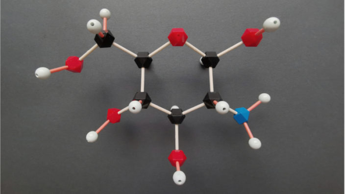
Glucosamine and chondroitin are both structural components of cartilage, the tissue that cushions the joints. Each of them is produced naturally in the body, but they are also available as dietary supplements.

Glucosamine is a naturally occurring polysaccharide that is found in cartilaginous joint tissues, bones, skin, ligaments, and nails, and it is involved in protein and lipid synthesis. It is contained inside the synovial fluid between the joints, where it helps to reduce friction on joint surfaces. Glucosamine is naturally present in some animal tissues, such as shellfish shells and animal bones, and even some types of fungi.

Chondroitin is a less well-studied molecule than glucosamine but is frequently paired with it, as some research suggests that it has anti-inflammatory properties. Chondroitin occurs naturally in animal tissue, especially in the joints and other connective tissue. However, these sources are much lower than the typical doses provided in chondroitin supplements.

Strangely, given its common usage for joint pain, it’s unclear whether glucosamine helps with osteoarthritis knee pain or in other joints. The results of previous studies also suggest that chondroitin isn’t helpful for pain from osteoarthritis in the knee, hip, or other joints.

No serious side effects have been reported in large, well-conducted studies of people taking glucosamine, chondroitin, or both for up to a 3-year period. However, glucosamine or chondroitin may interact with the blood-thinning drug warfarin, so caution is advised there.

The study results

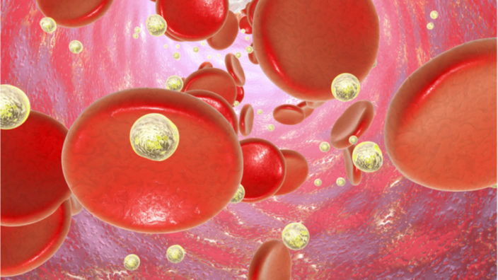
The new study looked at the data from over 16,000 participants who were taking glucosamine and chondroitin, with a particular focus on cardiovascular as well as all-cause mortality [1].

After controlling for age, supplementation with glucosamine and chondroitin was linked to a 65% reduction in cardiovascular mortality and a 39% reduction in all-cause mortality.

The researchers suggest that the most likely reason for this large influence on mortality is via the reduction of inflammation and the cytokines associated with it. In a previous study it was found that the level of C-reactive protein, a commonly used biomarker for systemic inflammation, was reduced by 23% in a test group taking glucosamine and chondroitin intake compared to a placebo group [2]. The same study showed that cytokine activity was also significantly reduced in the test group.

They also point out another study which showed that glucosamine influences the size of low-density lipoprotein and its ability to bind [3]. They suggest that this is possibly a way in which glucosamine may be able to reduce the development of atherosclerosis and reduce cardiovascular mortality.

Background: Limited previous studies in the United Kingdom or a single US state have demonstrated an association between intake of glucosamine/chondroitin and mortality. This study sought to investigate the association between regular consumption of glucosamine/chondroitin and overall and cardiovascular (CVD) mortality in a national sample of US adults.

Combined data from 16,686 participants in National Health and Nutrition Examination Survey 1999 to 2010, merged with the 2015 Public-use Linked Mortality File. Cox proportional hazards models were conducted for both CVD and all-cause mortality.

In the study sample, there were 658 (3.94%) participants who had been taking glucosamine/chondroitin for a year or longer. During followup (median, 107 months), there were 3366 total deaths (20.17%); 674 (20.02%) were due to CVD. Respondents taking glucosamine/chondroitin were less likely to have CVD mortality (hazard ratio [HR] = 0.51; 95% CI, 0.28-0.92). After controlling for age, use was associated with a 39% reduction in all-cause (HR = 0.61; 95% CI, 0.49–0.77) and 65% reduction (HR = 0.35; 95% CI, 0.20–0.61) in CVD mortality. Multivariable-adjusted HR showed that the association was maintained after adjustment for age, sex, race, education, smoking status, and physical activity (all-cause mortality, HR = 0.73; 95% CI, 0.57–0.93; CVD mortality, HR = 0.42; 95% CI, 0.23–0.75).

Conclusion

The results of this and other studies suggest that there is a strong association between glucosamine and chondroitin intake and a resulting decrease in cardiovascular and all-cause mortality.

It seems very likely that the mechanism in play here is a general reduction of systemic inflammation. Therefore, it stands to reason that measures that reduce such inflammation should be generally beneficial to tissue health and regeneration.

The usual caveats apply: if you are considering taking glucosamine and chondroitin, it should be done as part of developing a science-based personal longevity strategy. That said, glucosamine and chondroitin are cheap and readily available, and as long as you are prepared to proceed in a scientific and quantifiable manner, it may be worth considering.

We would like to ask you a small favor. We are a non-profit foundation, and unlike some other organizations, we have no shareholders and no products to sell you. All our news and educational content is free for everyone to read, but it does mean that we rely on the help of people like you. Every contribution, no matter if it’s big or small, supports independent journalism and sustains our future.

Literature

[1] King, D. E., & Xiang, J. (2020). Glucosamine/chondroitin and mortality in a US NHANES cohort. The Journal of the American Board of Family Medicine, 33(6), 842-847.

[2] Navarro, S. L., White, E., Kantor, E. D., Zhang, Y., Rho, J., Song, X., … & Lampe, J. W. (2015). Randomized trial of glucosamine and chondroitin supplementation on inflammation and oxidative stress biomarkers and plasma proteomics profiles in healthy humans. PloS one, 10(2), e0117534.

[3] Tannock, L. R., Little, P. J., Wight, T. N., & Chait, A. (2002). Arterial smooth muscle cell proteoglycans synthesized in the presence of glucosamine demonstrate reduced binding to LDL. Journal of lipid research, 43(1), 149-157.

Mouse eye

Reversing Cellular Age in Mice Restores Vision

Researchers at the Sinclair Lab at Harvard Medical School have restored lost vision to old mice, and mice with damaged retinal nerves, using partial cellular reprogramming. The aged cells can be reset to become younger again using this approach and are better at repairing damaged tissue.

The approach has huge potential in the treatment of age-related diseases, as it directly addresses and resets epigenetic alterations, one of the primary reasons we age.

The groundbreaking study, published in the journal Nature, is an important milestone on the path to translating the approach to humans and shows that it is possible to boost tissue regeneration in aged mammals making them more youthful.

Study co-author, Dr. David Sinclair, said in an article in Nature, “We set out with a question: if epigenetic changes are a driver of ageing, can you reset the epigenome?”, or, in other words, “Can you reverse the clock?”

This research builds on the previous research of Juan Carlos Izpisua Belmonte, who showed that it was possible to use partial reprogramming to reset cellular aging in mice back in 2016 [1]. Prior to this, it had been shown that cells could be reset this way in a petri dish, but it was a world first to show that it was possible to do the same in living animals.

The key to resetting the age of the cells is in triggering four genes to express the reprogramming factors Oct3/4, Sox2, Klf4 and c-Myc (OSKM), also known as Yamanaka factors. These factors are the basis of the well-known method of creating induced pluripotent stem cells (iPSCs), except unlike iPSCs, which are exposed to the reprogramming factors for a much longer time, these cells are only exposed for a transient period. This has the effect of resetting the functional age of a cell without it losing its identity and becoming fully reprogrammed as iPSCs are.

At the Sinclair Lab, researcher Yuancheng Lu was looking for a safer way to rejuvenate aged cells, as there were some concerns about using c-Myc, such as its propensity to induce cancer in certain circumstances. To reduce that risk, the researchers opted to remove c-Myc and used OSK only.

These three genes were placed into a viral vector and delivered to the target cells; the viral package also included a genetic switch that would allow OSK expression to be turned on when the mice were exposed to doxycycline, a common veterinary antibiotic, in their drinking water. In this manner, the researchers were able to turn the target genes on and off without stressing the mice, allowing them to reprogram the cells just long enough to reset their age.

The researchers chose to focus on the retinal nerves of the eye for this study. First, they injected the viral vector into the eye to find out if OSK would regenerate damaged nerves. Sure enough, expressing OSK in mice that had damaged eye nerves saw the tissue rejuvenate and repair itself. The team went on to show that vision was improved in mice with age-related vision impairment and in mice that experienced increased eye pressure, an emulation of glaucoma.

Finally, the approach was confirmed to have reset the epigenetic profile of aged cells back to a more youthful state in both mouse and human cells grown in the lab. The researchers are now exploring how aged cells retain an epigenetic memory of being young.

Ageing is a degenerative process that leads to tissue dysfunction and death. A proposed cause of ageing is the accumulation of epigenetic noise that disrupts gene expression patterns, leading to decreases in tissue function and regenerative capacity. Changes to DNA methylation patterns over time form the basis of ageing clocks, but whether older individuals retain the information needed to restore these patterns—and, if so, whether this could improve tissue function—is not known. Over time, the central nervous system (CNS) loses function and regenerative capacity. Using the eye as a model CNS tissue, here we show that ectopic expression of Oct4 (also known as Pou5f1), Sox2 and Klf4 genes (OSK) in mouse retinal ganglion cells restores youthful DNA methylation patterns and transcriptomes, promotes axon regeneration after injury, and reverses vision loss in a mouse model of glaucoma and in aged mice. The beneficial effects of OSK-induced reprogramming in axon regeneration and vision require the DNA demethylases TET1 and TET2. These data indicate that mammalian tissues retain a record of youthful epigenetic information—encoded in part by DNA methylation—that can be accessed to improve tissue function and promote regeneration in vivo.

Conclusion

This is another solid step forward on the road to translating partial cellular reprogramming to humans. Harvard has licensed the technology to Life Biosciences, a group of companies working on solving aging, which includes Dr. Sinclair on its scientific team.

While a cutting-edge approach like this is probably at least a decade away before reaching human trials, enthusiasm for partial cellular reprogramming is growing rapidly. There are many companies engaged in the approach, and it should not be long now before studies looking at other tissues and organs are published. Hopefully, these positive results will be replicated by other organizations in other tissues in the near future.

If researchers are right and epigenetic alterations are indeed a primary reason we age, resetting those alterations in humans could have a profound effect on our health and aging. Of course, there is a long way to go, but this is another step closer.

We would like to ask you a small favor. We are a non-profit foundation, and unlike some other organizations, we have no shareholders and no products to sell you. All our news and educational content is free for everyone to read, but it does mean that we rely on the help of people like you. Every contribution, no matter if it’s big or small, supports independent journalism and sustains our future.

Literature

[1] Ocampo, A., Reddy, P., Martinez-Redondo, P., Platero-Luengo, A., Hatanaka, F., Hishida, T., … & Araoka, T. (2016). In vivo amelioration of age-associated hallmarks by partial reprogramming. Cell, 167(7), 1719-1733.

researcher working with cell culture

A New Biomaterial for Thymic Cells

New research published in Biomacromolecules has successfully cultured thymic epithelial cells in vitro.

A 3D biomaterial for thymic epithelial cells

Tissue engineering and regenerative medicine attempts to regenerate tissues both in the lab and in vivo. While the fact that everyone experiences thymic involution should make it a prime target for regenerative medicine, very little research has been done in this area. As such, while the ideal biomaterials to culture various cells have been well investigated, much is still unknown about the behaviors of thymic cells. Thymic epithelial cells are one of the more populous cell types in the thymus and support the maturation of T cells. When isolated from the thymus and cultured in 2D, they exhibit several non-phenotypic behaviors. This limits the ability of researchers to use them to study the biology of the thymus or to regenerate thymic tissue.

Electrospinning is a manufacturing technique used for biomaterials in which the material is forced out of a very small nozzle by an electrical gradient. It is capable of forming highly porous structures made up of extremely small fibers. These 3D structures have been shown to be conducive to cell growth and mobility and the diffusion of nutrients throughout. A recent publication from a lab at the University of Minho in Portugal modified this technique and investigated its effects on cultured thymic epithelial cells. [1]

While electrospun fibers are architecturally highly beneficial for most cell types, they are often limited by their biological properties. Not all materials can be processed by electrospinning. One of the most common electrospinning materials, polycaprolactone, is not very biologically active. In this study, the researchers hypothesized that they could improve this material by functionalizing it with fibronectin, a key protein of the extracellular matrix in the thymus.

A step towards thymus regeneration

After electrospinning the polycaprolactone, they plasma treated their scaffolds and added amine groups by chemical treatment with hexamethylenediamine. They then chemically bound fibronectin to these newly formed amine groups. Each step of this process was optimized by the researchers, including treatment times and chemical concentrations, discovering the best manufacturing methods to create structures for thymic epithelial cell culture.

They then characterized the cultured cells via their metabolic activity, proliferation, and total protein deposition, with all three increasing for the seven days of the study. Cell attachments to the scaffold also increased throughout the study period. The cells, after being seeded on the surface, began to migrate towards the deeper portions of the scaffold. Lastly, the cultured cells’ expression of collagen IV and laminin, two key proteins deposited by this cell type in vivo, also increased throughout the experiment.

eFMs were successfully biofunctionalized through surface modification and subsequent immobilization of fibronectin, one of the major thymic ECM components. The FN-eFMs proved to be a suitable substrate for the culture of TECs, allowing cell adhesion, proliferation, and migration, together with the production of thymic ECM proteins such as collagen type IV and laminin. Overall, this study demonstrates that TECs showed an increased performance when cultured on 3D biofunctional structures than when cultured over 2D ones.

Conclusion

More research like this is sorely needed in the field of regenerative medicine. The effects of thymic involution may not be felt as directly as heart or liver failure, but it plays a large role in the progression of aging, including senescent cell accumulation and cancer. However, the results of this particular study should not be overstated. The researchers’ biomaterial (electrospun polycaprolactone functionalized with fibronectin) is potentially useful for thymus regeneration, but only minor improvements were seen relative to 2D culture on fibronectin and 3D culture on polycaprolactone without fibronectin. Additionally, seven days is a somewhat short length of time for cell cultures. While further culturing may have resulted in greater differences, that question remains unanswered. Finally, it is also unknown whether this biomaterial is an improvement over previously studied biomaterials, such as collagen or decellularized thymus extracellular matrix. Overall, there is a lot of room to expand on these initial positive findings.

We would like to ask you a small favor. We are a non-profit foundation, and unlike some other organizations, we have no shareholders and no products to sell you. All our news and educational content is free for everyone to read, but it does mean that we rely on the help of people like you. Every contribution, no matter if it’s big or small, supports independent journalism and sustains our future.

Literature

[1] Silva, C.S., Pinto, R.D., Amorim, S., Pires, R.A., Correia-Neves, M., … & Neves, N.M. (2020). Fibronectin-Functionalized Fibrous Meshes as a Substrate to Support Cultures of Thymic Epithelial Cells. Biomacromolecules, Online ahead of print. https://doi.org/10.1021/acs.biomac.0c00933

Small plate of food

A Key Gene Links Diet and Longevity

A key gene mediating the effect of dietary restriction on longevity has been identified, improving our understanding of the link between the two and raising the prospect of more targeted therapeutic interventions.

Less is more

Dietary restriction is one of the most reliable ways to increase longevity, increasing lifespan in species from fruit flies to rodents. Benefits can be seen from a range of dietary changes, such as intermittent fasting or caloric restriction, though restricting specific components of the diet, such as proteins or even specific amino acids, may be the most effective change.

Researchers at the Max Planck Institute for Biology of Ageing have pinned down a gene that plays a key role in this process, Sestrin1. The Sestrin genes – there are three versions of Sestrin in mammalian genomes and one in the Drosophila genome – encode proteins that detect amino acids and negatively regulate TORC1. TORC1 is part of the IGF/TOR nutrient-sensing network linking dietary restriction and longevity, but because this network is also involved in other processes, such as wound healing and insulin response, it may not be an ideal therapeutic target.

A specific sensor

The study began by investigating the effect of knocking out Sestrin in fruit flies and found that this reduced their increase in lifespan in response to dietary restriction, which isn’t really surprising, given that Sestrin regulates TOR. Next, the team engineered a version of Sestrin with a mutation in the amino acid binding region, preventing it from acting as an amino acid sensor. When cells starved of amino acids were fed the amino acid leucine, genes downstream of TORC1 were only activated if the cells had normal Sestrin, not the mutated version. In other words, Sestrin is a key amino acid sensor for the TORC1 pathway.

The researchers followed up with in vivo tests and found that flies with the mutated form of Sestrin  didn’t live longer on a restricted diet. More interesting still, the mutant flies lived longer than ordinary flies on a high-protein diet, but that was mainly due to the low survival of wild-type flies. Therefore,  it seems that Sestrin not only mediates the nutrient detection that makes dietary restriction beneficial but may also protect against the harmful effects of a protein-rich diet.

With a few more experiments, the researchers investigated how Sestrin mediates the effect of dietary restriction on lifespan. They found that it regulates gut homeostasis, specifically by modulating autophagy of intestinal stem cells to regulate turnover in the gut, helping keep it healthy. Raising Sestrin expression in intestinal stem cells led to a 10% increase in the lifespan of fruit flies.

Dietary restriction (DR) promotes healthy aging in diverse species. Essential amino acids play a key role, but the molecular mechanisms are unknown. The evolutionarily conserved Sestrin protein, an inhibitor of activity of the target of rapamycin complex 1 (TORC1), has recently been discovered as a sensor of amino acids in vitro. Here, we show that Sestrin null mutant flies have a blunted response of lifespan to DR. A mutant Sestrin fly line, with blocked amino acid binding and TORC1 activation, showed delayed development, reduced fecundity, extended lifespan and protection against lifespan-shortening, high-protein diets. Sestrin mediated reduced intestinal stem cell activity and gut cell turnover from DR, and stem cell proliferation in response to dietary amino acids, by regulating the TOR pathway and autophagy. Sestrin expression in intestinal stem cells was sufficient to maintain gut homeostasis and extend lifespan. Sestrin is thus a molecular link between dietary amino acids, stem cell function and longevity.

Conclusion

The identification of Sestrin as a key player linking dietary restriction and longevity makes it an attractive target for therapeutic intervention. There still isn’t good evidence that dietary restriction increases lifespan in humans, and even if there were, many people have trouble keeping to a restricted diet or might not be able to for medical reasons. Finding other ways to achieve the same effect thus seems like a promising path forward, and Sestrin provides one more tool with which to forge that path.

We would like to ask you a small favor. We are a non-profit foundation, and unlike some other organizations, we have no shareholders and no products to sell you. All our news and educational content is free for everyone to read, but it does mean that we rely on the help of people like you. Every contribution, no matter if it’s big or small, supports independent journalism and sustains our future.

Literature

[1] Lu J, Temp U, Müller-Hartmann A, Esser J, Grönke S, and Partridge L. Sestrin is a key regulator of stem cell function and lifespan in response to dietary amino acids. Nature Aging (2020), doi: 10.1038/s43587-020-00001-7
Disintegrating mind

Neurological Aging and the Lifespan Limit

Jeanne Louise Calment from France was the oldest person to grace this Earth, living a full life until 122 years of age [1]. She continuously exercised, taking up fencing in her 80s and cycling in her 100s. She claimed to have achieved such a long life through olive oil, port, and chocolate.

There are many speculations as to why she lived so long, from inherited traits to a  hypothesis that she may have faked her mother’s identity, which is buttressed by an analysis of facial features and a plausible reason for the switch [2]. However, assuming that she is truly 122 years old, her long survival was likely due to her exercise, eating habits, and inherited long lifespan [3].

While this is an interesting thing to ponder, a more interesting question for us is ‘Why did she die at 122?’ The answer isn’t definite, but there are some speculations, particularly one that the problem didn’t lie within mobility, or habits; instead, it lied within her brain.

A gradual depletion

Dr. Mora Francisco theorizes that by 120 years of age, even if we suffer from no other age-related diseases, the number of synapses (connections between neurons) in the neocortex, a location in the brain that controls cognition and movement, decreases to that of people with Alzheimer’s disease [4].

Dr. Francisco’s paper states that this can actually lead to a form of dementia without the common indicators. Key differences in advanced age lie within calcium homeostasis in neurons and decreases in synaptic density (thereby causing a decrease in different types of matter such as grey matter)  [5].

Calcium homeostasis seems to be increasingly dysregulated with age, caused by changes in the influx (redirection with age) to neurons. This adversely influences cell structure and many molecular pathways. [6]

In a study conducted in 2003, synaptic density was found to decrease in aged adults, resulting in lower neocortical density and the density of other affected areas within the brain [7]. This is because neurons gain all their synapses by the age of one, and the amount declines from there.

Stress may seem pretty inconsequential as a contributor to brain aging; however, Harvard researchers found in a 2019 study that lower neural excitation leads to longer lifespan [8]. They reported that it was caused by a gene regulator named ‘REST,’ which does its typical job during fetal development and is found to return to its post at an advanced age, in which it causes the brain to adapt to age. People who have higher levels of neural excitation have less of this regulator, as it suppresses genes involved in neural excitation.

Efforts to reverse the decline

Currently, there are multiple efforts to combat aging of the brain. Since it’s hard to get trials approved for brain aging, and the subject is very broad, the topic is being studied as its separate disorders, including Parkinson’s and various dementias.

For Parkinson’s, drugs to increase levels of dopamine in the brain have been proven effective in mice, such as through upregulating trace amine-associated receptor 5 (TAAR5). TAAR5 is shown to generally decline with age, so this study directly targets a side effect of aging [9].

To treat Alzheimer’s, Biogen is working on aducanumab, a drug that is intended to reduce beta amyloid in the brain [10]. Beta amyloid builds up in the brain during Alzheimer’s, making this a direct effort against a disease mechanism. Age-related amyloid buildup is a normal occurrence and happens on a smaller scale for people without Alzheimer’s [11]; therefore, in lower doses, this could become useful as a treatment for brain aging.

There are many efforts being led on this front, and it is suspected that we will see new research coming out soon on these topics of interest.

We would like to ask you a small favor. We are a non-profit foundation, and unlike some other organizations, we have no shareholders and no products to sell you. All our news and educational content is free for everyone to read, but it does mean that we rely on the help of people like you. Every contribution, no matter if it’s big or small, supports independent journalism and sustains our future.

Literature

[1] Robine, J.-M., Allard, M., Herrmann, F. R., & Jeune, B. (2019). The Real Facts Supporting Jeanne Calment as the Oldest Ever Human. The Journals of Gerontology: Series A, 74(Supplement_1), S13–S20. https://doi.org/10.1093/gerona/glz198

[2] Deigin, Y. (2019, March 1). Jeanne or Yvonne? New evidence in the Calment affair. Medium. https://yurideigin.medium.com/jeanne-or-yvonne-new-evidence-in-the-calment-affair-c35451f0d899

[3] Robin-Champigneul F. (2020). Jeanne Calment’s Unique 122-Year Life Span: Facts and Factors; Longevity History in Her Genealogical Tree. Rejuvenation research, 23(1), 19–47. https://doi.org/10.1089/rej.2019.2298.

[4] Mora F. (2013). Successful brain aging: plasticity, environmental enrichment, and lifestyle. Dialogues in clinical neuroscience, 15(1), 45–52.

[5] Mora, F., Segovia, G., & del Arco, A. (2007). Aging, plasticity and environmental enrichment: Structural changes and neurotransmitter dynamics in several areas of the brain. Brain Research Reviews, 55(1), 78–88. https://doi.org/10.1016/j.brainresrev.2007.03.011

[6] Toescu, E. C., Verkhratsky, A., & Landfield, P. W. (2004). Ca2+ regulation and gene expression in normal brain aging. Trends in Neurosciences, 27(10), 614–620. https://doi.org/10.1016/j.tins.2004.07.010

[7] Peter R., H. (1979). Synaptic density in human frontal cortex — Developmental changes and effects of aging. Brain Research, 163(2), 195–205. https://doi.org/10.1016/0006-8993(79)90349-4

[8] Zullo, J. M., Drake, D., Aron, L., O’Hern, P., Dhamne, S. C., Davidsohn, N., Mao, C.-A., Klein, W. H., Rotenberg, A., Bennett, D. A., Church, G. M., Colaiácovo, M. P., & Yankner, B. A. (2019). Regulation of lifespan by neural excitation and REST. Nature, 574(7778), 359–364. https://doi.org/10.1038/s41586-019-1647-8

[9] Efimova, E. V., Kozlova, A. A., Razenkova, V., Katolikova, N. V., Antonova, K. A., Sotnikova, T. D., Merkulyeva, N. S., Veshchitskii, A. S., Kalinina, D. S., Korzhevskii, D. E., Musienko, P. E., Kanov, E. V., & Gainetdinov, R. R. (2021). Increased dopamine transmission and adult neurogenesis in trace amine-associated receptor 5 (TAAR5) knockout mice. Neuropharmacology, 182, 108373. https://doi.org/10.1016/j.neuropharm.2020.108373

[10] Biogen. (n.d.). Alzheimer’s Disease. Retrieved November 18, 2020, from https://www.biogen.com/en_us/alzheimers-disease.html

[11] Rodrigue, K. M., Kennedy, K. M., & Park, D. C. (2009). Beta-Amyloid Deposition and the Aging Brain. Neuropsychology Review, 19(4), 436–450. https://doi.org/10.1007/s11065-009-9118-x

Nanoparticles in blood

CRISPR Successfully Deployed Against Two Cancer Types

Scientists have successfully tested a new nanoparticle-based delivery system for CRISPR kits, achieving drastic improvements in mouse models of glioblastoma and ovarian cancer [1].

A gift from our single-cell ancestors

CRISPR gene editing technology has been widely hailed as a potential game changer in medicine, including anti-cancer therapy. CRISPR’s ability to permanently disrupt tumor survival genes potentially constitutes a giant improvement over the long, exhausting, and toxic treatment protocols that exist today. However, to achieve this feat, CRISPR-based molecular systems need to be delivered to every cancer cell, which is not a trivial task.

Scientists gleaned CRISPR from bacteria. In nature, this evolutionarily developed defense mechanism allows bacteria to remember the DNA of bacteriophage viruses it encounters. The data is stored in the cell’s genome in sequences called CRISPR arrays. In the event of a new penetration by the same virus, the CRISPR system recognizes and cuts the viral DNA using Cas (CRISPR-associated proteins), rendering the virus harmless. It is essentially a primitive form of acquired immunity for single-cell organisms.

Several Cas proteins have been found in various bacteria. Cas9 is one of the most promising, since it can cleave nearly any DNA sequence. Unfortunately, it is also massive, making targeted cellular delivery of Cas9-based systems challenging. In previous research, the best results were achieved in the liver, with up to 60% gene editing efficacy (the percentage of cancer cells edited). In cancers that affect other tissues, the results have been less encouraging, such as 15% in lung cancer and 3% in melanoma.

Self-assembling weapons 

Most in vivo studies have used adeno-associated viruses (AAVs) to deliver CRISPR components to cells. Unfortunately, this delivery system is plagued by low carrying capacity, hepatotoxicity at high doses, long-term genotoxicity [2], and other effects. It also tends to provoke an immune response, as it is based on viral DNA [3]. This time, researchers decided to try a different approach: lipid nanoparticles (LNPs). In recent years, LNPs have gained popularity as a delivery vehicle for drug molecules [4], due to their superior cell-penetrating abilities and low toxicity. “Loaded” LNPs are assembled from a mixture of lipid molecules and cargo molecules (Cas9-coding mRNA and the “single-guide RNA” that tells Cas9 where to cut):

Using a novel library of lipid formulations, the researchers were able to develop a working delivery system. LNPs they had created successfully encapsulated CRISPR-Cas9 systems and delivered them to various cancer cells in vitro, with up to 98% gene editing efficacy. As for the mechanism of action, the researchers aimed for PLK1, a kinase that is essential for mitosis (cellular division). With PLK1 gene cut out, the cells were unable to divide. This resulted in cell cycle arrest and death by apoptosis.

LNPs treat brain and ovarian cancer

Of course, the ultimate test had to be done in vivo. For the first set of experiments, the researchers chose glioblastoma (GBM), a type of aggressive brain cancer. GBM is exceptionally lethal and resistant to treatment, mainly because the blood-brain barrier prevents conventional anti-cancer drugs from reaching the tumor. Only 5% of GBM patients survive for more than five years.

The researchers injected GBM cells into the hippocampi of mice. Following tumorigenesis, CRISPR-carrying LNPs were injected directly into the tumors, overriding the blood-brain barrier. A single injection resulted in 68% gene editing and high rates of apoptosis, while the surrounding healthy tissue remained mostly intact. Tumor growth was significantly reduced following the injection. 30% of mice survived for 60 days, which is when the experiment was terminated, while all the mice in the control group died within 40 days. The researchers claim that this is the highest survival improvement ever achieved in GBM by a single treatment.

Whenever a direct injection into a single non-metastasizing tumor is possible, the non-specificity of LNPs does not pose a problem. However, to engage metastatic cancer, a systemic injection is needed, so the treatment must ensure that CRISPR-carrying LNPs do not kill healthy cells. The scientists managed to coat LNPs with antibodies specific to various cancer cell types, and such LNPs zoom in on their targets while ignoring healthy cells. In a murine model of metastatic ovarian cancer, CRISPR-carrying LNPs were injected into the abdominal cavity (intraperitoneally). Despite being administered systemically, they delivered 82% of gene editing, strong tumor inhibition, and 80% increase in overall survival.

Conclusion

This research constitutes a potential breakthrough in cancer therapy. LNPs demonstrated their ability to deliver DNA-cleaving CRISPR kits directly to cancer cells even when administered systemically. Direct injection of CRISPR-carrying LNPs into brain tumors appears to be a workable way to override the blood-brain barrier and successfully attack treatment-resistant brain cancer. Although more must be done to establish the efficacy and low toxicity of LNP-based delivery systems, the concept has been proven.

We would like to ask you a small favor. We are a non-profit foundation, and unlike some other organizations, we have no shareholders and no products to sell you. All our news and educational content is free for everyone to read, but it does mean that we rely on the help of people like you. Every contribution, no matter if it’s big or small, supports independent journalism and sustains our future.

Literature

[1] Rosenblum, D., Gutkin, A., Kedmi, R., Ramishetti, S., Veiga, N., Jacobi, A. M., … & Lieberman, J. (2020). CRISPR-Cas9 genome editing using targeted lipid nanoparticles for cancer therapy. Science Advances, 6(47), eabc9450.

[2] Hanlon, K. S., Kleinstiver, B. P., Garcia, S. P., Zaborowski, M. P., Volak, A., Spirig, S. E., … & Lööv, C. (2019). High levels of AAV vector integration into CRISPR-induced DNA breaks. Nature communications, 10(1), 1-11.

[3] Ronzitti, G., Gross, D. A., & Mingozzi, F. (2020). Human Immune Responses to Adeno-Associated Virus (AAV) Vectors. Frontiers in Immunology, 11.

[4] Puri, A., Loomis, K., Smith, B., Lee, J. H., Yavlovich, A., Heldman, E., & Blumenthal, R. (2009). Lipid-based nanoparticles as pharmaceutical drug carriers: from concepts to clinic. Critical Reviews™ in Therapeutic Drug Carrier Systems, 26(6).

Rejuvenation Roundup November

Rejuvenation Roundup November 2020

Thanksgiving is over, but we’re still thankful that so many people have decided to help stop the ravages of age-related disease by researching the causes of aging and joining the organizations needed to bring this research to people, and we’re definitely thankful for everyone who chooses to directly support us.

LEAF News

Breanna Deutsch Wants You to Contact Lawmakers About Aging: Coming from the world of political communications, Breanna Deutsch, author of Finding the Fountain, highlights the role of public comment in creating public policy.

Rejuvenation Roundup Podcast

Ryan O’Shea of Future Grind hosts this month’s podcast, showcasing the events and research discussed here.

Helpful Information

Why I Hope to Be Alive at 75: Ezekiel Emanuel, the nation’s incoming COVID advisor, has expressed the opinion that it is a good idea to die at 75. Here, Editor-in-Chief Steve Hill disagrees with this method of dealing with age-related diseases.

Media Circus Surrounds Hyperbaric Oxygen Study: The media hype surrounding the results is nothing like the reality of the actual research paper, and this is another example of how shoddy journalism harms our field.

Send in the senolytics: Unity might have failed with UBX0101, but many other companies intend to make their own attempts at senescent cell removal, and Unity is trying again as well.

Research Roundup

The Extracellular Matrix Is Key to Heart Health: A new article sheds light on the differences between the extracellular matrix (ECM) of healthy people and people in end-stage heart failure.

C60 Does Not Improve Lifespan nor Healthspan: The findings of this study not only raise concerns about available C60 supplements, they also undermine the rationale for using them in the first place.

Researchers Use Lung Organoids to Study COVID: This highly impactful and timely research simultaneously makes major contributions to the field of lung regeneration and to our fight against the current global pandemic.

Aged Blood Makes Young Cells Old: Exposing young cells to aged blood causes their gene expression to become akin to aged cells.

Diluting Aged Blood Rejuvenates Old Brains: Aged blood can support rejuvenation if properly calibrated or, in this case, simply diluted.

Inflammation Decreases NAD+ During Aging: Researchers have discovered a link between senescent cells and the decline of NAD+ that occurs with aging.

Avoiding Osteoporosis with Alpha-Ketoglutarate: Research on rodents shows that alpha-ketoglutarate could be an effective way to treat or even prevent osteoporosis.

A New Scaffold for Liver Regeneration: This paper demonstrates that an alginate-gelatin-fibrin hydrogel formulation is a promising biomaterial for liver regeneration.

Gene Discovered to Alleviate Obesity: This particular gene removes some of the consequences of obesity without promoting actual weight loss.

Immune Protection for Insulin-Producing Cells: This is protection against the immune system, which both Type 1 and Type 2 diabetics need to retain working insulin-producing cells.

GrimAge Is the Best Clock for Predicting Death: Having an accelerated age according to the GrimAge epigenetic clock means that you are much more likely to die of any cause.

Linking Alzheimer’s Disease and Gut Bacteria: There is evidence that there is a bacterial element in some cases of Alzheimer’s disease, and a new study further reinforces this link.

Resveratrol Human Trial Results Published: The results of this study suggest that resveratrol may be beneficial for people suffering from heart failure.

Resveratrol improves motor function in patients with muscular dystrophies: Resveratrol decreases muscular and cardiac oxidative damage and improves conditions in animal models of muscular dystrophy.

The positive effect of spermidine in older adults suffering from dementia: The high dosage spermidine group results are far better than the achieved effects of currently available antidementia drugs. The placebo effect may be involved, so a follow-up study with a duration of 1 year is planned.

Physical activity and quality of life following astaxanthin in patients with heart failure: This compound was shown to increase self-reported physical activity and health-related quality of life in mental and physical ways.

Aspirin as a suppressor of cancer-related epigenetic aging: This study shows that, in colon tissue, methylation age in aspirin non-users increases with chronological age; however, in aspirin users, methylation age acceleration is suppressed according to epigenetic clocks.

Rapamycin-mediated mouse lifespan extension: Rapamycin at 42 parts per million in food increased survival significantly in both male and female middle-aged mice.

PPARγ agonists delay age-associated metabolic disease and extend longevity: One of these drugs reduces inflammation, fibrosis, and atrophy in aging tissues along with a host of other benefits.

Clinical Trial Examining Lactobacillus rhamnosus for Cognitive Functioning: Supplementation of this probiotic was associated with improved cognitive performance in middle-aged and older adults with cognitive impairment.

Senescent cell accumulation mechanisms inferred from parabiosis: Optimal treatments for removing senescent cells would match the time between treatments to the time it takes senescent cells to re-accumulate.

Circulating plasma factors involved in rejuvenation: This is a summary of blood factors that are associated with youthful and aged blood, including the effects of each one.

Telomerase improves motor function in a transgenic mouse model of Parkinson’s disease: Total, phosphorylated, and aggregated a-synuclein, which is associated with this disease, were significantly decreased in the hippocampus and neocortex of treated mice.

News Nuggets

OneSkin Has Released a Senotherapeutic Skin Cream: OneSkin recently released OS-01, a cosmetic skin care product that claims to reduce the activity of senescent cells according to typical cellular senescence biomarker

AI and Longevity Meet in a New Book: “Live Longer with AI” stretches far beyond the topic of AI and longevity to include everything from the basics of the biological machinery of aging to the ethics of immortality

Elevian Secures $15 Million in Equity Financing: This company is developing new drugs that can target the GDF11 pathway in order to spur rejuvenation of tissue.

Coming Up

Throughout this month, starting December 1st, we will be releasing a video from Ending Age-Related Diseases 2020 to the public! Subscribe to our YouTube channel to see the latest videos as they appear.

We would like to ask you a small favor. We are a non-profit foundation, and unlike some other organizations, we have no shareholders and no products to sell you. All our news and educational content is free for everyone to read, but it does mean that we rely on the help of people like you. Every contribution, no matter if it’s big or small, supports independent journalism and sustains our future.
Red wine

Resveratrol Human Trial Results Published

A human study on the popular supplement resveratrol on heart failure patients has been published, and the results are significant.

What is resveratrol?

Resveratrol, is a naturally occurring stilbenoid, a type of phenol that is present in various plants and foods, such as grapes, peanuts, cocoa, blueberries, raspberries, and cranberries. It is frequently mentioned as being a component of red wine rather than white wine, and this is because it is found in the grape skins that are used in the creation of red wine.

Resveratrol is known to increase the activity of SIRT1, often called the longevity gene, in multiple species, and it influences the development of fat cells. Given these qualities, resveratrol has been of interest as a potential candidate for treating cardiovascular disease. Indeed, there is human data suggesting that resveratrol supplementation decreases apolipoprotein-B and LDL cholesterol levels, whose excessive presence contributes to cardiovascular disease [1-2].

What did the study show?

The results of this study suggest that resveratrol may be beneficial for people suffering from heart failure [3]. Certainly, previous animal studies on this topic have achieved positive results, but, until now, the effects in people with heart failure had not been explored.

This was a small three-month human trial involving 60 participants, who were all suffering from heart failure and New York Heart Association class II-III symptoms for their condition. Class II is a category of mild symptoms, such as mild shortness of breath and/or angina with some limitations on performing ordinary activity. Class III symptoms include more serious limitations to regular activity and even difficulty during light activity, such as walking short distances.

For this study, half the participants were given a placebo, and the other half were given 100 milligrams of resveratrol every day for three months.

At the start and end of the study, the participants each had an echocardiogram, which uses ultrasound to show how the heart muscle and valves are functioning. In addition, a six-minute walk test, a quality of life questionnaire, a lung function test, and an RNA profile analysis were performed.

The results showed that the participants given resveratrol saw an improvement in systolic and diastolic left ventricular function and global longitudinal strain. This suggests that there was some improvement to heart function in these participants. This is supported by the increase of exercise capacity and improved ventilation metrics taken. There was also some improvement to participant quality of life according to the questionnaire they were given, though such things are subjective, of course.

The N-terminal prohormone of brain natriuretic peptide (NT-proBNP) in blood samples also showed a significant reduction. NT-proBNP levels go up when the heart cannot pump blood around the body as well as it should. The higher the level, the more likely that heart failure is present and the more severe it typically is.

There was also a marked reduction of galectin-3 in people treated with resveratrol. Galectin-3 is a proinflammatory lectin that can bind to β-galactoside and is secreted by activated macrophages. Galectin-3 is often used as a biomarker for heart failure.

Finally, in the resveratrol group, the levels of inflammatory cytokines IL-1 and IL-6 were significantly reduced. However, C-reactive protein and ferritin, whose elevated levels are commonly associated with heart failure, showed no significant change. There was also no significant change to white blood cell count, another biomarker of inflammation and heart failure.

The effects of resveratrol (RES) in heart failure have already been evaluated in animal models; however, in human clinical trials, they have not been confirmed yet. The aim of this study was to assess the effects of resveratrol treatment in systolic heart failure patients (heart failure with reduced ejection fraction or HFrEF). In this human clinical trial, 60 outpatients with NYHA (New York Heart Association) class II-III HFrEF were enrolled and randomized into two groups: receiving either 100-mg resveratrol daily or placebo for three months. At the beginning and at the end of the study echocardiography, a six-minute walk test, spirometry, quality of life questionnaire, lab test and RNA profile analysis were performed. The systolic and diastolic left ventricular function, as well as the global longitudinal strain, were improved significantly in the resveratrol-treated group (RES). Exercise capacity, ventilation parameters and quality of life also improved significantly in the RES group. In parallel, the cardiac biomarker levels (N-terminal prohormone of brain natriuretic peptide (NT-proBNP) and galectin-3) decreased in the treated group. The level of inflammatory cytokines decreased significantly after RES supplementation, as a consequence of the decreased expression level of leucocyte electron transport chain proteins. The main findings of our trial are that RES treatment added to the standard heart failure therapy improved heart function and the clinical condition by moderating the inflammatory processes in patients with HFrEF.

Conclusion

These results are mostly positive, though, as the researchers themselves correctly point out, this initial study has some limitations. As it has a small group and a fairly short follow-up period, it should be followed up by a larger-scale trial, ideally one with a longer treatment and follow-up period.

We would like to ask you a small favor. We are a non-profit foundation, and unlike some other organizations, we have no shareholders and no products to sell you. All our news and educational content is free for everyone to read, but it does mean that we rely on the help of people like you. Every contribution, no matter if it’s big or small, supports independent journalism and sustains our future.

Literature

[1] Magyar, K., et al. (2012). Cardioprotection by resveratrol: A human clinical trial in patients with stable coronary artery disease. Clinical Hemopheology and Microcirculation, 50(3): 179-187.

[2] Tomé-Carneiro, J., (2012). Consumption of a grape extract supplement containing resveratrol decreases oxidized LDL and ApoB in patients undergoing primary prevention of cardiovascular disease: a triple-blind, 6-month follow-up, placebo-controlled, randomized trial. Molecular Nutrition and Food Research, 56(5): 810-821.

[3] Gal, R., Deres, L., Horvath, O., Eros, K., Sandor, B., Urban, P., … & Habon, T. (2020). Resveratrol Improves Heart Function by Moderating Inflammatory Processes in Patients with Systolic Heart Failure. Antioxidants, 9(11), 1108.

Intestinal bacteria

Linking Alzheimer’s Disease and Gut Bacteria

In recent years, there has been evidence that the gut microbiota plays an important role in the development of Alzheimer’s disease. Certainly, there is evidence that there is a bacterial element in some cases of Alzheimer’s disease, and a new study further reinforces this link.

The researchers of this new study have shown a correlation, in humans, between populations of harmful bacteria in the gut microbiome and the development of the amyloid plaques in the brain seen in Alzheimer’s disease [1].

The research suggests that certain harmful bacteria present in the gut microbiome produce proteins that appear to modify the interaction between the immune and nervous systems and contribute to the development of Alzheimer’s disease.

The research team of one of the study authors, Giovanni Frisoni, director of the HUG Memory Centre and professor at the Department of Rehabilitation and Geriatrics of the UNIGE Faculty of Medicine, previously showed that the gut microbiota of Alzheimer’s patients is different from the microbiota of people who do not have the disease. They showed that the microbiotal diversity in these patients was altered, with certain populations of bacteria increasing in numbers while others fell.

They also previously showed a link between inflammatory signals in the blood, the elevated presence of certain types of gut bacteria, and Alzheimer’s disease. This led the researchers of this new study to try to find out if those inflammatory signals in the blood were acting as a go-between for the gut microbiota and the brain.

It is already known that the bacteria in the gut can communicate with and change how the immune system functions, which, in turn, alters interactions between the immune and nervous systems. These interactions, when harmful, can support neurodegeneration and the onset of conditions such as Alzheimer’s disease.

As a case in point, lipopolysaccharides, proteins present on the membranes of bacteria, are known to have proinflammatory properties and have been observed in and around the amyloid plaques and blood vessels of people with Alzheimer’s disease.

Gut bacteria can also interact with our metabolism through the production of short-chain fatty acids. Some short-chain fatty acids, such as butyrate, have been shown to have beneficial neuroprotective and anti-inflammatory properties, and in the case of butyrate, there is murine evidence that supplementing the loss of this particular short-chain fatty acid improves health. On the other hand, some short-chain fatty acids may have a harmful influence on metabolism and brain health.

Looking for the link

Knowing that interactions like this happen, the researchers investigated to see if inflammatory signals and other molecules from gut bacteria were linked to the onset of amyloid pathology. For the study, they enrolled a group of 89 participants aged between 65 and 85 years old, some with Alzheimer’s disease or other age-related neurodegenerative disorders.

They examined each participant using PET imaging and noted the presence of amyloid. Next, they took blood samples and examined the presence of inflammatory biomarkers and other proteins known to be produced by gut bacteria, which included lipopolysaccharides and short-chain fatty acids.

They found that there was a very strong correlation between some of these biomarkers and bacteria-produced proteins and the amount of amyloid plaque present in the brains of the study participants. The data showed that high levels of lipopolysaccharides in the blood were linked to large amyloid plaques in the brain.

The same was also true for the short-chain fatty acids acetate and valerate, whose elevated presence was associated with larger plaque deposits. However, high levels of butyrate were linked to reduced amyloid plaques.

The researchers now plan to identify the specific bacteria in the microbiota in order to ascertain which ones are responsible for promoting amyloid formation via the bloodstream.

We also discussed this paper with Dr. Michael Lustgarten, a microbiota expert from Tufts University, Boston. We also recommend checking out his YouTube channel for more microbiome information.

Metagenomic data support an association between certain bacterial strains and Alzheimer’s disease (AD), but their functional dynamics remain elusive. Objective:To investigate the association between amyloid pathology, bacterial products such as lipopolysaccharide (LPS) and short chain fatty acids (SCFAs: acetate, valerate, butyrate), inflammatory mediators, and markers of endothelial dysfunction in AD. Methods:Eighty-nine older persons with cognitive performance from normal to dementia underwent florbetapir amyloid PET and blood collection. Brain amyloidosis was measured with standardized uptake value ratio versus cerebellum. Blood levels of LPS were measured by ELISA, SCFAs by mass spectrometry, cytokines by using real-time PCR, and biomarkers of endothelial dysfunction by flow cytometry. We investigated the association between the variables listed above with Spearman’s rank test. Results:Amyloid SUVR uptake was positively associated with blood LPS (rho≥0.32, p≤0.006), acetate and valerate (rho≥0.45, p < 0.001), pro-inflammatory cytokines (rho≥0.25, p≤0.012), and biomarkers of endothelial dysfunction (rho≥0.25, p≤0.042). In contrast, it was negatively correlated with butyrate (rho≤–0.42, p≤0.020) and the anti-inflammatory cytokine IL10 (rho≤–0.26, p≤0.009). Endothelial dysfunction was positively associated with pro-inflammatory cytokines, acetate and valerate (rho≥0.25, p≤0.045) and negatively with butyrate and IL10 levels (rho≤–0.25, p≤0.038).

We report a novel association between gut microbiota-related products and systemic inflammation with brain amyloidosis via endothelial dysfunction, suggesting that SCFAs and LPS represent candidate pathophysiologic links between the gut microbiota and AD pathology.

Conclusion

The association between the gut microbiota and the formation of amyloid plaques seems pretty clear from this data.

The results also suggest that butyrate produced by gut bacteria from plant fiber, and potentially butyrate supplementation, could be a way to support brain health, as mouse studies have shown.

We would like to ask you a small favor. We are a non-profit foundation, and unlike some other organizations, we have no shareholders and no products to sell you. All our news and educational content is free for everyone to read, but it does mean that we rely on the help of people like you. Every contribution, no matter if it’s big or small, supports independent journalism and sustains our future.

Literature

[1] Marizzoni, M., Cattaneo, A., Mirabelli, P., Festari, C., Lopizzo, N., Nicolosi, V., … & Coppola, L. (2020). Short-chain fatty acids and lipopolysaccharide as mediators between gut dysbiosis and amyloid pathology in Alzheimer’s Disease. Journal of Alzheimer’s Disease, (Preprint), 1-15.

Death clock

GrimAge Is the Best Clock for Predicting Death

A new study published in the Journals of Gerontology has shown that the epigenetic clock GrimAge is a strong predictor of the effects of aging, particularly all-cause mortality: the likelihood of dying from any cause.

Did previous clocks use the wrong target?

As the researchers explain, the first-generation epigenetic clocks of Drs. Horvath (who contributed to this paper) and Hannum are very good at guessing calendar age: it is possible to take a cell sample, analyze it using one of these clocks, and accurately guess how old the donor is.

Ideally, it would be possible to use this kind of analysis to measure age acceleration effects. If such a clock is truly measuring the effects of aging, if a person is epigenetically older than the clock signifies, that person should be at greater risk for age-related diseases than a person who has not experienced such age acceleration.

Unfortunately, these two first-generation clocks are ill-suited for measuring the physical effects of age acceleration. This study has found that with these clocks, increases of epigenetic age over calendar age do not correlate with physical ability tests, cognitive tests, or the need for a patient to be prescribed multiple drugs (polypharmacy). Most importantly, they are not significantly correlated with all-cause mortality, making them practically useless for estimating the efficacy of a therapy that is designed to extend lifespan and healthspan.

This is where the second-generation clocks shine. The PhenoAge clock, as its name suggests, was trained against the phenotypes (effects) of aging, and the GrimAge clock was specifically trained against plasma proteins and the effects of smoking.

The study

A total of 490 people were tested as part of the epigenetic study, and they were tested for eight metrics: walking speed, grip strength, polypharmacy, the Fried Frailty Score, the Mini-Mental State Exam, the Montreal Cognitive Assessment, the Sustained Attention Reaction Time Task, and the Choice Reaction Time Task.

All four clocks were studied with this cohort, and the cohort was investigated for social mobility, which is a strong predictor of health outcomes, as well as other factors known to affect health, such as body mass index (BMI), smoking history, and alcohol consumption; while such things may affect epigenetics as well, it is of marginal value to have a clock that turns out to only successfully measure their influence.

In an simple, minimally adjusted analysis that did not control for these factors, the PhenoAge clock predicted four of the tested items, but “none of these associations survived multivariable adjustment.” GrimAge, by contrast, was associated with seven of the eight, and after multivariable adjustment, it still continued to predict three of them.

Most notably, GrimAge was very successful at predicting all-cause mortality, and none of the other clocks came close. People whose age acceleration, as measured by GrimAge, were one standard deviation above the mean were twice as likely to die over this 10-year study, and this association held true even when controlling for other factors.

Conclusion

The researchers state that GrimAge may already be a useful clinical endpoint for testing the efficacy of rejuvenation therapies that directly affect aspects of aging. If researchers already have a cellular biomarker that reliably correlates with a therapy’s effects on human beings, they can use it to evaluate therapies even in the earliest preclinical phases, which utilize human cells.

Furthermore, the researchers hypothesize that even more efficient epigenetic clocks can be developed. Such third-generation clocks would be built specifically to the purpose, being directly trained on frailty indices and similar predictors of mortality rather than on calendar age. In the future, we may have a large variety of clocks trained on individual age-related conditions and diseases; such clocks would be limited in scope but would provide specifically useful information on the efficacy of any given therapy.

We would like to ask you a small favor. We are a non-profit foundation, and unlike some other organizations, we have no shareholders and no products to sell you. All our news and educational content is free for everyone to read, but it does mean that we rely on the help of people like you. Every contribution, no matter if it’s big or small, supports independent journalism and sustains our future.
Elevian Logo

Elevian Secures $15 Million in Equity Financing

Elevian is a company that we have been keeping an eye on for a while, as it works on identifying blood factors that can spur regeneration. In particular, the company has been working with a naturally occurring molecule known as growth differentiation factor 11 (GDF11).

Previous research has shown that when aged mice are given GDF11, it reduces age-related cardiac hypertrophy, accelerates skeletal muscle repair, improves exercise capacity, improves brain function and cerebral blood flow, and improves metabolism [1-8].

The company is developing new drugs that can target the GDF11 pathway in order to spur rejuvenation of tissue. Its lead drug candidate, recombinant GDF11, has had positive results in preclinical models of Alzheimer’s disease, stroke, type 2 diabetes, heart failure, and obesity. Elevian is also focused on the development of novel proteins, antibodies and small molecule drugs that can also target the GDF11 pathway.

Today, we were pleased to hear that Elevian has closed a $15 million equity financing deal. The funding will be used to take recombinant GDF11 to the clinic and will initially be tested on people who have suffered from a stroke, but the company aims to expand its scope to include trials for other age-related diseases.

Among the various funding partners, we were delighted once again to see that Kizoo Ventures was one of the groups supporting this funding round. We really appreciate how Michael Greve and the rest of the team at Kizoo have been supporting many promising new biotech companies working on aging in recent years. He and his team are also the driving force behind the excellent annual Undoing Aging conferences, which bring our community together to exchange ideas and listen to the latest research.

In addition to the funding increase, Elevian has also expanded its staff and added Suzanne Fletcher and Dan Marshak to its board of directors. Dr. Yongting Wang has also joined as the director of Protein Biochemistry & Neuroscience.

We have included additional information from Elevian’s press release below.

In conjunction with the financing, Elevian has appointed Suzanne Fletcher and Dan Marshak to its board of directors. Fletcher is a general partner at Prime Movers Lab, investing in breakthrough scientific startups, with 20 years of experience working in the technology and investment space.  Biotech industry veteran Marshak is a former senior executive at PerkinsElmer, Cambrex Therapeutics and Osiris Therapeutics and sits on the board of Tecan Group, InVivo Therapeutics, and LifeVault Bio. Marshak will advise Elevian in its biopharmaceutical manufacturing program.

“Recombinant GDF11 is a novel therapy and approach to treating stroke and other age-related diseases. Elevian has assembled a team of excellent leaders to develop this multi-disease therapy,” said Fletcher. “We look forward to supporting this innovative company focused on treating devastating diseases.”

Elevian also expanded its executive leadership team. Yongting Wang, Ph.D. has joined as the director of Protein Biochemistry & Neuroscience. Dr. Wang leads Elevian’s protein biochemistry efforts, including the production and scale-up of rGDF11, as well as leading Elevian’s preclinical neuroscience development programs, including stroke. After a post-doc from MIT, Dr. Wang was formerly professor of neuroscience at Shanghai Jiaotong University, investigating the mechanism of injury and repair after ischemic stroke.

“The support and commitment from our new and existing investors demonstrate confidence in Elevian’s vision to treat multiple age-related diseases by targeting the GDF11 pathway,” said Mark Allen, M.D., CEO and co-founder of Elevian. “We are focused now on advancing our first drug, rGDF11 toward the clinic both to promote recovery after stroke and to treat obesity.  The expansion of our executive team and the additions to our board of directors will help to guide Elevian as we enhance our understanding of aging and age-related diseases.”

Dr. Allen and Elevian were recently featured in a Prime Movers Lab webinar exploring the top trends and technologies in human longevity, alongside Professor David Sinclair, Harvard Medical School, and Dr. Aubrey de Grey, SENS Research Foundation.

Original press release source

We would like to ask you a small favor. We are a non-profit foundation, and unlike some other organizations, we have no shareholders and no products to sell you. All our news and educational content is free for everyone to read, but it does mean that we rely on the help of people like you. Every contribution, no matter if it’s big or small, supports independent journalism and sustains our future.

Literature

[1] Conboy, I. M., Conboy, M. J., Wagers, A. J., Girma, E. R., Weissman, I. L., & Rando, T. A. (2005). Rejuvenation of aged progenitor cells by exposure to a young systemic environment. Nature, 433(7027), 760.

[2] Loffredo, F. S., Steinhauser, M. L., Jay, S. M., Gannon, J., Pancoast, J. R., Yalamanchi, P., … & Miller, C. M. (2013). Growth differentiation factor 11 is a circulating factor that reverses age-related cardiac hypertrophy. Cell, 153(4), 828-839.

[3] Sinha, M., Jang, Y. C., Oh, J., Khong, D., Wu, E. Y., Manohar, R., … & Hirshman, M. F. (2014). Restoring systemic GDF11 levels reverses age-related dysfunction in mouse skeletal muscle. Science, 344(6184), 649-652.

[4] Katsimpardi, L., Litterman, N. K., Schein, P. A., Miller, C. M., Loffredo, F. S., Wojtkiewicz, G. R., … & Rubin, L. L. (2014). Vascular and neurogenic rejuvenation of the aging mouse brain by young systemic factors. Science, 344(6184), 630-634.

[5] Walker, R. G., Poggioli, T., Katsimpardi, L., Buchanan, S. M., Oh, J., Wattrus, S., … & Thompson, T. B. (2016). Biochemistry and biology of GDF11 and myostatin: similarities, differences, and questions for future investigation. Circulation research, 118(7), 1125-1142.

[6] Walker, R. G., Czepnik, M., Goebel, E. J., McCoy, J. C., Vujic, A., Cho, M., … & Bernard, D. J. (2017). Structural basis for potency differences between GDF8 and GDF11. BMC biology, 15(1), 19.

[7] Walker, R. G., McCoy, J. C., Czepnik, M., Mills, M. J., Hagg, A., Walton, K. L., … & Harrison, C. A. (2018). Molecular characterization of latent GDF8 reveals mechanisms of activation. Proceedings of the National Academy of Sciences, 115(5), E866-E875.

[8] Ozek, C., Krolewski, R. C., Buchanan, S. M., & Rubin, L. L. (2018). Growth Differentiation Factor 11 treatment leads to neuronal and vascular improvements in the hippocampus of aged mice. Scientific reports, 8(1), 17293.

Health shield

Immune Protection for Insulin-Producing Cells

Recent research published in Nature Biomedical Engineering has shown a way to dramatically improve the feasibility of transplanting therapeutic cells, particularly pancreatic islet cells, while protecting them from the recipient’s immune response.

Shielding transplanted cells from the immune system

Because the functions of the pancreas are simple relative to other organs, most tissue engineering attempts to emulate the organ have also taken a simplistic approach. Rather than trying to regenerate a whole organ and the complex structure that accompanies it, most research has focused on the functional beta cells that become dysfunctional in patients with diabetes. While these beta cells can be isolated and manufactured, form clusters known as islets, and secrete insulin when transplanted, the recipient’s immune response ultimately limits their success. In type 1 diabetes, the patient’s immune system mistakenly attacks their own beta cells. In type 2 diabetes, the patient’s beta cells are dysfunctional and so cells from a different patient (non-autologous) must be used, which also elicits an immune response.

Much research has been done to develop implants that house the beta cells and protect them from the immune system while still allowing incoming nutrients and outgoing insulin. While this has been successful by using extremely small pore sizes that immune cells cannot penetrate, the body has other defense mechanisms. A fibrotic response can still wall off the entire implant, preventing nutrients from getting in and insulin from getting out. Researchers at MIT have recently demonstrated a material capable of preventing this fibrosis and allowing for long-term survival of the implant. [1]

First, the researchers demonstrated in vitro the ability of several cell types to survive, proliferate, and excrete therapeutic molecules, including fibroblasts, muscle cells, and kidney cells. They then optimized for pore size using kidney cells genetically primed to excrete erythropoietin (EPO) and placed the implants in the central gut (intraperitoneal) space of healthy mice. With 3 µm pores, both T-cells and macrophages from the immune system were able to penetrate the device. Interestingly, some macrophages were able to penetrate 1 µm pores, but not T cells, a phenomenon that has not been demonstrated before. Infiltration of these cells resulted in fewer kidney cells and less circulating EPO in the mice that received the implants. Pores of 0.8, 0.6, and 0.4 µm did not allow for immune cell infiltration but did support kidney cell survival and EPO excretion.

A new anti-fibrotic coating to prevent encapsulation

Next, the researchers tested five anti-fibrotic coatings on different devices along with an uncoated device as a control group. Two of the coatings showed no improvement, two moderately reduced fibrotic encapsulation of the device, and one showed almost no fibrotic encapsulation at all. The successful coating was a small molecule known as tetrahydropyran phenyl triazole (THPT). Kidney cells in the THPT device increased their EPO production up to 30 days and continued their therapeutic benefit with only a mild decrease during the length of the study: an extremely impressive 130 days.

A successful application in healthy mice using a robust cell line is one thing, but the researchers took their experiments one step further by implanting rat beta cells into mice with STZ-induced diabetes. In these mice, the implant maintained normal blood glucose levels indistinguishable from those of healthy mice without any immune suppression or additional therapeutic drugs. In implants without the THPT coating, these effects started to wear off at about 20 days. However, with the THPT coating, the effects of the implant were maintained for a median of 75 days. Afterwards, the implants were able to safely be removed surgically.

For >130 days, the device supported human cells engineered to secrete erythropoietin in immunocompetent mice, as well as transgenic human cells carrying an inducible gene circuit for the on-demand secretion of erythropoietin. Pancreatic islets from rats encapsulated in the device and implanted in diabetic mice restored normoglycaemia in the mice for over 75 days. The biocompatible device provides a retrievable solution for the transplantation of engineered cells in the absence of immunosuppression.

Conclusion

This study, particularly the researchers’ THPT coating, demonstrates considerable improvement in the delivery of therapeutic cells that require protection from the recipient’s immune system. While the most obvious application is to supplement the function of the pancreas (for type 1 diabetes in particular), this technology is not specific to the cell types tested in this study and could be utilized in a broad range of potential therapies. Such cellular factories of therapeutic compounds could be highly beneficial because they can react to their environment, such as beta cells reacting to glucose. Still, more research will be needed before these devices are ready for the clinic. The immune response in humans to THPT may vary from that of mice, and additional work will be needed to further extend the life of these implants in vivo.

We would like to ask you a small favor. We are a non-profit foundation, and unlike some other organizations, we have no shareholders and no products to sell you. All our news and educational content is free for everyone to read, but it does mean that we rely on the help of people like you. Every contribution, no matter if it’s big or small, supports independent journalism and sustains our future.
Literature

[1] Bose, S., Volpatti, L.R., Thiono, D., Yesilyurt, V., McGladrigan, C., … Langer, R., & Anderson, D.G. (2020). A retrievable implant for the long-term encapsulation and survival of therapeutic xenogeneic cells. Nature Biomedical Engineering, 4, 814-826.

Fat mouse

Gene Discovered to Alleviate Obesity

Researchers have discovered a chemical pathway that alleviates many harmful consequences of obesity while having little effect on obesity itself [1].

Obesity is bad for your lifespan. This is one of the fundamental truths of longevity science. Obesity drastically increases our susceptibility to cardiovascular diseases, type 2 diabetes, cancer, and other morbidities. It promotes inflammaging, the age-related chronic inflammation that causes significant, systemic harm.

The obesity epidemic hampers life expectancy [2] and healthspan [3] around the world. One of the ways in which obesity threatens our health is through metabolic syndrome – a cluster of conditions that includes increased blood pressure, high blood sugar, and abnormal cholesterol levels.

However, not all obese people develop metabolic dysfunction. Some individuals seem to be protected from the deleterious effects of obesity, and the reason for this is not well understood. This intriguing new research appears to have something to do with it.

Dealing with excess nutrients

Excessive nutrient supply induces bodily stress. The body has numerous mechanisms for coping with stress, but if a person gains weight and feels worse as a result, this can only mean that these coping mechanisms are overwhelmed. One of the known stress-resisting pathways includes the transcription factor Nrf2

The protein called Keap1 keeps a lid on Nrf2 activation. By silencing the Keap1-producing gene in mice, the researchers were able to increase Nrf2 levels. Two groups of mice, one with Nrf2 overexpressed and one wild-type, were each divided into two subgroups, one of which was fed a high-fat diet (HFD) while the other retained a regular diet. The researchers observed that the genetically modified mice showed improved insulin sensitivity and had a significantly lower percentage of body fat than their littermate controls.

However, Keap1 affects more than 200 genes, and not all these effects are beneficial. The genetically modified mice also showed elevated levels of NQO1, a gene that is one of the known downstream targets of Nfr2. NQO1 is, by itself, known to protect against diabetes and metabolic syndrome. This led the researchers to believe that many, if not all, of the protective effects achieved by Keap1 deletion were due to NQO1 overexpression.

Getting fat without the consequences

The scientists again created two groups of mice – one with the NQO1 gene overexpessed and one wild-type – divided into two subgroups each. The results were intriguing. While HFD-fed NQO1-transgenic mice gained excess weight on par with their overfed wild-type siblings, they suffered less from the deleterious effects of overeating. Plainly speaking, they overate and got fat but did not pay the usual price for it. This included physical abilities: coordination and treadmill endurance, while significantly reduced in overfed wild-type mice, were largely preserved in NQO1-transgenic mice fed the same diet.

Overexpression of NQO1 seemed to greatly increase the insulin sensitivity of mice, but there was more to it, as much of the havoc wreaked by obesity is due to the chronic inflammation that it causes. The mechanism includes infiltration of fat tissue by macrophages – immune cells that promote inflammation. NQO1-transgenic mice suffered from this infiltration much less than wild-type mice did. The fat was still there, but it was less inflamed due to the diminished presence of macrophages.

Interestingly, although elevated NQO1 presence was detected in several tissues but not in the liver, the transgenic mice seemed to be protected from hepatic steatosis (fatty liver), one of the usual hallmarks of obesity. NQO1 overexpression also improved lipid metabolism by increasing the availability of lipids widely considered beneficial, such as polyunsaturated fats.

The results of the study strongly suggest that NQO1 overexpression works by improving not just insulin sensitivity but also glucose, lipid, and NAD+ metabolism, and these effects are largely achieved by attenuation of mTOR signaling pathways [4]. This resembles the action of rapamycin, one of the most popular and well-researched compounds in the longevity field.

Compared to WT, HFD-fed NQO1-Tg mice exhibit enhanced activity of main pathways of glucose and NAD+ metabolism in liver, skeletal muscle, and adipose tissue of mice subjected to a 24-h fasting/3-h refeeding protocol. These observations are compatible with higher insulin sensitivity, improved whole-body glucose homeostasis, less oxidative stress, and higher sirtuin activity.

Conclusion

This research has uncovered a potential mechanism for alleviating the harmful effects of obesity, such as insulin resistance, chronic inflammation, and fatty liver. Considering how many familiar pathways are involved, this may be a sign that this line of longevity research is on the right track.

We would like to ask you a small favor. We are a non-profit foundation, and unlike some other organizations, we have no shareholders and no products to sell you. All our news and educational content is free for everyone to read, but it does mean that we rely on the help of people like you. Every contribution, no matter if it’s big or small, supports independent journalism and sustains our future.

Literature

[1] Di Francesco, A., Choi, Y., Bernier, M., Zhang, Y., Diaz-Ruiz, A., Aon, M. A., … & Pearson, K. J. (2020). NQO1 protects obese mice through improvements in glucose and lipid metabolism. npj Aging and Mechanisms of Disease, 6(1), 1-18.

[2] Vidra, N., Trias-Llimós, S., & Janssen, F. (2019). Impact of obesity on life expectancy among different European countries: secondary analysis of population-level data over the 1975–2012 period. BMJ open, 9(7), e028086.

[3] Walls, H. L., Backholer, K., Proietto, J., & McNeil, J. J. (2012). Obesity and trends in life expectancy. Journal of obesity, 2012.

[4] Lamming, D. W. (2016). Inhibition of the mechanistic target of rapamycin (mTOR)–rapamycin and beyond. Cold Spring Harbor perspectives in medicine, 6(5), a025924.

Media clown

Media Circus Surrounds Hyperbaric Oxygen Study

If you have been following the mainstream media recently, you have probably seen a story about hyperbaric oxygen treatment and claims that it can reverse aging. Unfortunately, the media hype surrounding the results is nothing like the reality of the actual research paper, and this is another example of how shoddy journalism harms our field.

Welcome to the media circus

Back in July, we talked about how hyperbaric oxygen therapy may reduce age-related cognitive decline in older people, which was based on the results of another study. A new publication from the same team of Israeli scientists led by Prof. Shai Efrati has further explored these original findings, and while the results are interesting, the media hype and marketing surrounding those results is frankly ridiculous and entirely unwarranted.

The researchers of this new study have shown the effects of hyperbaric oxygen therapy on the telomeres of immune cells and its influence over cellular senescence [1]. While the paper itself is appropriately scientific, the publicity resulting from this study is the exact opposite, showing no modesty or modicum of restraint.

One only has to look around to see utterly ridiculous media interpretations of these results. Yahoo proclaims ‘It Sure Looks Like Humans Have Found a Way to Reverse Aging’, Al Jazeera’s article is entitled ‘‘Backward in time’: Israeli scientists claim to reverse ageing’, and there are many more examples.

This is all par for the course when most media organizations cover our field, and, sadly, it only makes it harder for those of us who are working earnestly to support progress. It also once again serves to highlight the urgent need for journalists to learn to discern what study results actually mean instead of hyping stories to sell papers.

So, what did the new study really discover?

The actual results showed that hyperbaric oxygen therapy did show an increase of ~20% in average telomere length in certain immune cells. This is, on the surface, impressive, but the caveat is that the T cells examined have highly dynamic telomeres, and they can fluctuate considerably, depending on their situation. This does not mean that this change is not significant, but it is worth keeping in mind that similar changes can be observed in these cells as the result of regular exercise.

The other notable result is that biomarkers of cellular senescence also saw a ~35% fall in the same T cell population. Again, the problem with interpreting these results as rejuvenation or age reversal is that T cells are a poor choice of cell type to use for this kind of thing due to their highly dynamic nature. Unfortunately, they are a popular cell type to use in these sorts of studies, due to the ease of collection from the bloodstream.

These particular immune cells can have large variance in their telomere length based on the demand for cellular replication at that particular time. T cell populations replicate rapidly in the face of pathogens, and with each replication, the telomeres shorten, meaning that telomere lengths can vary in these cell populations from day to day. Infection and other environmental factors can play a key role in the status of T cell telomeres, and this is why they are not overly useful as aging biomarkers.

Finally, only 35 adults aged 64 and older were the subject group for this study, which makes this a pretty small study and certainly not large enough to draw conclusions of age reversal from.

Introduction: Aging is characterized by the progressive loss of physiological capacity. At the cellular level, two key hallmarks of the aging process include telomere length (TL) shortening and cellular senescence. Repeated intermittent hyperoxic exposures, using certain hyperbaric oxygen therapy (HBOT) protocols, can induce regenerative effects which normally occur during hypoxia. The aim of the current study was to evaluate whether HBOT affects TL and senescent cell concentrations in a normal, non-pathological, aging adult population.

Methods: Thirty-five healthy independently living adults, aged 64 and older, were enrolled to receive 60 daily HBOT exposures. Whole blood samples were collected at baseline, at the 30th and 60th session, and 1-2 weeks following the last HBOT session. Peripheral blood mononuclear cells (PBMCs) telomeres length and senescence were assessed.

Results: Telomeres length of T helper, T cytotoxic, natural killer and B cells increased significantly by over 20% following HBOT. The most significant change was noticed in B cells which increased at the 30th session, 60th session and post HBOT by 25.68%±40.42 (p=0.007), 29.39%±23.39 (p=0.0001) and 37.63%±52.73 (p=0.007), respectively.

There was a significant decrease in the number of senescent T helpers by -37.30%±33.04 post-HBOT (P<0.0001). T-cytotoxic senescent cell percentages decreased significantly by -10.96%±12.59 (p=0.0004) post-HBOT.

In conclusion, the study indicates that HBOT may induce significant senolytic effects including significantly increasing telomere length and clearance of senescent cells in the aging populations.

Conclusion

It seems highly unlikely that this approach is clearing senescent cells in the wider body; rather, these results are better explained by the dynamic nature of immune cells, a small study group, and a questionable choice of biomarkers. If hyperbaric oxygen treatment actually removed ~35% of senescent cells in the wider body, it would surely have been noticed by now, certainly if the results in mice are any indication. Senescent cells play a key role in multiple age-related diseases as well as chronic inflammation, and removing these cells in mice leads to rejuvenation; if the same were true of hyperbaric oxygen treatment, it should be apparent.

This really boils down to being a case of a poor choice of metrics being interpreted as meaningful reversal of cellular aging by generally clueless media organizations that are more interested in clickbait and selling papers than they are in actual scientific discovery and factual reporting. Promoting these results as conclusive proof of systemic age reversal only serves as publicity for people with a vested interest in selling hyperbaric oxygen therapy.

We would like to ask you a small favor. We are a non-profit foundation, and unlike some other organizations, we have no shareholders and no products to sell you. All our news and educational content is free for everyone to read, but it does mean that we rely on the help of people like you. Every contribution, no matter if it’s big or small, supports independent journalism and sustains our future.

Literature

[1] Hachmo, Y., Hadanny, A., Daniel-Kotovsky, M., Catalogna, M., Fishlev, G., Lang, E., … & Bechor, Y. (2020). Hyperbaric oxygen therapy increases telomere length and decreases immunosenescence in isolated blood cells: a prospective trial. Aging, 12.

The human liver with bubbles

A New Scaffold for Liver Regeneration

A new scaffold for liver regeneration has been characterized and published in the International Journal of Biological Macromolecules this week.

A need for transplants

Because of the early onset of many liver diseases relative to most age-related diseases, liver transplantations have the ability to dramatically extend the lives of patients. However, the limited availability of donated organs means that many people who need them remain on the waiting list until their diseases progresses too far and they lose their eligibility to receive one. Lab-grown livers are one proposed solution to this problem. Since most drugs are metabolized in the liver, lab-grown liver tissue is perhaps the most valuable tissue for in vitro, organ-on-a-chip testing during drug development.

Despite the innate ability of liver tissue to regenerate itself, harnessing this power in vitro has proven to be particularly difficult. The main cells of the liver, hepatocytes, are particularly sensitive to their environment and quickly lose their phenotype once outside natural liver tissue. Researchers at the Sree Chitra Tirunal Institute for Medical Sciences and Technology in India have recently published results working towards recreating the tissue microenvironment for hepatocytes in order to preserve their liver-specific behaviors [1].

Why fibrin?

Alginate and gelatin are two common hydrogels used for 3D cell culture of all tissue types, including the liver. They have complementary properties and are often used in conjunction with one another, but, thus far, haven’t been sufficient for tissue regeneration. To improve upon this formulation, the research team incorporated a third hydrogel known as fibrin. The researchers characterized this three-hydrogel system at various ratios of each component, looking at their mechanical properties, gelation kinetics, degradation profile, and 3D microarchitecture. They selected the best formulation based on these results, which was one which contained fibrin, to move forward into cellular testing.

HepG2 cells, a hepatocyte cell line commonly used in research, were cultured in the hydrogel and on 2D culture plastic. They were analyzed for their morphology and viability with excellent results in the fibrin combination hydrogel. Finally, liver-like functionality was assessed by Indocyanine green uptake, glycogen storage, Cytochrom P450, LDL uptake, ammonia clearance, and albumin synthesis. The cells grown on the fibrin combination hydrogel outperformed 2D culture by all measures, indicating that the hepatocytes maintained their phenotype.

In the present study, we explored the potential of fibrin incorporated hydrogel for hepatic tissue engineering. Fibrin has been used in various applications for providing better cellular proliferation and growth. Fibrin has several cell attachment pockets which help in cell adhesion. We observed that fibrin incorporated ADA-G hydrogel supports growth, proliferation, and functions of hepatic cells (HepG2 cell lines). The morphological analysis by SEM shows numerous interconnecting pores on lyophilized hydrogel which is an important property essential for cell in-growth and nutrition exchange. The hepatic cell function analysis(ICG uptake, albumin synthesis, CYP-P450) also revealed that cells on ADA-G-FIB hydrogels are metabolically active and performs natural hepatic cell function well when compared to the cells in the 2D environment. Thus ADA-G-FIB hydrogel investigated in this work may be used as an effective 3D system for liver tissue engineering applications.

Conclusion

These results demonstrate the researchers’ alginate-gelatin-fibrin hydrogel formulation to be a promising biomaterial for liver regeneration. However, it is well known that 3D hydrogel systems outperform 2D cell culture. Future studies will be needed to compare it to other hydrogel formulations before it can be considered a strong candidate in the area and move on to in vivo studies.

We would like to ask you a small favor. We are a non-profit foundation, and unlike some other organizations, we have no shareholders and no products to sell you. All our news and educational content is free for everyone to read, but it does mean that we rely on the help of people like you. Every contribution, no matter if it’s big or small, supports independent journalism and sustains our future.

Literature

[1] Rajalekshmi, R., Shaji, A. K., Joseph, R., Bhatt, A. (2020). Scaffold for liver tissue engineering: Exploring the potential of fibrin incorporated alginate dialdehyde–gelatin hydrogel. International Journal of Biological Macromolecules, in press. https://doi.org/10.1016/j.ijbiomac.2020.10.256