The Blog

Building a Future Free of Age-Related Disease

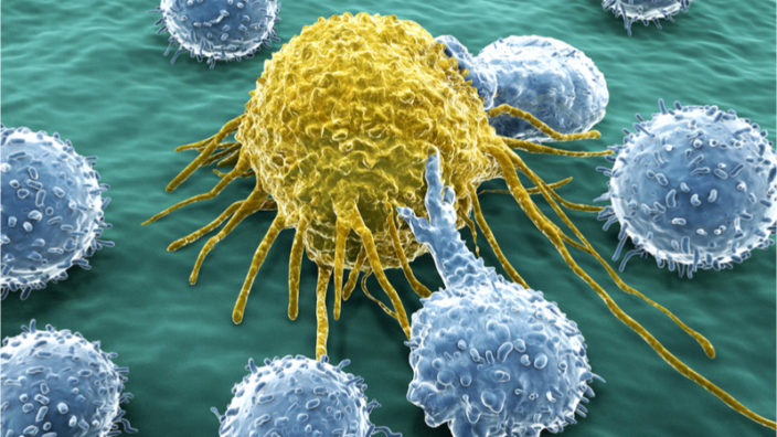
Partial Cellular Reprogramming Improves Memory in Old Mice

Today, we want to highlight another study that takes us a step closer to getting partial cellular reprogramming to the clinic.

Can we both have our cake and eat it?

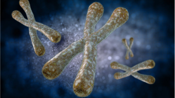
As previous studies [1] have taught us, partial cellular reprogramming is a balancing act of epigenetically rejuvenating cells without completely resetting them.

Building upon these studies, today’s new study shows how exposing cells to reprogramming factors in a living animal for just long enough can reverse age-related epigenetic alterations. The mice in this study had their cells engineered to react to doxycycline, a common antibiotic used in veterinary practice, in order to produce the OSKM reprogramming factors. The researchers found that giving the mice just enough exposure to OSKM improved their cognitive function without an increase in mortality during a four-month period.

In a behavioral test, wild-type and OSKM-expressing mice of the same age appeared to have a similar capacity to remember an object previously seen following a two-hour familiarization period. However, when the mice were shown the object again five days later, the wild-type mice had forgotten about it. On the other hand, the OSKM mice appeared to remember the object, suggesting superior memory retention. Moreover, there was also a correlation between the degree of induced OSKM expression in treated mice and their memory indices.

The researchers suggest that these results might be explained by the increase of OSKM-dependent proteins, which decline during aging, particularly the GluN2B subunit protein, which facilitates calcium permeability and increased synaptic potentiation – a critical function that declines with advancing age. The researchers found that the number of GluN2B-positive neurons in the OSKM mice had increased compared to the wild-type mice, which suggests changes to the neuronal plasticity of mature neurons.

There were no significant changes to ambulatory time in movement tests, nor were there any differences in anxiety in an open field test, which ascertains general movement and activity levels, anxiety, and the willingness to explore in scientific research. By nature, rodents are cautious, and when faced with open areas and bright lights, they have a tendency to hug walls or become stationary, both of which are anxiety responses. Any reduction of anxiety would be obvious, as the mice would be more inclined to move around and explore.

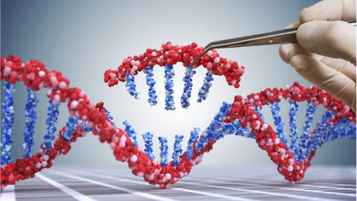
Post-translational epigenetic modifications take place in mouse neurons of the dentate gyrus (DG) with age. Here, we report that age-dependent reduction in H3K9 trimethylation (H3K9me3) is prevented by cyclic induction of the Yamanaka factors used for cell reprogramming. Interestingly, Yamanaka factors elevated the levels of migrating cells containing the neurogenic markers doublecortin and calretinin, and the levels of the NMDA receptor subunit GluN2B. These changes could result in an increase in the survival of newborn DG neurons during their maturation and higher synaptic plasticity in mature neurons. Importantly, these cellular changes were accompanied by an improvement in mouse performance in the object recognition test over long time. We conclude that transient cyclic reprogramming in vivo in the central nervous system could be an effective strategy to ameliorate aging of the central nervous system and neurodegenerative diseases.

Conclusion

Based on the results of previous studies, it is likely that the target cells are being rejuvenated and making positive changes to the signaling environment, cell behavior, reversing mitochondrial dysfunction, and epigenetic changes.

This is yet another step forward for partial cellular reprogramming in the context of age reversal in living animals and ultimately humans. While it is probably at least a decade if not more away from reaching human trials, these animal studies are paving the way for this to ultimately happen.

The road to rejuvenation is not going to be a short one; science, especially aging biology, is complex, and there are many challenges ahead to get partial cellular reprogramming to the clinic. Reversing cellular aging to prevent or reverse age-related diseases is going to be a long haul, not something that will happen overnight, but rather the work of decades.

However, it can be done and it is being done right now. The necessary studies to maximize safety when the approach finally does reach humans are being done right now, and, if successful, the results in people could be game-changing.

We would like to ask you a small favor. We are a non-profit foundation, and unlike some other organizations, we have no shareholders and no products to sell you. All our news and educational content is free for everyone to read, but it does mean that we rely on the help of people like you. Every contribution, no matter if it’s big or small, supports independent journalism and sustains our future.

Literature

[1] Ocampo, A., Reddy, P., Martinez-Redondo, P., Platero-Luengo, A., Hatanaka, F., Hishida, T., … & Araoka, T. (2016). In vivo amelioration of age-associated hallmarks by partial reprogramming. Cell, 167(7), 1719-1733.

DNA data

A Genetic Footprint of Aging

A new analysis has revealed a core set of genes involved in aging in both humans and mice. Together with a broader set of age-related genes assembled by the study [1], this resource will serve as a launching point for further investigations of the mechanisms behind aging and age-related diseases.

The heart of the matter

Numerous studies have tracked age-related molecular changes, with changes showing up in cellular metabolism and the transcriptome, proteome, and epigenome. While the accumulation of this data has improved our understanding of aging, no unifying picture has emerged. Some of the age-related changes seem to be specific to particular tissues, and it’s also not clear how many of these pathways are shared between different organisms.

An international team of researchers set out to answer these questions. They carried out a multi-layer and multi-tissue analysis of aging in male mice and then built on that by comparing their findings with gene expression data from human studies.

The first step was to characterize age-related genetic and epigenetic changes in different tissues of the same species. The researchers harvested livers, hearts, and quadriceps muscles from male mice of different ages. They then analyzed gene expression, DNA methylation, and three types of histone modification in these tissues.

Their analysis revealed differences in how these “omics” layers changed with age in these three tissues. Gene expression changes were most significant in the liver, while changes in the heart and muscle tissue were predominantly related to methylation and histones. Despite these differences, analysis showed that many of the genes affected by aging in the three tissues are involved in the same processes. While conclusions from one tissue type might not be applicable in another because different molecular factors are involved, the aging footprint is reflected in common biological processes in all three tissues.

Core age-related genes shared with humans

To figure out the core players in these shared processes, the team checked to see if the genes they had found shared binding sites for any transcription factors, genes that regulate the expression of other genes. They found a few binding motifs that were more common than would be expected, which pointed them towards a handful of transcription factors that may act as common regulators of the molecular changes associated with aging.

Having found these core age-related transcription factors, the next step was to check whether the same relationship exists in humans. The researchers used human gene expression data from other studies to investigate whether these transcription factors are affected by age. They found that many of them also had age-dependent expression in humans. Further analysis showed that variation in some of these genes was associated with differences in longevity, reinforcing the notion that they act as drivers of age-related processes in humans, though the authors note that these results are based on small samples and so “must be treated with caution and may not necessarily imply causality”.

Many genes and pathways have been linked to aging, yet our understanding of underlying molecular mechanisms is still lacking. Here, we measure changes in the transcriptome, histone modifications, and DNA methylome in three metabolic tissues of adult and aged mice. Transcriptome and methylome changes dominate the liver aging footprint, whereas heart and muscle globally increase chromatin accessibility, especially in aging pathways. In mouse and human data from multiple tissues and regulatory layers, age-related transcription factor expression changes and binding site enrichment converge on putative aging modulators, including ZIC1, CXXC1, HMGA1, MECP2, SREBF1, SREBF2, ETS2, ZBTB7A, and ZNF518B. Using Mendelian randomization, we establish possible epidemiological links between expression of some of these transcription factors or their targets, including CXXC1, ZNF518B, and BBC3, and longevity. We conclude that conserved modulators are at the core of the molecular footprint of aging, and variation in tissue-specific expression of some may affect human longevity.

Conclusion

This research has not found genes “for” aging or longevity. Rather, it has identified a core set of genetic regulators that are involved in aging in different tissues and species, although precisely how is a mystery awaiting further investigation. The study also serves as a rich source of data, a multi-omics, multi-tissue, multi-species analysis of genes involved in processes related to aging. The authors close by calling their work “a hypothesis-generating resource of candidate aging regulators” – an apt summary of their contribution, and we hope that future research will build upon it.

We would like to ask you a small favor. We are a non-profit foundation, and unlike some other organizations, we have no shareholders and no products to sell you. All our news and educational content is free for everyone to read, but it does mean that we rely on the help of people like you. Every contribution, no matter if it’s big or small, supports independent journalism and sustains our future.

Literature

Bou Sleiman, M., Jha, P., Houtkooper, R., Williams, R.W., Wang, X., and Auwerx, J. The Gener-Regulatory Footprint of Aging Highlights Conserved Central Regulators. Cell Reports, doi: 10.1016/j.celrep.2020.108203
Piglet

Generating Neural Progenitor Cells from iPSCs

The latest research published in the Journal of Tissue Engineering and Regenerative Medicine has reprogrammed pig fibroblast cells into neural progenitor cells.

Manufacturing Pig Brain Cells 

Brain regeneration is a novel and potentially groundbreaking strategy for improving brain health. Researchers have used these techniques both to stimulate the body’s own regeneration capabilities and to create miniature brain organoids, which can be used to study the pathology of neural tissues. However, unlike similar strategies applied to other tissues, the cells of neural tissues are especially difficult to work with. A biopsy cannot easily be taken from the brain like it can with bone marrow or skin. Furthermore, neural stem cells are notoriously difficult to culture and expand, even after isolation from a biopsy sample.

However, cellular reprogramming can take more common cell types, such as fibroblasts, and convert them to pluripotent stem cells using the Yamanaka factors (OCT4, SOX2, KLF4, and c-Myc). Recently, researchers at the University of São Paulo have used this technique in pigs to create induced pluripotent stem cells (iPSCs) and then differentiate them to neural progenitor cells. While this has been done in many studies using human and mouse cells, this process has been rarely studied in pigs, despite them being important both agriculturally and as preclinical models.The researchers optimized their technique and characterized the resulting iPSCs and neural progenitor cells. Both their reprogramming and differentiation was considered successful based on identifying markers of pluripotent stem cells, such as OCT4 and NANOG, and neural progenitor cells, such as Nestin and GFAP. This success means that other researchers can confidently utilize their methods for future porcine research. Furthermore, the research identified several differences between pigs and humans, potentially shedding light on the underlying physiological differences involved.

In this study, we were able to induce porcine embryonic fibroblasts to a pluripotentlike state. Generated piPSC exhibited the expected morphology, expressed endogenous pluripotency genes (pOCT4 and pNANOG), and were AP-, OCT4-, NANOG-, SSEA1-, and TRA1-60-positive, as detected by immunostaining; however, the three clonal colonies showed diverse expression profiles. Aiming to derive NPCs, two lines of piPSC were subjected to a neuronal induction protocol, and the obtained cells expressed pNestin and pGFAP and were positively stained for Nestin, β Tubulin III, and Vimentin, suggesting a commitment to neural differentiation. Further studies aiming to generate fully reprogrammed footprint-free piPSCs that are more suitable for specific cell-line differentiation are of great importance for better understanding, reprogramming, and characterization of porcine NPCs.

Conclusion

Pig physiology is surprisingly similar to that of humans, making swine a valuable preclinical model. While this is not a direct brain regeneration technique, the methods presented in this study can help researchers on their quest to better understand brain physiology, pathology, and neuroregeneration.

Telomeres

A New Mechanism of Telomere Lengthening in T Cells Discovered

An international collective of scientists has discovered a new cellular process: telomere transfer from antigen-presenting cells to T cells that boosts the latter’s lifespan and proliferative potential [1].

T cells, an important element of our adaptive immune system, are born as non-proliferating cells and, as such, do not experience telomere attrition. However, they do begin to actively proliferate upon activation by antigens, which is when telomere attrition starts taking its toll. Abnormally short telomeres are found in T cells from people who are infected with certain viral pathogens, such as HIV, or in chronically stimulated T cells from patients with inflammatory diseases. Telomere shortening in T cells is a major factor in immunosenescence.

The authors of this paper came across an intriguing phenomenon: as T cells contacted antigen-presenting cells (APCs), the average length of telomeres in the T cells increased by about 3000 base pairs. Simultaneously, the average length of telomers in the APCs decreased by the same amount. The dynamic of telomere growth/attrition over time showed a perfect reverse correlation.

T Cell and APC telomere interactions

After a series of experiments, the scientists found that the effect only existed when APCs were “loaded” with antigens and when T cells established physical contact with APCs via so-called immunological synapses.

It has been known that T cells are able to restore their telomeres by upregulating TERT (telomerase reverse transcriptase), though this mechanism works less and less well with each activation of a T cell [2]. Nevertheless, the researchers explored the opposite hypothesis: that contact with APCs somehow boosted TERT production in T cells instead of dampening it. Unsurprisingly, this was not the case, as T cells with the TERT gene knocked out still experienced telomere lengthening upon interactions with APCs.

Another mechanism of telomere lengthening is called ALT (alternative lengthening of telomeres) [3]. It is used by a broad range of cancer cells for unchecked replication, but this mechanism requires DNA synthesis. After the researchers inhibited all DNA synthesis in the T cells, they still demonstrated lengthening of telomeres, so ALT had to be ruled out as well.

It became evident that the telomere lengthening in T cells can only be caused by their synaptic interaction with APCs. The researchers detected telomere clustering (basically, congregation of telomeres in one place) in about 70% of the immunological synapses when antigens were present but only in 10% when they were not. When APCs were not coupled with T cells, no telomere clustering occurred at all. Therefore, APCs appear to cluster their telomeres at the synapse prior to telomere transfer.

Telomere DNA was then discovered outside the APCs. Since it could not be destroyed by a DNA-cleaving compound, the researchers suggested that these DNA fragments were enclosed in protective lipid vesicles, which they proved experimentally. Another compound found in the same vesicles was TZAP, the telomere-trimming factor that is apparently involved in the cleavage of the telomere fragments from their chromosomes inside APCs. The researchers found that silencing TZAP derailed the whole telomere transfer process.

By chemically labelling APC telomeres, the researchers proved that the telomere additions detected in the T cells indeed originated in the APCs. They were also able to isolate telomere-transporting vesicles. Transferred directly into T cells, these vesicles caused telomere lengthening, providing the final proof.

Conclusion

This discovery of a previously unknown cellular process has therapeutic potential. The researchers claim that a single APC-originated telomere transfer can rescue ultra-short telomeres in near-senescent T cells and boost their proliferation more effectively than TERT, which only adds 100-200 base pairs per activation. They also hypothesized that “a loss of telomere transfer capacity rather than a loss of telomerase is responsible for telomere shortening at the basis of ageing”. It may be possible to replicate and use this effect against various immunosenescence-related conditions.

We would like to ask you a small favor. We are a non-profit foundation, and unlike some other organizations, we have no shareholders and no products to sell you. All our news and educational content is free for everyone to read, but it does mean that we rely on the help of people like you. Every contribution, no matter if it’s big or small, supports independent journalism and sustains our future.

Literature

[1] Vaz, B., Vuotto, C., Valvo, S., D’Ambra, C., Esposito, F. M., Chiurchiù, V., … & Lanna, A. (2020). Intercellular telomere transfer extends T cell lifespan. bioRxiv.

[2] Barsov, E. V. (2011). Telomerase and primary T cells: biology and immortalization for adoptive immunotherapy. Immunotherapy, 3(3), 407-421.

[3] Cesare, A. J., & Reddel, R. R. (2010). Alternative lengthening of telomeres: models, mechanisms and implications. Nature reviews genetics, 11(5), 319-330.

Gene segment

Using CRISPR to Remove Mutated DNA to Defeat Cancer

Researchers have successfully used the CRISPR/Cas9 gene editing tool to destroy Ewing’s sarcoma and chronic myeloid leukemia tumor cells by targeting the fusion genes responsible for these tumors [1].

Fusion genes, a feature in many different types of cancer, arise when a mutation fuses two genes together, which typically happens when the DNA sequence between these genes is deleted. The resulting fusion gene is still functional in that it encodes proteins, but the proteins it encodes are different from those of the original genes, and this can have unintended consequences, including the development of cancer.

Fusion genes are present in various cancers, including prostate, breast, lung and brain, and up to around 20% of all cancers include these mutant genes.

Researchers from the Spanish National Cancer Research Center (CNIO) were able to use CRISPR/Cas9 editing techniques to cause targeted breaks in the nuclear DNA in mouse models of Ewing’s sarcoma and chronic myeloid leukaemia. They made two cuts in the introns, the non-coding areas of the gene, which are located at each end of the fusion gene.

The cancer cell then responds by attempting to repair the breaks in its DNA by joining the cut ends. This results in the complete removal of the fused gene located between the two cuts. Because the cancer cell depends on the fused gene for its continued survival, this repair, in effect, causes the cancer cell to destroy itself.

The team at CNIO will now be carrying out further studies to assess the efficiency and safety of this novel approach to see if it could be translated to humans. The researchers will also be testing the method on other types of cancers that are caused by fusion genes.

Fusion oncogenes (FOs) are common in many cancer types and are powerful drivers of tumor development. Because their expression is exclusive to cancer cells and their elimination induces cell apoptosis in FO-driven cancers, FOs are attractive therapeutic targets. However, specifically targeting the resulting chimeric products is challenging. Based on CRISPR/Cas9 technology, here we devise a simple, efficient and non-patient-specific gene-editing strategy through targeting of two introns of the genes involved in the rearrangement, allowing for robust disruption of the FO specifically in cancer cells. As a proof-of-concept of its potential, we demonstrate the efficacy of intron-based targeting of transcription factors or tyrosine kinase FOs in reducing tumor burden/mortality in in vivo models. The FO targeting approach presented here might open new horizons for the selective elimination of cancer cells.

Conclusion

While it is no doubt going to be some years before we see an approach like this being attempted in humans, if successful, the strategy could be potentially used to treat a range of fusion gene-based cancers. It also paves the way for other CRISPR/Cas9 gene editing therapies to be developed.

Another positive is that because these fusion genes are only found in cancer cells, they are highly specific targets, and attacking them has no effect on healthy cells. This means that gene therapy vectors could be broadly used to deliver CRISPR/Cas9 into tissues without harming healthy cells.

We would like to ask you a small favor. We are a non-profit foundation, and unlike some other organizations, we have no shareholders and no products to sell you. All our news and educational content is free for everyone to read, but it does mean that we rely on the help of people like you. Every contribution, no matter if it’s big or small, supports independent journalism and sustains our future.

Literature

[1] Martinez-Lage, M., Torres-Ruiz, R., Puig-Serra, P., Moreno-Gaona, P., Martin, M. C., Moya, F. J., … & Bueno, C. (2020). In vivo CRISPR/Cas9 targeting of fusion oncogenes for selective elimination of cancer cells. Nature Communications, 11(1), 1-14.

Taisho Pharmaceutical logo

Insilico Partners with Taisho for Senolytic Drug Discovery

Insilico Medicine has announced a historic deal with the Taisho Pharmaceutical Company, which is based in Japan, in a partnership to identify and develop new senolytic drugs.

The companies have agreed to collaborate on AI-powered drug discovery focused on identifying molecules to remove problematic senescent cells.

Founded in 1912, Taisho is focused on over-the-counter medicines and markets the brands Colac, Contac, Lipovitan-D, Pabron, Tempra, UPSA, and Vicks. It has also been successful with its antibiotic clarithromycin, which is used to treat respiratory infections and their consequences, such as pneumonia, skin problems including cellulitis, and ear infections.

This partnership suggests a growing interest of large pharmaceutical companies in senolytics, which naturally results in an influx of funding. Dr. Alex Zhavoronkov had the following to say about the deal:

Senescence is the most important process in our body that we tend to ignore as individuals and most of the pharmaceutical companies used to ignore. Until now. It is now clear that there is a clear connection between cellular senescence and a plethora of diseases, and there are next-generation AI tools that can be coupled with high-quality laboratory data to identify promising targets that have a clear-cut commercial opportunity.

We started building this platform in 2015, and it is now bearing fruit, as there is more interest in the pharmaceutical industry. The Taisho deal, for us, has historic significance. This is one of the largest and most visionary companies in Japan, a market with a substantial aging population.

Thursday, October 15, 2020 (9 am ET) – Insilico Medicine announced today that Taisho Pharmaceutical Co., Ltd. and Insilico have entered into a research collaboration to identify novel therapeutics against aging. Insilico Medicine will utilize both the target discovery and generative chemistry parts of its Pharma.AI platform in this collaboration. It will use its proprietary Pandomics Discovery Platform to identify novel targets for senolytic drugs and Chemistry42 platform for a molecular generation. This collaboration brings together Insilico’s state-of-art artificial intelligence (AI) technologies in drug discovery with Taisho’s expertise in drug development, aimed to extend the human healthspan.

‘We’re delighted to collaborate with Taisho Pharmaceutical, a well-recognized leader in the pharmaceutical industry and healthcare sector. It is believed that aging is a universal phenomenon that we cannot stop. However, emerging scientific evidence has shown that one may be able to reverse some of the age-associated processes. Through this collaboration, we will adopt our AI-powered drug discovery suites together with Taisho’s validation platform to explore the new space of anti-aging solutions’, said Jimmy Yen-Chu Lin, PhD, CEO of Insilico Medicine Taiwan, a fully-owned subsidiary of Insilico Medicine.

Under the terms of the agreement, Insilico Medicine will receive an upfront payment and milestone payments upon achievement of specified goals. Insilico Medicine will be responsible for early research phase target identification and molecular generation, and Taisho will work collaboratively with Insilico in validating the results in various in vitro and in vivo assays. Taisho has the exclusive option to acquire Insilico’s co-ownership of the successfully developed programs under agreed payment.

Conclusion

This is good news, particularly given that UBX0101, a drug from senolytic pioneers Unity Biotechnology, has recently failed in a human trial for osteoarthtiris. The reason is a matter of debate, but poor tissue choice, a flawed approach, the heterogeneity of senescent cell populations, and the trial’s reliance on the WOMAC scale are likely to be to blame.

Quite literally, the more shots on goal we can get, the better, as it increases the chances that something will succeed and reduce the impact that failures have. While this partnership may take some years to bear fruit in the form of drugs entering human trials, it is most welcome news for the field and represents the start of large pharmaceutical companies, and their vast funds and experience in drug development, taking an interest in our field.

We would like to ask you a small favor. We are a non-profit foundation, and unlike some other organizations, we have no shareholders and no products to sell you. All our news and educational content is free for everyone to read, but it does mean that we rely on the help of people like you. Every contribution, no matter if it’s big or small, supports independent journalism and sustains our future.
EGCG chemical

Uncovering the Origin of the SASP

A new study published in Nature shows a strong link between the protein G3BP1 and the SASP, a harmful byproduct of senescent cells.

Silencing the SASP, not stopping senescence

This study shows that silencing rasGAP SH3-binding protein 1 (G3BP1) expression in mice does not stop cells from becoming senescent. Instead, interfering with G3BP1 through epigallocatechin gallate (EGCG) interferes with cyclic GMP-AMP synthase (cGAS), whose cGAS-STING pathway is responsible for the type 1 IFN response, which is critical to the SASP.

The researchers observed this relationship directly by comparing the activities of cells with and without G3BP1. Cells without G3BP1 were unable to colocalize cGAS with the cytosolic chromatin fragments of senescent cells, thus stopping this pathway before it was started.

They confirmed their findings by supplementing cells with a downstream byproduct of cGAS. This supplement promoted the SASP even while GCBP1 was restricted, showing that this G3BP1/cGAS relationship is a primary cause of the production of these chemicals.

A double-edged sword

Simply turning off G3BP1 through EGCG is unlikely to be a desirable option for a broad therapy. Mice that cannot produce G3BP1 show symptoms of accelerated aging (progeria), including ataxia, and cells without it have difficulty proliferating. The researchers note that EGCG also affects other pathways, including Wnt and AKT, which are important to our biochemistry.

The researchers have also found evidence that G3BP1 is responsible for the production of Lamin B1, as cells without this protein are unable to produce it. This may explain the accelerated aging of mice without G3BP1, as a lack of lamins is associated with progeric diseases and genomic instability, a primary hallmark of aging.

Conclusion

While the researchers openly admit that this study offers only a partial understanding of one piece of the puzzle, this research represents an intermediate but critical step in our understanding of senescent cells and the SASP they create. Hopefully, it may be possible to build upon this research to develop therapies that interfere with the relevant pathways, and only the relevant pathways, in less harmful ways, stopping the deleterious effects of the SASP without causing side effects that increase the effects of aging in other areas.

We would like to ask you a small favor. We are a non-profit foundation, and unlike some other organizations, we have no shareholders and no products to sell you. All our news and educational content is free for everyone to read, but it does mean that we rely on the help of people like you. Every contribution, no matter if it’s big or small, supports independent journalism and sustains our future.
Fat cells

Fatty Tissue Generated on a Chip

Recent research published in the scientific journal Lab on a Chip sheds light on the behavior of adipocytes, the primary cells of our fat tissue.

Even though this tissue is often unwanted, regenerating adipose tissue is critical for reconstructive and cosmetic surgery, and being able to generate animal fat is critical for the creation of lab-grown meats. As the hormonal and endocrine functions of adipose tissue are implicated in aging pathways and age-related diseases, the study of these functions and how to modulate them could be incredibly important in tackling some aspects of aging. This is why researchers at Harvard are working to create fat-on-a-chip technology. [1]

Fattening Chips

Adipocytes, the main cells of our fat tissue, are especially difficult to study in two dimensions. By seeding and differentiating preadipocytes (the primary stem cell of fat tissue) on three-dimensional gelatin/polycaprolactone fibers, the researchers were able to grow adipocytes for twice as long and to a hundred times the size of two-dimensionally cultured cells. The size of adipocytes is of particular importance, as these cells grow dramatically during weight gain. In other words, our fat cells tend to get larger in size rather than quantity. Recapitulating this in vitro has proven to be a difficult challenge for researchers. While the cells in this study did not reach the size of cells in obese individuals, these results are a positive step towards achieving that goal.

Furthermore, the fat-on-a-chip system showed typical physiological responses to simulated meals and fasting. Glucose uptake increased dramatically when insulin was delivered to the cells, as was adiponectin secretion during fasting, which are both typical in vivo responses. The researchers also experimented with different patterning techniques and were able to further increase the similarity in cellular behaviors between their adipocytes and in vivo cells.

Taken collectively, this technology enables disease modeling and drug testing prior to in vivo validation and informs tissue engineering approaches for adipose reconstruction, aesthetic medicine, food and other applications for which adipocytes could be exploited.

Conclusion

The results of this study push us closer to being able to generate adipose tissue in a laboratory setting in the future. Additionally, this technology can be immediately utilized to help study the cellular physiology of adipocytes and better understand their role in diabetes and other age-related diseases. However, this model still has significant room for improvement. Just as animal models do not perfectly embody the conditions in humans, neither do these organ-on-a-chip technologies.

We would like to ask you a small favor. We are a non-profit foundation, and unlike some other organizations, we have no shareholders and no products to sell you. All our news and educational content is free for everyone to read, but it does mean that we rely on the help of people like you. Every contribution, no matter if it’s big or small, supports independent journalism and sustains our future.

Literature

[1] Pope, B.D., Warren, C.R., Dahl, M.O., Pizza, C.V., Henze, D.E. …, Parker, K.K. (2020). Fattening chips: hypertrophy, feeding, and fasting of human white adipocytes in vitro. Lab on a Chip, online ahead of print. DOI: 10.1039/d0lc00508h

T Cell Progenitors Boost Stem Cell Therapy Effectiveness

In a review paper, a group of scientists highlight a new strategy that addresses the shortcomings of stem cell therapy by adding T cell progenitors to the graft [1].

HSCT has a problem

Today, one of the few ways to alleviate immune system damage from chemotherapy and radiotherapy is a hematopoietic stem cell transplant (HSCT), which is the only stem cell therapy in widespread use. This treatment, which has been around for over 30 years, aims to replenish the pool of HSCs, the common progenitors of immune cells, and to restore the host’s immune defenses. However, there is a problem: the T cell population recovers much more slowly than that of other populations. This discrepancy gets more noticeable with age: in older patients, the population may never fully recover. T cell deficiency hampers the anti-cancer response of the immune system regenerated by HSCT and leaves the patient vulnerable to pathogens. To mitigate this effect, many clinics add donor T cells to the graft (transplant), but because T cells are so aggressive, foreign T cells can induce graft vs. host disease (GvHD), a potentially deadly autoimmune attack on vital organs. To account for this danger, the patient is given immunosuppressants. Sadly, this balancing act usually fails, as most patients who receive HSCT succumb either to cancer relapse or to GvHD within five years after transplantation.

ProT instead of T

The authors of this new paper highlight a novel possible solution to this problem. Research has shown that transplanted HSCs fail to differentiate into T progenitor cells (ProTs). Therefore, the idea is to grow ProTs in vitro and add them to the graft, essentially bypassing the dysfunctional process of ProT production in the host. In recent research conducted by two of the authors of this paper, ProTs began to appear in the thymi of mice one week after transplantation [2]. After two weeks, the number of thymocytes grew significantly, suggesting successful proliferation of the donor ProTs. The transplantation also promoted the reconstitution of the thymus, apparently restoring the crosstalk between thymic epithelial cells and thymocytes. In other research, T cells derived from transplanted ProTs exhibited early anti-tumor activity, shortening the time during which the mice who received chemotherapy or radiotherapy remained immunocompromised.

A major advantage of ProTs compared to full-grown T cells is that the former lack T cell receptors (TCRs). TCRs are used by T cells to bind to cells in the body and destroy them. As a result, ProTs are unable to induce GvHD. Compatibility requirements can be less strict, as ProTs undergo the full process of selection and tolerization in the thymus anyway. T cells derived from transplanted ProTs show little to no autoreactivity.

T cells also exhibit strong heterogenity. Our body uses gene rearrangement to create a great variety of T cells with more than one billion different receptors that can recognize and counter virtually any pathogen. The researchers report that in preclinical mouse models, donor ProTs have been shown to successfully recreate this variety, enabling the host organism to combat both cancer and infections.

On a less positive note, ProTs cannot fully alleviate the age-related decline of our immune system. Although aged mice still benefit from ProT-enhanced HSCT, and the ProT engraftment in the thymus is successful in both young and old animals, ProTs proliferate up to ten times slower in the latter group. This shows how important it is to keep looking for ways to delay or reverse age-related thymic involution.

Conclusion

HSCT is the flagship of stem cell therapy. Unfortunately, its effectiveness is limited due to the autoreactivity of transplanted T cells and age-related thymic involution. By partially alleviating these problems, the strategy outlined in this new paper can potentially provide cancer patients with better and more immediate immune protection.

We would like to ask you a small favor. We are a non-profit foundation, and unlike some other organizations, we have no shareholders and no products to sell you. All our news and educational content is free for everyone to read, but it does mean that we rely on the help of people like you. Every contribution, no matter if it’s big or small, supports independent journalism and sustains our future.

Literature

[1] Singh, J., Mohtashami, M., Anderson, G., & Zúñiga-Pflücker, J. C. (2020). Thymic engraftment by in vitro-derived progenitor T cells in young and aged mice. Frontiers in Immunology, 11, 1850.

[2] Singh, J., & Zúñiga-Pflücker, J. C. (2018). Producing proT cells to promote immunotherapies. International immunology, 30(12), 541-550.

Unity biotechnology logo

A New Senolytic Enters Human Trials

Unity Biotechnology has recently announced the launch of its Phase 1 study of UBX1325 for patients with diabetic macular edema. The drug is designed to remove problematic senescent cells from the body which accumulate as we get older and are a reason we age.

UBX1325 is a senolytic agent that targets the BCL-xL pathway to destroy senescent cells, much like Navitoclax, which is known to clear senescent cells via this pathway but has serious side effects; UBX1325 may even be a modified version of Navitoclax. The BCL-2 family of proteins includes BCL-1, BCL-w and BCL-xL and prevents the cell from entering apoptosis.

Diabetic macular edema is due to leaky blood vessels in the eye, causing fluid to accumulate in the macula, the central area of the retina. It is a complication of diabetic retinopathy and has an outcome similar to adult wet macular degeneration. The researchers at UNITY believe that senescent cells play a key role in the development of this disease and that it may be addressed by removing them with senolytics.

SAN FRANCISCO, Oct. 12, 2020 (GLOBE NEWSWIRE) — UNITY Biotechnology (“UNITY”) [NASDAQ:UBX], a biotechnology company developing therapeutics to extend healthspan by slowing, halting or reversing diseases of aging, today announced that the first patient has been dosed in a Phase 1 study of UBX1325 in patients with diabetic macular edema (DME).

“There is strong evidence of association between disease progression in DME and accumulation of senescent cells. There’s additional evidence that senescent cells secrete factors that can be damaging to the eye and lead to vascular leakage, a pathological hallmark of DME,” said Anirvan Ghosh, Ph.D., chief executive officer of UNITY. “Bcl-xL inhibition, a novel senolytic mechanism, has been shown in our preclinical tests to lead to an impact on senescent cells, reduction in vascular leakage, and improvement in retinal function. Our preclinical observations give us confidence in UBX1325 and we are excited to bring this therapeutic candidate into clinical development.”

The Phase 1, first-in-human, open-label, single-ascending dose study is designed to evaluate the safety, tolerability and pharmacokinetics of UBX1325 in patients with advanced DME. The trial is designed to enroll approximately 15 patients, with safety and tolerability data expected in the first half of 2021. UNITY anticipates initiating a proof of concept study in the first half of 2021.

About UBX1325

UBX1325 is a potent and selective small molecule inhibitor of Bcl-xL, an anti-apoptotic regulatory protein and a BCL-2 family member. Preclinically, UBX1325 has been shown to eliminate senescent cells and have a positive impact on inflammation, vascular leakage and visual function. UBX1325 is currently being evaluated in a first-in-human trial designed to treat patients with advanced diabetic macular edema. To learn more at clinicaltrials.gov about the Phase 1 clinical trial of UBX1325.

Will this time be different?

This announcement comes in the wake of disappointing news for UNITY Biotechnology, as its Phase 2 trial of UBX0101, another senolytic drug, failed to deliver the desired results. That study involved 183 patients with moderate-to-severe osteoarthritis of the knee given UBX0101, which targets the p53/MDM2 pathway, another way in which some senescent cells avoid apoptosis. Unfortunately, the study found no statistically significant difference in pain reduction between UBX0101 and placebo.

As with the previous clinical trial, success hinges a great deal on the tissue being targeted and the heterogeneity of the senescent cells within. It turns out that there are a number of different types of senescent cells, and they all use different pathways to evade destruction.

Success will depend on whether this drug targets the same pathway that the bulk of the senescent cells in the eye are using to survive. Let us hope that the right pathway has been chosen and more disappointing results can be avoided.

Skin peeling

Regenerating Skin With a Single Protein

Researchers have identified a factor present in the skin of baby mice that controls the formation of hair follicles during their early development. The factor is turned off in adult mice, but when it is turned back on, it allows them to regenerate their skin following injury without scarring, just like baby mice do.

Skin like a newborn

Researchers from Washington State University have recently published a study in the journal eLife showing how they conveyed the kind of skin regeneration only typically seen in baby mice to adult animals [1]. This has implications for treating wound healing following injury as well as addressing some aspects of skin aging.

The factor in question is called lymphoid enhancer-binding factor 1 (LEF1) and is a protein that, in humans, is encoded by the LEF1 gene. This particular protein is associated with papillary fibroblast cells, a type of cell found in the papillary dermis, the skin layer just below the surface which lends skin its tension and its familiar, youthful appearance.

The research team used RNA sequencing to compare the cells and gene expression in adult mice with baby mice. They used this technique to identify the LEF1 gene and wanted to see what would happen if the gene was switched on again in adult mice.

The LEF1 gene is active in baby mice during the first week of development and is then switched off once the skin and hair follicles have formed. The expression of LEF1, for the most part, remains turned off from this point onwards into adulthood. When researchers switched gene expression of LEF1 back on in the papillary fibroblast cells of adult mice, they found that these mice were able to heal wounds without scarring.

The researchers noted that the regenerated skin in the adult mice also included hair follicles and was able to form goosebumps, something that adult scar tissue is unable to do.

Scars are a serious health concern for burn victims and individuals with skin conditions associated with wound healing. Here, we identify regenerative factors in neonatal murine skin that transforms adult skin to regenerate instead of only repairing wounds with a scar, without perturbing development and homeostasis. Using scRNA-seq to probe unsorted cells from regenerating, scarring, homeostatic, and developing skin, we identified neonatal papillary fibroblasts that form a transient regenerative cell type that promotes healthy skin regeneration in young skin. These fibroblasts are defined by the expression of a canonical Wnt transcription factor Lef1 and using gain- and loss of function genetic mouse models, we demonstrate that Lef1 expression in fibroblasts primes the adult skin macroenvironment to enhance skin repair, including regeneration of hair follicles with arrector pili muscles in healed wounds. Finally, we share our genomic data in an interactive, searchable companion website (https://skinregeneration.org/). Together, these data and resources provide a platform to leverage the regenerative abilities of neonatal skin to develop clinically tractable solutions that promote the regeneration of adult tissue.

Conclusion

While more research is needed before an attempt to translate these findings to humans can be made, these results highlight how mammals can regenerate like other species do, given the appropriate stimulus. Many species such as salamanders do regenerate throughout their lives, but, in mammals, this ability is lost beyond the initial development phase.

The results of this study have implications for wound treatment as well as skin aging if its results can be translated to people.

We would like to ask you a small favor. We are a non-profit foundation, and unlike some other organizations, we have no shareholders and no products to sell you. All our news and educational content is free for everyone to read, but it does mean that we rely on the help of people like you. Every contribution, no matter if it’s big or small, supports independent journalism and sustains our future.

Literature

[1] Driskell, R., Phan, Q. M., Fine, G., Salz, L., Herrera, G., Wildman, B., & Driskell, I. M. (2020). Lef1 expression in fibroblasts maintains developmental potential in adult skin to regenerate wounds. bioRxiv.

Collagen

Creating Anti-Glucosepane Antibodies

New research takes us a step close to finding ways to remove the advanced glycation end-product known as glucosepane, a likely reason why our arteries stiffen as we age.

A possible solution to the problem

Some years ago, Aubrey de Grey from the SENS Research Foundation proposed that a possible solution to dealing with AGEs would be to find ways to break down the crosslinks, thus freeing up the trapped proteins and restoring tissue elasticity.

Until recently, glucosepane was very difficult to isolate, which is needed to develop drugs and therapies. This changed in 2015, when, thanks to funding from the SENS Research Foundation, the Spiegel lab perfected a method to create glucosepane on demand [1].

While it is not totally clear that AGEs are a primary cause of aging, some researchers such as Alexander Fedintsev and Alexey Moskalev believe that it is and presented evidence in support of it being a tenth hallmark of aging earlier this year [2].

Today, we want to highlight a new paper that concerns the development of antibodies specific for glucosepane [3]. This paper was published by a team of researchers, including Jonathan Clark from the Babraham Institute and David Spiegel from Harvard, who may be familiar to our regular readers. Once again, this research has been funded by the SENS Research Foundation, which continues to support this line of scientific inquiry.

Although there is ample evidence that the advanced glycation end-product (AGE) glucosepane contributes to age-related morbidities and diabetic complications, the impact of glucosepane modifications on proteins has not been extensively explored due to the lack of sufficient analytical tools. Here, we report the development of the first polyclonal anti-glucosepane antibodies using a synthetic immunogen that contains the core bicyclic ring structure of glucosepane. We investigate the recognition properties of these antibodies through ELISAs involving an array of synthetic AGE derivatives and determine them to be both high-affinity and selective in binding glucosepane. We then employ these antibodies to image glucosepane in aging mouse retinae via immunohistochemistry. Our studies demonstrate for the first time accumulation of glucosepane within the retinal pigment epithelium, Bruch’s membrane, and choroid: all regions of the eye impacted by age-related macular degeneration. Co-localization studies further suggest that glucosepane colocalizes with lipofuscin, which has previously been associated with lysosomal dysfunction and has been implicated in the development of age-related macular degeneration, among other diseases. We believe that the anti-glucosepane antibodies described in this study will prove highly useful for examining the role of glycation in human health and disease.

Conclusion

Highly specific antibodies for glucosepane are vitally important in order to develop potential drugs that can break down the offending AGEs, and without them, it would be impossible to progress a therapy. This paper is another important step forward for the development of a glucosepane breaker.

Currently, only one company is working on glucosepane breakers, and that is Revel Pharmaceuticals, which includes David Spiegel, one of the authors of this paper. Hopefully, this latest advance in our knowledge will encourage other AGE-breaker companies to launch and spark a surge of interest in this area in the same way that interest in senolytics has grown in recent years.

We would like to ask you a small favor. We are a non-profit foundation, and unlike some other organizations, we have no shareholders and no products to sell you. All our news and educational content is free for everyone to read, but it does mean that we rely on the help of people like you. Every contribution, no matter if it’s big or small, supports independent journalism and sustains our future.

Literature

[1] Draghici, C., Wang, T., & Spiegel, D. A. (2015). Concise total synthesis of glucosepane. Science, 350(6258), 294-298.

[2] Fedintsev, A., & Moskalev, A. (2020). Stochastic non-enzymatic modification of long-lived macromolecules-a missing hallmark of aging. Ageing Research Reviews, 101097.

[3] Streeter, M. D., Rowan, S., Ray, J., McDonald, D. M., Volkin, J., Clark, J., … & Spiegel, D. A. (2020). Generation and Characterization of Anti-Glucosepane Antibodies Enabling Direct Detection of Glucosepane in Retinal Tissue. ACS Chemical Biology.

Autophagy

Senescent Cells Have Less SIRT1 as a Result of Autophagy

Genes in the sirtuin family are conserved across a wide range of species and are involved in cellular metabolism, immune response, and aging. SIRT1 is known to decline during aging, but the mechanisms involved in this process were not known. Now, an international team has clarified how SIRT1 is regulated during cellular senescence [1].

The cause of decline

The researchers began by checking whether SIRT1 expression changes during senescence. Using RNA sequencing and quantitative PCR, they determined that the expression level remains the same even though there is less SIRT1 protein in senescent cells. In other words, the decline in SIRT1 happens through protein degradation rather than changes in gene expression.

Next, they treated cells with inhibitors of different protein degradation pathways to figure out which one is responsible for the breakdown of SIRT1. Blocking proteasome-mediated degradation didn’t stop SIRT1 levels from declining, but blocking lysosome-mediated degradation did. Since autophagy is one of the major lysosome-mediated processes in cellular senescence, it seemed a likely candidate for SIRT1 degradation.

To test this idea, the team knocked down the autophagy protein Atg7 and monitored SIRT1 during senescence. The knockdown not only prevented the SIRT1 decrease, it restored SIRT1 levels in already senescent cells. These findings clearly showed that the SIRT1 decrease in senescent cells happens via lysosome-mediated autophagy.

A partner in crime

The team used a combination of immunofluorescence and immunoprecipitation to tease apart the details of SIRT1 autophagy. They found that SIRT1 is normally in the nucleus but moves into the cytoplasm as cells become senescent. In the cytoplasm, SIRT1 colocalized with LC3, a protein involved in autophagy. Further work confirmed that SIRT1 and LC3 interact directly and that this interaction is required for SIRT1 autophagy during senescence.

In fact, the interaction between SIRT1 and LC3 becomes stronger during senescence. The study showed that this is due to decreased phosphorylation of SIRT1 in senescent cells, and the researchers even identified the specific SIRT1 amino acids involved in the interaction.

Finally, they measured SIRT1 protein levels in mice during natural aging and found a decrease in the spleen, testes, and thymus but not in other tissues. Treating mice with an autophagy inhibitor stopped the SIRT1 decline in old mice without changing the levels in young mice. They also repeated the experiment in human T cell cultures from older donors, demonstrating that the same mechanisms are at play in human cells.

SIRT1 (Sir2) is an NAD+-dependent deacetylase that plays critical roles in a broad range of biological events, including metabolism, the immune response and ageing1–5. Although there is strong interest in stimulating SIRT1 catalytic activity, the homeostasis of SIRT1 at the protein level is poorly understood. Here we report that macroautophagy (hereafter referred to as autophagy), a catabolic membrane trafficking pathway that degrades cellular components through autophagosomes and lysosomes, mediates the downregulation of mammalian SIRT1 protein during senescence and in vivo ageing. In senescence, nuclear SIRT1 is recognized as an autophagy substrate and is subjected to cytoplasmic autophagosome–lysosome degradation, via the autophagy protein LC3. Importantly, the autophagy–lysosome pathway contributes to the loss of SIRT1 during ageing of several tissues related to the immune and haematopoietic system in mice, including the spleen, thymus, and haematopoietic stem and progenitor cells, as well as in CD8+CD28− T cells from aged human donors. Our study reveals a mechanism in the regulation of the protein homeostasis of SIRT1 and suggests a potential strategy to stabilize SIRT1 to promote productive ageing.

Conclusion

This work deepens our understanding of SIRT1 and the role it plays in aging. Knowing how SIRT1 levels change opens the possibility of interventions to prevent it, which may be especially useful in dealing with immune cell aging. It’s also interesting to see autophagy involved in promoting senescence, since it’s generally considered a quality-control pathway that helps maintain health. This highlights the difference between bulk and selective autophagy, a topic that will likely reward further research. Likewise, clarifying the pathways that control SIRT1 phosphorylation to trigger its degradation during senescence may also prove a fruitful research avenue.

We would like to ask you a small favor. We are a non-profit foundation, and unlike some other organizations, we have no shareholders and no products to sell you. All our news and educational content is free for everyone to read, but it does mean that we rely on the help of people like you. Every contribution, no matter if it’s big or small, supports independent journalism and sustains our future.

Literature

  1. Xu, C., Wang, L., Fozouni, P., Evjen, G., Chandra, V., Jiang, J., Lu, C., Nicastri, M., Bretz, C., Winkler, J.D., Amaravadi, R., Garcia, B.A., Adams, P.D., Ott, M., Tong, W., Johansen, T., Dou, Z., and Berger, S.L. SIRT1 is downregulated by autophagy in senescence and ageing. Nature Cell Biology, doi: 10.1038/s41556-020-00579-5
Elderly teeth

A New Atlas of Dental Cells

A recent study published in Nature Communications has created an atlas for the various differentiated and stem cell populations found in our teeth.

Growing new teeth is not just for kids

Because of the many ways in which dental pathologies can come back to bite us and the many limitations of artificial implants and dentures, there is a significant interest in tooth regeneration. There are quite a few obstacles in the way of tooth regeneration, which combine the major difficulties of several other tissues. Similar to the gut and the skin, teeth are subjected to the unique stressors of the harsh external environment. Like bone and cartilage, their mechanical properties are critical to their success and must be recapitulated fully. Finally, their structure and the different regions of teeth are key components to their function. Directing regeneration in such an organized fashion is challenging in a number of other tissues, such as the kidneys.

Furthermore, we know only a small fraction of the biology of teeth and of their natural growth and regeneration. A recent publication resulting from a large collaboration of researchers led by the Medical University of Vienna has shed quite a bit of light in this arena [1]. Using the most advanced transcriptomics techniques (RNAseq, also known as Next-Generation Sequencing), this study examined cell populations in depth. The researchers focused on comparisons between the different regions within teeth, the differences between human and mouse teeth, and the differences between teeth that are actively growing/regenerating and teeth that are not.

Understanding our teeth through the power of RNA sequencing

RNA sequencing is a powerful tool. It can tell us a lot about a cell’s behavior at a given point in time based on the proteins which are being actively synthesized. Among many other findings, the researchers showed mouse incisors to be much more heterogeneous whereas human teeth tended to have a lot more variations within their cell populations. The results identify several cell types and cell populations previously uncharacterized in dental tissue as well as novel and more specific markers for dental mesenchymal stem cells (MSCs). Additionally, the differentiation of stem cells towards odontoblasts is characterized throughout the entire process, providing insights to the mechanisms underlying this cellular behavior. Finally, the studies identify several previously unidentified factors that are involved in natural tooth growth and regeneration in both human and mouse teeth.

Combined, these results have immediate impacts on our understanding of tooth biology and for the isolation and culture of dental cell populations. They also identify several novel points of intervention in the differentiation of dental stem cells and the natural generation of dental tissues. Additionally, they highlight some key differences between mouse and human teeth – knowledge that is notably important because mice are frequently used to test tooth regeneration strategies. However, it remains to be seen if these findings can be applied directly to improve approaches in dental tissue engineering and tooth regeneration.

Overall, we hope that the presented detailed and validated map of dental cell types, supplemented by human comparison, will serve as a key resource stimulating further studies of cell dynamics in tooth morphogenesis, also including reparative and regenerative therapies.

We would like to ask you a small favor. We are a non-profit foundation, and unlike some other organizations, we have no shareholders and no products to sell you. All our news and educational content is free for everyone to read, but it does mean that we rely on the help of people like you. Every contribution, no matter if it’s big or small, supports independent journalism and sustains our future.
Literature

[1] Krivanek, J., Soldatov, R. A., Kastriti, M. E., Chontorotzea, T., Herdina, A. N., …, Adameyko, I. (2020). Dental cell type atlas reveals stem and differentiated cell types in mouse and human teeth. Nature Communications, 11(4816). DOI: 10.1038/s41467-020-18512-7

Healthy elderly

Today’s Elderly Are Healthier Than a Generation Ago

A high-quality cohort study by Finnish scientists shows that today’s elderly are in better physical and cognitive shape than people of the same age were a generation ago [1,2].

Does longer also mean better?

One of the fears commonly evoked by the idea of life extension is that prolonging lifespan will not necessarily prolong healthspan. This is the concern that even if people are currently living longer, they have not become healthier, since the additional lifespan that modern medicine provides is burdened by illness and frailty. Some data, such as the rising prevalence of chronic diseases, seems to support this notion. On the other hand, there is a lot of anecdotal evidence that people today feel younger and healthier than their parents did at the same age, such as the idea that 70 is the new 60. Large-scale cohort studies could help to clear this up, but they are extremely hard to come by.

Two studies 30 years apart

A recent study by Finnish scientists is one of the most comprehensive and high-quality cohort studies ever performed. It is based on a study from 1989-1990 that was performed in the same city of Jyväskylä. Back then, 500 elderly Finns aged 75 or 80 (born in either 1910 or 1914) were tested for physical and cognitive abilities. Physical ability tests included maximal walking speed, maximal hand grip strength and knee extension strength, and respiratory function. The cognitive tests measured memory, cognition, verbal fluency, and reaction time. In this current study, the sample size has grown to 726, and the participants were born either in 1938-9 or 1942-3. In addition to both cohorts being genetically similar due to low levels of migration in this part of the country, the study’s great advantage is that the protocols were meticulously recreated three decades after the original study using the same or similar equipment.

At first glance, the results look promising, especially on the cognitive abilities front, where the later cohort convincingly outperformed the earlier one in almost every test. This is not surprising, though, as the cognitive advantage of later cohorts has been demonstrated by several studies and even has its own name: the Flynn effect [3].

There have been much fewer cohort studies of physical abilities, at times with opposing or inconclusive results. The Finnish study clearly demonstrates the advantage of the later-born cohort.

The later cohort showed markedly and meaningfully higher results in the maximal functional capacity tests, suggesting that currently 75- and 80-year old people in Finland are living to older ages with better physical functioning.

The difference between the earlier-born and the later-born cohorts was more pronounced among 75-year-old males and 80-year-old females. For instance, in the earlier cohort of 75-year-old men 48% percent showed grip strength lower than the threshold for increased risk of limited mobility, compared with only 27% in the later cohort. The results of the same test in women’s 80-year-old cohorts were 75% and 44%, accordingly.

However, these results come with many caveats, beginning with the rate of participation. In the first study, 77% of those contacted agreed to participate, while in the second, the response rate was just 44%. This caused the researchers to suspect that the second cohort was more prone to self-selection bias, and an effort was made to control for this effect.

Education, smoking and two world wars

The researchers note that numerous other factors could have affected the results. For instance, the increase in average strength can be partially explained by the increase in average height, which is a well-known worldwide phenomenon.

Apart from average height, the difference that stood out the most was in years of education, where the younger cohort had definitive advantage. Education correlates strongly with health, probably because more educated people tend to lead healthier lives, work less physically demanding jobs, and have access to better healthcare.

Another obvious factor was smoking. In the later cohort, fewer men indicated that they had ever smoked, which may have contributed to their superior health. Interestingly, the opposite was true for 75-year-old women. A possible cause is that people usually take up smoking when they are young, and in the 1930s, women who smoked were more frowned upon than 30 years later.

The researchers then delved even deeper into possible socioeconomic factors. They noted that people from the earlier cohort experienced two world wars and several periods of scarcity. The difference in average height can be explained by malnutrition and being engaged in child labor, which was widespread in Finland at that time. The 1940s marked the beginning of Finland as a welfare state, with measures like free school meals being introduced. On the other hand, environmental pollution, which also affects health and longevity, has increased over the last century around Jyväskylä due to urbanization.

Still, the results, even when controlled for variables like height and education level, were so statistically significant that the researchers view them as evidence that today’s elderly (at least in Finland) are healthier, stronger, more active and more cognitively sound than their parents were at the same age.

Conclusion

Quality cohort studies like this one, though still rare, will be increasingly available in the future. If sufficiently standardized and controlled for variables, they can help elucidate the true effects of modern lifestyles and advances in medicine on the public’s healthspan.

We would like to ask you a small favor. We are a non-profit foundation, and unlike some other organizations, we have no shareholders and no products to sell you. All our news and educational content is free for everyone to read, but it does mean that we rely on the help of people like you. Every contribution, no matter if it’s big or small, supports independent journalism and sustains our future.

Literature

[1] Koivunen, K., Sillanpää, E., Munukka, M., Portegijs, E., & Rantanen, T. (2020). Cohort differences in maximal physical performance: a comparison of 75-and 80-year-old men and women born 28 years apart. The Journals of Gerontology: Series A.

[2] Munukka, M., Koivunen, K., von Bonsdorff, M. B., Sipilä, S., Portegijs, E., Ruoppila, I., & Rantanen, T. (2020). Birth cohort differences in cognitive performance in 75-and 80-year-olds: a comparison of two cohorts over 28 years. Aging Clinical and Experimental Research, 1-9.

[3] Flynn JR (1987) Massive IQ gains in 14 nations: what IQ tests really measure. Psychol Bull 101:171