The Blog

Building a Future Free of Age-Related Disease

DNA clock

A DNA Methylation Clock to Measure Skin Age

A team of researchers have developed a tissue-specific clock that can measure the biological age of skin.

There is plenty of new evidence that different organs and tissues in our body age at different rates. In particular, the epigenetic state, the pattern of gene expression, of the cells that comprise these different organs and tissues can vary considerably. Changes to the methylation state are one of the ways that gene expression changes during aging, and they are part of the epigenetic alterations hallmark of aging.

Epigenetic alterations in aging include changes to methylation patterns, which generally correlate with a decrease in the amount of heterochromatin and an increase in chromosome fragility and transcriptional alterations (variance in gene expression), remodeling of chromatin (a DNA support structure that assists or impedes its transcription), and transcriptional noise.

By measuring the methylation state, it is possible to get a reasonably accurate view of how biologically old a particular tissue or organ is, based on its gene expression profile.

Today, we want to note a recent study in which researchers have developed a DNA methylation clock specifically for the aging of skin [1]. This is an important step forward, given that tissues and organs tend to age differently and at different rates, so specialized clocks that incorporate these differences are really a must for accurate measurement of aging and validation of interventions that seek to reverse aging.

DNA methylation (DNAm) age constitutes a powerful tool to assess the molecular age and overall health status of biological samples. Recently, it has been shown that tissue-specific DNAm age predictors may present superior performance compared to the pan- or multi-tissue counterparts. The skin is the largest organ in the body and bears important roles, such as body temperature control, barrier function, and protection from external insults. As a consequence of the constant and intimate interaction between the skin and the environment, current DNAm estimators, routinely trained using internal tissues which are influenced by other stimuli, are mostly inadequate to accurately predict skin DNAm age.

Conclusion

It makes sense that a skin-specific methylation clock would arrive first, given the ease of access to the largest organ in the body; if we had such a clock, testing interventions and changes to the age of the skin would be very simple.

The development of methylation and other kinds of epigenetic clocks has recently been an area of great interest to researchers, and, for many people, these types of aging biomarkers are really the gold standard for measuring changes to biological age.

However, it is still the early days of epigenetic clocks, and there is considerable work that needs to be done before highly accurate clocks can be developed. Currently, they can be somewhat hit and miss, depending on the tissue type being examined, and, in some cases, it is not entirely clear what exactly these clocks are measuring and what their results mean in the context of aging.

The need for highly accurate aging biomarkers has never been more urgent, especially as there are now therapies targeting the aging processes in human trials and others are poised to join them. The need to accurately quantify changes to biological age has never been more pressing than it is right now.

We would like to ask you a small favor. We are a non-profit foundation, and unlike some other organizations, we have no shareholders and no products to sell you. All our news and educational content is free for everyone to read, but it does mean that we rely on the help of people like you. Every contribution, no matter if it’s big or small, supports independent journalism and sustains our future.

Literature

[1] Boroni, M., Zonari, A., de Oliveira, C. R., Alkatib, K., Cruz, E. A. O., Brace, L. E., & de Carvalho, J. L. (2020). Highly accurate skin-specific methylome analysis algorithm as a platform to screen and validate therapeutics for healthy aging. Clinical Epigenetics, 12(1), 1-16.

Diagram of NADH

Why NAD+ Declines During Aging – Part 1

There has been considerable debate around the age-related decline of NAD+, particularly regarding whether or not it occurs in humans as it does in other species, such as mice. Today, as part of a two-part special, we are going to be taking a look at some of the evidence for the age-related decline of NAD+ in humans and some of the reasons why this likely happens.

What is NAD+?

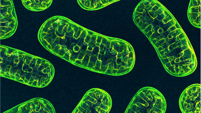
NAD+ is a molecule that is present in all living cells and is essential for a myriad of cellular processes, including regulating metabolism, performing signaling, facilitating DNA repair and blood vessel growth, and regulating some aspects of aging.

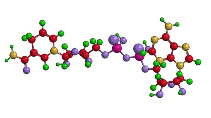
In the body, NAD+ is created from simple building blocks, such as the amino acid tryptophan, and it is created in a more complex way via the intake of food containing nicotinic acid (NA), nicotinamide riboside (NR), nicotinamide mononucleotide (NMN), and other NAD+ precursors.

These different pathways ultimately feed into a salvage pathway, which recycles them back into the active NAD+ form. This salvage pathway, shown on the bottom right of the diagram, includes NAM, NMN, NAD+, and their associated steps.

The evidence that NAD+ declines with age

A 2012 study reported that NAD+ levels decrease dramatically during aging in skin, and while the exact level of decline was not worked out, the average level of concentration seems to fall by a minimum of 50% during adult aging. The difference between newborns and adults, for example, appears to be several-fold lower, according to the study data [1].

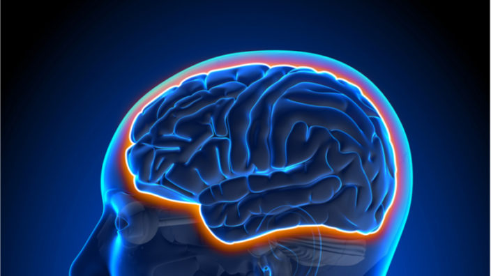
For the brain, two studies used MRI to examine NAD+ and how its level changes during aging. The first study, conducted back in 2015, saw researchers measuring intracellular NAD+ and NADH concentrations and the NAD+/NADH ratio in the human brain, detecting the age-dependent changes in NAD contents and the redox state associated with aging [2].

The second study from 2019 also concluded that an age-dependent decline of the NAD+ levels in the brain was also observed [3]. Taken together, these two studies suggest that there is a decline of NAD+ in the brain of between ~10% to ~25% between the period of young adulthood and old age.

Another study has come along to muddy the waters and contradict the previous two [4]. In biology, nothing is ever simple, and according to this 12-person, placebo-controlled randomized trial, there was no appreciable difference in NAD+ levels in the brain between young and old trial participants.

The researchers of this new study suggest that their results show that NAD+ decline is not associated with chronological aging per se in human muscle or brain, though they do suggest that brain and muscle tissue can benefit from supplementation with nicotinamide riboside, a precursor of NAD+.

It should be noted that this study is currently in preprint on Biorxiv and has not as yet passed peer review. It is also worth noting that at least one of the authors (Charles Brenner) has a stake in patents and the company that owns and licenses the sale of nicotinamide riboside.

For the liver, a previous study showed that liver samples from people aged 60 had around 30% lower concentrations of NAD+ compared to people aged 45 [5].

Finally, the level of NAD+ present in the bloodstream has also been shown to decrease rapidly during aging, according to the results of a 2019 study [6]. This more recent study improved upon an older study [7], which suggested that there was only a small decrease in NAD+ levels; this was due to the newer study using a superior methodology compared to the older one, thus giving a more accurate view of what happens to NAD+ in the bloodstream during aging.

It should be noted that NAD+ levels in blood are typically many times lower than tissue levels, and it is still not clear how important NAD+ is in the blood are and what function or significance its presence has.

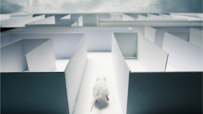
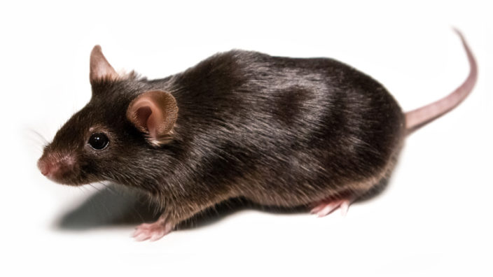
These findings are also supported by a wealth of animal studies and suggests that an age-related decline of NAD+ is common across a range of species, which includes mice and humans.

Some reasons why NAD+ declines with age

Based on both human and animal studies, it seems pretty clear that NAD+ levels do decline during aging in multiple tissues and in blood. However, the big question here is: why does NAD+ decline because of aging?

There are a number of mechanisms that have the potential of reducing NAD+ and could be part of this age-related decline.

Inflammaging and CD38

The most obvious and direct way in which NAD+ is reduced is via the activity of an NAD+ consuming enzyme linked to inflammation called CD38, which destroys NAD+, and its activity steadily rises during aging as systemic inflammation levels increase.

CD38 is a membrane-bound NADase that hydrolyzes NAD+ to nicotinamide and (cyclic-)ADP-ribose. It is associated with immune responses and energy metabolism, but it is also a NADase whose levels rise with aging, with a corresponding increase in NADase activity and a decrease of NAD+.

Therapies that reduce inflammation, particularly the age-related chronic systemic inflammation known as “inflammaging”, could reduce the presence of CD38, thus offering the potential to increase NAD+ [8].

A new class of drugs known as senolytics, which purge the body of inflammatory senescent cells, is an example of a therapy that has the potential to reduce systemic inflammation and could improve NAD+ levels as a consequence.

It is worth noting at this point that it is still unclear whether or not NAD+ synthesis continues at the same rate in older individuals as younger ones, and while it seems clear that inflammaging and CD38 play a key role in consuming NAD+ as we age, there are also a number of other culprits that are involved.

DNA damage and PARPs

Poly-ADP-ribose polymerases (PARPs) are a group of related proteins involved in a number of cellular processes, such as DNA repair, genomic stability, and the programmed cell death known as apoptosis, which damaged cells undergo to remove themselves from the system.

PARP is a key driver of NAD+ catabolism, a set of metabolic processes that break down large molecules, including breaking down food molecules to provide energy and molecular components for anabolic reactions, using that released energy to repurpose the broken-down molecules for growth.

Specifically, PARP1 activity can be increased further in response to DNA damage and genotoxic stress, and given that DNA damage typically increases with age, that would mean more PARP activity and thus more NAD+ consumption [9]. In this way, PARP1 activity may serve as a regulator of NAD+ and mediate its age-related decline as the result of ever-increasing genomic instability that comes with age.

This suggests that therapies that can reduce PARP activity may be a viable approach here; certainly, in animal models of DNA damage, there is NAD+ depletion that can be reversed by inhibiting PARP [10-11]. Other approaches, such as NAD+ repletion via precursor supplementation, may also help to address excessive PARP consumption.

SARM1

The SARM1 protein is another way in which NAD+ levels may be depleted during aging. It was recently demonstrated that SARM1 is able to cleave NAD+, creating NAM, ADP ribose and cyclic ADP ribose [12-13]. Activation of SARM1 has been shown to reduce the available NAD+ pool, but when its activity is blocked, it reduces diabetic peripheral neuropathy and addresses axonopathy in animal models [14-16].

Axonopathy refers to a functional or structural defect in the axon or its terminal, and it is believed to be a precursor for the development and progression of various neurodegenerative diseases. For this reason, SARM1 has become a potential target for drug development; however, there is, at present, little data for SARM1 activity in neurons in the context of aging.

SARM1 is also expressed in other tissues, including the liver and kidneys, and its function in that context of age-related changes to its activity is as yet not fully understood, nor is how that function changes during aging. For this reason, it is still not clear if it plays a central role in age-related NAD+ decline.

Conclusion

This is the end of part one, and we will be delving further into the potential sources of NAD+ depletion in part two, which is coming soon.

We would like to ask you a small favor. We are a non-profit foundation, and unlike some other organizations, we have no shareholders and no products to sell you. All our news and educational content is free for everyone to read, but it does mean that we rely on the help of people like you. Every contribution, no matter if it’s big or small, supports independent journalism and sustains our future.

Literature

[1] Massudi, H., Grant, R., Braidy, N., Guest, J., Farnsworth, B., & Guillemin, G. J. (2012). Age-associated changes in oxidative stress and NAD+ metabolism in human tissue. PloS one, 7(7), e42357.

[2] Zhu, X. H., Lu, M., Lee, B. Y., Ugurbil, K., & Chen, W. (2015). In vivo NAD assay reveals the intracellular NAD contents and redox state in healthy human brain and their age dependences. Proceedings of the National Academy of Sciences, 112(9), 2876-2881.

[3] Bagga, P., Hariharan, H., Wilson, N. E., Beer, J. C., Shinohara, R. T., Elliott, M. A., … & Detre, J. A. (2020). Single-Voxel 1H MR spectroscopy of cerebral nicotinamide adenine dinucleotide (NAD+) in humans at 7T using a 32-channel volume coil. Magnetic Resonance in Medicine, 83(3), 806-814.

[4] Elhassan, Y. S., Kluckova, K., Fletcher, R. S., Schmidt, M., Garten, A., Doig, C. L., … & Wilson, M. (2019). Nicotinamide riboside augments the human skeletal muscle NAD+ metabolome and induces transcriptomic and anti-inflammatory signatures in aged subjects: a placebo-controlled, randomized trial. BioRxiv, 680462.

[5] Zhou, C. C., Yang, X., Hua, X., Liu, J., Fan, M. B., Li, G. Q., … & Wang, P. (2016). Hepatic NAD+ deficiency as a therapeutic target for non-alcoholic fatty liver disease in ageing. British journal of pharmacology, 173(15), 2352-2368.

[6] Clement, J., Wong, M., Poljak, A., Sachdev, P., & Braidy, N. (2019). The plasma NAD+ metabolome is dysregulated in “normal” aging. Rejuvenation research, 22(2), 121-130.

[7] Guest, J., Grant, R., Mori, T. A., & Croft, K. D. (2014). Changes in oxidative damage, inflammation and [NAD (H)] with age in cerebrospinal fluid. PLoS One, 9(1), e85335.

[8] Tarragó, M. G., Chini, C. C., Kanamori, K. S., Warner, G. M., Caride, A., de Oliveira, G. C., … & Jurk, D. (2018). A potent and specific CD38 inhibitor ameliorates age-related metabolic dysfunction by reversing tissue NAD+ decline. Cell metabolism, 27(5), 1081-1095.

[9] Wang, G., Han, T., Nijhawan, D., Theodoropoulos, P., Naidoo, J., Yadavalli, S., … & McKnight, S. L. (2014). P7C3 neuroprotective chemicals function by activating the rate-limiting enzyme in NAD salvage. Cell, 158(6), 1324-1334.

[10] Fang, E. F., Scheibye-Knudsen, M., Brace, L. E., Kassahun, H., SenGupta, T., Nilsen, H., … & Bohr, V. A. (2014). Defective mitophagy in XPA via PARP-1 hyperactivation and NAD+/SIRT1 reduction. Cell, 157(4), 882-896.

[11] Scheibye-Knudsen, M., Mitchell, S. J., Fang, E. F., Iyama, T., Ward, T., Wang, J., … & Mangerich, A. (2014). A high-fat diet and NAD+ activate Sirt1 to rescue premature aging in cockayne syndrome. Cell metabolism, 20(5), 840-855.

[12] Summers, D. W., Gibson, D. A., DiAntonio, A., & Milbrandt, J. (2016). SARM1-specific motifs in the TIR domain enable NAD+ loss and regulate injury-induced SARM1 activation. Proceedings of the National Academy of Sciences, 113(41), E6271-E6280.

[13] Essuman, K., Summers, D. W., Sasaki, Y., Mao, X., DiAntonio, A., & Milbrandt, J. (2017). The SARM1 Toll/interleukin-1 receptor domain possesses intrinsic NAD+ cleavage activity that promotes pathological axonal degeneration. Neuron, 93(6), 1334-1343.

[14] Cheng, Y., Liu, J., Luan, Y., Liu, Z., Lai, H., Zhong, W., … & Huang, R. (2019). Sarm1 gene deficiency attenuates diabetic peripheral neuropathy in mice. Diabetes, 68(11), 2120-2130.

[15] Gilley, J., Ribchester, R. R., & Coleman, M. P. (2017). Sarm1 deletion, but not WldS, confers lifelong rescue in a mouse model of severe axonopathy. Cell reports, 21(1), 10-16.

[16] Gerdts, J., Brace, E. J., Sasaki, Y., DiAntonio, A., & Milbrandt, J. (2015). SARM1 activation triggers axon degeneration locally via NAD+ destruction. Science, 348(6233), 453-457.

Are Epigenetic Clocks Ready for Prime Time?

Reason, author of the anti-aging blog Fight Aging and CEO of Repair Biotechnologies, still isn’t convinced that methylation is very useful in assessing biological age or the efficacy of various therapeutics, despite the wide use of methylation-based clocks.

He surprised me at this year’s Longevity Therapeutics conference in San Francisco by speaking out about the utility of the now widely used epigenetic/methylation clocks pioneered by Steve Horvath, Morgan Levine, and others. I attended the conference on behalf of the Lifespan Extension Advocacy Foundation (LEAF) for my second year in a row, and I enjoyed the sessions and the discussions.

I asked Reason a couple of questions about epigenetic clocks in that session and circled back with him later in this email conversation.

Why are many scientists so gung ho about using methylation clocks in aging studies if the scientific support is lacking? For example, in my recent interview with David Sinclair, I asked about the broader issue of whether epigenetic changes are a cause or consequence of aging, and he was clear in arguing that they are a cause.

Everyone is using methylation clocks because (a) producing that data is a part of trying to make them useful, and (b) it is really useful to convince investors, grant writers, etc who have an unsophisticated view of this.

Are we on the right track to make methylation clocks useful in aging studies, or are the required follow-up calibration studies just not being done yet?

I think the community is on the right track, more or less, but they aren’t being rigorous enough. It is too ad hoc, and this will be slower.

David Sinclair conducted the research showing that epigenetic change is downstream of double-strand breaks. If that bears out, it is only a cause in the looser sense of the word cause. Not a root cause. A point of intervention a few items removed from the cause.

Josh Mitteldorf has argued that it probably wasn’t HGH that led to thymus gland regrowth in the TRIIM trial and that the other four factors (metformin, Vitamin D, DHEA and zinc) were probably more impactful. Do you think that what he argues has any merit?

I don’t think Mittledorf’s argument holds water, given the other evidence for growth hormone to produce thymic regrowth in animal models, but it is testable.

What biomarkers do you consider useful at this time, if not methylation clocks? Please elaborate.

Which biomarkers of aging are useful at this time for rejuvenation therapies: probably some of them, but whether or not that is the case has yet to be proven. People will (and probably should) take the reasonable position that composite biomarkers of frailty are meaningful and that we should pay attention to approaches that can reverse them. But all of the others have yet to be shown to be good enough that one can test new approaches to rejuvenation against them and be prepared to stand by the results.

How do you explain the success of clocks like GrimAge at predicting timing of disease and time to death if methylation clocks aren’t measuring anything meaningful?

All these items, such as GrimAge, are measuring something related to aging. The reason why we can’t just then use them to test any new approach is that we don’t know whether or not these clocks (or other biomarkers) are measuring everything relating to age. Rejuvenation therapies will fix specific line items, reverse specific forms of damage. Since aging is a network of interacting forms of damage, it is perfectly possible to get good correlations with biomarkers that are only sensitive to the consequences of one or two forms of damage or are overly sensitive to one specific consequence. So you really don’t know whether what you observe in the biomarker results are in any way reflective of how useful any given potential rejuvenation therapy might be.

This is very true for the epigenetic clocks. It is a reasonable position to suggest that frailty rolls up pretty much everything in aging, and therefore we should expect results on frailty to be indicative of how useful a rejuvenation therapy is across the board – but that still may or may not be usefully true. It is only plausible.

Now, for specific age-related diseases, one can of course see whether or not something works. There are very clear goals and metrics. If you have a potential rejuvenation therapy, and it fixes arthritis (as is the hope for senolytics), then you put it through the FDA process for arthritis and let the longer battle of discovery relating to everything else in aging play out thereafter. Which is pretty much what everyone will do in the present generation of companies in the longevity industry.

Can you elaborate on the basis for your thymus rejuvenation approach and how it’s developing? What biomarkers will you use to test effects?

Our work on regrowth of the thymus is based on upregulation of FOXN1, a master regulator of thymic growth and function. Its expression declines with age, and that is the proximate reason as to why the thymus atrophies and the supply of T cells declines. So we build gene therapies to produce greater expression of FOXN1, which causes the thymus to increase its cellularity and start turning out more useful T cells. We’re presently past the proof of principle stage in mice and working on the formulation of the therapy. Simple biomarkers are fairly well established for anything to do with a faltering immune system; a great deal of work has taken place in connection with HIV in the past few decades, for example. One uses flow cytometry to count immune cells with various different surface markers indicating different populations: in our case, we want to see more naive T cells in the usual proportions, split between CD4+ and CD8+ types that are effective at tackling pathogens and errant cells. This is not so different from the HIV therapies in which researchers and clinicians are looking for CD4+ cell counts as a measure of how damaged the immune system has become and how much it might have recovered following treatment.

We would like to ask you a small favor. We are a non-profit foundation, and unlike some other organizations, we have no shareholders and no products to sell you. All our news and educational content is free for everyone to read, but it does mean that we rely on the help of people like you. Every contribution, no matter if it’s big or small, supports independent journalism and sustains our future.
Hyperbaric oxygen therapy

Hyperbaric Oxygen Therapy May Reduce Cognitive Decline

A group of Israeli scientists led by Prof. Shai Efrati investigated the effects of hyperbaric oxygen therapy (HBOT) on age-related cognitive decline in healthy older adults, and the results were promising.

The brain is the most metabolically active organ in the body, but it has only limited intracellular energy storage [1]. In absence of local fuel reserves, to sustain neuronal metabolism, the brain depends mostly on cerebral blood flow (CBF), which is why CBF is constantly redirected to the currently active brain regions. CBF accounts for as much as 15% of cardiac output and 20% of resting total oxygen consumption, even though the brain itself comprises just 2% of body mass. CBF dysfunction has been firmly linked to age-related cognitive decline [2].

What is HBOT?

In HBOT, the patient breathes pure oxygen while being subjected to high air pressure, greater than that found naturally on Earth, in a pressure chamber. This results in more oxygen being delivered to the tissues, which promotes wound healing, infection mitigation, and angiogenesis, the creation of new blood vessels.

HBOT is a well-established treatment for a handful of conditions. The list of FDA-approved conditions for HBOT includes necrotizing infections, decompression sickness, non-healing diabetes-related wounds,  and even radiation burns. It is also popular among patients with TBI (traumatic brain injury) for its supposed ability to ameliorate various lingering effects of that condition, such as dizziness and cognitive problems. As with many unconventional treatments, HBOT clinics often peddle their product as effective against a plethora of other conditions, including cancer. Prof. Efrati says HBOT has not been proven to prevent or treat cancer.

Prof. Efrati is well-known in Israel and abroad as a pioneer of HBOT. In his clinic, based in Shamir Medical Center, he uses upgraded protocols that add to the basic process, such as repeated fluctuations in oxygen levels, as patients take their masks on and off during the session to alter oxygen consumption. According to him, this technique boosts HBOT efficacy by utilizing what is known as the hyperoxic-hypoxic paradox:

It is now understood that the combined action of intermittent shifts in oxygen concentration induces many of the cellular mechanisms needed for regeneration – the same mechanisms hypoxia triggers but without the hazardous consequences of actual hypoxia.

In particular, this protocol upregulates hypoxic induced factor (HIF). HIF variants HIF-1α and HIF-2α induce the release of vascular endothelial growth factor (VEGF), which promotes angiogenesis and stem cell proliferation. Simply put, when the human body senses a lack of oxygen, it tries to compensate by creating new blood vessels in order to more efficiently distribute the dwindling precious resource.

HBOT and age-related cognitive decline

Rigorous research of HBOT has been appearing more frequently in recent years, yielding evidence that HBOT can improve cognitive functions in post-stroke, TBI and anoxic brain patients even years after the actual incident [3]. However, the current study [4] was the first to examine the effects of the therapy on “normal” age-related cognitive decline.

The researchers enlisted several dozen healthy elderly adults and divided them equally into a study group and a control group. The measurements were taken after 12 weeks of HBOT, during which the participants took 60 sessions or “dives”, as HBOT enthusiasts call them. The most striking, statistically highly significant improvements reported in the study involved attention and information processing speed. The authors also reported a significant increase in the HBOT group’s energy levels compared to the control. Several other improvements, such as in visual memory, were found notable but not statistically significant. The authors hypothesize that further research with larger sample sizes could elucidate the magnitude of these additional effects.

Changes in CBF as a result of the treatment were also measured. Minor changes in the overall CBF were detected, but there were localized significant alterations as well: CBF increased in some regions associated with various aspects of cognitive function. Using statistical methods, the authors found a moderate correlation between the changes in CBF and the cognitive function improvement in particular patients. This finding is consistent with previous research that showed age-related functional decline to be related to reduced perfusion in specific cortical regions rather than to a global reduction in CBF.

One of the problems with HBOT research is setting up a proper control group. Prof. Efrati concedes that it is virtually impossible to meet a double-blind standard, since the treatment produces a clear sensory input (pressure fluctuations). The researchers could be potentially blinded to the groups, but the patients themselves could not be.

Whether and when HBOT will be approved as a therapy for age-related diseases remains to be seen. Meanwhile, home-owned soft chambers have been gaining popularity, but Prof. Efrati strongly warns against them:

The treatment protocol used in our studies cannot be followed with a soft chamber. There is no adequate quality control of the air that flows in, and privately owned chambers bear a high risk of contamination and fire. In short, they are both rather ineffective and unsafe.

Asked about his main takeaway from the study, Prof. Efrati strikes an upbeat note:

It means that so-called ‘normal aging’ should not be taken as a given. With appropriate treatment, age-related functional decline can be reversed.

Conclusion

This research is currently the most vigorous attempt to determine the efficacy of HBOT in treating age-related cognitive decline. It is especially important in the context of longevity research, since such decline is sometimes considered normal rather than a condition that should be studied and confronted. The research also adds to our understanding of the importance of CBF in cognitive function.

We would like to ask you a small favor. We are a non-profit foundation, and unlike some other organizations, we have no shareholders and no products to sell you. All our news and educational content is free for everyone to read, but it does mean that we rely on the help of people like you. Every contribution, no matter if it’s big or small, supports independent journalism and sustains our future.

Literature

[1] Tarumi, T., & Zhang, R. (2018). Cerebral blood flow in normal aging adults: cardiovascular determinants, clinical implications, and aerobic fitness. Journal of neurochemistry, 144(5), 595-608.

[2] Chen, J. J., Rosas, H. D., & Salat, D. H. (2011). Age-associated reductions in cerebral blood flow are independent from regional atrophy. Neuroimage, 55(2), 468-478.

[3] Boussi-Gross, R., Golan, H., Volkov, O., Bechor, Y., Hoofien, D., Schnaider Beeri, M., … & Efrati, S. (2015). Improvement of memory impairments in poststroke patients by hyperbaric oxygen therapy. Neuropsychology, 29(4), 610.

[4] Amir, H., Malka, D. K., Gil, S., Rahav, B. G., Merav, C., Kobi, D., … & Erez, L. (2020). Cognitive Enhancement of Healthy Older Adults Using Hyperbaric Oxygen: A Randomized Controlled Trial. Aging, 12(13).

Interview with Stephanie Lederman

Stephanie Lederman Discusses AFAR and Funding

Since 1992, Stephanie Lederman has been the Executive Director of the American Federation of Aging Research, which was founded back in 1981 with the mission of supporting and advancing healthy aging through biomedical research. She is a fellow of the New York Academy of Medicine and a member of the New York Academy of Sciences, and she holds a Master’s degree from Boston University.

We had the opportunity to interview Ms. Lederman at International Perspectives in Geroscience, a conference hosted at Weizmann Institute of Science (Israel) on September 4-5.

I’m happy to meet the representative of one of the oldest nonprofit organizations in our field. Before we discuss the projects of AFAR, could you please tell us what sparked your interest in this field and why did you finally choose to work with a nonprofit?

My whole career has been with nonprofits. I have an advanced degree in Public Health. My interest has always been to help the public feel healthier, not only as they age, as I’ve worked with children and youth in different areas. My career has been in a nonprofit, giving back to society, so I’ve always had an interest. I worked at one point for the American Red Cross on a health promotion program for healthy aging. Since then, I’ve became quite interested in the population, so here I am at AFAR for 25 years.

Could you please tell us about AFAR’s main activities?

Our main activities are to fund the young and inspiring and inspire young investigators to work in the field of the biology of aging. From the very beginning, although we do other things as well, we have funded over $200 million since the beginning of our first grant for the biology of aging. Back at a time people really didn’t know what that was about. Of course, we all know that it’s to understand how we age through the aging process, which will ultimately help us to delay and perhaps even eliminate many of the diseases of aging.

Funding for basic research remains one of the main concerns for our community. We understand that the pool of rejuvenation biotechnology companies is actually created by investigating the underlying mechanisms of aging and then selecting promising interventions in preclinical studies. Only then can a new biotechnology startup be formed to bring the research into human trials. Without basic science, there cannot be any science at all. However, funding for basic studies is the main thing that fundamental researchers of aging complain about most of the time. In your opinion, what are the main reasons for this situation?

I believe that the general public doesn’t quite understand the breakthroughs that have occurred in aging science. They only understand diseases, even though in many of the disease areas, like cancer, for example, there haven’t been the breakthroughs that the general public has been led to believe. I think the aging community has to do a better job of explaining what the breakthroughs have been and will be in the coming years, and only then will the general public begin to support it.

Some longevity activists are trying to use radical messaging that tries to intrigue potential supporters with a tenfold increase in lifespan instead of explaining the relationship between aging and disease along with the possibility of making the very processes of aging the actual therapeutic targets. What’s your way of overcoming the initial public skepticism about the usefulness of aging research?

I believe what we try to tell the general public is that a better world for everyone is in the picture. We don’t exaggerate what there might be; we don’t know what there might be. It’s much better to talk realistically, not negative, but realistically, so that people are unsuspicious, and they don’t think that “Oh, you’re doing this because you’re trying to sell a drug.” Or “You’re making it up because this is the fountain of youth, and we’ve heard that for thousands of years.” We at AFAR try to kind of take a middle ground so that people will believe us that aging science is a credible science. It was something that we hoped for, and now we can promise results.

What kinds of organizations or researchers are eligible for your grants’ support, and how much support are you usually able to provide to different types of grantees?

We have a range of programs, and everyone is eligible, if they have a good scientific proposal. We have peer review, but we are very interested in those proposals that are risky, because we get money from the private sector; the government takes care of those that are not as risky. So we look for those that are risky, but we do fund all over the world now. We’re looking for mainly those who are starting out; very often, it’s their breakthrough grant, and we allow them then to get more funding and be an established investigator.

What kind of topic is the most attractive for AFAR? Is there a favorite area of research, such as a focus on a specific hallmark of aging and its related conditions?

Every year, we just put out a request for proposals, and the best science gets funded. It’s different every year; we don’t really specifically say that we want something in telomeres or autophagy or anything like that; we just get whatever idea is coming at that particular time.

There is a problem that is often covered even in research papers, which is that a younger researcher has usually less chances to be funded or provided with a grant. Does this problem arise within AFAR?

We try to go for the younger investigator. Our basic mission is to fund young investigators in the biology of aging. Our founder’s premise was that we need to bring people into the field. That has always been from day one, to try to get people that need the money, if we see people that are kind of tied. The one that needs the money the most, because they’re younger or they’re not as established or they’re not in an institution that may have a lot more resources, we try to give that person the benefit of the doubt.

If you don’t mind me asking, what is the success rate of the grant applications within AFAR? What share of research gets funded, and what’s the total amount of funding that you allocate each year to aging research?

We have a two-tier process and administrative review, and that goes out to about 300 investigators to look at their one-page proposal. If it passes that, then they’re asked to come with a full proposal. Of those people that put in a full proposal, 50% get funded. If you’re looking at from the first part, there’s so many, but it’s hard to judge the percentage and we’re giving out now, in all of our grants, only about 4 or 5 million. We’ve given out more depending upon what year and what foundations; when the Ellison Foundation was supporting us, we gave out more. We’re trying to attract new foundations and new philanthropy to the field, so there’s plenty of more people that can get funded that, unfortunately, we’re not able to fund.

You’re doing a great job anyway; 5 million is a lot, especially for basic studies. I read that you participate in a program of supporting women in science, which I’m really happy about. There is a common misconception that women are less predisposed to scientific work and can’t be true leaders in their respective fields as much as men are. Your own example actually disproves that, quite obviously. Yet I often find these ideas in the air, even coming from my fellow longevity ladies. Could you please share your vision of the situation?

Yes, we’re a big believer in women in science; we have done many activities in that regard. I see the future, especially from the applications that we’re receiving and the people that we’re funding. The future is women. You know, the men have been around for a while; they’re there. Even at this conference, you could see that it was mainly men, I think there were one or two women, but the people in the audience were a majority of women. So we’re optimistic that women are being attracted to this field.

AFAR itself is actively advocating for research on aging on many levels. One such project was support for the TAME study, which investigates the potential of metformin for extending healthy lifespan in people without diabetes. Could you please tell our readers more about the advocacy projects apart from TAME?

Yes. So TAME obviously is very important because if aging is an indication, there will be new drugs. So that’s very important. But we are advocating on behalf of many scientists, we do conferences, media briefings, and webinars. If someone comes to us with an idea, we very often will try to advocate on their behalf. We’re not an advocacy organization, but we like to educate the general public, foundations, the world of philanthropy, and the government in a variety of different ways. We do a lot more than people realize, because we don’t spend a lot of money on advertising, but we’re doing a lot. People may not hear about it. The main initiative is TAME, because it’s been all over the news, but there are many other things as well.

What message would you like to share with people who are considering becoming advocates for healthy longevity or working on fundraising for science?

I would say that the science is now for this field, that it’s a relatively new field, but the excitement over the last 10 years, from what I’ve seen in the amount of time that I’ve been involved, is extremely exciting. There is a lot in the air right now. I think people could have the hope and realize the promise but without the exaggeration of living to 500. One thing I do want to add as well is that a healthier population will benefit society. If there are more older people living longer, there are many things that they could be doing to help society, They’re not a burden; they are really a prize for us, and I think we need to reap the benefits of an older population.

Is there a take-home message for our readers that you would like to offer?

I started off with the message of “A better world for older people is a better world for everyone.” I would say that it’s not just about aging. As you get older, it’s the life course. The work and the research that the field is doing right now will benefit everyone from childhood to older age. It’s not just about “Let’s find something for an old person.” It’s for everybody.

We would like to ask you a small favor. We are a non-profit foundation, and unlike some other organizations, we have no shareholders and no products to sell you. All our news and educational content is free for everyone to read, but it does mean that we rely on the help of people like you. Every contribution, no matter if it’s big or small, supports independent journalism and sustains our future.
DNA mutation

Genomic Instability May Directly Lead to Diseases of Aging

A new perspective published in Cell explains a direct link between genomic instability and age-related diseases [1].

Somatic mutations

Mutation is normally explained as the driving force behind evolution, as changes in the germ lines of organisms result in beneficial and harmful mutations to organisms. The organisms with the harmful mutations die off and the ones with the beneficial mutations pass them on to future generations, resulting in speciation and the diversity of life on Earth we see today.

However, the germ line, which is responsible for the creation of future generations, is not the only thing that mutates. Mutations also occur within organisms as well, as DNA is damaged over time. This damage is known as genomic instability, and it is one of the primary hallmarks of aging, leading to multiple negative downstream effects.

The authors of this review refer to the resulting differences between cells as somatic mosaicism, and they describe how new genetic sequencing technology is able to categorize these mutations. As they explain, these mutations are actually more common per cell than germline mutations, as germ cells have defenses against mutation that somatic cells do not.

Mutating towards aging and cancer

The researchers point out the relationship between aging and heritable genetic diseases, as some diseases require both inherited DNA and somatic mutations to occur. They also hypothesize that genetic mutation may be behind cells’ loss of ability to properly process proteins and to remove themselves when they are diseased; according to this hypothesis, these mutant cells directly lead to proteostasis diseases such as Alzheimer’s.

Additionally, a form of evolution happens within somatic cells as well. Mutations that favor the mutant cell over ordinary cells can be selected for. Upon exposure to UV radiation, skin cells can mutate, and these mutant skin cells can outcompete their ordinary counterparts. These cells are not yet cancerous, but they are more prone to cancer than normal skin cells. This perspective cites data that these sorts of mutations also occur in the lungs, leading to COPD in smokers, and when they occur in blood and arterial cells, they may be a partial cause of atherosclerotic diseases in all individuals.

The reviewers discuss the role of two other hallmarks of aging, telomere attrition and epigenetic alterations, in leading to the transcriptional noise that differentiates cells that should be identical.

Abstract

Age-related accumulation of postzygotic DNA mutations results in tissue genetic heterogeneity known as somatic mosaicism. Although implicated in aging as early as the 1950s, somatic mutations in normal tissue have been difficult to study because of their low allele fractions. With the recent emergence of cost-effective high-throughput sequencing down to the single-cell level, enormous progress has been made in our capability to quantitatively analyze somatic mutations in human tissue in relation to aging and disease. Here we first review how recent technological progress has opened up this field, providing the first broad sets of quantitative information on somatic mutations in vivo necessary to gain insight into their possible causal role in human aging and disease. We then propose three major mechanisms that can lead from accumulated de novo mutations across tissues to cell functional loss and human disease.

Conclusion

Obviously, a direct link between genomic instability and age-related diseases is not good news, as genomic instability is one of the most difficult hallmarks to do anything about; intensive gene therapy would be required to restore genomically compromised somatic cells back to their original state, assuming that they cannot be replaced by genomically uncompromised stem cells.

However, in science, we must go where the research leads us. If this perspective is correct and aging is more directly driven by genomic instability than most researchers believe, then more efforts should be put towards genomic solutions that directly address this particular upstream hallmark of aging.

Literature

[1] Vijg, J., & Dong, X. (2020). Pathogenic Mechanisms of Somatic Mutation and Genome Mosaicism in Aging. Cell182(1), 12–23. https://doi.org/10.1016/j.cell.2020.06.024

We would like to ask you a small favor. We are a non-profit foundation, and unlike some other organizations, we have no shareholders and no products to sell you. All our news and educational content is free for everyone to read, but it does mean that we rely on the help of people like you. Every contribution, no matter if it’s big or small, supports independent journalism and sustains our future.

Interview with Alexander Fedintsev

A New Hallmark of Aging Proposed

Two researchers, Alexander Fedintsev and Alexey Moskalev, have published an expansive review paper in which they propose a new hallmark of aging [1].

The authors draw our attention to a phenomenon that is sometimes overlooked in the context of aging: the accumulation of damage to long-lived molecules caused by the non-enzymatic chemical reactions of glycation, carbonylation, and carbamylation. With time, these modifications inflict extensive damage to our bodies. The authors go as far as to cautiously suggest that this phenomenon might be the upstream cause of several known hallmarks of aging, such as cellular senescence.

Although the authors dedicate a fair share of their attention to intracellular damage, such as histones being susceptible to glycation, their focus is on the stiffening of the extracellular matrix (ECM), which stems from the accumulation of cross-links between long-lived molecules, mainly collagen and elastin. As these molecules have a lifespan comparable to that of a human, the damage is effectively irreparable due to the slow turnover. Some researchers suggest that the decrease in tissue elasticity puts a 100- to 120-year upper limit on the functioning of our cardiovascular and pulmonary systems, which would be in line with our observations [2].

Age-related ECM stiffening seems to inhibit the proliferation of stem cells and even cause cellular senescence. Amazingly, old, senescent cells can be rejuvenated by transferring them into young ECM. The authors make an interesting connection between the mechanistic qualities of the ECM and intra- and intercellular chemical reactions. One of their boldest claims is that the physical stiffness of the ECM is mistaken by cells for fibrosis. This cellular misunderstanding triggers a fibrosis-fighting mechanism that involves cellular senescence. Fighting senescence is one of the major goals of longevity research, and considerable progress has been made, but the authors suggest that we might have been looking for answers in the wrong places. In another example, mesenchymal stem cells (MSCs) tend to differentiate into bone cells when cultured on a stiff matrix. This, the authors claim, may explain the phenomenon of tissue calcification.

ECM stiffness, of course, can wreak havoc in more than one way. For instance, when ECM stiffness increases due to glycation, it significantly reduces T cell proliferation. Adding to the troubles of the immune system, migrating cells, such as immune cells, can experience nuclear membrane rupture when squeezing through tight spaces, which results in cellular death or DNA breakage. Cross-links facilitate this phenomenon by reducing ECM permeability.

Arterial stiffness resulting from ECM aging, the authors suggest, may by itself explain hypertension, because baroreceptors (the receptors in our blood vessels that react to blood pressure), contrary to what their name suggests, react to strain rather than to pressure. As vascular walls get stiffer, they strain less under the same pressure, tricking the receptors into miscalculating desirable blood pressure.

ECM affects intracellular processes via integrins, proteins that attach the cellular cytoskeleton to the ECM. Integrins’ reaction to increased ECM stiffness leads to a surge in ROS (reactive oxidative species) production.

ECM stiffness may play a role in tumor formation by promoting angiogenesis. Developing their mechanistic argument, the authors suggest that “a physical cue devoid of chemical specificity” – i.e. ECM stiffening – may be enough to change certain cell phenotypes into cancerous ones.

As the authors explain, it may also contribute to age-related cognitive decline in at least two ways. First, arterial stiffening may lead to BBB (brain-blood barrier) disruption. Second, ECM stiffening in the brain may impede the formation of new synapses, which simply lack room to thrive, and thus reduce neuroplasticity.

Accumulation of AGEs (advanced glycation products) leads to lung tissue stiffening, causing breathing difficulties in old age. Last but not least, AGEs can be devastating to our cartilage and bone tissue.

Although the authors admit that reversing ECM stiffness is going to be an uphill battle, they do suggest a battery of possible interventions. AGE inhibitors have been shown to extend the lifespan of some species. Caloric restriction, one of our best weapons in our fight against aging, is known to reduce the rate of AGE accumulation. Unfortunately, AGE inhibitors can slow the rate of ECM degradation but cannot rejuvenate the matrix. Other promising avenues are boosting the production of elastin and restricting protein consumption.

Since a paper that declares the discovery of a new hallmark of aging is by all means a major event, we decided to interview one of the authors, Alexander Fedintsev.

What made you focus on this particular problem? How did you realize that this might be a new hallmark of aging? Why do you think this has been overlooked?

Many other researchers, not just me, have been aware of protein cross-links for years. The idea that non-enzymatic cross-links are the primary cause of aging was proposed by Johan Björksten in 1942 but, ever since, their role went largely underestimated. Aubrey De Grey did a great job describing glycation in his book, yet many researchers still think that cross-links are responsible only for arterial stiffening (a factor that contributes to hypertension) and wrinkle formation. The damage of cross-linking to long-lived macromolecules is not even mentioned in the most famous and cited paper in biogerontology, The Hallmarks of Aging [3]. This is what initially caught my attention, along with the fact that while many scientists work on clearing senescent cells, restoring mitochondrial function, epigenetic rejuvenation, et cetera, only a handful center on ECM damage. I am worried by this, especially since developing a cross-link breaker is an extremely hard problem, one that might require major efforts to crack.

Your paper says that non-enzymatic modifications of long-lived macromolecules give rise to virtually all hallmarks of aging. It sounds almost like you have discovered the ultimate upstream cause of aging.

As I said, this was first proposed more than half a century ago by Björksten. However, at that time, there was little to no evidence to back it up. Today, we have a much better understanding of the molecular mechanisms behind the pathogenicity of cross-links and adducts. Two different views of the subject exist. Let’s call the first one the weak ECM hypothesis. It states that the damage to long-lived macromolecules is just one of the drivers of aging, though a particularly important one. The strong ECM hypothesis postulates that we cannot solve the problem of aging solely by attempts on cellular rejuvenation since cellular aging is caused mainly by ECM degeneration. Inversely, research shows that cells are rejuvenated when transferred into young ECM. So, yes, ECM rejuvenation might be the key.

In our paper, we stick mostly to the weak ECM hypothesis and demonstrate that ECM degradation is an important contributor to the aging of all organs and systems, important enough to be considered a new hallmark of aging. Moreover, it seems that damage to the ECM (and other long-lived molecules) might even cause some of the other hallmarks, such as senescent cell accumulation.

Several years ago, I stumbled upon a paper [4] that claimed that in yeast, all intracellular damage is cleared during gametogenesis, effectively resetting biological age. Nothing surprising about that, but, stunningly, according to the paper, this age resetting does not necessarily require meiosis! Naturally, we should not conclude that cells can completely self-repair from a single study that can be faulty or inapplicable to mammalian cells. But if this is applicable to humans, then our somatic cells might have the potential to completely rejuvenate themselves. I asked myself: “Let’s say it’s true. What might be the cause of aging then?” And the answer was the extracellular matrix. It goes like this: ECM proteins like collagen and elastin have enormously long lifespans during which they accumulate uncontrolled non-enzymatic modifications via glycation, carbonylation, and carbamylation. This leads to the formation of toxic adducts. These, in turn, cause sterile systemic inflammation and cross-links that stiffen the matrix. The resistance to the ECM-degrading proteases (like collagenase) increases, the protein turnover slows, and damage accumulation accelerates. Contrary to intracellular damage, there are no known enzymes (at least, not in mammals) that can neutralize adducts and break cross-links, so, currently, we are helpless.

Still, the evidence in favor of the strong ECM hypothesis has been mounting. Neurons transplanted from mice into rat brains can live up to 40% longer than a mouse. Mouse erythropoietic stem cells being serially transplanted to younger mice could live almost 3 times longer than the species’ maximal lifespan. This suggests that cells might have a much greater potential for self-rejuvenation than previously thought, and what might be limiting this ability to regenerate is their microenvironment and ECM in particular.

Stem cells from old mice were shown to be rejuvenated by young ECM [5], while old ECM negatively influences stem cells from young mice. Stiff ECM negatively impacts neuronal progenitors [6]. In addition, ECM is causatively linked to cellular senescence.

Speaking of which, you seem to suggest a new explanation for cellular senescence: that this is basically a mechanism for fibrosis prevention. Can you explain this to our readers?

The fibrosis-mediating role of cellular senescence was discovered long ago. It makes perfect sense in the context of the cell cycle arrest and SASP (senescence-associated secretory phenotype). It’s essentially a part of our wound-healing process. The cell cycle arrest is needed to reduce the amount of collagen that the cells synthesize. Matrix metalloproteinases included in SASP destroy excessive collagen, while proinflammatory cytokines attract immune cells to finalize the cleanup. The anti-fibrotic nature of cellular senescence has been demonstrated in multiple studies. So, the question is, if cellular senescence is indeed an antifibrotic program, why is it dramatically upregulated during the aging process? My bet is that cells can sense the excess of collagen via ECM stiffening, and if the stiffness exceeds a certain threshold, this might launch the cellular senescence sequence. But the increase in stiffness is exactly what happens during normal aging due to non-enzymatic cross-link formation! So, in terms of stiffness, glycated, aging ECM may be indistinguishable from fibrotic ECM. This erroneously triggers the wound-healing senescence program in the absence of any actual wound or fibrosis. There is a brilliant study that demonstrates the role of the ECM in cellular senescence: the scientists created mice with a mutation that makes the collagen produced by their cells resistant to collagenase (which is very similar to what happens during normal aging). This caused severe premature senescence along with decreased lifespan, kyphosis, osteoporosis, and hypertension [7]. Amazingly, interaction with ECM from young cells is sufficient to restore aged, senescent cells to an apparently youthful state [8].

I also think now that ECM non-enzymatic modifications may be a major cause of age-related cognitive decline. Our cognitive ability depends on neuroplasticity – the ability to form, maintain, and destroy connections between neurons. Each neuron is surrounded by the perineuronal net – a special form of ECM. To make way for a new synapse, this ECM needs to be degraded by microglia cells. But what if the macromolecules in the perineuronal net also become more resistant to matrix-degrading enzymes with age? It would hamper the ability to make new synapses, decreasing neuroplasticity. Of course, the perineuronal net is made mostly of proteoglycans and hyaluronic acid, rather than collagen or elastin, but proteoglycans are also subject to glycation and cross-link formation [9].

Do you think you have built a bulletproof case, or is it more of a hypothesis, a suggestion?

I think that the weak ECM hypothesis can be considered proven, while the strong ECM hypothesis is still a hypothesis. However, focusing on rejuvenating the ECM is a win-win approach anyway.

If your idea becomes widely recognized, what do you expect to happen? What implications would it have on medical research, on the development of new therapeutic approaches?

If the strong ECM hypothesis is true, we could reverse all hallmarks of aging and age-related pathologies solely by targeting the ECM degradation. If only the weak hypothesis is true, we still need to find a way to rejuvenate the ECM. No matter how far we progress in other directions, ECM degradation will still be limiting our lifespan and healthspan.

Have you received any feedback from other longevity researchers? Specifically, the authors of the original 2013 paper on the hallmarks of aging? Do you have an idea of what their reaction might be?

I have received positive and constructive feedback from Aubrey de Grey. Professor Alexey Moskalev was inspired and joined as a co-author, which I appreciate a lot. I also got quite positive feedback from reviewers and some other researchers. I haven’t received any feedback from the authors of The Hallmarks of Aging; however, I think that most researchers do recognize (though maybe underestimate) the problem we describe in our paper. Many researchers, though, will probably have trouble accepting the strong ECM hypothesis, since it represents a serious paradigm shift.

SENS research shows that glucosepane may be the most problematic of all AGEs. You seem to agree with this view: according to your paper, glucosepane is the chief culprit behind cross-link formation in the ECM.

Glucosepane is indeed the most abundant AGE in the human body, and this might pose a huge problem. First, we need to find an appropriate animal model to test potential drugs that can break cross-links. The current most popular models – mice and rats – are unsuitable since most cross-links in their ECM have a methylglyoxal origin. Second, glucosepane cross-links, it seems, lack any weak points, contrary to the alpha-diketone cross-links that could be broken by alagebrium. Finally, I personally don’t think we can create an enzyme that breaks glucosepane cross-links simply because enzymes are large molecules unable to fit between collagen fibrils, which cross-links pull even tighter together. Additionally, glucosepane is a non-fluorescent AGE, so it cannot be detected using non-invasive equipment like an AGE reader.

Speaking of potential obstacles in discovering therapies for ECM stiffening, do you think they might be insurmountable, or is it just a matter of time before we find ways to effectively reverse this process?

I personally don’t believe that breaking cross-links is impossible, but I think this is an extremely difficult task. It might take decades to develop a glucosepane breaker in a single lab.

Do you think the longevity research field, in general, is moving in the right direction? Are you optimistic about its future?

Not really. I only know one lab that is working on developing a glucosepane breaker. This is an extremely hard problem, and we need joint efforts of multiple labs around the world to solve it. I’m unimpressed by senolytics since, currently, the results are on par with caloric restriction. Think about it: we cannot outperform a method that has been around for more than a century! We also lack good candidates for gene therapy that would slow the rate of aging. Yes, that’s right: most manipulations with genes in mice that extend lifespan do so by decreasing age-independent mortality rather than the rate of aging [10]! So, I would say we do need a change of focus.

We would like to ask you a small favor. We are a non-profit foundation, and unlike some other organizations, we have no shareholders and no products to sell you. All our news and educational content is free for everyone to read, but it does mean that we rely on the help of people like you. Every contribution, no matter if it’s big or small, supports independent journalism and sustains our future.

Literature

[1] Fedintsev, A., & Moskalev, A. (2020). Stochastic non-enzymatic modification of long-lived macromolecules-a missing hallmark of aging. Ageing Research Reviews, 101097.

[2] Robert, L., Robert, A. M., & Fülöp, T. (2008). Rapid increase in human life expectancy: will it soon be limited by the aging of elastin?. Biogerontology, 9(2), 119-133.

[3] López-Otín, C., Blasco, M. A., Partridge, L., Serrano, M., & Kroemer, G. (2013). The hallmarks of aging. Cell, 153(6), 1194-1217.

[4] Ünal, E., Kinde, B., & Amon, A. (2011). Gametogenesis eliminates age-induced cellular damage and resets life span in yeast. Science, 332(6037), 1554-1557.

[5] Sun, Y., Li, W., Lu, Z., Chen, R., Ling, J., Ran, Q., … & Chen, X. D. (2011). Rescuing replication and osteogenesis of aged mesenchymal stem cells by exposure to a young extracellular matrix. The FASEB Journal, 25(5), 1474-1485.

[6] Segel, M., Neumann, B., Hill, M. F., Weber, I. P., Viscomi, C., Zhao, C., … & Sharma, A. (2019). Niche stiffness underlies the ageing of central nervous system progenitor cells. Nature, 573(7772), 130-134.

[7] Vafaie, F., Yin, H., O’Neil, C., Nong, Z., Watson, A., Arpino, J. M., … & Pickering, J. G. (2014). Collagenase-resistant collagen promotes mouse aging and vascular cell senescence. Aging Cell, 13(1), 121-130.

[8] Choi, H. R., Cho, K. A., Kang, H. T., Lee, J. B., Kaeberlein, M., Suh, Y., … & Park, S. C. (2011). Restoration of senescent human diploid fibroblasts by modulation of the extracellular matrix. Aging cell, 10(1), 148-157.

[9] Pokharna, H. K., & Pottenger, L. A. (1997). Nonenzymatic glycation of cartilage proteoglycans: an in vivo and in vitro study. Glycoconjugate journal, 14(8), 917-923.

[10] de Magalhães, J. P., Thompson, L., de Lima, I., Gaskill, D., Li, X., Thornton, D., … & Palmer, D. (2018). A reassessment of genes modulating aging in mice using demographic measurements of the rate of aging. Genetics, 208(4), 1617-1630.

Mitochondria

The First Precision Gene Editing Tool for Mitochondrial DNA

Researchers have developed what can be described as the first precision gene editing tool for mitochondrial DNA (mtDNA). This new tool is different from the well-known CRISPR/Cas9 method of gene editing and should make the study of mitochondrial biology and mitochondrial diseases easier.

This new discovery is a game changer

Prior to this, creating mouse models of mitochondrial disease for study and drug development has been very challenging; this transformative technology has the potential to make creating such models far easier.

The related study, which was published in the journal Nature, showcases the new mitochondrial gene editing tool [1]. Mutations within mtDNA can cause a variety of unusual and poorly understood conditions, and while CRISPR/Cas9 and related gene editing technologies have allowed researchers to edit DNA faster and easier than before, they only work on the DNA stored in the cell nucleus, the home of the majority of our genetic information.

CRISPR/Cas9 gene editing technologies cannot be used to edit mtDNA, as they cannot access it properly. CRISPR/Cas9 uses a small guide RNA to home the Cas9 enzyme in on a specific target spot on the genome, where it can then cut both strands of DNA. However, no one has figured out how to move the guide RNA into the mitochondria, meaning that it is not possible to edit mtDNA in the same way.

This new discovery came when the researchers were studying how bacteria fight each other using toxins, specifically a group of bacterial toxins known as deaminases, which can damage DNA and RNA bases by removing nitrogen-containing parts from them. Normally, these deaminases target single strands of DNA or RNA; however, they found that a particular cytidine deaminase enzyme, DddA, had no effect on single-stranded DNA or RNA.

After much testing with no success, they discovered that DddA did not work on single-strand DNA or RNA but in fact did work on double-strand DNA. It was assumed that deaminases only worked on single-strand DNA or RNA and that was their purpose in this context, so it was therefore a real surprise to find that DddA bucked this trend.

Taming the beast

The researchers realized that this unusual quirk might be useful for gene editing, in particular for editing mtDNA, as it could bypass the problem of getting the RNA guide inside the mitochondria.

However, it was not as simple as just using DddA directly, due to it being a naturally occurring bacterial toxin; if left to run amok, it could destroy DNA everywhere it encountered it. In order to use DddA to edit mtDNA, the researchers had to find a way to stop DddA from changing mtDNA until the desired target location was reached.

To do so, they split the protein into two inactive pieces; they used 3D imaging data to work out how to cut the protein into two parts in such a way that, when divided, they were harmless, but the rejoined protein was able to resume its original function.

The team joined each part of the DddA protein to customizable DNA-targeting proteins that do not require guide RNA to locate their target. These customizable DNA-targeting proteins are designed to bind to specific regions of DNA, which then brings the two parts of the DddA protein together again. This allowed DddA to work as a precision gene editing tool, preventing it from going on the rampage and destroying all nearby DNA.

They tested the approach to make changes to specific mitochondrial genes, which resulted in fully functional mitochondria, with the exception of the gene that they had edited to cause a defect.

The next step for the researchers will be to search for other bacterial deaminases that could potentially be developed into gene editors. The team hopes that such tools could allow for more effective modeling of mitochondrial diseases as well as assist the development of therapies to address them.

Bacterial toxins represent a vast reservoir of biochemical diversity that can be repurposed for biomedical applications. Such proteins include a group of predicted interbacterial toxins of the deaminase superfamily, members of which have found application in gene-editing techniques. Because previously described cytidine deaminases operate on single-stranded nucleic acids, their use in base editing requires the unwinding of double-stranded DNA (dsDNA)—for example by a CRISPR–Cas9 system. Base editing within mitochondrial DNA (mtDNA), however, has thus far been hindered by challenges associated with the delivery of guide RNA into the mitochondria. As a consequence, manipulation of mtDNA to date has been limited to the targeted destruction of the mitochondrial genome by designer nucleases. Here we describe an interbacterial toxin, which we name DddA, that catalyses the deamination of cytidines within dsDNA. We engineered split-DddA halves that are non-toxic and inactive until brought together on target DNA by adjacently bound programmable DNA-binding proteins. Fusions of the split-DddA halves, transcription activator-like effector array proteins, and a uracil glycosylase inhibitor resulted in RNA-free DddA-derived cytosine base editors (DdCBEs) that catalyse C•G-to-T•A conversions in human mtDNA with high target specificity and product purity. We used DdCBEs to model a disease-associated mtDNA mutation in human cells, resulting in changes in respiration rates and oxidative phosphorylation. CRISPR-free DdCBEs enable the precise manipulation of mtDNA, rather than the elimination of mtDNA copies that results from its cleavage by targeted nucleases, with broad implications for the study and potential treatment of mitochondrial disorders.

Conclusion

The arrival of a new tool that overcomes the limitations of CRISPR/Cas9 in the context of mitochondrial gene editing is a very welcome development indeed. Mitochondrial dysfunction is one of the hallmarks of aging, so having tools that could repair damaged mtDNA as well as address mitochondrial diseases has a huge amount of potential to be truly transformative, just as the discovery of CRISPR/Cas9 has been.

We would like to ask you a small favor. We are a non-profit foundation, and unlike some other organizations, we have no shareholders and no products to sell you. All our news and educational content is free for everyone to read, but it does mean that we rely on the help of people like you. Every contribution, no matter if it’s big or small, supports independent journalism and sustains our future.

Literature

[1] Mok, B. Y., de Moraes, M. H., Zeng, J., Bosch, D. E., Kotrys, A. V., Raguram, A., … & Mougous, J. D. (2020). A bacterial cytidine deaminase toxin enables CRISPR-free mitochondrial base editing. Nature, 1-7.

A Blood Factor Boosts Neurogenesis and Cognition

By now, the race to discover regenerative factors present in blood has been underway for a number of years, with numerous researchers seeking a “secret sauce” in blood that has the potential to rejuvenate aged tissues and organs.

A fork in the road

It is clear from previous animal studies going back over a decade that it is possible to rejuvenate an animal via factors found in blood. Researchers such as Irina and Michael Conboy first demonstrated back in 2005 that rejuvenation was possible by joining aged and young mice together to share circulatory systems, which is known as parabiosis [1].

The aged mice were shown to benefit from exposure to the younger systemic environment, and their tissues were rejuvenated. This led to the conclusion that one or two things had happened: that factors in young blood had rejuvenated the old mice or that parabiosis had caused the dilution of inhibitory, pro-inflammatory factors, which stop tissue from repairing properly and accumulate with age.

What we are now seeing is a divergence of approaches, with scientists broadly following one of the above two paths to rejuvenation. The Conboys in particular believe that what you remove from old blood is more important than what you put in, and they are busy developing a blood filtering therapy that can “scrub” aged blood of the handful of primary pro-aging factors that prevent tissue regeneration.

Groups such as Alkahest and Elevian are busy searching for the secret sauce in young blood in the hope that identifying key factors might lead them to rejuvenation.

Fooling the brain into thinking that it has exercised

Researchers led by Dr. Saul Villeda at the University of California, San Francisco have published a new study that builds on the second path and the search for beneficial factors present in blood. These researchers demonstrate that a particular factor in the blood of aged mice that exercise regularly can convey the beneficial effects of exercise on neurogenesis and cognition in the brains of aged mice.

To trace the factor responsible for this, the researchers took the blood from active old and middle-aged mice that frequently ran on their cage wheels and then transfused it to old mice that had no wheel and were sedentary.

The sedentary mice that were given the blood a total of eight times during a 3-week period were almost as good at learning and memory tests, including maze navigation, as the active mice. A control group of old mice that were sedentary and got blood from another group of sedentary old mice saw no such improvement.

The researchers noted that the inactive mice that got blood from the active mice also grew around twice as many new neurons in the hippocampus, the area of the brain associated with learning and memory. This increased neurogenesis was similar to that observed in active, exercising mice.

The researchers identified the factor responsible by examining the blood of the active mice and seeing which factors increased when they exercised. This led them to identify an enzyme called glycosylphosphatidylinositol specific phospholipase D1 (Gpld1) which is created in the liver.

To see if this was the factor responsible, the researchers injected the Gpld1 gene into the tail vein of aged, inactive mice, causing that enzyme to increase in production. After a period of 3 weeks, their cognitive performance and neurogenesis was similar to the active, exercising mice.

Intriguingly, the researchers did not find a large amount of Gpld1 in the brains of the active mice, which suggests that it does not cross the blood-brain barrier. It is likely that its influence on the brain derives from cleaving other proteins from the membranes of cells, allowing the freed molecules to enter the bloodstream and thus lower systemic inflammation.

A reduction of inflammation, especially in the brain, would likely result in increased neurogenesis and improved cognition, as supported by previous studies showing that a reduction of systemic inflammation spurs rejuvenation.

Finally, they also examined a group of elderly people who exercised regularly and compared them with an inactive group and found that Gpld1 levels in the blood of the active group of people was significantly higher. This is particularly promising and suggests that the mouse results seen here have good potential to translate to people.

Dr. Villeda and his team now aim to find a drug that can mimic these beneficial effects and that could be given to older people who are too frail to exercise.

Reversing brain aging may be possible through systemic interventions such as exercise. We found that administration of circulating blood factors in plasma from exercised aged mice transferred the effects of exercise on adult neurogenesis and cognition to sedentary aged mice. Plasma concentrations of glycosylphosphatidylinositol (GPI)–specific phospholipase D1 (Gpld1), a GPI-degrading enzyme derived from liver, were found to increase after exercise and to correlate with improved cognitive function in aged mice, and concentrations of Gpld1 in blood were increased in active, healthy elderly humans. Increasing systemic concentrations of Gpld1 in aged mice ameliorated age-related regenerative and cognitive impairments by altering signaling cascades downstream of GPI-anchored substrate cleavage. We thus identify a liver-to-brain axis by which blood factors can transfer the benefits of exercise in old age.

Conclusion

There can be little doubt that blood factors have the potential to rejuvenate aged tissue and organs, and likely the first fruits of this work will arrive in the not-too-distant future. The debate around removing factors from old blood versus putting them in is also likely to be resolved before too long, and it could always be the case that both approaches have an effect. Time and further experimentation will tell.

We would like to ask you a small favor. We are a non-profit foundation, and unlike some other organizations, we have no shareholders and no products to sell you. All our news and educational content is free for everyone to read, but it does mean that we rely on the help of people like you. Every contribution, no matter if it’s big or small, supports independent journalism and sustains our future.

Literature

[1] Conboy, I. M., Conboy, M. J., Wagers, A. J., Girma, E. R., Weissman, I. L., & Rando, T. A. (2005). Rejuvenation of aged progenitor cells by exposure to a young systemic environment. Nature, 433(7027), 760-764.

Red meat

Trimethylamine May be Involved in Vascular Aging

Researchers suggest that trimethylamine, a metabolic byproduct that is created when bacteria in the gut break down foods such as red meat, may be a key player in vascular aging.

Trimethylamine and vascular aging

Some researchers believe that trimethylamine damages our arteries, according to a new study conducted by researchers at the University of Colorado Boulder [1]. The researchers suggest that excessive trimethylamine production could be a key risk factor for heart disease.

When we consume foods that contain L-carnitine and choline, the bacteria in our gut quickly break it down into a form ready for the body to consume; however, while doing so, they metabolize it into trimethylamine.

Carnitine is encountered in many animal products, although red meat has the highest known levels. For example, a typical 4-ounce steak has an estimated amount of between 56 and 162 milligrams of carnitine. Carnitine is also present in lower amounts in chicken, milk and other dairy products, fish, beans, and avocado. Choline can be found in fish, beef, chicken and other poultry, eggs, and some beans and nuts.

After the food is broken down, the trimethylamine next reaches the liver, where it is converted into trimethylamine-N-Oxide (TMAO), which then enters the bloodstream and travels around the body. Previous studies have shown that people who have higher blood levels of TMAO are at far greater risk of stroke or heart attack.

The researchers examined the blood and arterial health of 101 older people as well as 22 young adults. They discovered that TMAO levels typically increase with advancing age due to changes to the bacteria in the gut.

This is consistent with other studies showing that the bacterial populations in the gut microbiome change during aging, from being helpful to becoming a more harmful composition as we get older. In this case, the bacteria that produce trimethylamine increase as we age, and thus the level of TMAO in the bloodstream rises.

They found that the adults with high blood levels of TMAO had significantly worse arterial health and function compared to adults with lower amounts. People with higher TMAO in their arteries had increased levels of oxidative stress, inflammation, and tissue damage to the arterial wall lining.

Next, the researchers studied the effects of TMAO on mice, and the results were quite alarming. Mice that were directly given TMAO experienced rapid aging of their blood vessels. Following several months of TMAO supplementation, the 12-month-old mice resembled 27-month-old mice, which is the equivalent of a 35-year-old person looking like an 80-year-old.

There was also some initial data that suggested mice with higher levels of TMAO also experience impaired learning and cognition.

The researchers also report that mice given a compound called dimethyl butanol, which is present in olive oil, vinegar, and red wine, experienced a reduction of vascular dysfunction. This suggests that dimethyl butanol somehow inhibits the creation of TMAO, possibly via the inhibition or slowing of the growth of trimethylamine-producing bacteria populations in the gut microbiome.

The researchers’ next step is to explore other compounds that could potentially reduce the production of TMAO and slow down vascular aging.

Age-related vascular endothelial dysfunction is a major antecedent to cardiovascular diseases. We investigated whether increased circulating levels of the gut microbiome-generated metabolite trimethylamine-N-oxide induces endothelial dysfunction with aging. In healthy humans, plasma trimethylamine-N-oxide was higher in middle-aged/older (64±7 years) versus young (22±2 years) adults (6.5±0.7 versus 1.6±0.2 µmol/L) and inversely related to brachial artery flow-mediated dilation (r2=0.29, P<0.00001). In young mice, 6 months of dietary supplementation with trimethylamine-N-oxide induced an aging-like impairment in carotid artery endothelium-dependent dilation to acetylcholine versus control feeding (peak dilation: 79±3% versus 95±3%, P<0.01). This impairment was accompanied by increased vascular nitrotyrosine, a marker of oxidative stress, and reversed by the superoxide dismutase mimetic 4-hydroxy-2,2,6,6-tetramethylpiperidin-1-oxyl. Trimethylamine-N-oxide supplementation also reduced activation of endothelial nitric oxide synthase and impaired nitric oxide-mediated dilation, as assessed with the nitric oxide synthase inhibitor L-NAME (NG-nitro-L-arginine methyl ester). Acute incubation of carotid arteries with trimethylamine-N-oxide recapitulated these events. Next, treatment with 3,3-dimethyl-1-butanol for 8 to 10 weeks to suppress trimethylamine-N-oxide selectively improved endothelium-dependent dilation in old mice to young levels (peak: 90±2%) by normalizing vascular superoxide production, restoring nitric oxide-mediated dilation, and ameliorating superoxide-related suppression of endothelium-dependent dilation. Lastly, among healthy middle-aged/older adults, higher plasma trimethylamine-N-oxide was associated with greater nitrotyrosine abundance in biopsied endothelial cells, and infusion of the antioxidant ascorbic acid restored flow-mediated dilation to young levels, indicating tonic oxidative stress-related suppression of endothelial function with higher circulating trimethylamine-N-oxide. Using multiple experimental approaches in mice and humans, we demonstrate a clear role of trimethylamine-N-oxide in promoting age-related endothelial dysfunction via oxidative stress, which may have implications for prevention of cardiovascular diseases.

Conclusion

If this connection between excessive TMAO levels and vascular aging turns out to be correct in humans and can be replicated in other independent studies then it suggests a possible therapeautic intervention point. TMAO levels certainly appear to rise as we age as do the populations of bacteria in the microbiome which produce Trimethylamine, so they may be onto something here, though there are likely nuances requiring further study. If TMAO turns out to be a problem as suggested by the researchers approaches that could be potentially developed, such drugs to block its activity or even adjusting the microbiome, could offer a solution to this problem.

We would like to ask you a small favor. We are a non-profit foundation, and unlike some other organizations, we have no shareholders and no products to sell you. All our news and educational content is free for everyone to read, but it does mean that we rely on the help of people like you. Every contribution, no matter if it’s big or small, supports independent journalism and sustains our future.

Literature

[1] Brunt, V. E., Gioscia-Ryan, R. A., Casso, A. G., VanDongen, N. S., Ziemba, B. P., Sapinsley, Z. J., … & Seals, D. R. (2020). Trimethylamine-N-Oxide Promotes Age-Related Vascular Oxidative Stress and Endothelial Dysfunction in Mice and Healthy Humans. Hypertension, 76(1), 101-112.

Blood-brain barrier

Selectivity of the Blood-Brain Barrier Changes with Age

The mechanisms at work in the blood-brain barrier change with age, affecting the selectivity of this vital interface.

The blood-brain barrier

The blood-brain barrier is a layer of cells in the blood vessels of the brain that prevents molecules in the blood from crossing into the central nervous system. Selective transport across the barrier is important both to keep out pathogens and to maintain normal brain function, as malfunction of the blood-brain barrier is associated with a range of diseases, such as Alzheimer’s and meningitis.

For over 100 years, scientists have used tracer molecules to probe the blood-brain barrier. This research has shown that the barrier is impermeable to most macromolecules. However, previous studies have not explored how permeable the barrier is to the thousands of endogenous proteins floating in our blood.

How proteins get across

A team of researchers investigated precisely that question in a new study published in Nature [1]. The team used isotopes and fluorescent markers to label hundreds of proteins in the plasma of mice and then tracked their movement across the blood-brain barrier. They found that plasma proteins readily cross the barrier but that the process is controlled by a complex system of regulators. Using single-cell RNA sequencing, they identified genes correlated with enhanced or inhibited uptake, such as the transferrin receptor TFRC and the alkalaine phosphatase ALPL.

By carrying out the tests in both young and old mice, the researchers were able to spot a change in the performance of the blood-brain barrier with age. Roughly half as many plasma proteins crossed the barrier in older brains. This was linked with the down-regulation of genes related to uptake in older mice. Based on these findings and some transport experiments, the researchers concluded that receptor-mediated transport across the barrier decreases in older mice. At the same time, transport via lipid rafts increases. This change causes a shift from specific to non-specific transport, altering the selectivity of the blood-brain barrier.

Restoring the balance

The researchers also investigated the possibility of reversing these changes in order to help keep the brain healthy. The ALPL gene came up in their list as an inhibitor of TFRC, which promotes receptor-mediated transport. Treating aged brain cells with an ALPL blocker increased the expression of TFRC and made the cells better at taking up plasma. While this is still a far cry from fixing the problem, it demonstrates the potential of pharmacological intervention.

The vascular interface of the brain, known as the blood–brain barrier (BBB), is understood to maintain brain function in part via its low transcellular permeability. Yet, recent studies have demonstrated that brain ageing is sensitive to circulatory proteins. Thus, it is unclear whether permeability to individually injected exogenous tracers—as is standard in BBB studies—fully represents blood-to-brain transport. Here we label hundreds of proteins constituting the mouse blood plasma proteome, and upon their systemic administration, study the BBB with its physiological ligand. We find that plasma proteins readily permeate the healthy brain parenchyma, with transport maintained by BBB-specific transcriptional programmes. Unlike IgG antibody, plasma protein uptake diminishes in the aged brain, driven by an age-related shift in transport from ligand-specific receptor-mediated to non-specific caveolar transcytosis. This age-related shift occurs alongside a specific loss of pericyte coverage. Pharmacological inhibition of the age-upregulated phosphatase ALPL, a predicted negative regulator of transport, enhances brain uptake of therapeutically relevant transferrin, transferrin receptor antibody and plasma. These findings reveal the extent of physiological protein transcytosis to the healthy brain, a mechanism of widespread BBB dysfunction with age and a strategy for enhanced drug delivery.

Conclusion

This study identifies and characterizes yet another way the body eventually begins to malfunction. Mice are not humans, of course, but if the results hold in human cells, we’ll have one more piece of the aging puzzle. With this groundwork in place, researchers can look into ways to prevent or reverse this process, keeping the barrier from malfunctioning so that the brain can stay healthy.

We would like to ask you a small favor. We are a non-profit foundation, and unlike some other organizations, we have no shareholders and no products to sell you. All our news and educational content is free for everyone to read, but it does mean that we rely on the help of people like you. Every contribution, no matter if it’s big or small, supports independent journalism and sustains our future.

Literature

[1] Yang, A.C., Stevens, M.Y., Chen, M.B., Lee, D.P., Stähl, D., Gate, D., … Wyss-Coray, T. (2020) Physiological blood-brain transport is impaired with age by a shift in transcytosis. Nature, doi:10.1038/s41586-020-2453-z

Frailty and Age-Related Immune Decline Reversed in Mice

Frailty and the decline of the immune system are two typical features of aging, but new research shows that both can be halted and even somewhat reversed using a novel cell-based therapy.

Frailty and immunosenescence

Frailty is something that frequently affects older people, and it ultimately results in the loss of independence and quality of life for many people. Everyday tasks such as getting around become a challenge, and trips and falls can become deadly for people suffering from frailty.

Along with frailty, aging is accompanied by the gradual decline of the immune system, a process known as immunosenescence, in which immune cells cause inappropriate inflammation or become dysfunctional and unable to do their jobs. This makes older people more vulnerable to infectious diseases, as the immune system becomes increasingly less able to fight back against invading pathogens.

This puts older people at a higher level of risk during cold and flu season and during outbreaks such as COVID-19. Vaccinations are also typically less effective in the elderly as their immune systems just do not work that well, so they normally have the highest mortality rates when infectious diseases are circulating.

In the last few decades, life expectancy has continued to rise steadily, but that increase is not always accompanied by good health. Healthspan, the period of life that an individual spends healthy and free from disease, is the focus of researchers at the University of Bern as well as the University Hospital Bern, and they are aiming to find ways to increase healthspan in people. With a population increasingly reaching ever more advanced age it has become a matter of urgency to help older people maintain health for as long as possible.

Belly fat plays a key role in inflammaging

Inflammaging, the chronic low-grade systemic inflammation that typically accompanies aging, is thought to be instrumental in aging, and researchers have long proposed that inflammaging influences the rate of aging and contributes to the development of various age-related diseases. This persistent background of inflammation also leads to increasingly poor tissue repair and degeneration as we grow older

Inflammaging has many sources, including senescent cells and their pro-inflammatory secretions, cell debris, microbial burden, and immunosenescence. The researchers of a recent study have shown that visceral adipose tissue, known as belly fat, is also a key contributor to inflammaging.

The researchers showed that immune cells known as eosinophils have a central role in regulating inflammation and aging via their contribution to inflammaging. Eosinophils are white blood cells that are typically found circulating in the bloodstreams of both mice and humans, but they are also resident in belly fat.

In healthy people, between 1-5% of all white blood cells in the body are eosinophils, which are created in the bone marrow, circulate in the bloodstream, and generally migrate into various tissues where they reside for a short time. Eosinophils contain a package of around 200 granules of enzymes and proteins which burst open (degranulate) to release their payloads when triggered. During normal operation, these pro-inflammatory cells act almost like tiny grenades, moving to the target and exploding to destroy nearby bacteria, parasites, and so on.

The eosinophils resident in belly fat also appear to have an additional and previously unknown role, helping to regulate and maintain local immune system homeostasis, or balance. However, as we age, the number of eosinophils present in the belly fat declines, and the number of pro-inflammatory macrophages, immune cells that eat waste, rises. As a result of this shift in immune cell distribution, belly fat becomes a source of pro-inflammatory signals, which then contribute to the overall background of systemic inflammation.

Rejuvenating belly fat

After establishing the key role eosinophils play in regulating inflammation and immune system balance, the researchers explored the possibility of rejuvenating visceral adipose tissue in order to restore the balance of immune cell types back to that of younger individuals. They transferred eosinophils from young mice into old mice, which reversed both local and systemic inflammation.

The research team noted that the transplanted eosinophils homed in on the adipose tissue, which had a rejuvenating effect on the old mice. The mice experienced an improvement to physical fitness during endurance and grip tests and were less frail. Perhaps most fascinating was that their vaccination response improved, which suggests that the immune system was rejuvenated to a significant level.

The researchers are now planning to use these findings to develop and translate therapies with the aim of rejuvenating the immune system, reducing inflammation, and to promote increased healthspan in humans.

Adipose tissue eosinophils (ATEs) are important in the control of obesity-associated inflammation and metabolic disease. However, the way in which ageing impacts the regulatory role of ATEs remains unknown. Here, we show that ATEs undergo major age-related changes in distribution and function associated with impaired adipose tissue homeostasis and systemic low-grade inflammation in both humans and mice. We find that exposure to a young systemic environment partially restores ATE distribution in aged parabionts and reduces adipose tissue inflammation. Approaches to restore ATE distribution using adoptive transfer of eosinophils from young mice into aged recipients proved sufficient to dampen age-related local and systemic low-grade inflammation. Importantly, restoration of a youthful systemic milieu by means of eosinophil transfers resulted in systemic rejuvenation of the aged host, manifesting in improved physical and immune fitness that was partially mediated by eosinophil-derived IL-4. Together, these findings support a critical function of adipose tissue as a source of pro-ageing factors and uncover a new role of eosinophils in promoting healthy ageing by sustaining adipose tissue homeostasis.

Conclusion

Once again, this demonstrates that aging is not a rigid or one-way set of processes and that it has a great deal of malleability. Of the various approaches to aging being developed, the rejuvenation of the immune system seems a solid strategy indeed. By maintaining our immune systems as we grow older through therapies such as this, we could maximize our healthspans for longer and, most importantly, healthier lives.

We would like to ask you a small favor. We are a non-profit foundation, and unlike some other organizations, we have no shareholders and no products to sell you. All our news and educational content is free for everyone to read, but it does mean that we rely on the help of people like you. Every contribution, no matter if it’s big or small, supports independent journalism and sustains our future.

Spam

Repetitions in Our DNA React to Anti-Aging Interventions

A group of scientists from Colorado State University has discovered that repetitive elements (REs) in our DNA increase with aging and react to known longevity-promoting interventions [1].

Are REs genomic parasites?

At the dawn of genomic research scientists were stunned to learn that as much as 95% of our genome is non-coding: it does not code for proteins that our body needs in order to operate. A lot of what initially had been labeled as “junk DNA” was later revealed to carry out multiple important functions, such as regulating gene expression. However, up to two-thirds of our genome consists of REs, which are patterns of DNA that have multiple copies across the genome. Of these, retrotransposable elements (RTEs) comprise the majority. As their name suggests, RTEs can multiply via a process reminiscent of “copy and paste”, inserting their new copies back into the genome at various locations. Using intricate molecular machinery, RTEs transcribe themselves into RNA and back into a fresh copy of their DNA sequence, which is then inserted at a new location. Although RTEs, being under constant evolutionary pressure, tend to balance their need for propagation with the host’s survival [2], they can still disrupt genes and influence gene expression, and they have been linked to multiple disorders, some of which are age-related [3]. These short sequences of DNA, which are clearly acting as units of natural selection, may be the best illustration of Richard Dawkins’ “selfish gene” theory.

Still, under normal conditions, most REs are inactive. They reside in genomic regions that are chromatinized (wound into a tight structure called chromatin), whereas for a DNA sequence to be transcribed, the region it resides in has to be unwound. But the volume of RE transcriptions seems to rise with age, probably due to chromatin instability that is a major aging-related phenomenon. It may seem that increased RE transcriptions are a consequence rather than a cause of aging, but the authors of the paper note that REs have been linked to other hallmarks of aging, such as oxidative stress and cellular senescence, and hence may play a causative role. Notably, at least one study showed that inhibiting retrotransposition extends lifespan in mice [4].

REs react to interventions

The researchers hypothesized that if the increase in RE transcriptions is indeed age-related, it should react to known longevity-promoting interventions. To test their hypothesis, they first analyzed an RNA-sequencing dataset from an experiment in which mice were subjected to lifelong caloric restriction (CR), which is probably the most effective life-prolonging intervention that we know of. The researchers found a statistically significant increase in RE transcripts with age, which was largely attenuated by CR. They proceeded to analyze the effect of a set of somewhat shorter, but still long-term in mouse years, 8-month interventions that included CR, rapamycin, and acarbose treatments. All treatments brought about significant, global reductions in RE transcripts. This is consistent with the notion that rapamycin and acarbose mimic CR and work via similar pathways. Using a separate dataset, the authors found that a high-fat diet, which is known to reduce lifespan, also significantly promotes RE transcription.

Yet another interesting experiment that the authors conducted involved transgenic mice with the growth hormone receptor knocked out, a known way to increase longevity. The animals, as expected, showed less RE activity than their genetically unmodified peers.

Although various interventions resulted in somewhat different patterns of RE reduction, there was a significant overlap: 518 specific RE transcripts were downregulated and 92 were upregulated in all of the experiments.

Exercise lowers RE expression

The RNA-sequencing data on long-term anti-aging interventions in humans is scarce, so the researchers opted for the next best thing: a dataset that included young and older sedentary adults as well as older adults that have been habitually exercising for at least 5 years. In line with their hypothesis, the researchers found that sedentary older adults showed a much larger volume of RE expressions than young adults, but this effect seemed to be attenuated by regular exercise. Maximal aerobic exercise capacity (VO2 max), which is a major physiological predictor of longevity, also correlated heavily with RE transcription levels.

Collectively, our results support the growing idea that global RE dysregulation may be an important mechanism of aging (and not simply an adverse effect of the process). Reversing age-related RE transcript accumulation may be necessary for healthy aging, as our present findings show that health/lifespan-enhancing interventions consistently reduce RE expression.

Conclusion

REs may be the “orphans” of longevity research compared to coding sequences, but there is a growing body of evidence that their increased transcription with age may play a role in aging-related diseases. The current research seems to reinforce this notion. Though more work should be done to clarify the connection between REs and aging, the general idea that RE transcripts increase in volume mainly due to aging-related chromatin destabilization, and consequently contribute to various adverse effects, seems plausible.

We would like to ask you a small favor. We are a non-profit foundation, and unlike some other organizations, we have no shareholders and no products to sell you. All our news and educational content is free for everyone to read, but it does mean that we rely on the help of people like you. Every contribution, no matter if it’s big or small, supports independent journalism and sustains our future.

Literature

[1] Wahl, D., Cavalier, A. N., Smith, M., Seals, D. R., & LaRocca, T. J. (2020). Healthy aging interventions reduce non-coding repetitive element transcripts. bioRxiv.

[2] Bourque, G., Burns, K. H., Gehring, M., Gorbunova, V., Seluanov, A., Hammell, M., … & Mager, D. L. (2018). Ten things you should know about transposable elements. Genome biology, 19(1), 1-12.

[3] Prudencio, M., Gonzales, P. K., Cook, C. N., Gendron, T. F., Daughrity, L. M., Song, Y., … & Baker, M. C. (2017). Repetitive element transcripts are elevated in the brain of C9orf72 ALS/FTLD patients. Human molecular genetics, 26(17), 3421-3431.

[4] Simon, M., Van Meter, M., Ablaeva, J., Ke, Z., Gonzalez, R. S., Taguchi, T., … & Neretti, N. (2018). Inhibition of retrotransposition improves health and extends lifespan of SIRT6 knockout mice. bioRxiv, 460808.

Interview with Edwina Rogers

Edwina Rogers – Effective Advocacy for Longevity

We had the opportunity to interview Edwina Rogers, CEO of the Global Healthspan Policy Institute and a professional public health advocate supporting aging research. Edwina has degrees in corporate finance and law. She worked for four U.S. Presidents and two U.S. Senators, helping to shape national policy regarding healthcare. We caught up with her at International Perspectives in Geroscience, a conference hosted at Weizmann Institute of Science (Israel) in September 2019, in order to discuss the principles of effective advocacy.

You have served in public policy positions and the United States, including the White House, and in private international sectors for more than 20 years, but now you’re the founder and leader of the Global Healthspan Policy Institute. What’s made you focus your attention on the problem of aging?

That’s a very good question. I have been involved in mainly health care policy, that might not be so easy to tell from my bio or my resume. But, in the White House, I worked on adding prescription drugs to Medicare In the U.S., and Medicare is the program for people who are 65 years old, the health insurance program for them, and it used to not cover prescription drugs.

In 2001 and 2002, that was a big project I worked on in the White House, and in order to do that, you have to learn how to build coalitions to get a major piece of legislation through. That’s very expensive and has to get through the House and the Senate. Then, when I left the White House, I started working in the Senate, and I also did health care policy but all kinds of health care policy, everything from global AIDS legislation to mental health parity, things like that.

When I left the Senate, I got very involved in healthcare policy for corporate America, and in corporate America, of course, they want to keep their employees happy, healthy, and working as long as possible, especially if they have special talents and they’re productive. That led me to this initiative about the Patient Centered Medical Home, which is an integrated healthcare model that is very well advanced in Denmark. We wanted to implement it in the United States, so we used a coalition to do that, and that was very successfully implemented in the United States. It improves health outcomes and lowers costs. Basically, it’s an extension of that project and that concept. Global Healthspan Policy Institute is a think tank but also an international coalition to change public policy.

While the community acknowledges they need to change regulations related to the development of rejuvenation biotechnologies, many people don’t really know where to start. As an experienced policymaker, can you briefly describe the steps to take in order to become an effective advocate for healthy life extension technologies and make a real impact on laws and regulation? What does the career of an advocate look like?

That’s also a good question. I think that the main thing that you need to do is education of the decision makers, so we stay in front of decision makers. Decision makers would be people in the executive branch who work on regulations but then also the legislators in Congress and in the Senate, members of Parliament. It’s very similar in every country.

We do briefings and outreach to the decision makers. For example, with aging, most people that I’ve met with, they were not aware at all that aging was something that could be controlled and manipulated. Therefore, it’s something that should be researched, and there should be funding for aging research. We’ve had some good success educating the right people. We’ve had people that are sympathetic and understanding of the issue. It’s been surprising that some people in high positions, especially in the U.S., in the Medicare system and Medicaid system, who are physicians, they’ll say to us that aging is something that’s a natural process, you can’t change it at all. These people are MDs, so just telling you that story right there lets you know how much education we have to do to get people up to speed on the whole aging process.

How exactly does one prepare for this stage of negotiations and education of the decision makers?

Okay. Well, the best way to prepare would be to have policy statements, position papers, and explanatory documents that are written and tailored for elected officials and their staff. They are not journal articles; they are not research articles in peer-reviewed journals. It is a document that looks quite differently. It’s much more in layman’s terms; it wouldn’t be anything that you’ve seen here today at this conference. It would be things like frequently asked questions, a briefing paper, that is short, maybe at the eighth grade reading level without a lot of technical terms. So you have documents like that that you have on your website that you also distribute.

There are also subscription services where you can email all the members of Congress and their staff, and senators. We send out emails, we send out a weekly newsletter every Thursday, with articles, short articles. We write short summaries, and then we link to the fuller articles. These are articles that are not so scientific in nature, but they describe a lot of the advancements that are happening in the aging area and aging research. We’ll just pick three or four of the most important articles every week, summarize them, and write a few of our own. We send out a newsletter every Thursday to members of Congress and then anybody else who subscribes to the newsletter. So, it’s just a steady drumbeat.

Social media is important, but less important for what we do, because what really matters is if you can get in front of one or two key decision makers and get a lot done, versus having a big social movement. But that’s also very important, it’s just not exactly what we do.

If I hear you right, the main thing is to get educated yourself, to have the facts at your fingertips, and to have these facts delivered in a condensed way, as a sort of a summary, to present the facts to the decision makers in order to inform them very quickly and without too much terminology.

Yes, and we kind of call it an elevator pitch. A lot of times, when you get in front of a Senator or Congressman, you might just have three or four minutes to get them interested, so you have to have your elevator pitch ready to go, and then you can have longer conversations after you get them interested. Those are the kinds of things that we’ve worked on.

Another really important thing is, in order to get a seat at the table and get clout, you need to be part of a coalition, a big coalition, and the bigger, the better. You have to make sure that all the stakeholders are at the table. An ideal coalition for the aging community is longevity or healthspan. We chose to use the word healthspan because longevity is a little bit dicey when you’re talking to members of Congress. Because they’re thinking, oh, well, you’re trying to make sure everybody lives longer, but what are we going to do about the cost of their retirement programs, because in the U.S., we’re set at a retirement age. It’s not going up, in order for it to go up, we would have to have legislation.

If you think about an ideal coalition, you would want to have consumer advocacy groups involved, they will be very important. By consumer, I mean patient advocacy groups, that they are not left out. There’s not a big market for patient advocacy groups on aging, so we have to make the market, they’re not there. But other types of patient advocacy groups are available, but they’re all disease specific at the moment, and you need major employers, you need health plans, you need local governments, state governments, national governments, all coming together. Hospitals, providers, physicians, researchers, universities, the bigger, the better. The more diverse, the better.

Sounds very interesting. One of the projects that you’re leading involves supporting the TAME study. Could you please tell us about the main milestones that have been achieved so far in this project? What’s its current situation [as of September 2019], and what are your future plans for further support of the TAME study?

Well, TAME is a big project, the cost is about $64 million, and the principal investigators have been looking for ways to scale back the cost. It has to be done over five years and X number of sites, but we’re very fortunate that the Food and Drug Administration in the US approved a study on the first drug that could slow down the aging process itself. So, when you’re looking for $64 million, that’s a lot of money to look for, and there’s no way that anyone can make money, because the drug they’re looking at is Metformin, which is a generic drug, which costs like a few pennies. So there’s no pharmaceutical interest or money involved in that particular study.

But we are very fortunate in that, over the last two or three years, the NIH has shown great interest, and they have basically committed to more than half of the money for that particular clinical trial, and so the gap now is much smaller. The money that we’re looking for is now less than $20 million and not the $60 million between the private funds that have been pledged. AFAR is very active on this, of course; Dr. Nir Barzilai is the principal investigator.

The final gap of money that we need to start the study and kick it off most likely will be coming from private foundations, but we did have that one bit of luck finally, with NIH agreeing to pull some money from Alzheimer’s and from cancer, and from the National Institute of Aging and cobble it together and have some seed money to get it going. I would say that the funding should be complete on that, within the next two years.

Is there a way in which people from our community could actually help with this project?

I think that if more broad support was shown, even though it’s not a lot of money, I think that it would be very helpful to get foundations on board. Right now, we have the NIH, so the US government is doing their part. Then we have quite a few private foundations that have already committed with us to close this gap. We need more private foundations and maybe even corporate money, but the piece that we don’t have is the general public’s support. Even though maybe the general public support is $10,000, it could be made from 500 people. That makes it sort of a very different formula, where it shows that there is also public support, public understanding, and public knowledge of what it is that we’re trying to do, and then that would only increase the chances of getting the commitments from the last few foundations. So that would be my advice.

What are, say, three to five main political bottlenecks that have to be addressed by healthy life extension advocates in order to accelerate the development of rejuvenation biotechnology as a field? What’s your vision on how to solve these bottlenecks?

Well, probably the main bottleneck for aging research would be the lack of funding, where it’s not top of the list. And then the second bottleneck, If I had to prioritize them would be the lack of information; most people don’t understand how aging research is so incredibly important that it touches on more than 14 chronic diseases.

It’s not just disease specific, but it’s hard to take that leap, because it’s something that you can’t see: it’s not cancer, it’s not a broken arm, it’s not so specific. It’s gradual, and it’s long term. I think that the funding and also the education, educating the general public, just having more awareness about the aging process, and that it can be manipulated, it can be measured, you can measure it with a baseline, you could have interventions, and there are currently interventions that people in the movement know about but not anyone hardly outside of the movement, outside of medical providers.

I think that it’s now ready to become mainstream; more journalists interested would certainly be helpful to working on the decision makers, members of Parliament, members of Congress; having them all briefed up on this is very important. So dissemination of information, and then just the general public.

It would be great if we got Hollywood interested in this and we had a mainstream blockbuster movie that touched on aging research. Something like that can really turn the whole movement around, and it could just take off. Things like that happen; there are other examples. For example, in the gay community, they had some good success with having interest in Hollywood and made it mainstream and raised the visibility of the issue. There are other examples like that were just backwater; nobody really knew about the particular project or issue that needed to be addressed, and then a movie pops out about it. Then it’s like a household word; everybody’s pretty up to speed on it.

There are often concerns related to the possibility that access to rejuvenation treatments will not be universal. People are afraid that the treatments will be prohibitively expensive and that elites will keep the information secret. While I doubt that scientific knowledge can be truly hidden from the public, I still share the concern regarding the price tag, at least for some of the treatments, What can the public do to set the prices for new treatments as low as possible from the political point of view?

I see that concern, and I understand it, but I’m less worried about that. Because, as we already know, the gold star of the best treatments available right now is to actually diet and exercise. I think that most humans don’t know about the diet and exercise. That’s not something that is being kept secret, but it’s actually confusing information; people will be told, Okay, you need to exercise, but what does that mean? We spent about a year and a half going through all the clinical trials on exercise with regard to aging, and longevity and life extension, and the ones that outperform placebo.

We have had meetings with everything from life insurance companies, to health insurance companies, and they already have a lot of information. We know that the sort of optimum amount of exercise to make yourself more healthy and extend your life would be movement, try to get something like 7,500 steps a day, and then more intensive exercise at 150 minutes per week. We already know that that’s optimal.

And the research shows that, well, if you do 300 minutes a week, you really don’t get any improvement over the 150 minutes, you might get 1% improvement. Also, with the steps, a lot of people will get a Fitbit or something, and they know that they’re supposed to be doing steps, and it’s programmed with 10,000. 10,000 might not be obtainable for most people, and then they become discouraged, so they don’t know that you actually get your full benefit from 7,500. Anything above that is nice and fine, and it’s not harmful, but you’re not getting the benefit in your healthspan, in your lifespan, and then you’re short of that, you get maybe 60% of the benefit and 40% of the benefit, depending on how many steps on average you’re able to do. That’s an example on the exercise side.

On the diet side, of course, we know about the fasting, you coordinate fasting, you’re trying to let your body rest and not eat for a minimum of 12 hours a day, 14 will be better, 16 would be great. Beyond that, you could get into some issues; depends on your own personal health. For intermittent fasting, there are some costs associated with programs like, for example, Prolong, where you have supplements that your body cannot detect that are food. You can still eat, you can have three meals a day, you’re not eating a lot, but your body thinks you’re not eating at all, so it can rejuvenate itself.

So those are just some examples of ways that you can greatly improve your chances of living longer and increasing your healthspan, but they actually don’t cost any money. It’s just a matter of information and knowledge. Now, some of the other treatments, for example, making sure you have enough vitamin D, that’s not expensive either. Metformin has some really good science behind it; for some people, not for everybody, it costs maybe a penny or two a day. I’m currently not aware of a proven, fantastic, expensive treatment program that a billionaire could go off to, other than some of these basic things that we’re talking about, and live another 5 or 10 years and have an advantage over the rest of us.

I’m asking this question exactly for this reason: we are currently in a period of transition from when we have almost nothing except for lifestyle interventions to when we’re going to have a lot of different treatments technically available. A lot of these therapies might actually be considered to be life-saving treatments, so they should be available to the public. How can a public advocate contribute to reducing prices if some of these treatments are going to be very expensive in the beginning? We all know that the price will go down, but it might not go down very quickly

Let’s say there’s a drug that was developed by a big pharmaceutical company, in Western Europe or in the US, and it’s quite expensive. There won’t be anything major that public advocates could do other than put pressure on their governments to negotiate with the pharmaceutical companies to try to get the prices down. Some governments don’t recognize their patents when they do develop things like that. Or they’ll threaten them, basically, that if you don’t sell it to us at a discount, then we’re going to not recognize your patent, and we’re going to make it available to our people anyway. Of course, I don’t think that’s very acceptable, there has to be some way to make sure that the patent is recognized and the company that developed it is appropriately compensated for their discovery, and they’re fine, but that’s already the case with every disease in every drug around the world.

For example, AIDS, when the first drug came out, that prolonged life and people could live with AIDS, it was very expensive, and Africa needed it. That was what the Global AIDS Bill was about, to try to get that drug into Africa at a discounted rate so that the people there could have access to it. It’s a problem, but it’s a problem that I hope we get to deal with, because right now, we don’t even have that problem. Because we don’t have anything that is proven that we’re all trying to get access to; there’s not anything at the moment that is expensive that we could be fighting for. But the minute there is something, I think it will be very hard to keep it from humanity. If it’s something that is known to save lives and improve them drastically. Probably, there’ll be big pressure for it to be in the public good and open access.

Would you like to talk just a little bit about what you are doing for yourself to maintain your health over your lifespan?

Definitely, the steps and the exercise, I talked about the 7500 steps, the 150 minutes of intensive exercise, and the fasting, so that’s really good information. In the past, maybe it’s 11 o’clock at night, and I haven’t gone to sleep, maybe I’m going to go downstairs and have a cup of hot chocolate. Now I know that I should not do that. Instead, I’m trying to limit my intake of food in the shortest period of time each day and doing the longer fast periods with this diet that I mentioned. So I do use Prolong, it’s a five-day diet that mimics fasting, where your body can’t detect that you’ve eaten anything, and all the numbers come back much better when I measure things. I also use something called Inside Tracker. Inside Tracker has a program that is called Inner Age, and it tells you what your biological age is, what your age is with regard to your biomarkers. It also sends you tips every day, you need to add nuts or avocado to your diet, you need to sleep more. Sleep is very important, that’s a big part of living longer.

There was one post during the poster session that talks about psychological age. The basic point there is that feeling content, feeling happy, having friends, and having meaning and purpose in life can also help.

I keep a list on my cell phone of like 50 things that extend life that are known to be true. Some of them are quirky, for example, having a late-model car that has all the new safety technology on it. Lane changing warnings and blind spot warnings in your car. Many people die from falling, actually, when they get older, so there are things you can do to make sure that you don’t fall. Most people fall in the shower. I heard this brilliant speech one time that Jared Diamond gave in California. He talked about how we spend all this money researching this topic and that topic in healthcare, and most of us are going to die from falling in our shower.

So let’s don’t overlook the easy fixes to extend life, preventing auto accidents, falling in the shower, breaking a hip, things like that. That’s why I said I have this list of 50 things from the research that we gathered that outperforms placebo.

Sounds great. Do you have a take-home message for our readers?

Well, I think that my message for your readers would be that they’re on the right track with advocating for healthy life extension. There’s so many issues these days that you could be an advocate for that you could spend your precious spare time on, but I think that this one is very significant. I feel good about it, this one is my favorite, and I think that it’s something that people can get behind and feel good about. They can share this information with their loved ones. Most people are generally interested in improving their health and living happy, long, full lives, and that’s what we’re talking about here.

We would like to ask you a small favor. We are a non-profit foundation, and unlike some other organizations, we have no shareholders and no products to sell you. All our news and educational content is free for everyone to read, but it does mean that we rely on the help of people like you. Every contribution, no matter if it’s big or small, supports independent journalism and sustains our future.
Neurons

Parkinson’s Disease Symptoms Reversed in Mice

Researchers have found that blocking the activity of a single gene causes other cell types to become dopamine-producing neurons, a type of neuron that Parkinson’s disease destroys.

What is Parkinson’s disease?

Parkinson’s disease is a progressive, age-related, neurodegenerative disease that mainly affects the motor system and thus movement. Symptoms start gradually; sometimes it begins with a barely noticeable tremor in just one hand. These tremors and shakes are common, and the disorder also commonly causes stiffness and difficulty of movement.

The symptoms affecting motor skills are the result of the death of specialized dopamine-producing neurons in the region of the brain known as the substantia nigra, a region of the midbrain.

The reasons why these cells die is poorly understood, but it certainly involves the accumulation and formation of waste proteins into Lewy bodies in the neurons. The end result is that the loss of these neurons leads to a lack of dopamine in the brain, which then causes the telltale symptoms of impaired motor skills.

It has long been suggested that a possible approach to curing Parkinson’s disease could be to replace the lost dopamine-producing neurons, and a number of researchers have been developing ways to achieve this. Recently published research takes this idea beyond the typical idea of stem cell transplants and explores an exciting new solution to the problem.

Creating new neurons to replace losses

Dr. Xiang-Dong Fu and his team at the University of California San Diego School of Medicine recently published a new study that builds upon a fortunate discovery that they made some time ago [1].

They discovered several years back that inhibiting the PTB gene using siRNA or even deleting the gene entirely in connective tissue cells called fibroblasts caused them to change into neurons. This got them thinking about the idea of using this discovery as a foundation for a therapy that might replace lost neurons in the brain and potentially treat a range of neurodegenerative diseases in which neurons are destroyed.

They found that when they used PTB inhibition in a mouse model of Parkinson’s disease, astrocyte cells in the brain transformed into neurons. Astrocytes are “star-shaped” cells that reside in the brain and are the most common of the glial cells in the brain that are closely associated with neuronal synapses. They support the brain by helping to regulate the transmission of electrical impulses within the brain.

Following PTB inhibition, the symptoms of Parkinson’s vanished in the mice, which suggests that the conversion of astrocytes allowed some of them to replace some of the dopamine-producing neurons in that area of the brain lost to Parkinson’s disease.

The mice returned to normal function within three months after just one treatment and remained free from the symptoms of Parkinson’s disease for the remainder of their lives.

The team’s next step will be to refine its methods and test the approach in a different mouse model of Parkinson’s, one which uses a genetic modification to emulate the disease. In this study, the team used a molecule to poison the dopamine-producing neurons, causing the mice to develop symptoms similar to Parkinson’s disease. They have also patented their approach to ultimately test it in human trials in the future.

Parkinson’s disease is characterized by loss of dopamine neurons in the substantia nigra. Similar to other major neurodegenerative disorders, there are no disease-modifying treatments for Parkinson’s disease. While most treatment strategies aim to prevent neuronal loss or protect vulnerable neuronal circuits, a potential alternative is to replace lost neurons to reconstruct disrupted circuits. Here we report an efficient one-step conversion of isolated mouse and human astrocytes to functional neurons by depleting the RNA-binding protein PTB (also known as PTBP1). Applying this approach to the mouse brain, we demonstrate progressive conversion of astrocytes to new neurons that innervate into and repopulate endogenous neural circuits. Astrocytes from different brain regions are converted to different neuronal subtypes. Using a chemically induced model of Parkinson’s disease in mouse, we show conversion of midbrain astrocytes to dopaminergic neurons, which provide axons to reconstruct the nigrostriatal circuit. Notably, re-innervation of striatum is accompanied by restoration of dopamine levels and rescue of motor deficits. A similar reversal of disease phenotype is also accomplished by converting astrocytes to neurons using antisense oligonucleotides to transiently suppress PTB. These findings identify a potentially powerful and clinically feasible approach to treating neurodegeneration by replacing lost neurons.

Conclusion

This approach is very promising, though we should be cautious here, as mice are not people and these results may not translate. These researchers are taking their time to test the approach in another mouse model of the disease to see if they can achieve similar results. The field is littered with failed attempts to reverse Parkinson’s, as promising results in individual animal models have failed to pan out, so doing the experiment again in another mouse model is a wise move.

That said, it is a proof of concept that replacing lost neurons could work as a way to cure Parkinson’s, even if it does not translate directly and without any modification for human use. The exhaustion of specialized cell populations is thought to be one of the reasons we age, so it is good to see a working solution in action.

We would like to ask you a small favor. We are a non-profit foundation, and unlike some other organizations, we have no shareholders and no products to sell you. All our news and educational content is free for everyone to read, but it does mean that we rely on the help of people like you. Every contribution, no matter if it’s big or small, supports independent journalism and sustains our future.

Literature

[1] Qian, H., Kang, X., Hu, J., Zhang, D., Liang, Z., Meng, F., … & Devaraj, N. K. (2020). Reversing a model of Parkinson’s disease with in situ converted nigral neurons. Nature, 582(7813), 550-556.