The Blog

Building a Future Free of Age-Related Disease

Balance

Removing Some Senescent Cells May Have Consequences

Senescent cell accumulation is a likely reason we age, but removing some populations of specialized cells that have turned senescent may have negative consequences, according to a new study.

What are senescent cells?

As you get older, more and more of your cells enter into a state known as cellular senescence. Senescent cells no longer divide or support the tissues of which they are part; instead, they secrete a variety of proinflammatory cytokines, chemokines, and extracellular matrix proteases, which, together, form the senescence-associated secretory phenotype, or SASP.

The SASP increases the level of chronic inflammation, leads to impaired tissue repair, and is linked to the progression of various age-related diseases. If that is not bad enough, their presence can cause nearby healthy cells to become senescent after exposure to the SASP.

Senescent cells normally destroy themselves via a programmed process called apoptosis, and they are also removed by the immune system; however, the immune system weakens with age, and increasing numbers of senescent cells escape this process and begin to accumulate in all the tissues of the body.

By the time people reach old age, significant numbers of these senescent cells have built up, causing chronic inflammation and damage to surrounding cells and tissue. The accumulation of senescent cells is thought to be one of the proposed reasons we age.

Removing senescent cells may not be without some risks

One proposed solution to the problem of senescent cell accumulation and the resulting pro-inflammatory SASP is their therapeutic removal through a new class of drugs known as senolytics. Various past animal studies have shown that the removal of senescent cells appears to be beneficial to health and, in some studies, increases healthy lifespan.

These results have been replicated enough times in the past few years that senolytics should be considered a viable approach to treating aging and its resulting age-related diseases.

However, despite these promising results and the current efforts underway to translate them to people in clinical trials, there are some researchers who caution against senolytic, as senescence is partially an adaptive response to aging, and disturbing it could be harmful.

Another concern is that some specialized cell populations do not replace themselves, such as heart cells and, later in life when the thymus shrinks and barely works, T cells. The concern is that removing these specialized cells, even though they are senescent and clearly causing harm via their secretions, could lead to worse consequences. Stem cell exhaustion is another proposed reason we age, and it is plausible that removing highly specialized populations of cells without providing for their replacements could accelerate that aging process.

Today, we want to highlight a new study that suggests a specialized population of liver endothelial cells could be one such cell population at risk from senolytics [1].

The accumulation of senescent cells can drive many age-associated phenotypes and pathologies. Consequently, it has been proposed that removing senescent cells might extend lifespan. Here, we generated two knockin mouse models targeting the best-characterized marker of senescence, p16Ink4a. Using a genetic lineage tracing approach, we found that age-induced p16High senescence is a slow process that manifests around 10–12 months of age. The majority of p16High cells were vascular endothelial cells mostly in liver sinusoids (LSECs), and to lesser extent macrophages and adipocytes. In turn, continuous or acute elimination of p16High senescent cells disrupted blood-tissue barriers with subsequent liver and perivascular tissue fibrosis and health deterioration. Our data show that senescent LSECs are not replaced after removal and have important structural and functional roles in the aging organism. In turn, delaying senescence or replacement of senescent LSECs could represent a powerful tool in slowing down aging.

Conclusion

Does removing these specialized but senescent populations of cells outweigh the harm that they would otherwise do by their secreted SASP? It seems doubtful, given the broadly positive results that previous studies have shown when senolytic therapies are used systemically. However, removing them may open another can of worms and, in the long term, may accelerate another aging process, such as stem cell exhaustion.

If this is the case, then replacing senescent cells is something that should be done in combination with senolytic therapies that remove them; fortunately, that is something that has been suggested long ago and is part of the proposed toolkit for treating aging, which has solutions for every reason we age.

We would like to ask you a small favor. We are a non-profit foundation, and unlike some other organizations, we have no shareholders and no products to sell you. All our news and educational content is free for everyone to read, but it does mean that we rely on the help of people like you. Every contribution, no matter if it’s big or small, supports independent journalism and sustains our future.

Literature

[1] Grosse, L., Wagner, N., Emelyanov, A., Molina, C., Lacas-Gervais, S., Wagner, K. D., & Bulavin, D. V. (2020). Defined p16High Senescent Cell Types Are Indispensable for Mouse Healthspan. Cell Metabolism.

Egg cell

NAD+ Restores Mitochondrial Function in Egg Cells

Researchers have discovered that a lack of NAD+ is responsible for mitochondrial dysfunction in egg cells (oocytes), leading to decreased fertility with age.

Mitochondria in oocytes

Given how much it must divide to create a human being, the oocyte is the largest cell in the human body, and it contains far more mitochondria than any other cell as well; most cells have a few hundred or thousand, and the oocyte contains a hundred thousand. The mitochondria are the powerhouses of our cells, and it is no surprise that the oocyte requires a considerable amount of power.

This also means that mitochondrial dysfunction, one of the hallmarks of aging, has a significant effect on these cells. In a new paper, researchers hypothesize that increased reactive oxygen species (ROS) production by damaged mitochondria leads to genetic dysfunction in oocytes, leading to infertility and other problems.

The role of NAD+

Nicotinamide adenine dinucleotide (NAD+) is a topic that we have covered extensively, as it is responsible for many critical functions in our cells. As NAD+ has been shown in previous work to improve mitochondrial function in other cell types [1], the researchers decided to see if it would improve the function of oocytes as well.

To test this hypothesis, they chose to administer nicotinamide riboside (NR), a well-known precursor of NAD+, to 8-month-old female mice. Mice of this age that had been given NR for four months had their NAD+ increased substantially over ordinary 12-month-old mice, and multiple other metrics, including follicle number, ovulation potential, oocyte count, ovary size, and litter size, were improved as well. However, administration of NAD+ did not fully return such fertility-related metrics to those of 2-month-old mice.

While such things have been covered in previous research, this study highlights the role of the mitochondria in the viability of oocytes. Supplementation of NR was shown to promote healthy mitochondrial function, as it increases mitophagy, the process by which damaged mitochondria are destroyed in order to leave room for healthy mitochondria, and increases the expression of genes responsible for mitochondrial development.

Abstract

Loss of follicles together with decreased oocyte quality and quantity contribute to age-associated ovarian senescence and infertility. Although underlying mechanisms for ovarian senescence are still unknown, mitochondrial dysfunctions have been reported. Here, we showed age-dependent decreases in ovarian Nicotinamide Adenine Dinucleotide (NAD+) levels in mice whereas supplementing aging mice with nicotinamide riboside (NR), an NAD+ precursor, increased ovarian NAD+ content. We found that increases in ovarian NAD+ levels in aging mice led to increased number of ovarian follicles and ovulatory potential as well as increased live birth rate. NR supplementation also reduced levels of reactive oxygen species and decreased spindle anomalies in aging oocytes, together with increased mitochondrial membrane potential (??m) and decreased mitochondrial clustering. In addition, NR supplementation improved ovarian mitochondrial energy metabolism. Our data suggested that supplementation with NAD+ precursors in vivo and in vitro could be potential therapeutic approaches for treating age-related ovarian infertility.

Conclusion

This study builds upon previous research, making it clear that NAD+ plays a critical role in aging at the cellular level. However, this is a mouse study, and the effects of NR, or any other precursor of NAD+, on human fertility have not yet been tested. Furthermore, more research is, as always, needed to analyze the upstream causes that lead to the depletion of NAD+ as we age and what interventions are effective against them.

We would like to ask you a small favor. We are a non-profit foundation, and unlike some other organizations, we have no shareholders and no products to sell you. All our news and educational content is free for everyone to read, but it does mean that we rely on the help of people like you. Every contribution, no matter if it’s big or small, supports independent journalism and sustains our future.

Literature

[1] Zhang, H., Ryu, D., Wu, Y., Gariani, K., Wang, X., Luan, P., … & Schoonjans, K. (2016). NAD+ repletion improves mitochondrial and stem cell function and enhances life span in mice. Science, 352(6292), 1436-1443.

Niacin-rich foods

Niacin May Prove Useful Against Brain Cancer

A possible new role for niacin has been discovered in the fight against brain cancer.

An old vitamin 

Niacin is a form of vitamin B3 and has been a popular supplement since its discovery in 1937 by the researcher Conrad Elvehjem. Niacin is actually nicotinic acid, and it is possible to create it through the oxidation of nicotine using nitric acid. However, nicotine is well known as being the highly addictive chemical found in tobacco, and so the supplement industry dodged this negative association by marketing it as niacin, which is formed from the words NIcotinic ACid vitamIN.

Niacin is present in a variety of foods, including chicken, tuna, turkey, peanuts, coffee, kidney beans, pork, and bacon. Meats typically have the highest niacin content by a large margin; however, this may not be practical for dietary reasons. Niacin is widely and inexpensively available as a supplement.

While other forms of B3, such as nicotinamide mononucleotide (NMN) and nicotinamide riboside (NR), have enjoyed the spotlight in recent years, it seems that an old favorite, niacin, is making somewhat of a comeback.

Niacin and NAD+ biology

Niacin, like NMN and NR have been of interest to people in the context of its potential as an anti-aging supplement due to its critical role in the creation of Nicotinamide adenine dinucleotide (NAD+), a coenzyme present in all living cells. NAD+ is vital for the production of ATP, a type of universal energy critical to cellular function and life itself. NAD+ is also involved in cellular signaling, facilitates redox reactions, carries electrons from one reaction to another, and regulates the immune system.

NAD+ is created from simple building blocks, such as the amino acid tryptophan, and it is created in a more complex way via the intake of food that contains nicotinic acid (niacin) or other NAD+ precursors, such as NMN and NR. These different pathways ultimately feed into a salvage pathway, which recycles them back into the active NAD+ form.

As we age, the levels of NAD+ available to us fall due to rising levels of chronic systemic inflammation, which leads to deregulated metabolism, a decline of cell function and efficiency, and increasingly poor tissue regeneration and upkeep. Some researchers suggest that by boosting the levels of NAD+ back to more youthful levels, we might somewhat mitigate the effects of aging to support healthy longevity.

Teaching an old dog new tricks

A few days ago, we talked about the results of a human trial for niacin in the context of mitochondrial myopathy, a mitochondrial disease that causes prominent muscular problems, and we observed its benefits in the healthy control group. It seems that niacin has a few more tricks up its sleeve, as a new cancer study shows [1].

Researchers at the Cumming School of Medicine (CSM) at the University of Calgary have shown that niacin combined with chemotherapy spurs immune cells to attack glioblastoma, a type of brain tumor. This resulted in a significant slowing down of disease progression in the mice, with untreated mice living for just 40 days while mice given the combination therapy lived for 150 days.

Glioblastoma is a highly aggressive form of brain cancer, and there are currently no fully effective solutions to it. Typically, people with glioblastoma die within 14 to 16 months of diagnosis, and that is even with chemotherapy and radiation therapy. A key reason why this form of cancer is so deadly is that it takes control of the immune system and makes it work for the tumor rather than attacking it on sight.

The researchers here discovered that just niacin alone increased the survival time of mice; when combined with the chemotherapy drug temozolomide, which is typically used to treat glioblastoma, there was a significant increase in survival time.

They found that the niacin was able to reset the microglia, monocytes, and macrophages that are normally hijacked by glioblastoma and reactivate them so that they stopped supporting the tumor and instead attacked it as they should naturally do.

Glioblastomas are generally incurable partly because monocytes, macrophages, and microglia in afflicted patients do not function in an antitumor capacity. Medications that reactivate these macrophages/microglia, as well as circulating monocytes that become macrophages, could thus be useful to treat glioblastoma. We have discovered that niacin (vitamin B3) is a potential stimulator of these inefficient myeloid cells. Niacin-exposed monocytes attenuated the growth of brain tumor–initiating cells (BTICs) derived from glioblastoma patients by producing anti-proliferative interferon-α14. Niacin treatment of mice bearing intracranial BTICs increased macrophage/microglia representation within the tumor, reduced tumor size, and prolonged survival. These therapeutic outcomes were negated in mice depleted of circulating monocytes or harboring interferon-α receptor–deleted BTICs. Combination treatment with temozolomide enhanced niacin-promoted survival. Monocytes from glioblastoma patients had increased interferon-α14 upon niacin exposure and were reactivated to reduce BTIC growth in culture. We highlight niacin, a common vitamin that can be quickly translated into clinical application, as an immune stimulator against glioblastomas.

Conclusion

Needless to say, we do not encourage people to rush out and buy niacin as a method of treating cancer, and researchers will need to confirm dosage, delivery and length of time as part of a properly controlled human trial before we can know for sure if these mouse results translate to people.

In the fight against cancer, the immunotherapy approach is the most promising compared to traditional drugs,so anything that can help a compromised immune system get back to doing what it should, namely killing cancer cells and destroying tumors, is a positive move.

Our immune system is the ultimate living drug, so the more we can do to improve it and keep it working efficiently, the better outcomes can be expected in combating cancer and other diseases that compromise the immune system.

While we have known about niacin for over 80 years it seems it is not yet finished revealing its secrets and this is another example of new uses for old drugs and compounds. As more advances in drug screening and discovery arrive, we may find that many old favorites get a new lease on life as we find that they have previously unknown utility for treating diseases.

We would like to ask you a small favor. We are a non-profit foundation, and unlike some other organizations, we have no shareholders and no products to sell you. All our news and educational content is free for everyone to read, but it does mean that we rely on the help of people like you. Every contribution, no matter if it’s big or small, supports independent journalism and sustains our future.

Literature

[1] Sarkar, S., Yang, R., Mirzaei, R., Rawji, K., Poon, C., Mishra, M. K., … & Yong, V. W. (2020). Control of brain tumor growth by reactivating myeloid cells with niacin. Science Translational Medicine, 12(537).

Osteoclast

A New Likely Mechanism Behind Osteoporosis Discovered

A group of Japanese scientists has found that advanced glycation end products (AGEs) tend to accumulate in osteoblasts with age, inducing apoptosis via endoplasmic reticulum stress. This mechanism likely contributes to the development of osteoporosis [1].

AGEs and age

Advanced glycation end-products, fittingly abbreviated as AGEs, are proteins or lipids that have had sugars attached to them via the process of glycation. They tend to accumulate in our bodies with time and play a pathogenic role in the development of various age-related diseases, such as diabetes, atherosclerosis, and Alzheimer’s disease [2].

AGEs have become a popular target of aging research in recent years. One of the aspects that may have received relatively little attention is their role in the development of osteoporosis, a widespread and potentially deadly age-related disease caused by the deterioration of bone matter.

Bone health heavily depends on the intricate balance between two processes: bone deposition (the buildup of bone tissue by osteoblasts) and bone reposition (the eating away of bone tissue by osteoclasts). AGEs accumulating in the bone extracellular matrix have already been shown to impede osteoblastic activity, but little has been known about the impact of the intracellular AGE accumulation in osteoblasts themselves. The current research seeks to elucidate this question to help scientists devise more effective ways of fighting osteoporosis.

Osteoblast apoptosis is thought to be induced by endoplasmic reticulum (ER) stress [3]. Each ER, a cellular organelle that visually resembles folded fabric, is responsible, in part, for protein folding. When the process is disrupted, and the accumulation of unfolded proteins in the ER is detected, the unfolded protein response (UPR) is triggered, and the folding of proteins slows to allow for maintenance. However, if things do not go back to normal soon, the UPR opts for apoptosis. Since AGEs are known inducers of ER stress [4], the researchers hypothesized that the accumulation of AGEs in osteoblasts causes apoptosis via this cellular mechanism.

To check this hypothesis, the researchers treated two lines of osteoblasts, one murine and one human, with glycolaldehyde (GA), which is a precursor to AGEs, and assessed the accumulation of carboxymethyllysine (CML), a well-known AGE. CML was also chosen because its levels were reported to be elevated in osteoporotic patients [5]. As a result of GA treatment, CML contents in the osteoblasts increased significantly. In each cell line, the expression of Bcl-2, an anti-apoptotic protein, declined, while the expression of Bax, a pro-apoptotic protein, went up. The number of cells positive for caspase-3 (a major pro-apoptotic enzyme and one of the best markers of apoptosis) was significantly increased by GA treatment.

Intracellular AGEs and apoptosis

The next step was to determine that apoptosis had been induced by AGEs rather than by a different mechanism. By assessing CML concentration over time, the researchers found that caspase-3 activation immediately followed CML accumulation, strongly suggesting that intracellular CML directly induces caspase-3 activation. The researchers then measured the effect of aminoguanidine (AMG), a well-known inhibitor of AGE formation. AMG did slow AGE formation while also improving cell viability and reducing apoptosis. These results suggest that CML accumulation was indeed strongly involved in the apoptotic process that the researchers had witnessed.

The involvement of ER stress

To put in place the final piece of the puzzle, the scientists had to prove that intracellular AGEs induced osteoblast apoptosis specifically via ER stress. CML-modified proteins were detected within the ER, which was a good sign for the researchers, if not for the cells. Several important ER stress markers increased following GA treatment. An additional clue was that treatment with 4-PBA, a chemical chaperone that promotes protein folding and relieves ER stress, was found to mitigate the apoptosis. There are several ER stress signaling pathways that can induce apoptosis, and the researchers were not able to precisely identify the culprit, but they concluded, with a high degree of certainty, that the apoptosis they observed was indeed caused by ER stress.

Human samples

Finally, to determine whether intracellular AGEs accumulate with increasing age in osteoblasts in vivo, CML accumulation was measured in osteoblasts taken from human vertebrae. A younger and an older group were compared. The median percentage of CML-positive osteoblasts turned up to be significantly higher in the older group than in the younger group (28% and 9%, respectively). The researchers also compared the prevalence of apoptotic osteoblasts in two groups using a TUNEL assay. The median percentage of TUNEL-positive osteoblasts was much higher in the older group. Statistical analysis then demonstrated that the proportions of CML-positive and TUNEL-positive osteoblasts correlated significantly. Moreover, a significant overlap was discovered between CML-positive and TUNEL-positive osteoblasts (i.e. a high number of both AGE-affected and apoptotic cells).

Conclusion

The researches have conclusively proved that AGEs accumulate in human osteoblasts with age and promote osteoblastic apoptosis via ER stress. This discovery can lead to the development of new osteoporosis treatments based on existing and yet-to-be-discovered AGE inhibitors and ER stress mitigators.

We would like to ask you a small favor. We are a non-profit foundation, and unlike some other organizations, we have no shareholders and no products to sell you. All our news and educational content is free for everyone to read, but it does mean that we rely on the help of people like you. Every contribution, no matter if it’s big or small, supports independent journalism and sustains our future.

Literature

[1] Suzuki, R., Fujiwara, Y., Saito, M., Arakawa, S., Shirakawa, J., Yamanaka, M., … Nagai, R. (2020). Intracellular accumulation of advanced glycation end-products induces osteoblast apoptosis via endoplasmic reticulum stress. Journal of Bone and Mineral Research.

[2] Vistoli, G., De Maddis, D., Cipak, A., Zarkovic, N., Carini, M., & Aldini, G. (2013). Advanced glycoxidation and lipoxidation end products (AGEs and ALEs): an overview of their mechanisms of formation. Free radical research, 47(sup1), 3-27.

[3] Park SJ, Kim KJ, Kim WU, Oh IH, Cho CS. Involvement of endoplasmic reticulum stress in homocysteine-induced apoptosis of osteoblastic cells. J Bone Miner Metab. 2012;30(4):474-484.

[4] Adamopoulos, C., Farmaki, E., Spilioti, E., Kiaris, H., Piperi, C., & Papavassiliou, A. G. (2014). Advanced glycation end-products induce endoplasmic reticulum stress in human aortic endothelial cells. Clinical chemistry and laboratory medicine, 52(1), 151-160.

[5] Hein, G., Wiegand, R., Lehmann, G., Stein, G., & Franke, S. (2003). Advanced glycation end-products pentosidine and N?-carboxymethyllysine are elevated in serum of patients with osteoporosis. Rheumatology, 42(10), 1242-1246.

Niacin

Niacin Increases NAD+ Significantly in Human Trial

Researchers have concluded a human trial using niacin, a very old vitamin discovered over 80 years ago, and have shown that it can increase the presence of an energy compound essential to cellular function.

Niacin and NAD+

Niacin is a form of water-soluble vitamin B3 and has been known since American biochemist Conrad Elvehjem identified it in 1937. Niacin was originally called nicotinic acid because it can be created by the oxidation of nicotine with nitric acid. However, people know nicotine to be the addictive chemical in tobacco, so the name niacin was adopted, which comes from the words NIcotinic ACid vitamIN.

There has been considerable interest in supplements that have the ability to increase NAD+ levels in recent years, and niacin is one such supplement that can do this. NAD+ biology plays an important role in energy metabolism, so before we dive into the results of a new human study that we want to spotlight today, we should discuss NAD+ and why it is important in the context of metabolism and aging.

Nicotinamide adenine dinucleotide (NAD) is a coenzyme found in all living cells. It is a dinucleotide, which means that it consists of two nucleotides joined through their phosphate groups. One nucleotide contains an adenine base, and the other contains nicotinamide.

In metabolism, NAD facilitates redox reactions, carrying electrons from one reaction to another. This means that NAD is found in two forms in the cell; NAD+ is an oxidizing agent that takes electrons from other molecules in order to become its reduced form, NADH. NADH can then become a reducing agent that donates the electrons it carries. The transfer of electrons is one of the main functions of NAD, though it also performs other cellular processes, including acting as a substrate for enzymes that add or remove chemical groups from proteins in post-translational modifications.

NAD+ is created from simple building blocks, such as the amino acid tryptophan, and it is created in a more complex way via the intake of food that contains nicotinic acid (niacin) or other NAD+ precursors. These different pathways ultimately feed into a salvage pathway, which recycles them back into the active NAD+ form.

An old compound given a new lease of life

The results of a new human trial using niacin shed new light on its role in NAD+ biology [1].

The trial participants were given a steadily increasing dose of niacin, starting at 250 mg/day to 750-1000 mg/day over a 4-month period, then a 10-month follow-up treatment period. The participants were organized into a study group of individuals with mitochondrial myopathy and a control group of healthy age-matched people consisting of two healthy people for each patient with mitochondrial myopathy. All the study participants were placed on the same niacin supplementation regimen.

The researchers report that niacin treatment increased muscle NAD+ levels by 1.3-fold at 4 months and 2.3-fold after 10 months in the study group. The control group saw no increase of NAD+, suggesting its levels are highly controlled in skeletal muscle tissue and will only increase in patients suffering from lower than normal levels of NAD+ experienced in conditions such as mitochondrial myopathy and potentially aging which also effects mitochondrial fuction. This is also important, as prior to this study, it was unknown if niacin supplementation improved NAD+ in muscle or if it is filtered out and simply elevated NAD+ in the liver.

The level of whole blood NAD+ was also increased by supplementation by 7.1-fold in the study group and 5.7 in the control group after 4 months compared to the participants’ baseline. At the 10-month mark, the increase was 8.2-fold compared to the baseline. This shows that niacin supplements are not simply filtered out by the liver; in fact, they reach the bloodstream in significant amounts to elevate NAD+ levels there.

Niacin supplementation also appears to improve body composition. Participants saw a decrease in whole-body fat percentage in controls and increased muscle mass in both the control and study group. After 10 months of niacin supplementation, the patients demonstrated improved muscle strength.

The researchers also observed that hepatic fat was halved and visceral fat was reduced by a quarter. These types of fat deposits are both linked to an increased risk of metabolic syndrome. So while the niacin had no effect on the surface fat around the body, it did impact significantly on the unhealthy fat surrounding the organs.

Finally, niacin has been linked to increasing blood glucose levels, a risk factor for developing conditions such as diabetes. The results of the study showed that niacin did increase fasting glucose levels in both study and control groups following 4 months of supplementation. However, glycosylated hemoglobin, which reflects long-term glucose levels, was not affected.

So, while niacin does appear to increase glucose levels according to the results of this study, the benefits appear to significantly outweigh any negatives that a slight elevation of glucose may have, as other studies support [2].

The other side effect of note following 4 months of niacin supplementation was that there was a slight decrease in hemoglobin concentration in the participants; however, this never fell below normal healthy ranges during the study.

NAD+ is a redox-active metabolite, the depletion of which has been proposed to promote aging and degenerative diseases in rodents. However, whether NAD+ depletion occurs in patients with degenerative disorders and whether NAD+ repletion improves their symptoms has remained open. Here, we report systemic NAD+ deficiency in adult-onset mitochondrial myopathy patients. We administered an increasing dose of NAD+-booster niacin, a vitamin B3 form (to 750-1,000 mg/day; clinicaltrials.gov NCT03973203) for patients and their matched controls for 10 or 4 months, respectively. Blood NAD+ increased in all subjects, up to 8-fold, and muscle NAD+ of patients reached the level of their controls. Some patients showed anemia tendency, while muscle strength and mitochondrial biogenesis increased in all subjects. In patients, muscle metabolome shifted toward controls and liver fat decreased even 50%. Our evidence indicates that blood analysis is useful in identifying NAD+ deficiency and points niacin to be an efficient NAD+ booster for treating mitochondrial myopathy.

Conclusion

These results give food for thought when we consider NAD+ biology in the context of metabolism and aging. The price of other precursor supplements that may increase NAD+ in humans, such as NR and NMN, are very high compared to niacin, which is cheap and readily available. These results show that niacin can elevate human NAD+ in muscle and blood and potentially in other tissues as well.

Needless to say, while these results are intriguing, we do not endorse supplement use or any product or supplement vendor, and all discussion here is for scientific interest.

We would like to ask you a small favor. We are a non-profit foundation, and unlike some other organizations, we have no shareholders and no products to sell you. All our news and educational content is free for everyone to read, but it does mean that we rely on the help of people like you. Every contribution, no matter if it’s big or small, supports independent journalism and sustains our future.

Literature

[1] Pirinen, E., Auranen, M., Khan, N. A., Brilhante, V., Urho, N., Pessia, A., … & Haimilahti, K. (2020). Niacin cures systemic NAD+ deficiency and improves muscle performance in adult-onset mitochondrial myopathy. Cell Metabolism.

[2] Goldberg, R. B., & Jacobson, T. A. (2008, April). Effects of niacin on glucose control in patients with dyslipidemia. In Mayo Clinic Proceedings (Vol. 83, No. 4, pp. 470-478). Elsevier.

45th birthday

Why I Am Still Future Positive on My Birthday

Don’t let your birthday get you down and remind you that you are getting older; there are plenty of reasons to be cheerful about the future and your place in it, especially as science is now turning its attention to aging.

Another trip around the sun

So here I am again, another birthday just gone and another journey around the sun under my belt, so I thought I would do my customary musing about the future as I do every year.

For me, the idea of getting older does not bother me; what concerns me is the ill health and loss of independence that accompanies aging. It is absolutely irrefutable at this point that advancing age leads to developing the various diseases of old age to the point that one of them eventually kills you. No matter how well you live, no matter how carefully you diet and work out, sooner or later (considerably later if you exercise often), aging is going to catch up with you.

This year was actually a bit of a milestone for me as I have reached level 45, and while I wish I could stop earning experience points now and stay at this level, this is not an option; after all, nothing can stop the advance of time.

Okay, but what about stopping aging? Well, it turns out that this is unfortunately also unlikely to happen, yet.

I say yet, because as a journalist, I am keenly aware that many researchers are now engaged in research and unlocking the secrets behind aging and developing ways to allow us to get chronologically older without becoming biologically older. Your biological age is really the most important thing because it determines how aged your body really is; chronological age is just a number on your driver’s license.

So it is that my birthday finds me perfectly happy to grow chronologically older; I am just less keen on getting biologically older. That’s the trick: while it is not possible to stop advancing in chronological age, which is simply a measurement of how many times the world has orbited the sun since you were born, it is becoming increasingly clear that biological age is another matter entirely, and there is a real chance that science might do something about that.

Staying future positive

During my professional life, people frequently ask me how close we are to defeating aging. This is a perfectly understandable and fair question, of course; the problem with the question is that it’s also very hard to answer given how complex it really is.

A pretty big reason for this is because while aging is not too complex to understand, and indeed we will fully understand it sooner or later, there are a number of things that we need to tackle. It turns out there are at least nine reasons we age and possibly some other reasons that we don’t fully appreciate or even know about yet. This makes developing therapies to target these root causes very challenging, and there are many pitfalls and problems that can arise during research and development.

However, that does not mean that aging is too complex to understand for us to eventually bring it under medical control, which really seems to be just a question of time at this point. It’s no longer a matter of if we can do something about aging; it’s just a matter of how soon.

To help people to keep track of progress, we have created the Rejuvenation Roadmap, a database that charts promising therapies and how close they are to being available. Also, while I am always more than happy to talk about the progress happening in the field, I also urge people to explore the field themselves so that they can learn and evaluate for themselves rather than simply taking my word for it.

During my work, I speak to hundreds of researchers in the field as well as supporters of the research and members of the general public, who often know nothing about the field. I encounter a range of responses, including pessimism, which occurs surprisingly more often than I expect from people in the immediate and supposedly supportive community rather than the general public. In many cases, I find that the researchers are more upbeat about our chances of success than anyone else.

It is perfectly natural to be cautious about the unknown, but there comes a point when that caution can morph into unwarranted pessimism. We should, of course, fully acknowledge the challenge that defeating aging represents, but, at the same time, we should avoid being excessively negative given that there is plenty of reason to be future positive, which is why I remain so despite another year having passed.

Today, I am going to take a look at what I consider the most currently promising approach against aging: senolytics.

The future might be closer than you think

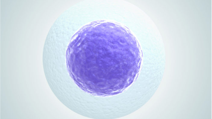
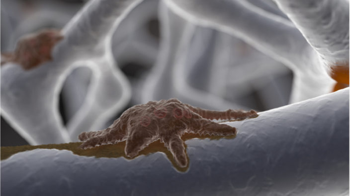
Senescent cells are aged or damaged cells that should destroy themselves via a process known as apoptosis but, for various reasons, do not do so; instead, they hang around, sending out inflammatory signals that harm nearby healthy cells, block effective tissue repair, and contribute to numerous age-related diseases. One proposed solution is to remove these problem cells by causing them to enter apoptosis, as originally intended, using senolytic drugs and therapies.

Senolytics are probably the most advanced of the therapies that directly address an aging process in order to prevent age-related disease and keep tissues and organs working more youthfully.

UNITY Biotechnology has been the leader of the pack since interest in developing senolytics burst into a frenzy of research a few years ago, and the company has been conducting human trials of its lead candidate drug (UBX0101) since earlier this year.

UBX0101 blocks the activity of the P53 pathway, which some senescent cells use to evade apoptosis and cling to life; by inhibiting this pathway, it causes these cells to self-destruct.

In February 2020, UNITY announced the launch of a Phase 2 study of UBX0101 in patients with moderate to severe osteoarthritis of the knee. This trial will evaluate three different doses (0.5 mg, 2 mg and 4 mg) of UBX0101.

Also, in March 2020, UNITY announced that it is were launching a Phase 1b study of UBX0101 in patients with moderate to severe osteoarthritis of the knee. This trial will explore the safety, tolerability and initial efficacy of a single 8.0 mg dose or multiple doses (two 4.0 mg doses separated by one month) of UBX0101.

We anticipate that the results of both of these human trials will be announced in the second half of 2020.

UNITY also has another candidate drug (UBX1967) closely behind; this drug targets the BCL2 pathway, another way senescent cells escape apoptosis, and it is slated to enter human trials for the treatment of age-related macular degeneration, diabetic macular edema, diabetic retinopathy, and glaucoma. UBX1967 is currently undergoing the investigational new drug (IND) application process, which is the first step in the drug review process by the U.S. Food and Drug Administration prior to launching human trials.

UNITY is not the only game in town when it comes to senolytics; Cleara Biotech is another biotech company working on this problem with a FOXO4-DRI peptide known as Proxofim. Proxofim was discovered by Dr. Peter de Keizer and has been in development since 2015 as a senolytic. In past studies, aged mice treated with Proxofim saw health benefits, including improved fitness, a glossier coat, and improved renal function.

However, the drug is not ready for human trials yet and is currently on its third generation of refinement. Cleara is now working hard to improve its potency, safety, and selectivity as part of its iPROX program to create an improved version. It is the company’s hope that the greatly refined and safer iPROX drug will allow translation to humans and the launch of clinical trials. Proxofim is likely a few years away from reaching human trials, but it is certainly something worth keeping an eye on as things develop.

Perhaps the most interesting company working on senolytics for me personally is SIWA Therapeutics, which is also working on senescent cell removal and has been busy developing 318H, a humanized monoclonal antibody. According to the company, 318H is being developed to slow or even reverse aging by removing harmful senescent cells but can also target cancer cells.

A few years ago, researchers at SIWA discovered that senescent and cancerous cells both have an advanced glycation end product (AGE) marker on their surfaces; this is due to both kinds of cells being oxidatively stressed and having abnormally high levels of glycolysis, which is known as the Warburg effect.

They then designed a monoclonal antibody specific to the AGE marker, which binds to the cells of aggressive cancers and senescent cells. They tested a mouse version of 318H in mice, and it removed senescent cells and promoted muscle regeneration. It also significantly reduced cancer metastasis in a model mouse of breast cancer.

If SIWA is successful with this immunotherapeutic approach to senescent cells and cancer, it would be an absolute game changer and allow for the effective treatment of cancer and a slew of age-related diseases associated with the accumulation of senescent cells.

There are a number of other interesting companies working on senolytic therapies including Oisín Biotechnologies, Senolytic Therapeutics, and Foxbio, to name a few; as time passes, others will almost certainly join them.

Based on the progress so far, it looks like senolytics are most likely to be the first true rejuvenation therapeutics in the sense that they directly target an aging process, and, in the next few years, we may see some of the first generation of these therapies arrive. When that day arrives, it has the potential to change how the world sees and treats aging, and that day will be a great day indeed. Stay future positive; we are getting closer to our goal!

We would like to ask you a small favor. We are a non-profit foundation, and unlike some other organizations, we have no shareholders and no products to sell you. All our news and educational content is free for everyone to read, but it does mean that we rely on the help of people like you. Every contribution, no matter if it’s big or small, supports independent journalism and sustains our future.
CellAge logo

Update from CellAge

Today is one of those days when we must deliver some bad news. Science is hard, particularly when it concerns the biology of aging, and things do not always go to plan. There are no guarantees that a project will succeed, and, quite simply, the more shots on goal we can get, the more chance that one of them will get through.

The news today is an example of when things do not work out as hoped, and it is with a heavy heart that we report this update from the CellAge team, which worked on a project that the lifespan.io community helped to raise funds for.

We are writing to regretfully inform you that our time at CellAge has come to an end. In the coming weeks, we will be closing down the project and the company. While it breaks our hearts to do so, unfortunately this is the only way forward at this time. We have contacted our crowdfunding partner LEAF and have returned all of the unused crowdfunding funds – we believe that it will be put to good use in accordance with the LEAF mission.

For the last few years, the CellAge team has been working with the aim to develop synthetic promoters that would be able to specifically identify senescent cells. We have partnered with a number of different organisations designing R&D plans and developing methods to achieve this goal. While we had a number of operational hurdles along the way, we have made progress. We believe that it is possible to build on top of our work; therefore, we will share the results and details of our findings with the wider scientific community and the general public.

We would like to thank everyone for their support and enthusiasm around this project. While this specific project has reached its conclusion, we remain passionate about improving healthspan and the quality of human life. We will continue to put our energy and hard work towards this goal.

Sincerely,

CellAge Team

While we are disappointed to hear this news from the project team, we should never shy away from reporting the truth, no matter how unpleasant or unpalatable it might be. Aging research is hard, and developing a successful company to take a project to market is a huge challenge; this time it did not succeed, but the next time it might. We will keep on trying until we win.

We would like to ask you a small favor. We are a non-profit foundation, and unlike some other organizations, we have no shareholders and no products to sell you. All our news and educational content is free for everyone to read, but it does mean that we rely on the help of people like you. Every contribution, no matter if it’s big or small, supports independent journalism and sustains our future.
Blood filter

Alkahest Human Trial to Address Cognitive Impairment

Alkahest has announced plans to launch a Phase 2 clinical trial that aims to address cognitive impairment and takes the unusual approach of using an external medical device that filters a harmful protein from the blood of end-stage renal disease patients.

The medical device, known as an AKST1210, will be connected in-line with a haemodialysis machine. The AKST1210 is designed to filter out and remove a protein called Beta-2 microglobulin (B2M), which was identified in a 2015 study by Dr. Saul Villeda, who confirmed its role as a pro-aging factor present in old blood [1].

B2M is an immune-associated protein with harmful, pro-inflammatory activity, and its presence has been shown to impair cognitive ability in animal studies. B2M is a problem for patients on long-term hemodialysis, as it can aggregate into amyloid fibers that deposit in joint spaces, a disease known as dialysis-related amyloidosis, and is thought to contribute to cognitive impairment in patients on hemodialysis for end-stage renal disease.

The following is an extract from Alkahest’s official press release:

SAN CARLOS, Calif., June 01, 2020 (GLOBE NEWSWIRE) — Alkahest Inc., a clinical stage biotechnology company focused on discovering and developing transformative therapies to treat age-related diseases, today announced the initiation of a clinical study to explore the use of an extracorporeal medical device to remove excess Beta-2 microglobulin (B2M) from circulation for the treatment of cognitive impairment in patients undergoing hemodialysis for end stage renal disease (ESRD).

“A large proportion of individuals with end-stage renal disease who receive hemodialysis have cognitive impairment, which may be associated with reduced ability for self-care, poor adherence to dietary and fluid restrictions, and poor outcomes,” said Karoly Nikolich, Ph.D., chief executive officer of Alkahest.  “Alkahest’s preclinical research has demonstrated that Beta-2 microglobulin, which is present at higher levels in older individuals, is drastically elevated in patients undergoing dialysis and may contribute to the high prevalence of cognitive impairment in these individuals. By reducing the amount of B2M in the plasma, we hope to introduce an effective way to lessen this impairment and allow patients on hemodialysis for ESRD to achieve improved treatment outcomes and quality of life.”

AKST1210-201 is a randomized, double-blind, feasibility and tolerability study investigating the use of the AKST1210 device connected to the dialysis circuit in 20 subjects with ESRD and cognitive impairment over a period of 20 weeks. Key objectives are safety and tolerability, as well as feasibility of conducting clinical trials with AKST1210 in this setting. Secondary objectives include measurement of removal of B2M, as well as improvements in clinical measures of cognitive, functional and quality of life assessments.

Conclusion

The B2M protein is one of a number of proteins that have been observed to rise with age, and reducing its presence may help reduce the risk of cognitive impairment for patients on hemodialysis. If the trial is successful, then Alkahest has selected a good therapeutic target with an approach that should be relatively easy to implement.

We would like to ask you a small favor. We are a non-profit foundation, and unlike some other organizations, we have no shareholders and no products to sell you. All our news and educational content is free for everyone to read, but it does mean that we rely on the help of people like you. Every contribution, no matter if it’s big or small, supports independent journalism and sustains our future.

Literature

[1] Smith, L. K., He, Y., Park, J. S., Bieri, G., Snethlage, C. E., Lin, K., … & Wheatley, E. G. (2015). [beta] 2-microglobulin is a systemic pro-aging factor that impairs cognitive function and neurogenesis. Nature medicine, 21(8), 932-937.

Journal Club Joao Pedro de Magalhaes

The CellAge Database with Dr. João Pedro de Magalhães

On Tuesday, June 30, the Journal Club returns, and our special guest Dr. João Pedro de Magalhães will be giving a talk and taking questions about the CellAge database.

Dr. João Pedro de Magalhães and his team at the University of Liverpool recently published a new study in which they used a systems biology approach to examine and better understand cellular senescence and its various pathways involved, which resulted in the creation of the CellAge database [1].

The CellAge database contains information about 279 genes that are linked to the process of cellular senescence. This resource will provide researchers with a valuable tool for better understanding aging and developing therapies that address cellular senescence.

We develop CellAge, a manually curated database of 279 human genes driving cellular senescence, and perform various integrative analyses. Genes inducing cellular senescence tend to be overexpressed with age in human tissues and are significantly overrepresented in anti-longevity and tumor-suppressor genes, while genes inhibiting cellular senescence overlap with pro-longevity and oncogenes. Furthermore, cellular senescence genes are strongly conserved in mammals but not in invertebrates. We also build cellular senescence protein-protein interaction and co-expression networks. Clusters in the networks are enriched for cell cycle and immunological processes. Network topological parameters also reveal novel potential cellular senescence regulators. Using siRNAs, we observe that all 26 candidates tested induce at least one marker of senescence with 13 genes (C9orf40, CDC25A, CDCA4, CKAP2, GTF3C4, HAUS4, IMMT, MCM7, MTHFD2, MYBL2, NEK2, NIPA2, and TCEB3) decreasing cell number, activating p16/p21, and undergoing morphological changes that resemble cellular senescence.

Join us on Tuesday, June 30 at noon EDT, 5 PM UK time on our Facebook page to catch the livestream, or if you are a Lifespan Hero, you can join us on the show; details on how to do that will be emailed to you.

We would like to ask you a small favor. We are a non-profit foundation, and unlike some other organizations, we have no shareholders and no products to sell you. All our news and educational content is free for everyone to read, but it does mean that we rely on the help of people like you. Every contribution, no matter if it’s big or small, supports independent journalism and sustains our future.

Literature

[1] Avelar, R. A., Ortega, J. G., Tacutu, R., Tyler, E. J., Bennett, D., Binetti, P., … & Shields, S. (2020). A multidimensional systems biology analysis of cellular senescence in aging and disease. Genome Biology, 21, 1-22.

DNA clock

New Epigenetic Clock Can Help Discover Anti-Aging Drugs

Researchers have proposed a new epigenetic clock designed to measure subtle epigenetic changes in vitro [1]. They hope that their discovery will help to expedite the creation of new anti-aging drugs.

Measuring biological age

Aging research is all about the difference between chronological and biological age. The former is simply the passage of time, but researchers can try and slow the advance of the latter. One of the necessary instruments is a clock that can reliably measure the biological age of organisms.

In recent years, several such clocks have been devised based on different principles, including age-related changes in blood composition and telomere shortening. However, probably the most reliable to date are epigenetic clocks, which analyze changes in gene expression [2]. The principle was laid out in a seminal paper by Steve Horvath et al. in 2013 [3]. Epigenetic clocks are usually based on the methylation of CpG sites, areas in the genome where a cytosine nucleotide (C) is followed by a guanine nucleotide (G), which can be either a single “CG” or a recurring sequence such as “CGCGCG”. Cytosines in such sequences can have methyl groups added to them (methylation). Each C can be either methylated or not. A strong correlation has been observed between CpG methylation profile and biological age, although the mechanism of this correlation is not fully understood.

Since there are tens of millions of CpG sites across the genome, epigenetic clocks use only a fraction of them. Researchers fine-tune their clocks by carefully choosing the sites that show the strongest correlation with biological age. For instance, Horvath’s original multi-tissue clock and the skin and blood clock that he and his group developed in 2018 [4] use 353 and 391 sites, respectively.

Designing a clock for cultured cells

Epigenetic clocks have rarely been used for identifying changes in the biological age of cultured cells, as epigenetic changes in cultured cells accumulate faster than in the human body. In vitro experiments could greatly expedite aging research and make it more cost-effective, but the unavailability of reliable epigenetic clocks for such experiments is a major hurdle that this research strives to overcome.

The scientists decided to create a clock that would measure minor epigenetic changes in cultured cells in order to use them as a yardstick for the creation of novel anti-aging drugs. They began with obtaining human mammary fibroblasts (HMF) from a healthy 16-year old donor and culturing them from passage (division) 10 to 20, and such cells enter senescence after 29 passages. Methylation was measured after every other passage at 850,000 CpG sites.

The researchers then tested three of the existing epigenetic clocks: Horvath’s original multi-tissue clock from 2013, the skin and blood clock, and the PhenoAge clock developed by Levine at al. in 2018 [5]. All the clocks showed higher numbers at the end of the experiment than at the beginning, reflecting the aging of the cells involved, but the results were too crude. As the researchers concluded, “none of the existing clocks was ideally suited to accurately measure subtle anti-ageing drug potential in human primary cells in vitro“.

To design a clock that would measure minor changes in methylation that occur between passages, the researchers introduced a second set of cells in addition to the aforementioned HMFs: human dermal fibroblasts (HDF) obtained from a different donor, with a different proliferative span and a different rate of methylation changes. After sifting through hundreds of thousands of CpG sites, the researchers had identified 2,543 that showed the strongest correlation with the aging of both samples, and then narrowed the pool down even more to just 42 predictor CpGs that they used to build what they called CellAgeClock.

The novel clock had been tested on 26 additional cell samples and was able to correctly identify the passage number most of the time, which is impressive given the subtlety of epigenetic changes between passages. The researchers then tried to interpret the readings of other clocks in terms of the number of passages but failed to get consistent results.

Since the original aim was to create a tool that would help discover new geroprotective drugs, the researchers wanted to see how well CellAgeClock measures the rejuvenating power of existing ones. They tested the well-researched rapamycin alongside three newer compounds: torin2, Dactolisib/BEZ235, and trametinib. The clock confirmed the anti-aging prowess of rapamycin and revealed that torin2, Dactolisib/BEZ235, and, to a lesser extent, trametinib, have rejuvenating potential as well. The results were also recreated in vivo by demonstrating that the drugs indeed prolong the lifespan of drosophila flies.

Conclusion

The researchers infer from these results that the epigenetic clock they have devised can successfully predict, based on experiments in vitro, the rejuvenating effect of novel drugs in vivo. If their conclusion is correct, CellAgeClock might indeed make the process of discovering new anti-aging drugs faster and cheaper. However, since only two cell types were used both to design and test the clock, and since three out of four drugs that it was tested on inhibit the mTOR pathway, it remains to be seen whether the positive results can be reproduced for different cell and drug types.

We would like to ask you a small favor. We are a non-profit foundation, and unlike some other organizations, we have no shareholders and no products to sell you. All our news and educational content is free for everyone to read, but it does mean that we rely on the help of people like you. Every contribution, no matter if it’s big or small, supports independent journalism and sustains our future.

Literature

[1] A CellAgeClock for expedited discovery of anti-ageing compounds. Celia Lujan, Eleanor J. Tyler, Simone Ecker, Amy P. Webster, Eleanor R. Stead, Victoria E. Martinez Miguel, Deborah Milligan, James C. Garbe, Martha R. Stampfer, Stephan Beck, Robert Lowe, Cleo L. Bishop, Ivana Bjedov bioRxiv 803676

[2] Horvath, S., & Raj, K. (2018). DNA methylation-based biomarkers and the epigenetic clock theory of ageing. Nature Reviews Genetics, 19(6), 371.

[3] Horvath, S. (2013). DNA methylation age of human tissues and cell types. Genome biology, 14(10), 3156.

[4] Horvath, S., Oshima, J., Martin, G. M., Lu, A. T., Quach, A., Cohen, H., … & Wilson, J. G. (2018). Epigenetic clock for skin and blood cells applied to Hutchinson Gilford Progeria Syndrome and ex vivo studies. Aging (Albany NY), 10(7), 1758.

[5] Levine, M. E., Lu, A. T., Quach, A., Chen, B. H., Assimes, T. L., Bandinelli, S., … & Whitsel, E. A. (2018). An epigenetic biomarker of aging for lifespan and healthspan. Aging (Albany NY), 10(4), 573.

Rejuvenation Roundup May

Rejuvenation Roundup May 2020

May has brought us a lot of interesting advances in the world of rejuvenation, and we at lifespan.io have moved forward with projects of our own, including our upcoming Ending Age-Related Diseases 2020 conference, an accelerated Journal Club schedule with more speakers, a roundtable discussion, and our new general science series focused on the potential issues of a world without aging: Science to Save the World! Here’s everything that happened in May.

LEAF News

As part of our commitment to supporting the development of the longevity industry, we have included a recording of Developing a Biotechnology Startup in the Rejuvenation Field, a workshop hosted by Kelsey Moody of Ichor Therapeutics, in the on-demand section of our upcoming conference, Ending Age-Related Diseases 2020. All registered participants get access to it!

Rejuvenation Roundup Podcast

Ryan O’Shea of Future Grind hosts this month’s podcast, showcasing the events and research discussed here.

Interviews

The Moonshot Venture Fellowship: Dr. Ole Mensching of Apollo Health Ventures discusses the Moonshot Venture, a new initiative to fund fledgling biotechnology initiatives.

The Paper That Helped Change WHO’s Position on Aging: This interview with Daria Khaltourina discusses the role of the International Longevity Alliance in changing the position of the World Health Organization in relation to aging.

Research Roundup

Modulating Macrophages to Prevent Disc Degeneration: Polarizing macrophages towards the M2 healing type from the M1 inflammatory type has been shown to prevent the degeneration of spinal discs, which is caused by unwanted apoptosis.

Resistance Training in Middle Age Doubles Muscle NAD+ Levels: Training muscles through weightlifting has been shown to double NAD+, a critical compound for life, in muscle.

Metformin Enhances Autophagy and Alleviates Inflammaging: Metformin downregulates the activity of the TH17 cytokine family, which is a cause of age-associated inflammation.

NAD+ and the Circadian Rhythm: New research suggests that NAD+, a critical molecule that impacts many aspects of life, may have a role in maintaining the natural fluctuations of our bodies.

lifespan.io Appears on the Damage Report: lifespan.io President Keith Comito discusses aging, the ultimate nonpartisan issue, with John Iadarola of The Young Turks.

A New Method for Detecting Mitophagy: Preserving cells through fixation alters their chemistry but makes it much easier to do large-scale testing. A new method of detecting mitophagy, the act of recycling damaged mitochondria, can withstand this fixation process.

SYK Inhibitors May Be a New Class of Senolytics: Although its mechanism of action is unknown, the spleen tyrosine kinase inhibitor R406 seems to preserve immune function while removing senescent cells.

A Senolytic Prodrug For Multiple Cell Types: The prodrug SSK1 targets β-galactosidase, a major feature of senescent cells, in order to remove them.

The CellAge Database Charts Cellular Senescence: The Human Aging Genomic Resources database is sponsoring CellAge, a database that is charting 279 genes associated with cellular senescence.

A Vaccine for Senescent Cells: Using a vaccine rather than senolytic drugs may be an efficient method of removing senescent cells, and their heterogeneity would likely prevent such a vaccine from detrimentally removing them all.

Eliminating senescent chondrogenic progenitor cells enhances chondrogenesis: Distraction arthroplasty, which generates intermittent hydrostatic pressure, is an effective treatment for osteoarthritis, but senescent cells interfere with this treatment. Removing these cells improves its effectiveness.

A CellAgeClock for expedited discovery of anti-ageing compounds: In order to discover new compounds that reverse the hallmark of epigenetic alterations, researchers have developed this in-depth epigenetic clock.

Prediction of Chronological and Biological Age From Laboratory Data: A new panel of 356 blood biomarkers, among diverse groups, shows promise for determining biological age.

GDF11 improves survival and promotes recovery after ischemic stroke in aged mice: Growth differentiation factor 11 has been shown to promote brain repair and prevent glial overproliferation in a mouse model of stroke.

Improvement of muscular atrophy by AAV–SaCas9-mediated myostatin gene editing in aged mice: Using an adeno-associated virus to deliver a CRISPR-based gene therapy to deplete myostatin from muscle tissue has been shown to be effective against sarcopenia in this mouse model..

Cell Therapy Shows Promise in Parkinson’s: A human Parkinson’s patient had his own skin cells reprogrammed into dopaminergic progenitor cells in order to treat his disease, and the results were positive.

Infusion of Plasma from Exercised Mice Ameliorates Cognitive Dysfunction: While it does not cure the basic mechanisms behind Alzheimer’s, injecting plasma from exercised mice into mice with Alzheimer’s disease has been shown to prove therapeutic benefits.

Allo-priming as a universal anti-viral vaccine: Patented, allogeneic immune cells primed to fight back against viral infection may serve as a method of defending the elderly against future pandemics.

The Anti-Ageing Effects of a Natural Peptide Discovered by Artificial Intelligence: A machine learning system discovered peptide RTE62G, which was subsequently proven in further testing to have beneficial effects on the extracellular matrix.

New Possible Treatment for Osteoarthritis Reported: Researchers have identified two molecules that, when taken together, have been shown to at least partially reverse osteoarthritis in mice.

Extracellular Vesicles Inhibit Glucocorticoid-Induced Osteonecrosis of the Femoral Head: By releasing chemicals that promote growth and prevent unwanted apoptosis, extracellular vesicles derived from stem cells have been shown to prevent degeneration of the top of the hip bone in a rat model.

Mesenchymal Stem Cells Repair Bone Marrow Damage of Aging Rats: MSCs have been shown to be vital in repairing damage inflicted by age-related bone marrow degeneration in a rat model.

Effects of Metformin on Lifespan, Cognitive Ability and Inflammatory Response in a Short-Lived Fish: Metformin has been shown to improve critical biomarkers in annual fish, potentially offering promise for mammals as well.

Senescence and Longevity of Sea Urchins: Sea urchins have extreme regenerative and anti-senescence abilities; this review discusses how they differ from mammal species.

News Nuggets

The Challenge of Including Aging in the ICD: Dr. Stuart Calimport and Dr. Alexander Zhavoronkov discuss the idea of including aging in the International Classification of Diseases.

Jim Mellon Donates £1 Million to Aging Research: Investment mogul Jim Mellon has donated a record-breaking 1 million British pounds to Oriel College in the United Kingdom.

Coming Up

Ending Age-Related Diseases 2020: Come and join us on August 20-21! With panels, discussions, and appearances from many of the major luminaries in the field, including Dr. Aubrey de Grey, Dr. Judith Campisi, Dr. Steve Horvath, and Dr. Vadim Gladyshev, our online conference promises to be an event to remember for a very, very long time.

We would like to ask you a small favor. We are a non-profit foundation, and unlike some other organizations, we have no shareholders and no products to sell you. All our news and educational content is free for everyone to read, but it does mean that we rely on the help of people like you. Every contribution, no matter if it’s big or small, supports independent journalism and sustains our future.

Vaccine

A Vaccine for Senescent Cells

Could a vaccine approach to removing harmful senescent cells that accumulate with age be an alternative to senolytic drugs, which destroy them? Some researchers certainly believe it might and have published a new mouse study showing just that.

What are senescent cells?

As you age, increasing numbers of your cells enter into a state known as senescence. Senescent cells do not divide or support the tissues of which they are part; instead, they emit a range of potentially harmful chemical signals that encourage nearby healthy cells to enter the same senescent state. Their presence causes many problems: they reduce tissue repair, increase chronic inflammation, and can even eventually raise the risk of cancer and other age-related diseases.

Senescent cells normally destroy themselves via a programmed process called apoptosis, and they are also removed by the immune system; however, the immune system weakens with age, and increasing numbers of senescent cells escape this process and begin to accumulate in all the tissues of the body.

By the time people reach old age, significant numbers of these senescent cells have built up, causing chronic inflammation and damage to surrounding cells and tissue. Senescent cells only make up a small number of total cells in the body, but they secrete proinflammatory signals including, cytokines, chemokines, and extracellular matrix proteases, which, together, form the senescence-associated secretory phenotype, or SASP. The accumulation of senescent cells is thought to be one of the reasons we age.

A vaccine-based approach to senescent cells

Today, we want to highlight a new study that is taking a slightly different approach to senescent cell accumulation in the form of a vaccination [1]. These researchers demonstrate that a vaccination against a cell surface marker is a viable approach to dealing with the senescent T cells present in fat tissue.

The researchers in the study we are talking about today show that vaccination against a surface marker present on the surfaces of senescent T cells living in fat tissue can spur the rest of the immune system to see them as a problem and seek and destroy them. This should be beneficial given the level of evidence that excessive fat tissue produces an increased amount of senescent cells, and as a result, an elevated level of systemic inflammation from the secreted SASP, which disrupts metabolism and impairs healthy tissue function.

A vaccine would provide a constantly active way to remove senescent cells, but that might not be ideal in humans. This is because cellular senescence is a useful process and is only a problem during aging, which is when they accumulate, get out of control, and become harmful. Cellular senescence is a safety net against cancer, and it facilitates wound healing and tissue repair when it functions correctly. If a vaccine were to target all senescent cells and immediately remove them, this could be problematic for wound healing and regeneration. Therefore, removing all senescent cells using a constantly active mechanism such as a vaccine may not be wise unless we are certain that the targeted cells serve no useful function.

However, it should be noted that due to the heterogeneity of senescent cell populations, the surface marker that this vaccine uses only appears on a subset of these cells, so the treatment would be unlikely to target all senescent cells at once; this is much like the present use of senolytics, which target different pathways used by subsets of senescent cells. In other words, it is unlikely that a single senolytic or vaccine could target all senescent cells at once due to their diversity in characteristics, such as surface markers and pro-survival pathways used to evade apoptosis.

The heterogeneity of senescent cell populations is a hot topic for researchers in recent months and there is even a SASP Atlas, which aims to comprehensively categorize the various secretions that the various senescent cell types make [2]. There is also the excellent HAGR cellular senescence database CellAge, which is charting the interactions, gene expressions, and complexities of senescent cells. Databases like this will, without a doubt, help researchers make the most informed decisions about future targets for senescent cell removal, including vaccines.

Senotherapy targeting for senescent cells is designed to attenuate age-related dysfunction. Senescent T cells, defined as CD4+ CD44high CD62Llow PD-1+ CD153+ cells, accumulate in visceral adipose tissues (VAT) in obese individuals. Here, we show the long-lasting effect of using CD153 vaccination to remove senescent T cells from high-fat diet (HFD)-induced obese C57BL/6J mice. We administered a CD153 peptide-KLH (keyhole limpet hemocyanin) conjugate vaccine with Alhydrogel (CD153-Alum) or CpG oligodeoxynucleotide (ODN) 1585 (CD153-CpG) and confirmed an increase in anti-CD153 antibody levels that was sustained for several months. After being fed a HFD for 10–11 weeks, adipose senescent T cell accumulation was significantly reduced in the VAT of CD153-CpG-vaccinated mice, accompanied by glucose tolerance and insulin resistance. A complement-dependent cytotoxicity (CDC) assay indicated that the mouse IgG2 antibody produced in the CD153-CpG-vaccinated mice successfully reduced the number of senescent T cells. The CD153-CpG vaccine is an optional tool for senolytic therapy.

Conclusion

It is possible to make the case that removing senescent and dysfunctional T cells from fat tissue could be useful, as such damaged immune cells likely serve no useful purpose and instead add to systemic inflammation. As we learn more about the heterogeneity of senescent cell populations, it may turn out to be the case that there are other populations of senescent cells that are not useful in any way and could have surface markers that allow a vaccine-based approach to destroy them as well. However, that would need to be tested in further studies to establish this.

Would a vaccine approach to clearing senescent cells be superior to periodic senolytic treatment to purge the cells? That remains to be seen, although the direction of the research here is certainly intriguing and gives some food for thought.

We would like to ask you a small favor. We are a non-profit foundation, and unlike some other organizations, we have no shareholders and no products to sell you. All our news and educational content is free for everyone to read, but it does mean that we rely on the help of people like you. Every contribution, no matter if it’s big or small, supports independent journalism and sustains our future.

Literature

[1] Yoshida, S., Nakagami, H., Hayashi, H., Ikeda, Y., Sun, J., Tenma, A., … & Rakugi, H. (2020). The CD153 vaccine is a senotherapeutic option for preventing the accumulation of senescent T cells in mice. Nature communications, 11(1), 1-10.

[2] Basisty, N., Kale, A., Jeon, O., Kuehnemann, C., Payne, T., Rao, C., … & Schilling, B. (2019). A Proteomic Atlas of Senescence-Associated Secretomes for Aging Biomarker Development. bioRxiv, 604306.

HAGR logo

The CellAge Database Charts Cellular Senescence

The Human Ageing Genomic Resources (HAGR) is a collection of curated databases and tools designed to help researchers study the genetics of human aging through modern approaches such as functional genomics, network analyses, systems biology and evolutionary analyses.

One of the many databases in the HAGR family is CellAge, which contains information about 279 genes that are linked to the process of cellular senescence. As cells age and become too damaged to keep working in a healthy manner, most of them destroy themselves via an internal self-destruct mechanism known as apoptosis. Unfortunately, as we age, more and more of these broken cells evade apoptosis and cling to life using various pro-survival pathways. The accumulation of the resulting senescent cells is thought to be one of the reasons we age.

The problems begin once these senescent cells build up to excessive levels and start harming tissue repair and nearby healthy cells by secreting a range of inflammatory signals. Collectively, these inflammatory signals form a pro-inflammatory cocktail known as the senescence-associated secretory phenotype (SASP). The SASP contributes to inflammaging, the chronic, systemic inflammation typically seen in older people. This smoldering background of inflammation paves the way for various age-related diseases to develop and retards tissue repair and regeneration.

Perhaps worst of all, senescent cells and the inflammatory SASP that they produce can also encourage nearby healthy cells to also become senescent. Initially, there are only a relatively small number of senescent cells present in organs and tissues, but even a few can have a catastrophic effect on healthy tissue, and their numbers grow quickly as they poison healthy cells nearby.

The CellAge database is the brainchild of Dr. João Pedro de Magalhães and his team at the University of Liverpool. In a new study, the researchers demonstrated how they used a systems biology approach to examine and better understand cellular senescence and the various pathways involved in the process [1].

During the study, the team created models of protein-protein interactions and co-expression networks to track the complex interactions that occur during cellular senescence, the result of which allowed them to identify likely cellular senescence regulators. Using siRNAs, the researchers discovered 26 potential candidates that could induce at least one biomarker of cell senescence in 13 genes, causing those cells to undergo changes that resemble cellular senescence.

We develop CellAge (https://genomics.senescence.info/cells), a manually curated database of 279 human genes driving cellular senescence, and perform various integrative analyses. Genes inducing cellular senescence tend to be overexpressed with age in human tissues and are significantly overrepresented in anti-longevity and tumor-suppressor genes, while genes inhibiting cellular senescence overlap with pro-longevity and oncogenes. Furthermore, cellular senescence genes are strongly conserved in mammals but not in invertebrates. We also build cellular senescence protein-protein interaction and co-expression networks. Clusters in the networks are enriched for cell cycle and immunological processes. Network topological parameters also reveal novel potential cellular senescence regulators. Using siRNAs, we observe that all 26 candidates tested induce at least one marker of senescence with 13 genes (C9orf40, CDC25A, CDCA4, CKAP2, GTF3C4, HAUS4, IMMT, MCM7, MTHFD2, MYBL2, NEK2, NIPA2, and TCEB3) decreasing cell number, activating p16/p21, and undergoing morphological changes that resemble cellular senescence.

Overall, our work provides a benchmark resource for researchers to study cellular senescence, and our systems biology analyses reveal new insights and gene regulators of cellular senescence.

Conclusion

A deeper understanding of cellular senescence and its interactions will certainly help the development of senolytic drugs that target senescent cell accumulation, so this database is a welcome addition to the research arsenal.

We would like to ask you a small favor. We are a non-profit foundation, and unlike some other organizations, we have no shareholders and no products to sell you. All our news and educational content is free for everyone to read, but it does mean that we rely on the help of people like you. Every contribution, no matter if it’s big or small, supports independent journalism and sustains our future.

Literature

[1] Avelar, R. A., Ortega, J. G., Tacutu, R., Tyler, E. J., Bennett, D., Binetti, P., … & Shields, S. (2020). A multidimensional systems biology analysis of cellular senescence in aging and disease. Genome Biology, 21, 1-22.

A New Method for Detecting Mitophagy

Researchers from the Laboratory for Cell Function Dynamics in Japan have developed a new method for detecting mitophagy, the process of recycling damaged mitochondria.

The Necessity of Mitophagy

Our mitochondria are the powerhouses of our cells, turning the food we eat into ATP, the form of chemical energy that our cells use. However, over time, our mitochondria become damaged, thus harming our cells’ basic ability to function. One method that our cells use to stave off this problem is called mitophagy, which destroys and recycles these damaged mitochondria so that new ones can take their place. The failure of cells to perform mitophagy leads to mitochondrial dysfunction, which is one of the hallmarks of aging.

Therefore, much research has been devoted to stimulating mitophagy, as researchers attempt to rid the cells of bad mitochondria without harming healthy ones.

Fixed Cells Make High Throughput Possible

Fixation is any process that keeps cells in an immobile state, whether through the use of a chemical such as formaldehyde or sub-freezing temperatures. However, fixation affects cellular chemistry, and, as the researchers of this study explain, mt-mKeima, a commonly used compound that is currently used to detect the occurrence of mitophagy, is sensitive to fixation. Therefore, they sought out and developed mito-SRAI, a new method of detecting mitophagy that was insensitive to fixation, making it suitable for high-throughput cell assays, which often use fixed cells.

The researchers also demonstrated that mito-SRAI is not susceptible to being destroyed by the cell’s proteosome, making it ideal for use in living samples.

Finally, and probably most interestingly, the researchers used their novel method along with a host of related techniques to screen 76,000 compounds for potential therapeutic use, testing them for specificity and toxicity. A compound called T-271 was the researchers’ top choice after this selection process, as it appears to have the unique property of inducing mitophagy in damaged mitochondria without harming healthy ones, although considerably more refinement and analysis of this compound is required in order to progress to in vivo and human trials.

Summary

Dysfunctional mitochondria accumulate in many human diseases. Accordingly, mitophagy, which removes these mitochondria through lysosomal degradation, is attracting broad attention. Due to uncertainties in the operational principles of conventional mitophagy probes, however, the specificity and quantitativeness of their readouts are disputable. Thorough investigation of the behaviors and fates of fluorescent proteins inside and outside lysosomes enabled us to develop an indicator for mitophagy, mito-SRAI. Through strict control of its mitochondrial targeting, we were able to monitor mitophagy in fixed biological samples more reproducibly than before. Large-scale image-based high-throughput screening led to the discovery of a hit compound that induces selective mitophagy of damaged mitochondria. In a mouse model of Parkinsons disease, we found that dopaminergic neurons selectively failed to execute mitophagy that promoted their survival within lesions. These results show that mito-SRAI is an essential tool for quantitative studies of mitochondrial quality control.

Conclusion

This paper did not go into extensive detail about T-271, and it is possible that the researchers will further analyze this compound in a future paper. The fact that the researchers were able to screen 76,000 compounds once developing this novel detection method shows how far we have come in our ability to perform biological research.

It also shows that such fundamental research into creating better biomarkers is the sort of basic research that is necessary for the development of mitophagic and other therapies. Being able to productively use fixed cells in high-throughput assays to directly analyze the effectiveness of a therapy is an enormous boon for the rapid and successful deployment of therapies that target not only this particular hallmark of aging but other disorders as well.

We would like to ask you a small favor. We are a non-profit foundation, and unlike some other organizations, we have no shareholders and no products to sell you. All our news and educational content is free for everyone to read, but it does mean that we rely on the help of people like you. Every contribution, no matter if it’s big or small, supports independent journalism and sustains our future.
Keith on The Damage Report

lifespan.io Appears on the Damage Report

As part of our mission to engage the wider public audience, lifespan.io President Keith Comito has recently appeared on the Damage Report, a politics and current events show hosted by John Iadarola, political host for the online news network The Young Turks.

Regardless of anyone’s political leanings, it is important that we engage with the general public and remind everyone that aging, and its accompanying diseases, is a problem that everyone has in common, regardless of if they consider it to be one.

Aging is the ultimate nonpartisan issue. We all have a dream of when the vaccine is coming out, but vaccines don’t work with elderly people’s compromised immune systems. For them to work, we need to improve immune system protection.

Keith was invited to talk about the connection between COVID-19 and aging and how developing technologies that directly address aging in order to reverse age-related diseases could make Medicare for All more cost-effective and dramatically reduce healthcare costs in general.

If we fund immune system regeneration, and it’s successful, not only would it make everyone less likely to get COVID-19 but could cut the death toll down to 30% of what it is right now.

Keith makes a great point: if the immune systems of older people could be boosted or even rejuvenated so that they work as they did in youth, COVID-19 would have a harder time infecting them and putting their lives at risk, and they would also be protected from a myriad of other diseases, both infectious and age-related.

We have already seen some promising human trial results that show that it is possible to achieve this in humans, and there are other approaches in development that seek to rejuvenate the immune system in different ways.

Aging is a primary risk factor for a multitude of diseases, both infectious and age-related; therefore, it makes total sense to shift the focus of medical research towards developing the technologies that directly target the aging processes in order to improve healthy longevity and reduce the strain on healthcare and social security systems. It’s time that we start to take healthcare and how we treat age-related diseases in a new direction.

We would like to ask you a small favor. We are a non-profit foundation, and unlike some other organizations, we have no shareholders and no products to sell you. All our news and educational content is free for everyone to read, but it does mean that we rely on the help of people like you. Every contribution, no matter if it’s big or small, supports independent journalism and sustains our future.