The Blog

Building a Future Free of Age-Related Disease

Narrow artery

Two Proteins Allow LDL Cholesterol into Our Cells

Researchers have found out how LDL cholesterol is able to enter our cells with the help of two proteins that form a doorway into the cell. The findings have ramifications for treating heart disease, which is currently the world’s greatest killer.

Revealing how LDL gets into our cells

We have all likely heard about good and bad cholesterol, with high-density lipoprotein (HDL) cholesterol being considered good and low-density lipoprotein (LDL) cholesterol being bad. While this popular summary is somewhat simplistic given that some LDL cholesterol is needed for the body to function correctly, it is true that too much LDL in the bloodstream can be a problem.

LDL cholesterol tends to build up in the walls of our arteries, causing a reduction of blood flow, which typically leads to heart disease, the end result of which is a heart attack or stroke.

In a new study, researchers from the University of New South Wales have discovered how LDL enters our cells by identifying two proteins that function like a gateway in the membrane wall to permit access [1]. This pair of membrane proteins also presents a potential target for drugs that can block their activity and help lower LDL cholesterol levels in the bloodstream.

The researchers made this discovery while investigating the role of two proteins, NPC1 and NPC2, and how they facilitate the transport of LDL cholesterol into the cell. Thanks to the use of highly advanced imaging equipment, they were able to look at the proteins close to the atomic level and see exactly how they transport the LDL inside the cell.

The lysosomes act as a protective barrier in our cells and work like garbage disposal units, breaking down unwanted proteins and destroying invaders. NPC1 and NPC2 essentially act like a key to a door, working in unison to usher the LDL cholesterol into the cell without it being destroyed by the lysosomes.

LDL is important for our cells and tissues, including muscle growth and development; however, too much in the bloodstream can clog our arteries and lead to the development of heart disease. By creating drugs that target NPC1 and NPC2, there is the potential to increase how much LDL cells uptake.

Lysosomal cholesterol egress requires two proteins, NPC1 and NPC2, whose defects are responsible for Niemann-Pick disease type C (NPC). Here, we present systematic structural characterizations that reveal the molecular basis for low-pH-dependent cholesterol delivery from NPC2 to the transmembrane (TM) domain of NPC1. At pH 8.0, similar structures of NPC1 were obtained in nanodiscs and in detergent at resolutions of 3.6 A˚ and 3.0 A˚, respectively. A tunnel connecting the N-terminal domain (NTD) and the transmembrane sterol-sensing domain (SSD) was unveiled. At pH 5.5, the NTD exhibits two conformations, suggesting the motion for cholesterol delivery to the tunnel. A putative cholesterol molecule is found at the membrane boundary of the tunnel, and TM2 moves toward formation of a surface pocket on the SSD. Finally, the structure of the NPC1-NPC2 complex at 4.0 A˚ resolution was obtained at pH 5.5, elucidating the molecular basis for cholesterol handoff from NPC2 to NPC1(NTD).

Conclusion

Now that the researchers understand how LDL enters the cell with the help of NPC1 and NPC2, they can begin to develop drugs that target these membrane proteins and encourage our cells to uptake more LDL and reduce the amount present in the bloodstream. If successful, this has considerable potential for treating excessive LDL cholesterol and addressing heart disease.

We would like to ask you a small favor. We are a non-profit foundation, and unlike some other organizations, we have no shareholders and no products to sell you. All our news and educational content is free for everyone to read, but it does mean that we rely on the help of people like you. Every contribution, no matter if it’s big or small, supports independent journalism and sustains our future.

Literature

[1] Qian, H., Wu, X., Du, X., Yao, X., Zhao, X., Lee, J., … & Yan, N. (2020). Structural Basis of Low-pH-Dependent Lysosomal Cholesterol Egress by NPC1 and NPC2. Cell.

Rejuvenation Roundup June

Rejuvenation Roundup June 2020

This has been a productive month for rejuvenation biotechnology companies, longevity researchers, and us here at lifespan.io. Let’s take a look at what’s happened in June.

LEAF News

Why I Am Still Future Positive on My Birthday: Board member Steve Hill shares his musings on his birthday, discussing where we are and where we go from here.

Can Aging Research Prevent the Next Pandemic?: Aging weakens the immune system, leaving us susceptible to communicable diseases.

Journal Club

The CellAge Database with Dr. João Pedro de Magalhães: CellAge is a database that contains 279 genes that are linked to cellular senescence, and Dr. Magalhães came to Journal Club to explain this database to us.

Interviews

Diluting Blood Plasma Rejuvenates Old Mice: Irina and Michael Conboy return for an interview explaining how diluting the aging factors blood plasma promotes rejuvenation.

Rejuvenation Roundup Podcast

Ryan O’Shea of Future Grind hosts this month’s podcast, showcasing the events and research discussed here.

Research Roundup

New Epigenetic Clock Can Help Discover Anti-Aging Drugs: By measuring changes to epigenetics, CellAgeClock can help determine the epigenetic effects of a great many compounds.

A New Likely Mechanism Behind Osteoporosis Discovered: Misfolded proteins cause osteoblasts to lose function, resulting in bone that cannot be rebuilt.

Removing Some Senescent Cells May Have Consequences: Some senescent cells are responsible for maintaining blood-tissue barriers, and removing these cells leads to health problems and physical deterioration.

Hair Graying May Be Reversible: Like tree tings, gray hairs can show what happened to a hair over the course of its growth, and this phenomenon has been used to show that reducing stress can restore color to hair in some cases.

Gut Bacteria Can Predict Your Biological Age: Gut bacteria can serve as a biomarker of aging, and a deep learning algorithm has been built to find out how.

The Gene LMNA’s Effect on Aging: The LMNA gene is responsible for lamins, which protect the nucleus against genomic damage. Mutations to this gene can lead to accelerated aging.

Deploying Immunotherapy Against Senescent Cells: Using CAR-T to send the immune system against cancer cells is a new form of therapy; using the same technique against senescent cells may also be effective.

The Role of the Immune System in Removing Senescent Cells: Inflammation, the SASP, and cellular senescence are all closely related.

A Strong Link Between Alzheimer’s and Senescence: While senescence is difficult to detect in neurons, due to the fact that mature neurons do not divide, senescence may be a critical part of the Alzheimer’s cascade.

A Biomarker Based on Senescent Cell Secretions: As you age, senescent cells secrete the inflammatory SASP, which can turn other cells senescent as well. Using the SASP as a biomarker may prove useful in the clinic.

Reversing Atherosclerosis by Resetting Gut Bacteria: Introducing cyclic peptides to reset the gut microbiomes of mice fed a high-fat diet reduced their cholesterol by more than a third.

Nanoparticles Deliver Twin Drug Payload for Atherosclerosis: Delivering both cyclodextrin and statin to atherosclerotic lesions has been shown to improve cardiovascular health in mice.

New Evidence of Common Genetic Causes in Age-Related Diseases: Algorithms and statistical analysis have discovered the role of genes in causing age-related diseases.

Pilot Study Results Suggest Epigenetic Age Reversal: Ponce de Leon Health has published a press release in which it claims that its supplement, Rejuvant, reverses epigenetic age by 8.5 years.

Transient metabolic improvement in obese mice treated with navitoclax or dasatinib/quercetin: There is a link between obesity and senescent cells, and treating these cells with senolytics has been shown to be effective in mice.

Bone marrow mesenchymal stem cells improve thymus and spleen function of aging rats: Multiple biomarkers associated with aging were improved through the application of these particular stem cells.

Niacin Increases NAD+ Significantly in Human Trial: The human body is good at turning the vitamin niacin into the vital compound NAD+.

NAD+ Restores Mitochondrial Function in Egg Cells: Oocytes are enormous cells with a lot of mitochondria, and NAD+ has been shown to restore the function of these mitochondria in older ages.

Niacin May Prove Useful Against Brain Cancer: A type of cancer called a glioblastoma hijacks immune cells, but treatment with niacin disrupts this process.

Plasma endothelial cells-derived extracellular vesicles promote wound healing in diabetes: These extracellular vesicles were shown to inhibit cellular senescence in diabetic skin fibroblasts.

Small Extracellular Vesicles Have GST Activity and Ameliorate Senescence-Related Tissue Damage: These particular vesicles, when taken from young donors, have been shown to have multiple anti-aging effects, including reducing the effects of reactive oxygen species.

Inhibition of Neural Stem Cell Aging Through the Transient Induction of Reprogramming Factors: Transient OSKM has been previously shown to encourage stem cells to stay youthful without losing cellular specialization, and this experiment shows that it works in neural stem cells as well.

Reversing a model of Parkinson’s disease with in situ converted nigral neurons: Turning astrocytes into neurons was shown to be effective in a model of this neurodegenerative disease.

The mitochondrial derived peptide humanin is a regulator of lifespan and healthspan: Organisms, including human beings, with naturally higher levels of the peptide humanin have considerably longer lives than organisms with lower levels.

Identification of longevity compounds with minimized probabilities of side effects: Combining two separate databases, DrugAge for geroprotectors and SEP-L1000 for side effects, allows researchers to focus their efforts on the drugs that might provide the most benefits with the fewest risks.

Discovery of fifteen plant extracts and identification of cellular processes to prolong the lifespan of budding yeast: Yeast is a traditional model of aging, and these compounds have been found to target pathways associated with the aging processes.

Telmisartan and Rosuvastatin Synergistically Ameliorate Dementia and Cognitive Impairment in Older Hypertensive Patients With Apolipoprotein E Genotype: APOE-e4 is a particularly dangerous allele to carry, but new research has identified drugs that can alleviate some of the problems associated with it.

Gray whale transcriptome reveals longevity adaptations associated with DNA repair and ubiquitination: Whales are some of the longest-lived animal species known, and this research shows that their longevity is at least partially due to their ability to repair genetic damage.

News Nuggets

Alkahest Announces a Human Trial to Address Cognitive Impairment: We have previously discussed the possibility of removing unwanted compounds from aged blood, and Alkahest is conducting a Phase 2 clinical trial to do exactly this with the compound B2M.

Update from CellAge: Board member Steve Hill reports some bad news from the team at CellAge. Unfortunately, science is hard especially in the context of aging research, and there is always the danger that things do not go according to plan; sadl,y on this occasion, this is the case, and it highlights why we must take as many shots on goal as possible.

Human Augmentation Institute: Rejuvenation Roundup Podcast host Ryan O’Shea will speaking at the Humanity Plus Post-Pandemic Summit, a free digital event taking place on July 7-8, 2020. Ryan’s talk will focus on his efforts with a new non-profit organization, the Human Augmentation Institute, which was founded to uphold bodily autonomy and ensure that any efforts in human augmentation are done ethically, safely, and responsibly.

We would like to ask you a small favor. We are a non-profit foundation, and unlike some other organizations, we have no shareholders and no products to sell you. All our news and educational content is free for everyone to read, but it does mean that we rely on the help of people like you. Every contribution, no matter if it’s big or small, supports independent journalism and sustains our future.

Ponce de Leon Health Logo

Pilot Study Results Suggest Epigenetic Age Reversal

Today, we want to highlight a press release from Ponce de Leon Health that talks about the results of a pilot consumer trial that the company has recently concluded. The company claims that it has been successful in reducing the epigenetic age of participants by an average of 8.5 years with its dietary supplement Rejuvant.

Ponce de Leon Health initially worked with Dr. Brian Kennedy, who was, at the time, based at the Buck Institute for Research on Aging, searching for compounds that were generally recognized as safe (GRAS) but that had the potential to influence aging in mammals. The company screened over 300 GRAS compounds and identified compounds that could modulate a number of pathways that are linked to aging. These compounds affected the mTOR pathway, blocked the proinflammatory secretions made by senescent cells, affected genomic stability pathways, aided in ammonia detoxification, and supported protein homeostasis.

Dr. Kennedy subsequently joined Ponce de Leon Health as its Chief Scientific Officer, and the company has been busy testing and preparing to translate these findings to people. Its strategy has been to test its products on mammalian models that closely emulate human aging in order to give the best chance of translating beneficial results to us.

For the consumer testing discussed in this press release, the company gave participants Rejuvant and measured their epigenetic ages using DNA methylation testing. This supplement contains a proprietary form of calcium alpha-ketoglutarate, which the FDA considers to be GRAS. The company believes that Rejuvant works by slowing down the rate of age-related DNA methylation and reducing the inflammation caused by senescent cells, two proposed reasons why we age.

FERNANDINA BEACH, Fla., June 29, 2020 /PRNewswire/ — Ponce De Leon Health, Inc. (“Ponce de Leon”), a longevity company focused on reversing epigenetic aging, today announced the results of pilot research suggesting that Rejuvant® LifeTabs®, reduced the biological age of customers by an average of 8.5 years. The biological age was assessed by way of a DNA biomarker test that measures DNA methylation. Of the subjects in the analysis, two-thirds showed a statistically significant reduction in biological age. The analysis was based on data from subjects who had taken Rejuvant LifeTabs for a period of four to six months. Rejuvant, a dietary supplement, is Ponce de Leon’s first commercial product.

The data were independently compiled by Dr. Yelena Budovskaya, Ph.D., co-founder and lead scientist of TruMe Labs, a pioneer in the emerging field of epigenetic aging testing. Ponce de Leon is a distributor of TruMe’s TruAge Index, a take-at-home, non-invasive epigenetic age test. The subjects included in the analysis represent all 17 customers who had taken a TruAge test at the time of their first purchase of Rejuvant, as well as a second test after taking the supplement for four to six months. The TruAge Index measures DNA methylation, which is growing in acceptance by longevity researchers as an important biomarker of biological aging.

All formulations of LifeTabs contain the proprietary active ingredient LifeAKG™, which was shown in recent preclinical animal studies to increase lifespan (the length of life) and healthspan (the length of healthy life) in mammalian models. Ponce de Leon has eight patents pending for inventions arising from its research and development activities.

Ponce De Leon is currently enrolling participants for a larger, randomized, double-blind, placebo-controlled clinical trial, to be conducted at Indiana University Medical Center later this year.

The Company’s Chief Medical Officer, Francis R. Palmer, M.D., stated that “although at this stage we only have consumer data, these findings are very promising. We are only aware of one other published trial where researchers were able to show a reduction in biological age in humans as measured by DNA methylation. In this other trial, modest reductions in biological age were observed in a small number of subjects who were treated with a combination of prescription drugs for a year. The Rejuvant results are suggestive at this time but it showed a greater average reduction in biological age, achieved in six months, using non-prescription compounds.” Added Palmer, “we expect our human clinical trial results to corroborate the encouraging data we have now, and we believe this will have a meaningful impact on both the scientific and consumer communities.”

The Company’s CEO and Founder, Tom Weldon, commented “the pilot research data appears to be consistent with our earlier mammal results but we appreciate the need for a more robust study to validate this data on lifespan and healthspan research. We look forward to conducting that study later this year and adding to these positive findings. The potential for Rejuvant to reverse epigenetic aging, as measured by DNA methylation testing and other measurable biological markers, may profoundly benefit not only the individuals who take the product, but also the economies and societies in which they participate and live.”

Conclusion

Obviously, this has yet to be proven conclusively in human trials, and the company is busy preparing to launch a larger-scale trial later this year to that end. Until those results are published, we are going to reserve judgment, but the initial animal data is certainly promising, and the concept of slowing epigenetic changes down is plausible.

Finally, CEO and founder Tom Weldon of Ponce de Leon Health will be presenting a talk at the Ending Age-Related Diseases 2020 online conference hosted by lifespan.io on August 20-21, offering you a chance to learn more about this line of research.

We would like to ask you a small favor. We are a non-profit foundation, and unlike some other organizations, we have no shareholders and no products to sell you. All our news and educational content is free for everyone to read, but it does mean that we rely on the help of people like you. Every contribution, no matter if it’s big or small, supports independent journalism and sustains our future.

New Evidence of Common Genetic Causes in Age-Related Diseases

By using a large UK health databank and advanced methods of statistical analysis, scientists have discovered genetic ties between various age-related diseases and proposed several candidate drugs for upstream intervention.

Building age-of-onset profiles of diseases

The very fact that multitudes of age-related diseases (ARDs) exist alludes to more than just a coincidence. One of the foremost goals of longevity research is to discover the common causes of various ARDs even in the absence of an obvious connection. A group of researchers has recently published a paper [1] that attempts to do just that with powerful statistical tools and a vast genomic database.

The researchers chose the UK Biobank, which stores genetic and health information on nearly half a million people, as their dataset. They identified age-of-onset profiles for 116 diseases and divided them into four different clusters based on these profiles.

Clusters of age-related diseases

The first cluster (top left) is the most representative of ARDs. Diseases in this cluster begin to appear much more frequently after the age of 40, the incidence rate rising rapidly. Out of 25 diseases assigned to this cluster, 11 were cardiovascular, and the rest included such conditions as diabetes, osteoporosis, and cataracts, so  there were no surprises here. The second cluster (top right) contains 51 diseases with prevalence beginning to rise at the age of 20, but at a slower rate. The diseases in this cluster are the most diverse: 17 of them were musculoskeletal, 13 were gastrointestinal, and there were others, such as anemia, deep venous thrombosis, thyroid problems, and depression. This cluster convincingly illustrates the overall susceptibility to diseases rising with age. The third cluster (bottom left) contains 30 diseases distributed mostly equally across various age cohorts, including immunological, neurological, musculoskeletal, gastrointestinal, and respiratory diseases in roughly equal proportion. Interestingly, all the diseases in this cluster have an immune component. Finally, the fourth cluster (bottom right) consists of a smaller number of diseases that mostly affect children, the largest group being infectious diseases. The authors did not include cancer in their analysis, explaining that the interaction between genetic and environmental factors in cancer probably differs significantly from non-cancerous diseases with a similar age-of-onset profile. Another serious limitation was the age of the individuals, which was capped at 65 in this particular dataset. The scientists left out epigenetic factors as well.

Common genetic associations

Next, the researchers identified associations between genetic variants and the diseases. Almost 94,000 polymorphic sites (DNA loci that can have multiple variants in the population) showed associations with at least one disease, and 80 diseases had at least one significant association with a polymorphic site. Though 47% of those “significant” variants were connected to more than one disease, just 9% were associated with multiple diseases from different clusters. This suggests that many diseases with similar age-of-onset profiles have common genetic roots. Since the scientists controlled for disease categories, causal relationships between diseases, and co-occurrences, they concluded that the shared genetic associations that they discovered point to a common etiology. The rigorous control for other variables probably resulted in the elimination of some true positives, meaning that the actual correlation should be even stronger. Importantly, genetic associations to the diseases from clusters 1 and 2 had an abnormal proportion of known aging-related genes. This puts the results in line with our previous knowledge.

To prove that their findings can have concrete therapeutic implications, the scientists identified several existing drugs that can target common genetic causes of multiple ARDs. Among the significant hits were 14 drugs already approved for various, mostly age-related conditions, including diabetes, osteoporosis, and cardiovascular diseases. One of the drugs, prunetin, has already been shown to extend the lifespan of drosophila flies [2]. The scientists note that these results can lead to a more targeted approach to ARD treatment, alleviating the all-too-familiar phenomenon of polypharmacy, the use of multiple prescription drugs, in the elder population.

Two evolutionary theories of aging

In the opening lines of their paper, the researchers point out that aging is sometimes described as an evolutionary paradox, since it should have been eradicated by natural selection due to its immense adverse effect on an organism. Yet this is a paradox in name only, as at least two evolutionary theories of aging exist [3]. The first one explains aging via mutation accumulation. It postulates that mutations that are only harmful later in life are less susceptible to natural selection: a gene that contains such a mutation would already have been passed on to the next generation by the time it exerts its deleterious effect on the organism. According to the second theory, antagonistic pleiotropy, the same genes that induce diseases in old age might still be selected for reproduction if they are beneficial earlier in life. In short, nature only cares about our health for as long as we can reproduce. The researchers report that they have found evidence in favor of both theories, though results supporting the antagonistic pleiotropy theory were more limited.

Conclusion

In addition to the fascinating results that support the upstream approach to aging, this study constitutes a tour de force of employing big data methods in longevity research. As more genetic and health information accumulates in public databases, we are in for a long-overdue surge in studies like this one that employ statistics and machine learning to great effect. The study is also far from being purely theoretical; its authors have demonstrated that identifying common genetic causes of various age-related diseases can quickly bring tangible benefits in the form of more targeted pharmaceutical interventions.

We would like to ask you a small favor. We are a non-profit foundation, and unlike some other organizations, we have no shareholders and no products to sell you. All our news and educational content is free for everyone to read, but it does mean that we rely on the help of people like you. Every contribution, no matter if it’s big or small, supports independent journalism and sustains our future.

Literature

[1] Donertas, H. M., Fabian, D. K., Valenzuela, M. F., Partridge, L., & Thornton, J. M. (2020). Common genetic associations between age-related diseases. medRxiv.

[2] Piegholdt, S., Rimbach, G., & Wagner, A. E. (2016). The phytoestrogen prunetin affects body composition and improves fitness and lifespan in male Drosophila melanogaster. The FASEB Journal, 30(2), 948-958.

[3] Gavrilov, L. A., & Gavrilova, N. S. (2002). Evolutionary theories of aging and longevity. TheScientificWorldJOURNAL, 2.

Using a meter

A Biomarker Based on Senescent Cell Secretions

Researchers have published a new study suggesting that some of the inflammatory signals secreted by senescent cells could be used as a biomarker to measure aging and health.

What are senescent cells?

As you age, increasing numbers of your cells enter into a state known as senescence. Senescent cells do not divide or support the tissues of which they are part; instead, they emit a range of harmful chemical signals known as the senescence-associated secretory phenotype, or SASP. The SASP contains a complex cocktail of factors including cytokines, chemokines, proteins, growth factors, and more.

This secreted SASP is what makes senescent cells such a problem, as it not only contributes to the chronic systemic inflammation known as inflammaging, it can also encourage nearby healthy cells to become senescent as well. This means that a relatively small number of senescent cells in tissue can have a very large impact on the health and function of that tissue.

The presence of senescent cells and their SASP has been implicated in a myriad of age-related diseases and conditions; this is why researchers are currently developing senolytics, therapies that can remove them from the body.

Using the SASP as a biomarker of aging

The researchers of a new study make the case for collectively using factors SASP to form a biomarker of aging and health [1]. Within the complex cocktail of secreted proteins and molecules that make up the SASP, the researchers identified a total of seven factors that they believe give a reliable indication of biological age and relative health.

These factors are also easy to test given that they can be sampled via a simple blood draw and then fed into diagnostic testing equipment. This is critical because, in order to be clinically useful, biomarkers should be both cost-effective and accurate.

Produced by senescent cells, the senescence-associated secretory phenotype (SASP) is a potential driver of age-related dysfunction. We tested whether circulating concentrations of SASP proteins reflect age and medical risk in humans. We first screened senescent endothelial cells, fibroblasts, preadipocytes, epithelial cells, and myoblasts to identify candidates for human profiling. We then tested associations between circulating SASP proteins and clinical data from individuals throughout the life span and older adults undergoing surgery for prevalent but distinct age-related diseases. A community-based sample of people aged 20–90 years (retrospective cross-sectional) was studied to test associations between circulating SASP factors and chronological age. A subset of this cohort aged 60–90 years and separate cohorts of older adults undergoing surgery for severe aortic stenosis (prospective longitudinal) or ovarian cancer (prospective case-control) were studied to assess relationships between circulating concentrations of SASP proteins and biological age (determined by the accumulation of age-related health deficits) and/or postsurgical outcomes. We showed that SASP proteins were positively associated with age, frailty, and adverse postsurgery outcomes. A panel of 7 SASP factors composed of growth differentiation factor 15 (GDF15), TNF receptor superfamily member 6 (FAS), osteopontin (OPN), TNF receptor 1 (TNFR1), ACTIVIN A, chemokine (C-C motif) ligand 3 (CCL3), and IL-15 predicted adverse events markedly better than a single SASP protein or age. Our findings suggest that the circulating SASP may serve as a clinically useful candidate biomarker of age-related health and a powerful tool for interventional human studies.

Conclusion

This SASP-based biomarker could potentially prove useful in quantifying the presence and level of senescent cells, along with their subsequent health risks in older people. It could also have potential in measuring the reduction of said senescent cells, following senolytics and other therapies aimed at removing them from the body.

Quite simply, the more high-quality biomarkers of aging we have available, the easier it will be to confirm the efficacy of a therapy aimed at addressing the reasons we age. Using the components of the SASP to measure biological age, health, and risk factors for age-related diseases seems like a promising avenue of research.

We would like to ask you a small favor. We are a non-profit foundation, and unlike some other organizations, we have no shareholders and no products to sell you. All our news and educational content is free for everyone to read, but it does mean that we rely on the help of people like you. Every contribution, no matter if it’s big or small, supports independent journalism and sustains our future.

Literature

[1] Schafer, M. J., Zhang, X., Kumar, A., Atkinson, E. J., Zhu, Y., Jachim, S., … & Kotajarvi, B. (2020). The senescence-associated secretome as an indicator of age and medical risk. JCI insight, 5(12).

Atherosclerotic artery

Nanoparticles Deliver Twin Drug Payload for Atherosclerosis

Cyclodextrins have been of particular interest in the context of treating atherosclerosis, so today we want to highlight some interesting research that uses a novel delivery method to combat this disease.

Nanoparticles improve drug delivery

Cyclodextrins describe a group of cyclic oligosaccharides, which have a varied range of uses in the food, pharmaceutical, drug delivery, agricultural, and chemical industries due to their ability to solubilize and stabilize guest compounds.

In recent years, there has been an increasing interest in the use of nanoparticles in the context of drug delivery. In March 2020, researchers published a study showing how they encapsulated cyclodextrin molecules in order to deliver them to atherosclerotic lesions [1].

The researchers showed that polymeric cyclodextrin with a diameter of approximately 10 nanometers accumulates around 14 times more within atherosclerotic plaque than conventional cyclodextrin does. This is good news as it makes it possible to reduce plaque sizes using lower doses of cyclodextrin, making it more cost-effective and more efficient.

Building on this initial research, the results of a follow-up study have been published in the journal ACS Nano. Those researchers had used both cyclodextrin and statin molecules encapsulated in nanoparticles to deliver both to atherosclerotic lesions [2]. The researchers refined their process during this study to create nanoparticles with a diameter of 100 nanometers, making it even more efficient.

Upon arrival, the payload is released into the lesion, and the cyclodextrin binds the cholesterol molecules present in the lesion to it. This binding of the cholesterol appears to aid the macrophages present at the lesion site, presumably due to it binding at least some of the oxidized cholesterols.

Oxidized cholesterols and their byproducts, such as 7-ketocholesterol, are a real problem for macrophages to break down, and macrophages cannot metabolize 7-ketocholesterol at all. The macrophages keep on absorbing it until they grow into giant bloated and dying foam cells, which then contribute to the mass of atherosclerotic plaque.

The combination of cyclodextrin and statin appears to cause the reduction of cholesterol levels in established plaque in mice. Statins also seem to inhibit the formation of foam cells, which, again, suggests that the indigestible oxidized cholesterols and byproducts are being somewhat reduced in abundance. These results strongly suggest that the synergy of cyclodextrin and statin can address the inflammation and cholesterol-laden environment present within plaques.

Atherosclerotic plaques exhibit high deposition of cholesterol and macrophages. These are not only the main components of the plaques but also key inflammation-triggering sources. However, no existing therapeutics can achieve effective removal of both components within the plaques. Here, we report cargo-switching nanoparticles (CSNP) that are physicochemically designed to bind to cholesterol and release anti-inflammatory drug in the plaque microenvironment. CSNP have a core–shell structure with a core composed of an inclusion complex of methyl-β-cyclodextrin (cyclodextrin) and simvastatin (statin), and a shell of phospholipids. Upon interaction with cholesterol, which has higher affinity to cyclodextrin than statin, CSNP release statin and scavenge cholesterol instead through cargo-switching. CSNP exhibit cholesterol-sensitive multifaceted antiatherogenic functions attributed to statin release and cholesterol depletion in vitro. In mouse models of atherosclerosis, systemically injected CSNP target atherosclerotic plaques and reduce plaque content of cholesterol and macrophages, which synergistically leads to effective prevention of atherogenesis and regression of established plaques. These findings suggest that CSNP provide a therapeutic platform for interfacing with cholesterol-associated inflammatory diseases such as atherosclerosis.

Conclusion

The development of another way to address atherosclerosis is welcome given the lack of current solutions. These animal results also give hope that biotech company Underdog Pharmaceuticals‘ approaches to bust oxidized cholesterol using a modified form of cyclodextrin may also work.

We would like to ask you a small favor. We are a non-profit foundation, and unlike some other organizations, we have no shareholders and no products to sell you. All our news and educational content is free for everyone to read, but it does mean that we rely on the help of people like you. Every contribution, no matter if it’s big or small, supports independent journalism and sustains our future.

Literature

[1] Kim, H., Han, J., & Park, J. H. (2020). Cyclodextrin polymer improves atherosclerosis therapy and reduces ototoxicity. Journal of Controlled Release, 319, 77-86.

[2] Kim, H., Kumar, S., Kang, D. W., Jo, H., & Park, J. H. (2020). Affinity-Driven Design of Cargo-Switching Nanoparticles to Leverage a Cholesterol-Rich Microenvironment for Atherosclerosis Therapy. ACS Nano.

Villi and bacteria

Reversing Atherosclerosis by Resetting Gut Bacteria

A new study has seen researchers alter the balance of harmful bacteria in the gut microbiome to reduce cholesterol and reverse atherosclerosis in mice fed a high-fat Western diet.

What is the gut microbiome?

The gut microbiome describes the varied community of bacteria, archaea, eukarya, and viruses that inhabit our gut. The four bacterial phyla of Firmicutes, Bacteroidetes, Proteobacteria, and Actinobacteria comprise 98% of the intestinal microbiome.

The microbiome is a complex ecosystem that regulates various aspects of gut function along with the immune system, the nutrient supply, and metabolism. It also helps to control the growth of pathogenic bacteria, protects from invasive microorganisms, and maintains the intestinal barrier. Essentially, the microbiome exists in a delicate balance, and if that balance is disturbed, it can lead to a decline of health and the development of disease.

As we age, the composition and diversity of the microbiome changes, as the beneficial bacteria populations tend to decline and populations of harmful bacteria typically increase in numbers. One emerging hypothesis is that these changes to the gut microbiome lead to detrimental changes elsewhere in the body and could potentially be the origin point of inflammaging, the chronic low-grade smoldering background of inflammation typically observed in older people.

Resetting the microbiome with cyclic peptides

A new study published by researchers at Scripps in the journal Nature Biotechnology has shown how the gut microbiome of mice can be reset to a healthier state using molecules that they have developed [1]. Perhaps the most exciting of all, resetting the microbiome significantly slowed down the progression of atherosclerosis in the treated animals.

The researchers used molecules known as peptides to slow down the growth of harmful gut bacteria, which usually increase in numbers during aging. The mice were given a high-fat diet to develop high cholesterol and accelerate the onset of atherosclerosis. Treatment with the peptides shifted the balance of bacteria in the gut microbiome, which reduced cholesterol levels and greatly slowed down the accumulation of fatty deposits in the arterial plaques that are typical in atherosclerosis.

Normally, our cells regulate the microbiome and keep harmful bacteria under control and helpful bacteria healthy by using a varied collection of secreted molecules, including antimicrobial peptides to keep things in check.

The researchers of this study used cyclic peptides, which are polypeptide chains which contain a circular sequence of bonds. These naturally occurring peptides are generally antimicrobial, and they have a variety of medical applications, including as antibiotics and immunosuppressants. To see if they could find ways to beneficially remodel the microbiome, the researchers screened a library of cyclic peptides.

Using a mouse strain that is genetically vulnerable to high cholesterol levels, they gave them a Western diet, which caused detrimental changes to the microbiome and rapidly elevated blood cholesterol levels while hastening the onset of atherosclerosis. They then sampled the microbiomes of the mice and treated each sample with a different cyclic peptide that had passed the screening process. A day following this treatment, the research team sequenced the bacterial DNA present in the samples to see which peptide, if any, had spurred a beneficial change in the microbiome.

As luck would have it, they identified two peptides that appeared to slow the growth of harmful gut bacteria in the samples. The resulting changes shifted the microbiome balance and diversity back to what is seen in mice that eat a healthy diet.

When they used these peptides on the mice fed a high-fat Western diet, they discovered, after two weeks of treatment, an impressive ~36% reduction of cholesterol compared to the untreated control mice. After a period of ten weeks, the mice had around a 40% size reduction of atherosclerotic plaques in their arteries compared to the control group.

According to the researchers, the successful cyclic peptides had interfered with the outer membranes of the bacteria to slow or even halt their growth. The research team has a catalogue of these peptides, which they have been developing over the course of several years, and claim that the ones they have are harmless to mammalian cells and only interact with bacteria.

Another plus on the safety side is that the cyclic peptides pass through the gut but do not enter the bloodstream and can be taken orally; for example, the mice in this study were given the peptides via their drinking water, meaning that administration is simple and safe. The next step for the researchers is to test their cyclic peptides on mouse models of type 2 diabetes to see if this metabolic condition, which is associated with the microbiome, can be addressed.

The gut microbiome is a malleable microbial community that can remodel in response to various factors, including diet, and contribute to the development of several chronic diseases, including atherosclerosis. We devised an in vitro screening protocol of the mouse gut microbiome to discover molecules that can selectively modify bacterial growth. This approach was used to identify cyclic d,l-α-peptides that remodeled the Western diet (WD) gut microbiome toward the low-fat-diet microbiome state.

Daily oral administration of the peptides in WD-fed LDLr-/- mice reduced plasma total cholesterol levels and atherosclerotic plaques. Depletion of the microbiome with antibiotics abrogated these effects. Peptide treatment reprogrammed the microbiome transcriptome, suppressed the production of pro-inflammatory cytokines (including interleukin-6, tumor necrosis factor-α, and interleukin-1β), rebalanced levels of short-chain fatty acids and bile acids, improved gut barrier integrity and increased intestinal T regulatory cells. Directed chemical manipulation provides an additional tool for deciphering the chemical biology of the gut microbiome and might advance microbiome-targeted therapeutics.

Conclusion

There is a growing appreciation for the role of the gut microbiome in health and aging and the development of therapies that acknowledge that we are symbiotic organisms that rely on the health of our resident bacteria. Our microbiome has the potential to keep us healthy or harm us, depending on the delicate balance within, and understanding this and being able to manipulate it to promote health could pay big dividends in the near future.

We would like to ask you a small favor. We are a non-profit foundation, and unlike some other organizations, we have no shareholders and no products to sell you. All our news and educational content is free for everyone to read, but it does mean that we rely on the help of people like you. Every contribution, no matter if it’s big or small, supports independent journalism and sustains our future.

Literature

[1] Chen, P. B., Black, A. S., Sobel, A. L., Zhao, Y., Mukherjee, P., Molparia, B., … & Pinto, A. F. (2020). Directed remodeling of the mouse gut microbiome inhibits the development of atherosclerosis. Nature Biotechnology, 1-10.

Alzheimer's disease

A Strong Link Between Alzheimer’s and Senescence

In a recent study, researchers from the Buck Institute have shown that cellular senescence, one of the hallmarks of aging, is partially responsible for Alzheimer’s disease.

Cellular senescence is unusual in the brain. As the researchers explain, the normal method for determining whether or not a cell is senescent is to prove that it is no longer capable of cellular division. However, mature neurons do not divide at all, whether senescent or not. Therefore, the researchers of this study propose a unique method of assessing neuronal senescence:

1. Multiple senescence markers need to be used to assess senescence in neurons; 2. The mechanism of action of any identified senescence-inducing stressor should be consistent with that in mitotically-competent cells; and 3. The phenotype should still persist after the senescence-inducing stressor has been removed.

Proteostasis

Through a complicated manufacturing process, our cells use DNA as the template to create proteins, the basic parts that allow our cells to fulfill all of their functions.

Sometimes, however, these proteins do not fold properly. This can be caused by a failure of chaperones that promote proper protein formation, and the resulting misfolded proteins are not always properly destroyed in the lysosomes, the cellular organelles responsible for cleaning up unwanted intracellular debris.

The main pathology of Alzheimer’s disease is that misfolded tau proteins within the cell, along with amyloid beta proteins outside the cell, clump together and form aggregates that clog and destroy neurons. As the researchers of this study explain, one of the most recent hypotheses is that soluble amyloid beta oligomers (AßO), precursors of amyloid beta aggregates, are primarily responsible for Alzheimer’s.

The role of senescence

In this study, the researchers propose that cellular senescence is a major part of the Alzheimer’s cascade. They hold that amyloid beta oligomers promote senescence, directly leading to accumulation of the SASP in the aging brain, which then causes other neurons to become senescent as well. The researchers claim that this cascade, rather than the conventionally understood cascade of amyloid beta aggregation, may be responsible for much of the brain cell loss associated with Alzheimer’s. They also hold that tau pathology itself, rather than being a direct result of Alzheimer’s, may actually be caused by cellular senescence and that immune response may also play a significant role in Alzheimer’s pathology, particularly when the blood-brain barrier is leaky.

Due to their postmitotic status, the potential for neurons to undergo senescence has historically received little attention. This lack of attention has extended to some non-postmitotic cells as well. Recently, the study of senescence within the central nervous system (CNS) has begun to emerge as a new etiological framework for neurodegenerative diseases such as Alzheimer’s disease (AD) and Parkinson’s disease (PD). The presence of senescent cells is known to be deleterious to non-senescent neighboring cells via development of a senescence-associated secretory phenotype (SASP) which includes the release of inflammatory, oxidative, mitogenic, and matrix-degrading factors. Senescence and the SASP have recently been hailed as an alternative to the amyloid cascade hypothesis and the selective killing of senescence cells by senolytic drugs as a substitute for amyloid beta (Aß) targeting antibodies. Here we call for caution in rejecting the amyloid cascade hypothesis and to the dismissal of Aß antibody intervention at least in early disease stages, as Aß oligomers (AßO), and cellular senescence may be inextricably linked. We will review literature that portrays AßO as a stressor capable of inducing senescence. We will discuss research on the potential role of secondary senescence, a process by which senescent cells induce senescence in neighboring cells, in disease progression. Once this seed of senescent cells is present, the elimination of senescence-inducing stressors like Aß would likely be ineffective in abrogating the spread of senescence. This has potential implications for when and why AßO clearance may or may not be effective as a therapeutic for AD. The selective killing of senescent cells by the immune system via immune surveillance naturally curtails the SASP and secondary senescence outside the CNS. Immune privilege restricts the access of peripheral immune cells to the brain parenchyma, making the brain a safe harbor for the spread of senescence and the SASP. However, an increasingly leaky blood brain barrier (BBB) compromises immune privilege in aging AD patients, potentially enabling immune infiltration that could have detrimental consequences in later AD stages. Rather than an alternative etiology, senescence itself may constitute an essential component of the cascade in the amyloid cascade hypothesis.

Conclusion

This study, like many before it, shows that biology is a complicated set of interactions and that even a proteostasis-based disease such as Alzheimer’s may not solely stem from a single hallmark of aging.

Most importantly, however, it provides an explanation for the failure, or very limited success, of treatments that solely focus on amyloid. If the researchers are correct in that cellular senescence is responsible for, or is a major part of, the Alzheimer’s cascade that leads to dementia and death, a combination therapy of senescent cell removal (whether through senolytics or some other method) and treating tau and amyloid pathology at its root may be effective against Alzheimer’s disease.

We would like to ask you a small favor. We are a non-profit foundation, and unlike some other organizations, we have no shareholders and no products to sell you. All our news and educational content is free for everyone to read, but it does mean that we rely on the help of people like you. Every contribution, no matter if it’s big or small, supports independent journalism and sustains our future.
White blood cells

Role of the Immune System in Removing Senescent Cells

Could the immune system itself be the best way to purge senescent cells, which accumulate as we age, from the body? A new review takes a look at the role of the immune system in removing these problematic cells.

What are senescent cells?

As you age, increasing numbers of your cells enter into a state known as senescence. Senescent cells do not divide or support the tissues of which they are part; instead, they emit a range of potentially harmful chemical signals that encourage nearby healthy cells to enter the same senescent state. Their presence causes many problems: they reduce tissue repair, increase chronic inflammation, and can even eventually raise the risk of cancer and other age-related diseases.

Senescent cells normally destroy themselves via a programmed process called apoptosis, and they are also removed by the immune system; however, the immune system weakens with age, and increasing numbers of senescent cells escape this process and begin to accumulate in all the tissues of the body.

By the time people reach old age, significant numbers of these senescent cells have built up, causing chronic inflammation and damage to surrounding cells and tissue. Senescent cells only make up a small number of total cells in the body, but they secrete proinflammatory cytokines, chemokines, and extracellular matrix proteases, which, together, form the senescence-associated secretory phenotype, or SASP.

Reviewing the immune system’s role in senescent cell accumulation

Based on past conversations with Dr. Andrei Gudkov and Dr. Alexandra Stolzing, we wondered if there were potentially better long-term solutions to senescent cells than senolytics?

Both of these leading aging researchers suggested that a potential long-term solution to senescent cells was, instead of periodically destroying them with apoptosis-inducing drugs known as senolytics, to use the immune system to clear them out.

This makes sense given that the immune system can be considered a “living drug” and, when working properly, is a highly efficient and adaptable defense system against invading pathogens, cancer, and other conditions. Unfortunately, like all our biological systems, aging takes its toll, and our immune systems begin to falter and fail in later life.

Immunosenescence, the steady deterioration of the immune system brought on by advancing age, is likely to be a major culprit in allowing senescent cells to accumulate; however, of course, it may not be the sole reason.

Rising levels of chronic inflammation, known as inflammaging, play a key role in causing cells to become dysfunctional and impairing tissue repair and regeneration. Inflammaging has multiple sources, including senescent cell accumulation, cell debris, immunosenescence, inappropriate activation of the immune system, increased microbial burden, and changes to the microbiome.

It is not beyond the realm of plausibility to consider that with the rise of inflammaging comes the decline of the immune system and the efficient clearance of unwanted senescent cells, nor is it unreasonable to think that finding ways to restore the immune system to more youthful function would be a good strategy to deal with senescent cells.

Dr. Stolzing, renowned senescent cell researcher Dr. Judith Campisi, and a number of other scientists have recently joined forces and published an open access review of what is currently known about the role of the immune system in the clearance of senescent cells [1].

Cellular senescence is an essentially irreversible arrest of cell proliferation coupled to a complex senescence-associated secretory phenotype (SASP). The senescence arrest prevents the development of cancer, and the SASP can promote tissue repair. Recent data suggest that the prolonged presence of senescent cells, and especially the SASP, could be deleterious, and their beneficial effects early in life can become maladaptive such that they drive aging phenotypes and pathologies late in life. It is therefore important to develop strategies to eliminate senescent cells. There are currently under development or approved several immune cell-based therapies for cancer, which could be redesigned to target senescent cells. This review focuses on this possible use of immune cells and discusses how current cell-based therapies could be used for senescent cell removal.

Conclusion

Restoring and even boosting the performance of the immune system in the context of senescent cell removal seems like a solid long-term approach to dealing with senescent cells and one that is rather more elegant than the somewhat crude and periodic purging of these cells using senolytics. That said, either senolytics or immunotherapies targeting senescent cells have the potential to work, so the arrival of either would be welcome.

We are already starting to see immunotherapies for cancer being repurposed for senescent cell removal, with CAR-T being the latest in a number of similar approaches. Ultimately, a rejuvenated immune system would be the ideal outcome and the likely long-term solution to senescent cells, and we are looking forward to seeing more progress in this area of research.

We would like to ask you a small favor. We are a non-profit foundation, and unlike some other organizations, we have no shareholders and no products to sell you. All our news and educational content is free for everyone to read, but it does mean that we rely on the help of people like you. Every contribution, no matter if it’s big or small, supports independent journalism and sustains our future.

Literature

[1] Kale, A., Sharma, A., Stolzing, A., Desprez, P. Y., & Campisi, J. (2020). Role of immune cells in the removal of deleterious senescent cells. Immunity & Ageing, 17(1), 1-9.

Deploying Immunotherapy Against Senescent Cells

Researchers are starting to test the use of immunotherapies, normally used for treating cancer, to target senescent cells, which accumulate in our bodies as we age and are implicated in many age-related conditions.

What are senescent cells?

As you age, increasing numbers of your cells enter into a state known as senescence. Senescent cells do not divide or support the tissues of which they are part; instead, they emit a range of potentially harmful chemical signals that encourage nearby healthy cells to enter the same senescent state. Their presence causes many problems: they reduce tissue repair, increase chronic inflammation, and can even eventually raise the risk of cancer and other age-related diseases.

Senescent cells normally destroy themselves via a programmed process called apoptosis, and they are also removed by the immune system; however, the immune system weakens with age, and increasing numbers of senescent cells escape this process and begin to accumulate in all the tissues of the body.

By the time people reach old age, significant numbers of these senescent cells have built up, causing chronic inflammation and damage to surrounding cells and tissue. These senescent cells are a key process in the progression of aging.

One solution to the problem of senescent cell accumulation is to remove them using drugs or other therapies; such removal therapies are collectively known as senolytics.

Training the immune system to seek and destroy senescent cells

For decades, the foundation of cancer treatment has consisted of surgery, chemotherapy, radiation therapy, and targeted drug therapies. However, immunotherapies, which recruit and boost the power of a patient’s immune system to attack cancer cells, have risen to prominence to the point that many people in the cancer research community are calling them the “fifth pillar” of cancer treatment.

Chimeric antigen receptor (CAR-T) therapy is a type of immunotherapy that involves collecting a patient’s own immune cells, modifying them, and returning them to the patient to help fight a particular disease.

The backbone of CAR-T therapy are the T cells, the workhorses and soldiers of our immune system, which are primarily created in the thymus and trained in the lymph nodes. T cells play a central role in organizing our immune response and recruit other immune cells to help them destroy infected cells and invading pathogens.

During the CAR-T process, the collected T cells are genetically modified to produce receptors on their surfaces called chimeric antigen receptors (CARs). These receptors enable the T cells to identify and bind to a specific protein or antigen on target cells, usually tumor cells.

Once engineered to express the antigen-specific CAR, the collected T cells are then multiplied (expanded) in number, eventually reaching the hundreds of millions. The final step is then to reintroduce these supercharged immune cells back to the patient, where their newly engineered receptors help them to identify and destroy the target cells, which have the antigen on their surfaces.

While typically deployed against various cancers, the approach is now being explored for its utility against other diseases. The researchers of a new study have taken a CAR-T immunotherapy approach to removing senescent cells [1]. The team identified that urokinase-type plasminogen activator receptor (uPAR), a cell-surface protein, is widely activated in senescent cells and is a suitable target for CAR-T therapy to ablate the problematic senescent cells.

Cellular senescence is characterized by stable cell-cycle arrest and a secretory program that modulates the tissue microenvironment. Physiologically, senescence serves as a tumour-suppressive mechanism that prevents the expansion of premalignant cells and has a beneficial role in wound-healing responses. Pathologically, the aberrant accumulation of senescent cells generates an inflammatory milieu that leads to chronic tissue damage and contributes to diseases such as liver and lung fibrosis, atherosclerosis, diabetes and osteoarthritis. Accordingly, eliminating senescent cells from damaged tissues in mice ameliorates the symptoms of these pathologies and even promotes longevity. Here we test the therapeutic concept that chimeric antigen receptor (CAR) T cells that target senescent cells can be effective senolytic agents. We identify the urokinase-type plasminogen activator receptor (uPAR) as a cell-surface protein that is broadly induced during senescence and show that uPAR-specific CAR T cells efficiently ablate senescent cells in vitro and in vivo. CAR T cells that target uPAR extend the survival of mice with lung adenocarcinoma that are treated with a senescence-inducing combination of drugs, and restore tissue homeostasis in mice in which liver fibrosis is induced chemically or by diet. These results establish the therapeutic potential of senolytic CAR T cells for senescence-associated diseases.

Conclusion

The immune system is the ultimate living drug, and immunotherapies take that to the next level by making it stronger and better able to combat diseases. Using the same approach to address senescent cell accumulation and the myriad of age-related diseases that such accumulation facilitates is a logical next step for the technique.

This approach presents a viable alternative to searching for senolytic drugs, and if our own immune systems can be trained to go after unwanted senescent cells using common biomarkers present in senescent cell populations, our own cells become a senolytic therapy in their own right. In the not too-distant future, such a therapy combined with other immune system-boosting therapies and perhaps thymus rejuvenation could provide robust disease resistance in the elderly.

We would like to ask you a small favor. We are a non-profit foundation, and unlike some other organizations, we have no shareholders and no products to sell you. All our news and educational content is free for everyone to read, but it does mean that we rely on the help of people like you. Every contribution, no matter if it’s big or small, supports independent journalism and sustains our future.
Clock goes too fast

The Gene LMNA’s Effect on Aging

LMNA, a gene coding for lamins, whose mutations cause many developmental diseases, is linked to Hutchinson-Gilford Progeria (HGP) [1]. HGP is a disease that causes premature aging and is currently affecting around 390 children. Their average life expectancy is around 13 years, but some affected individuals can live to 20 years.

Nuclear lamins are fibrous proteins found in the nuclear envelope, and they contribute to the cell’s structure and play a key role in the regulation of transcription of DNA to RNA. They form the nuclear lamina in the interior of the nuclear envelope with inner nuclear membrane proteins.

Researchers have speculated that lamins have a link to aging [2], as LMNA mutation causes premature aging. Currently, there is evidence for a link between lamins and senescence, epigenetics, and genomic instability. There is not much current research being done in this area, but what we do know is very interesting.

A link between lamins and senescence

Cellular senescence, discovered in 1961 by Leonard Hayflick and Paul Moorhead, is a state in which cells no longer perform their functions, instead emitting a collection of harmful chemicals, known as the senescence-associated secretory phenotype (SASP), that turn other cells senescent. Senescence is primarily caused by telomere shortening and DNA damage, and senescent cells are known to contribute to multiple diseases, such as Alzheimer’s, Parkinson’s, and dementia.

In a study conducted by the Campisi Lab at the Buck Institute for Research on Aging, the nuclei of senescent cells were examined for lamin content [3]. It was found that cells undergoing senescence lacked lamin B1, a critical scaffold of the nucleus and a useful biomarker of aging.

The cells were induced to senesce via DNA damage, replicative exhaustion, and expression of oncogenes, cancer-associated genes. It was found that the activation of several tumor-suppressing pathways associated with senescence, including p53 and pRB, were enough to induce this loss of lamin B1.

However, any of the other positive regulators for senescence, such as the p38 mitogen-activated protein kinase, nuclear factor-κB, ataxia telangiectasia–mutated kinase, or reactive oxygen species signaling pathways, did not create this effect, suggesting a link between lamins and cancer.

Lamins and genomic instability

Genomic instability is characterized by a high frequency of mutations in cells during division, and it has been linked to both cancer and aging. Additionally, it is found to be linked to replicative senescence and can potentially induce it in somatic cells.

In a study conducted by researchers in the University of Toronto Cell and Systems Biology Department [4], it was found that the lack of a proteinase responsible for the development of lamin A increased genomic instability, such as DNA damage, and it caused chromosomal aberrations.

This lack of a proteinase also impaired the p53 binding protein 1, which repairs DNA damage, resulting in a slower and less effective response along with more DNA damage.

Lamins and epigenetic changes

Lamin loss is correlated with epigenetic changes and damage over time. In one study, researchers found that nuclear membranes became misshapen over time, causing harmful epigenetic changes. It was found that the overwhelming cause was the gradual loss of lamins.

Right now, researchers are trying to deliver new lamins to cells to increase their overall lifespan and to keep the nuclear membranes in shape.

Conclusion

There is ample evidence for the involvement of lamins in aging and cancer; however, research and demographic studies still need to be conducted to confirm a significant link, as we have only reached the tip of the iceberg. Many labs working on specific topics within aging are linking their topics to lamins, and substantial progress is being made.

We would like to ask you a small favor. We are a non-profit foundation, and unlike some other organizations, we have no shareholders and no products to sell you. All our news and educational content is free for everyone to read, but it does mean that we rely on the help of people like you. Every contribution, no matter if it’s big or small, supports independent journalism and sustains our future.

Literature

[1] Sinha, J. K., Ghosh, S., & Raghunath, M. (2012). Progeria: A rare genetic premature ageing disorder. Indian Journal of Medical Research.

[2] Rodriguez, S., & Eriksson, M. (2010). Evidence for the Involvement of Lamins in Aging. Current Aging Science, 3(2), 81–89. doi: 10.2174/1874609811003020081

[3] Freund, A., Laberge, R.-M., Demaria, M., & Campisi, J. (2012). Lamin B1 loss is a senescence-associated biomarker. Molecular Biology of the Cell, 23(11), 2066–2075. doi: 10.1091/mbc.e11-10-0884

[4] Liu, B., Wang, J., Chan, K. M., Tjia, W. M., Deng, W., Guan, X., … Zhou, Z. (2005). Genomic instability in laminopathy-based premature aging. Nature Medicine, 11(7), 780–785. doi:10.1038/nm1266

[5] Whitton, H., Singh, L. N., Patrick, M. A., Price, A. J., Osorio, F. G., López‐Otín, C., & Bochkis, I. M. (2018). Changes at the nuclear lamina alter binding of pioneer factor Foxa2 in aged liver. Aging cell, e12742.

Clock for dinner

Gut Bacteria Can Predict Your Biological Age

Can we use the bacteria in our gut to measure how biologically old we are? Some researchers certainly think we can and have developed a microbiome clock that examines the bacteria living in our guts to predict our age.

The microbiome

The gut microbiome is a complex and ever-changing ecosystem populated by a myriad of archaea, eukarya, viruses, and bacteria. Four microbial phyla, Firmicutes, Bacteroides, Proteobacteria, and Actinobacteria, make up 98% of the total population of the intestinal microbiome.

The microbiome is a complex ecosystem that regulates various aspects of gut function along with the immune system, the nutrient supply, and metabolism. It also helps to control the growth of pathogenic bacteria, protects from invasive microorganisms, and maintains the intestinal barrier.

It is well documented that the gut microbiome experiences considerable changes during the aging process with the diversity and number of beneficial bacteria declining, which typically accompanies an increase of harmful bacteria. These detrimental changes are thought by some researchers to be the origin point of inflammaging, the chronic systemic inflammation typically seen in older people that impairs healthy tissue repair and supports the development of a number of age-related diseases.

The gut microbiome is a highly complex ecosystem and can be influenced not only by aging but also by diet, lifestyle, smoking, exercise, alcohol consumption, and environmental conditions. Additionally, everyone has significantly different microbiomes with regional and even ethnic differences being present. This has made past attempts at creating a microbiome clock to ascertain a person’s biological age a challenge.

Building a microbiome aging clock

Researchers from the laboratory of Dr. Vadim Gladyshev at Harvard Medical School and Insilico Medicine joined forces for a new study, which analyzed the data from 13 studies on the human gut microbiome and aggregated it to see if developing an aging biomarker based on the microbiome was plausible [1].

The team trained a deep neural network using over 1000 microflora samples, and the resulting clock predicted the age of the person with a mean margin of error of 5.9 to 6.8 years.

The findings support the idea that the microbiome does change in a somewhat predictable manner in the context of aging and that it may be possible to refine the clock further for much higher accuracy. The researchers’ next step will be to focus on specific populations of bacteria to see which ones have an influence over the rate at which we age. This should allow microbiome clocks of increasing accuracy to be developed.

The human gut microbiome is a complex ecosystem that both affects and is affected by its host status. Previous metagenomic analyses of gut microflora revealed associations between specific microbes and host age. Nonetheless there was no reliable way to tell a host’s age based on the gut community composition. Here we developed a method of predicting hosts’ age based on microflora taxonomic profiles using a cross-study dataset and deep learning. Our best model has an architecture of a deep neural network that achieves the mean absolute error of 5.91 years when tested on external data. We further advance a procedure for inferring the role of particular microbes during human aging and defining them as potential aging biomarkers. The described intestinal clock represents a unique quantitative model of gut microflora aging and provides a starting point for building host aging and gut community succession into a single narrative.

Conclusion

This represents a good first attempt at creating a microbiome aging clock, and subsequent refinement may allow the creation of a more accurate clock able to predict a person’s age within a lower margin of error.

We would like to ask you a small favor. We are a non-profit foundation, and unlike some other organizations, we have no shareholders and no products to sell you. All our news and educational content is free for everyone to read, but it does mean that we rely on the help of people like you. Every contribution, no matter if it’s big or small, supports independent journalism and sustains our future.

Literature

[1] Galkin, F., Mamoshina, P., Aliper, A., Putin, E., Moskalev, V., Gladyshev, V. N., & Zhavoronkov, A. (2020). Human gut microbiome aging clock based on taxonomic profiling and deep learning. iScience, 101199.

Global pandemic

Can Aging Research Prevent the Next Pandemic?

The world is reeling from the SARS-CoV-2 (COVID-19) pandemic, which has both had severe economic and humanitarian effects. As we continue to practice social distancing, we should ask ourselves: How can we avoid similar pandemics in the future?

Aging and infectious disease are linked

As we have discussed previously, aging is a major contributor to COVID-19 mortality rates. As this graph shows, your risk of dying to COVID-19 increases exponentially with age. It rises from 0.2% at the age of 20 to 8% at the age of 70 – an increase of 40 times!

This age-related mortality risk is not unique to COVID-19; other infectious diseases, such as the flu [1], also show an increase in their mortality rates as aging progresses. In addition to an increased mortality risk upon infection, the elderly are more likely to be infected by – and thus spread – pathogens, as has been shown with tuberculosis [2]. Because of this, it could be argued that any efforts to protect the elderly population could also protect the rest of the population, including the very young, from infection, which could arguably have effects that include the reduction of infant mortality.

These effects would work through herd immunity in the same way that a vaccine does. In fact, some therapies that work to reverse aspects of aging could improve the effectiveness of vaccines. This is because the immune system is weakened as we age, and this weakened immune system has a weaker response to vaccination [3]

The aged immune system is weaker because aging influences the immune system in multiple ways.

An important part of the immune system is its ability to respond directly to specific viruses or bacteria that infect us, which is why we can gain immunity to a pathogen after catching it and why vaccinations work. These specific responses rely, in part, on a particular type of immune cell – a ‘helper T cell’ – recognizing the pathogen and telling the rest of the immune system what antibodies are needed to kill it [4]. As we age, the ‘factory’ that matures these cells, the thymus, turns to fat and stops functioning in a process called involution [5], while the stem cells that contribute to the creation of these cells decrease their activity [6].

To make things worse, senescent cells, popularly known as zombie cells, build up in the tissues of the body as aging progresses [7]. These cells release a mixture of chemicals into the body that both cause inflammation and inhibit parts of the immune system [8], particularly the parts that are most harmed through the above changes.

It isn’t all bad news, though! While damage caused by aging does cause parts of the immune system to fall apart, scientists are rising to the challenge and developing drugs which may reverse this damage and restore the immune system. It’s still too early to be certain, but many of these scientists are now testing their drugs in humans, meaning that they may only be a few short years from the clinic if their trials are successful. lifespan.io has created the Rejuvenation Roadmap, a project that tracks the progress of aging research.

Rejuvenating the thymus

One of the most advanced efforts in this area is the effort to restore the thymus. Right now, two companies are trying to do this:

Repair Biotechnologies is trying to regenerate the thymus by activating a protein called FOXN1, which has successfully been used to regenerate the thymi of mice in other experiments [9]. Though the functional benefits of this approach in human infection are unknown, its research is promising, though it is focused on animals at the moment.

Intervene Immune is similarly trying to regenerate the thymus but is attempting to do so by injecting human growth hormone into patients [10]. Human growth hormone is known to have serious side effects, including increased risk of some forms of cancer [11]. Intervene Immune intends to reduce these side effects by injecting its patients with a range of other drugs, and – at least relative to the scale of its first human trial – it seems to have been successful in preventing the serious side effects previously seen in response to human growth hormone injection. It made headlines when treatment with its drug cocktail actually reversed markers of biological aging in humans [12] and may fully prove the effectiveness and safety of its therapy after two more human trials.

Between these two companies, Intervene Immune is the only one running human trials and is currently offering its therapeutic to the public through its website. However, the jury is still out on how effective or safe its therapy truly is, so it may be worth waiting a few more years to make sure it actually does have benefits without serious risks.

Other approaches are in the works

Some scientists are even starting to discuss the possibility of increasing the number of functional immune cells by destroying senescent immune cells [13]. Some immune cells deactivate themselves through negative feedback loops [14], so the hope is that the destruction of senescent versions of these cells could allow the remaining cells to activate and replicate, restoring active immune cell numbers. However, this has only been considered recently, and there has been very little progress in this area.

Other groups, such as the Lynne Cox lab, are looking at the possibility of inhibiting the inflammatory [15] and immunosuppressive [16] secretions of senescent cells in order to help restore immune functionality to a younger state. This research is still at an early stage, though it could have broad positive effects. It is certainly an approach that we will keep a close eye on!

Although this is promising, it is important to remember that there have been failures in the past. ResTORbio recently attempted to use a drug that inhibited mTOR (and thus improved the housekeeping function of autophagy in cells [17]) to restore immune function. Although initial human results were promising, a larger-scale study that we reported on previously found the drug to have no significant positive effects. This goes to show that, while optimism is warranted in some cases, it should be tempered with the knowledge that, in science, nothing is certain.

Conclusion

In the end, beyond its more obvious results, aging can contribute to death from, and infection by, multiple types of pathogens. Much like vaccination provides herd immunity and reduced symptoms from a particular disease, therapeutics to restore the aged immune system may provide a level of herd immunity against a wide range of diseases, increase the effectiveness of many drugs and vaccines, and even reduce the annual death toll from infections such as the common cold, which are mild in the young but deadly in the elderly. Hopefully, by the end of the 2020s, we will see the first of these therapies commercially available in clinics, though this is not a certainty, and which will succeed and which will fail are currently unknown. Still, there is reason for optimism and hope that rejuvenation therapies will stop the next pandemic from killing anywhere near as many people as COVID-19 has.

We would like to ask you a small favor. We are a non-profit foundation, and unlike some other organizations, we have no shareholders and no products to sell you. All our news and educational content is free for everyone to read, but it does mean that we rely on the help of people like you. Every contribution, no matter if it’s big or small, supports independent journalism and sustains our future.

Literature

[1] Cromer, D., van Hoek, A. J., Jit, M., Edmunds, W. J., Fleming, D., & Miller, E. (2014). The burden of influenza in England by age and clinical risk group: a statistical analysis to inform vaccine policy. Journal of Infection, 68(4), 363-371.

[2] Thomas, T. Y., & Rajagopalan, S. (2001). Tuberculosis and aging: a global health problem. Clinical infectious diseases, 33(7), 1034-1039.

[3] Grubeck-Loebenstein, B., Della Bella, S., Iorio, A. M., Michel, J. P., Pawelec, G., & Solana, R. (2009). Immunosenescence and vaccine failure in the elderly. Aging clinical and experimental research, 21(3), 201-209.

[4] Janeway Jr, C. A., Travers, P., Walport, M., & Shlomchik, M. J. (2001). B-cell activation by armed helper T cells. In Immunobiology: The Immune System in Health and Disease. 5th edition. Garland Science.

[5] Dixit, V. D. (2010). Thymic fatness and approaches to enhance thymopoietic fitness in aging. Current opinion in immunology, 22(4), 521-528.

[6] Kim, M. J., Kim, M. H., Kim, S. A., & Chang, J. S. (2008). Age-related deterioration of hematopoietic stem cells. International journal of stem cells, 1(1), 55.

[7] López-Otín, C., Blasco, M. A., Partridge, L., Serrano, M., & Kroemer, G. (2013). The hallmarks of aging. Cell, 153(6), 1194-1217.

[8] Saleh, T., Tyutynuk-Massey, L., Cudjoe Jr, E. K., Idowu, M. O., Landry, J. W., & Gewirtz, D. A. (2018). Non-cell autonomous effects of the senescence-associated secretory phenotype in cancer therapy. Frontiers in oncology, 8, 164.

[9] Bredenkamp, N., Nowell, C. S., & Blackburn, C. C. (2014). Regeneration of the aged thymus by a single transcription factor. Development, 141(8), 1627-1637.

[10] Fahy, G. M., Brooke, R. T., Watson, J. P., Good, Z., Vasanawala, S. S., Maecker, H., … & Horvath, S. (2019). Reversal of epigenetic aging and immunosenescent trends in humans. Aging cell, 18(6), e13028.

[11] Swerdlow, A. J., Cooke, R., Beckers, D., Borgström, B., Butler, G., Carel, J. C., … & Ecosse, E. (2017). Cancer risks in patients treated with growth hormone in childhood: the SAGhE European Cohort Study. The Journal of Clinical Endocrinology & Metabolism, 102(5), 1661-1672.

[12] Fahy, G. M., Brooke, R. T., Watson, J. P., Good, Z., Vasanawala, S. S., Maecker, H., … & Horvath, S. (2019). Reversal of epigenetic aging and immunosenescent trends in humans. Aging cell, 18(6), e13028.

[13] Kasakovski, D., Xu, L., & Li, Y. (2018). T cell senescence and CAR-T cell exhaustion in hematological malignancies. Journal of hematology & oncology, 11(1), 91.

[14] Popmihajlov, Z., & Smith, K. A. (2008). Negative feedback regulation of T cells via interleukin-2 and FOXP3 reciprocity. PloS one, 3(2).

[15] Alimbetov, D., Davis, T., Brook, A. J., Cox, L. S., Faragher, R. G., Nurgozhin, T., … & Kipling, D. (2016). Suppression of the senescence-associated secretory phenotype (SASP) in human fibroblasts using small molecule inhibitors of p38 MAP kinase and MK2. Biogerontology, 17(2), 305-315.

[16] Ruhland, M. K., Loza, A. J., Capietto, A. H., Luo, X., Knolhoff, B. L., Flanagan, K. C., … & Schaffer, A. (2016). Stromal senescence establishes an immunosuppressive microenvironment that drives tumorigenesis. Nature communications, 7(1), 1-18.

[17] Jung, C. H., Ro, S. H., Cao, J., Otto, N. M., & Kim, D. H. (2010). mTOR regulation of autophagy. FEBS letters, 584(7), 1287-1295.

Graying hair

Hair Graying May Be Reversible

A team of researchers have discovered a novel way to study the process of hair graying, finding it potentially reversible and linked to psychological stress.

The abundant anecdotal evidence of hair graying overnight (or, as Shakespeare put it in Henry IV: “Thy father’s beard is turned white with the news”), may have finally received a scientific certificate of authenticity. A group of researchers set out to compile a comprehensive model of hair graying and discovered that individual hairs can rapidly gray as a result of psychological stress and then revert to their normal colored state [1].

Tree rings – only in hairs

As the authors note at the beginning of their paper: “Hair graying is a universal hallmark of aging, but its mechanisms are insufficiently understood and its reversibility in humans remains uncertain.” This natural phenomenon may indeed have been overlooked due to its triviality. What can be more natural than hair graying with age? To fill this void, the researchers decided to develop a quantitative approach to hair graying, including proteomic signatures for gray and pigmented hair. To do so, they digitized hair pigmentation patterns (HPPs) across the length of individual human hairs. Knowing the average speed of hair growth, they transformed HPPs into a temporal view of each hair’s development. This is somewhat similar to dendrochronology, a method of deducing the history of a tree from its rings that represent elapsed years.

In accordance with previous research, the scientists found that hair graying is the result of melanosome loss. Gray hairs contain 98% fewer melanosomes (organelles responsible for the synthesis, storage, and transport of melanin) than their normally pigmented peers.

To create their “tree ring” model, the researchers had analyzed individual hairs taken from 14 individuals of different genders, ages, and racial backgrounds. They were surprised to find a few instances of quick hair graying and reversal, and sometimes double reversal, all during one hair growth cycle that usually lasts 3-5 years. The reversals seemed to happen in every direction: some hairs started growing pigmented and then turned white, some started growing white and then reverted to the pigmented state. In double reversals, hairs would either start as white, revert to pigmented and then back to white, or vice versa, creating “striped” patterns.

Is stress responsible?

Among the 14, three individuals had multiple two-colored hairs collected within a month. In each case, the hairs were located a few centimeters from each other, yet their chronological patterns of graying turned out to be strikingly similar; graying and its reversal happened at roughly the same time in all hairs. The researchers then hypothesized that both greying and its reversal could have been triggered by abrupt changes in the levels of psychological stress.

Curiously, despite all the anecdotal evidence of stress causing gray hair, it has only been scientifically confirmed in mice [2]. Yet, stress has been linked to other aging-related processes in humans, such as telomere shortening and DNA methylation [3]. Using their chronological HPP model and stress assessment by the subjects, the researchers have been able to link graying/repigmentation events in two cases to distinct shifts in the levels of stress (separation from a spouse in one case). Given the small sample size, it is early to make definitive conclusions, but the researchers suggest that they have successfully demonstrated the robustness of their method. They also predict that HPP analysis may one day be used to provide information about the psychobiological state of the individual in the past.

Sadly, the researchers do not imply that hair graying can be artificially reversed by lowering stress levels. According to the study, this spontaneous reversal can only happen in relatively young individuals – or, rather, in relatively young hairs, since every hair’s biological state is unique. However, the mechanisms behind this phenomenon can potentially be utilized and amplified to revert hair graying, even in older people.

The proteomic profile of hair graying

Delving deeper into the process of graying, the researchers performed a comprehensive proteomic analysis of dark and gray hairs. They were able to isolate several proteins that were distinctively up- or down-regulated in gray hairs. Most of these proteins are linked to mitochondrial energy metabolism, such as NAD/NADH pathways, which play an important role in other aging processes as well. Some of these processes have already been shown to be reversible [4]. This correlates with this study’s findings that hair graying can be reversed as well. The researchers stopped short of marking any of the proteins as potential targets for intervention, but their work can provide a good starting point for future research.

Our data strongly support the notion that human aging is not a linear and irreversible biological process and may, at least in part, be halted or even reversed. Our method to map the rapid (weeks to months) and natural reversibility of human hair graying may thus provide a powerful model to explore the malleability of human aging within time scales substantially smaller than the human lifespan.

Conclusion

Although not life-threatening, as many other age-related changes are, hair graying is arguably the most symbolic one. The current findings demonstrate that hair graying is reversible, that it is linked to other aging processes, and, therefore, can potentially respond to similar treatments. This team’s proteomic analysis can help future researchers discover such treatments.

We would like to ask you a small favor. We are a non-profit foundation, and unlike some other organizations, we have no shareholders and no products to sell you. All our news and educational content is free for everyone to read, but it does mean that we rely on the help of people like you. Every contribution, no matter if it’s big or small, supports independent journalism and sustains our future.

Literature

[1] Rosenberg, A., Rausser, S., Ren, J., Mosharov, E., Sturm, G. M., Ogden, R. T., … & Picard, M. (2020). Human Hair Graying is Naturally Reversible and Linked to Stress. bioRxiv.

[2] Zhang, B. et al. Hyperactivation of sympathetic nerves drives depletion of melanocyte stem cells. Nature 577, 676–681, doi:10.1038/s41586-020-1935-3 (2020).

[3] Epel, E. S. et al. Accelerated telomere shortening in response to life stress. Proc Natl Acad Sci U S A 101, 17312–17315, doi:0407162101 [pii]10.1073/pnas.0407162101 (2004).

[4] Singh, B., Schoeb, T. R., Bajpai, P., Slominski, A. & Singh, K. K. Reversing wrinkled skin and hair loss in mice by restoring mitochondrial function. Cell Death Dis 9, 735, doi:10.1038/s41419-018-0765-9 (2018).

Irina and Michael Conboy

Diluting Blood Plasma Rejuvenates Old Mice

Back in 2005, Drs. Irina and Michael Conboy showed that joining the circulatory systems of young and old mice together in a procedure called parabiosis could rejuvenate aged tissues and reverse some aspects of aging in old mice.

Following this discovery, many researchers concluded that there must be something special in young blood that was able to spur rejuvenation in aged animals, and various companies have been trying to find out what. Indeed, we recently reported that researchers were apparently successful in halving the epigenetic age of old rats by treating them with Elixir, a proprietary mix of pro-youthful factors normally found in young blood.

However, a question still remains: was the rejuvenation the result of there being something beneficial in the young blood, or is it more a case of dilution of the harmful factors present in old blood?

Today, we want to spotlight a new study by Drs. Irina and Michael Conboy, which again lends more weight to the idea that the rejuvenation is most likely due to a dilution of pro-aging factors in old blood rather than there being any special sauce in young blood [1].

During the study, the research team discovered that by replacing half of the blood plasma in old mice with a saline and albumin mixture, the albumin replacing the lost protein that was removed when the original old blood plasma was taken, they could achieve a similar or even greater rejuvenation effect in brain, liver, and muscle tissues as joining two mice together through parabiosis or giving old mice young blood.

We had the opportunity to interview Drs. Irina and Michael Conboy about this new discovery and to see if we could get to the bottom of the mystery surrounding aged blood rejuvenation.

Steve: This recent paper builds on the 2015 paper of TGF beta, but it goes even further back to the days when you guys had a lab next door to Amy Wagers and Tony Wyss-Coray and you all shared the techniques, including the parabiosis technique.

Irina: Yes. Actually, I would like also to thank you, Elena, and the whole organization for highlighting our work and giving us an opportunity to speak in interviews.

Steve: You are very welcome. So, is this dilution? Is it what you put in that’s more important, is it what you take out, or is it both? I personally think that the evidence strongly suggests that it’s more what you take out, but that doesn’t necessarily mean that there isn’t good stuff in young blood.

Irina: Since our 2005 heterochronic parabiosis paper, many people jumped into this boat of young blood, thinking that the reason for rejuvenation is that there are less young factors in an old animal and we provided them. Meanwhile, all our work even leading to that paper suggested the opposite outcome: that there are excessive factors in old blood that are actually good proteins; for example, TGF beta. You cannot live without TGF beta. But, when people age, the levels of this protein become elevated, and they start doing counterproductive things for tissue repair, induce inflammation, increase fibrosis, and prevent proliferation of tissue stem cells. That was our point of view for the past 15 years, and every single paper that we published since was putting forward the general idea that it is not the young blood, it is the old blood that needs thought and attention.

Eventually, we decided to set up an unambiguous experimental system in which we can resolve the lack of young versus excess of old blood factors paradigm. As you mentioned, there might be a combination of things: something declines with age, and something else increases with age, but it does not mean that rejuvenation can operate by adding age-diminished molecules to old mammals with their age-elevated dominant inhibitors of tissue health and repair. Once we developed a small-circuit blood exchange, which is a well-controlled system, it’s not like parabiosis, now we were able to answer this question directly: If you dilute old blood, will there be rejuvenation? If you dilute young blood, will there be aging? Because if you hypothesize that, with age, you have diminishing positive factors in blood, if you dilute young blood, you will make mammals older because you have diminished their young factors. That was the premise of the paper, and we did not know what to expect.

We started out with a clean slate; let’s see what happens. What happened is that it seems that rejuvenation in all these previous experiments, such as parabiosis, exchanging old animals with young blood, and so forth, all of this works through blood dilution, through normalizing age-elevated circulating proteins. If you dilute the blood of young animals, they do not become older. It is not really that you have some decline with aging, it is that you have excess with aging. That was the major fork in the road where most people went in the direction of adding young blood fractions or factors (with lackluster clinical success). We went in the opposite direction from the same fork in the road. It is paradigm shifting, because if you cannot rejuvenate old mammals by young blood or fractions or proteins, then it’s better to stop doing that and move into something more productive that has a direct route to clinical translation, because therapeutic plasma exchange is already FDA approved.

Steve: Tony Wyss-Coray’s lab at Alkahest is about to do a human trial for a blood scrubber. It’s a medical device that goes in-line with dialysis, and it’s designed to remove B2M from the blood. That’s the one that Saul Villeda identified a while ago. Are they heading in the right direction?

Irina: They published a Nature Medicine paper saying that infusion of small volumes of young plasma into old mice is rejuvenative, and then they spent millions trying to apply this to people in three years of clinical trials, which unfortunately did not show much improvement in AD, if I’m not mistaken. Removing age-elevated molecules, might be a similar thing that therapeutic plasma exchange accomplishes in an already FDA-approved way. There was, in fact, a recent promising clinical trial with TPE that slowed progression of AD, and Dr. Dobri Kiprov was the clinical director of that trial. Now, with respect to specific molecules, Beta-2 microglobulin is an invariant chain of MHC class 1, which is present on most cells in your body and presents viral antigens, so, if a cell is infected by a virus, the immune system can recognize this. It is not really a conventional systemic factor; and the levels of B2M are a reflection of inflammation. If you have chronic inflammation, tissue will have more of this molecule on the surface of more cells. Some of that will be shed into circulation. So, a high B2M level is a marker for inflammation. Interestingly, we did not find that B2M increases with age in the bloodstream, but it becomes more prevalent locally in some parts of old, inflamed tissues; we published on this in our 2016 heterochronic blood exchange paper in Nature Communications.

Steve: It’s at least partly regulated by the NF-kB complex, which is part of inflammation.

Irina: Well if you think what this molecule is, B2M is a very well-known protein that is present on cell surfaces in combination with other proteins and has a defined function of presenting cells’ own peptides. In some diseases where inflammation is predominant, you might have so much MHC that the part that sheds into the bloodstream is now elevated. Will you reduce inflammation if you scrub that molecule from the bloodstream? Not likely. Inflammation will persist locally in tissues through elevated gene expression for this molecule and many other proinflammatory proteins, being regulated by NF-kB, TGF beta, interferon gamma, IL-6, etc. So, each cell will still produce more and more MHC if there is an inflammatory environment.

On top of that, the reason that we decided to publish our paper is that if you can reposition an already FDA-approved procedure to treat the disease, that repositioning will be much faster and less costly than the development of anything new. Therapeutic plasma exchange can accomplish the same thing as specific targeting of systemic Beta-2 microglobulin, plus, as we publish, it can simultaneously normalize a number of age-elevated products, not just B2M. Commercialization of TPE as a repositioned therapy will be much faster and more effective, as compared to additional intricate specific devices or instruments. We are starting a company, which is called IMYu, focusing on the application of therapeutic plasma exchange and defined pharmacology for preventing, attenuating and reversing diseases of later years; applying designer approaches for combating a number of inflammatory fibrotic, degenerative and metabolic pathologies.

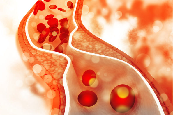
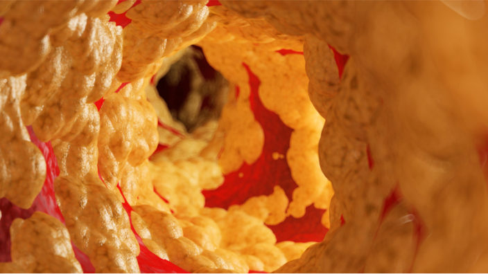
Steve: I was wondering whether restoring blood to a younger state would help reduce or prevent fibrotic tissue from forming. It seems it might?

Irina: We saw that it does. In the muscle, for example, there is less fibrosis, and we showed in the liver there was less fibrosis. So we showed therapeutic effects on fibrotic diseases.

Steve: With GDF-11. We still really don’t know what’s going on with that, even now.

Irina: GDF-11 is a member of the TGF beta family, and many members of that family promote blood vessel formation. So through that property, which never was disputed and was discovered before, there should be some improvement and tissue repair, but on the minus side, you also will promote growth of any tumor or precancerous mass. And GDF-11 has a very strong association with a number of human cancers, for example, colon cancer. GDF-11 typically is so minute that you cannot even find it in plasma. If you had a lot of it, you would promote blood vessel formation, but it also might promote cancers; and GDF-11 is not unique; you can use VEGF, which also improves tissue repair and promotes cancers as well.

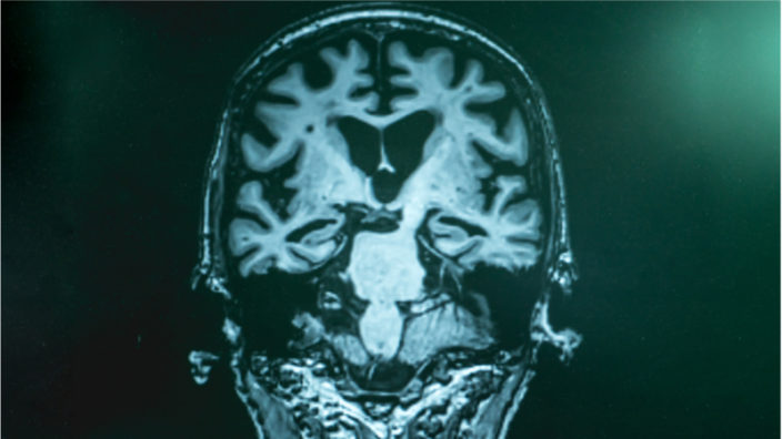
Steve: The one big question that people usually ask is, oh, okay, about muscle, bone and other tissues, but they always say, but what about the brain? You’ve got the blood-brain barrier, and the brain is weird, it doesn’t work like the rest of the body. What was the evidence for rejuvenation in the brain?

In our paper, we looked at the hippocampal neurogenesis, which is the formation of new neurons in the area of the brain that is responsible for learning and memory. We saw an eightfold increase in all the animals after neutral blood exchange, which is much higher than anything else that was observed either by heterochronic parabiosis or reported by young blood infusion. In old mice transfused with 50% young blood, we did not see an improvement in neurogenesis, which, again, puts a question mark on potency of young blood in old mammals. Does young blood by itself work as a medicine or not? If you dilute old blood by 50%, in neutral age blood exchange – with albumin-supplemented saline, there is quickly better neurogenesis. In our work in progress, we also see a reduction of neuro-inflammation by neutral blood exchange and improvement in animal cognition. We decided to have another dedicated study on those two parameters. That work is collaborative with professor Yi Zuo from UC Santa Cruz.

Steve: There’s a population of specialized stem cells in the hypothalamus, and some researchers think that, as they die, their ability to regulate hormones fails. Is that possibly what starts the cascade off?

Irina: I don’t think so. There are so many things that can start the cascade. For example, thymic involution, after which our immune system is not the same. Then, there’s a physical decline in the numbers of memory T cells and B cells because they have no telomerase, so once their telomeres shorten, they die. Overall damage to tissues everywhere and an increase of senescent cells, there are so many different directions. I don’t think that the only one is hypothalamus.

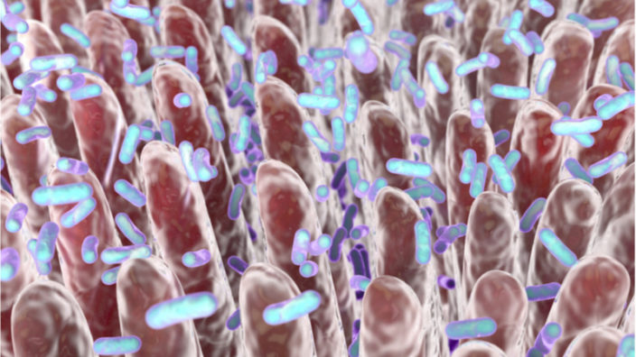
Steve: Changes to the microbiome as well.

Irina: Exactly, and that’s why I don’t think a silver bullet will be able to resolve diseases of aging. You know, therapeutic plasma exchange or neutral blood exchange are excellent in this regard because they molecularly recalibrate numerous factors simultaneously. They restore the health of numerous organs because multiple age-elevated determinants change to younger levels.

Steve: It’s a relatively low-hanging fruit by the look of it. Some people have asked if that technology would be easily scalable or cost-effective for a global audience. Wouldn’t it be, because it’s already approved?

Irina: It’s already approved. Dr. Kiprov was doing it in that clinic for 25 years. Anecdotally, people were telling him that they had more energy after this procedure was done. They feel younger, they feel better, and they do not succumb to viral illnesses that much. So, again, it’s just anecdotal evidence, but it certainly is worthy of attention in the current pandemic situation. People who underwent repetitive TPE did not get sick with the flu. This was back in 2017, 2018 before COVID, and they were from the age group 65 to 70, and 60% of flu hospitalizations were in this age group. None of the people who were in Dr. Kiprov’s care, even though they did not get the flu shot, got sick. So, better immunity is predicted as well.

Steve: They do transfusions a lot as well; there’s anecdotal evidence that you hear from time to time about people giving blood, and it makes them feel better as well.

Irina: Absolutely, yeah. I noticed in some recent publications when authors want to prove that young blood works, they have to just pump old animals with huge volumes of young blood. In actuality, de facto diluting the old blood. Even then, the data is highly variable and not as strong as if you take out some factors and replace them with a saline supplemented with albumin.

Steve: So we’re not saying it’s dilution, but it’s dilution.

Irina: Exactly.

Steve: That all feeds into the idea of inflammaging, as they call it, effectively all of the sources of inflammation that are creating this inflammaging are a major reason why we age. And if you remove that inflammation, things start working again, like NAD+, for example, that’s actively destroyed by CD38, which is present in the SASP. So, if you remove it now, levels go back up, and that’s why people may feel that they’re more energetic.

Irina: Another point is that the so-called young factors, of course, don’t disappear from our bodies. Where did they go? They’re still there. They’re suppressed by the age-elevated proteins. Once you remove that suppression, now, all of a sudden, you also have all of the young, systemic tissue determinants. They were not expressed appropriately but now they are, so you don’t need to add them back.

Steve: A lot of people are downstream of the problem. NAD levels drop, we’ll try and restore it with nicotinamide riboside, NMN, and other things like that. They’re all trying to compensate, and it does seem to have benefits, but obviously, it’s not a great long-term fix.

Irina: This is somewhat metaphorically similar to the application of insulin. You can not just keep exchanging blood and have good effects. In the same way, if somebody is diabetic, they need insulin to live, but if you keep injecting insulin, they’re going to die. This is a somewhat invasive procedure where your whole blood goes through a machine and the cells are returned to you and the plasma is diluted. It is not completely benign, and you cannot just keep doing it every day. So, to know how long the effects last and to know it for each individual is an additional know-how part that we found out from a couple of years of research and are studying further. I’m sure there will be competition, but I just want to warn everybody, do not go somewhere and get your blood replaced with saline. Wait until there are reputable clinical trials and people know a little bit more.

Steve: So, no popping over the border for a quick oil change, not advised at this point, although I suppose the principle is similar to changing the oil in the car.

Michael: Or like a fish tank. You get the poor goldfish in murky water and then you change the water to clean it up, and that perks up the fish.

Irina: As a principle, it’s better and safer than infusions of blood or bodily fluids, and it is more effective than any single factor, but we still need to know how to apply the procedure in a way that is effective for you. Because we’re not aging in the same way. We all age slightly differently.

Steve: There was a study out this year that identified three distinct types of ways that you age, and, based on your metrics, they gave you an ageotype, and it determines how and what kind of way you’re aging the fastest.

Irina: I don’t rely on those, to tell the truth, because I hear from others who have been told that they are seven years younger by these metrics than their chronological age, that they are surprised, as they feel quite old regardless of this “rosy” prognostication.

Steve: I feel like that. I was 45 the day that your paper was released, and I have mornings like that where everything hurts. Hopefully reducing systemic inflammation would mean fewer days like that.

Irina: Yeah, absolutely.

Steve: It is not just a question of how long can we live, but how well can we live. I think a lot of people are more focused on the healthspan aspect. I mean, another 10, 20 extra years, I’m not going to say no, but I want those 10 or 20 years to be healthy.

Irina: That’s why I don’t ever do a longevity study of how long a mouse lives. In many of those, you have an old mouse that is very sick and sits in the corner of the cage and shivers, but that is considered as a positive outcome because it did not die yet. We don’t do this type of work. We do rejuvenation work that starts with an old animal, and you analyze if you made it younger.

Steve: Using other examples of animals in nature, negligibly senescent animals in particular, we see that one of the reasons why they live so long is their ability to regenerate and repair. That’s probably why they live so long compared to us, although we don’t do badly. Then you’ve got super agers like the mole rat, the clam that lives right at the bottom of the ocean.

Irina: I don’t try to focus on either, because squirrels live as long as mole rats, and they’re a specialized case. All the squirrels that you see running around you outside, they live 20 to 30 years, while a rat, which looks exactly like a squirrel, lives only to three. So, there are many examples of enhanced longevity that we do not understand.

Steve: As far as I can tell, biological immortality, negligible senescence, or very long longevity tends to occur in extremely stable environments, such as at the bottom of the ocean where things don’t change much. If the environment doesn’t change, then there’s no environmental pressure or evolutionary pressure to change.

Irina: Clams are just so different from people that one interesting thing might be, again, a specialized case that explains how they live so long and cannot be applied to more complex systems like mammals. All of that, of course, deserves consideration and research. We should look into multiple directions, no doubt.

Steve: Comparative biology is interesting in itself. You never know what you might find out. I think you’re closing in on the situation, and it’d be interesting to see this dilution thing finally put to rest. It’s been going on a long time.

Irina: I agree.

Steve: And the actual practicality of it isn’t too bad. I recall from the interview in January that you mentioned that, because there’s so many feedback loops in the system, the reversed or younger blood state tends to be or could potentially be quite persistent for a while, so it’s not like we’re going to need to do it every week.

Irina: Yes, exactly, but you need to figure out when and how to do it. In conventional therapy plasma exchange, they remove almost all of your blood plasma, and a designer procedure could be applied where it is less harsh and has stronger positive effects. There’s still repositioning, so you should not try to use it right away. For example, if you have a fibrotic or inflammatory disease, you need to discuss with your doctor whether or not TPE is good for you. But to say that now everybody can just go and get rejuvenated, it’s a few steps removed from that; we still need to do the clinical trial. In the clinical trial, we will know so much more, then we can really prescribe the best procedure for each individual or group of people that have similar diseases.

Steve: That’s the key, because the FDA is definitely not going to approve anything for age reversal, but they will obviously go with things like disease modification. You could pick fibrotic diseases and say, “We’re going to go for this target, this is the endpoint.” That’s how you get it in.

Irina: Yes, that’s our business plan. To go for fibrotic diseases, this is our target and endpoint. For inflammatory diseases, this is a different target and endpoint. For type 2 diabetes or associated metabolic diseases, this is the target and endpoint. Once we have success for all those in clinical trials, then we feel strongly that the FDA will approve the modification or designer TPE for each group.

Steve: In its current state, it’s already approved anyway. This is the next-generation version of it. If you can demonstrate the disease reversal or modification, then that would be approved. Of course, once it’s approved, then it has lots of potential off-label use, and it goes from there.

Irina: They could compete with us, but by that time, we will have nextgen devices, products and services that are more effective and safe, and, of course, we are known names in the field.

Steve: The quantification systems and devices are getting more sophisticated now as well. Medical wearables are becoming extremely popular now with just the general public, things like that. It’s quite plausible that, in order to find out when you need to go in for your next oil change, you could have a little sensor or something implanted.

Irina: That’s exactly one of the future directions, a wearable analytical device, the direction which is illustrated in our recent paper in Lab on Chip. So yes, you can envision, not now but let’s say in seven – ten years, that you have a wearable device on your wrist, which tells you that, hey, your TGF beta is a little bit too high. This is the schedule for you to come and have the procedure.

Steve: It could be a bit like the little service light coming on your car dash. We talked about how, in the future, a filtering system could be reduced in size eventually to potentially be a wearable device?

Irina: I don’t think so. I don’t think that our filter system could be reduced to that. Maybe at some point, not soon.

Steve: Yeah, right now, you’d have to walk around with it on a trolley, which is a little bit inconvenient.

Irina: It’s a little inconvenient, but for somebody who has a degenerative pathology, it’s a better alternative than progressive health decline.

Elena: From what age do you think this kind of therapy might be useful for people? I’m asking because some of the rejuvenation potential of this therapy, so it seems will be very interesting for women who would like to extend their period of fertility, and they wonder also, as women naturally lose some blood every month, at least some of them do, does it make any sense to sort of replenish the blood with this kind of therapy?

Irina: Those are very interesting questions. We have a project that addresses effects on reproductive function, both in males and females, and there is at least anecdotal evidence of improvement. We believe that our approach will be helpful for extending reproductive health, but we have to try it first in experimental animals to answer this question. This specific age might differ from person to person, so that’s, again, personalized rejuvenation; this is our mission and know-how as well. With respect to the monthly blood loss, this is an amazing idea; I have never thought of it. Never really came to mind, but I have known that, for example, women are healthier than men for some time with respect to cardiovascular disease. I wonder how much of that comes from this effect of dilution of blood? I don’t know.

Steve: I would say estrogen also could have quite an effect on longevity. There was a study back in 2009 that showed how estrogen actually interacts with TERT and activates telomerase.

Irina: What if we start this treatment before the loss of estrogen or loss of testosterone, can we maintain those positive hormones for decades longer? Something to be tested.

Steve: Yes and they’ve already got implanted devices that can help regulate hormones as well.

Irina: The thing is, and this is again the major paradigm shift of our paper, it suggests that hormonal therapy was unsuccessful because of the aged combination of multiple factors, molecules and cells, that dominantly inhibit anything positive that you introduce. A metaphor is that if you have a cluster of rotten bananas, and you throw a healthy banana, a nice, good banana, into that cluster, is that going to help? You are not going to rejuvenate the cluster. The other bananas will rot, and your banana will rot as well. That applies not just to hormone replacement but also to cell transplantation, when people try to put young, healthy stem cells or tissues into old mammals. There is a need to reduce the inhibitory components of the aged environment, and there is a potential you don’t need to add anything because hormones do not disappear from our body. They simply are not expressed. They might be re-expressed again, once you remove their inhibitors.

Steve: Well, it deals with the central theme, which is inflammation. Inflammation interrupts signaling and normal function, makes the immune system not work properly, reduces NAD, it does all those things.

Irina: This plethora of things which go wrong with aging is dealt with at once. Kind of like how antibiotics are dealing with bacterial infections, as a class.

Steve: Somebody once said to me, if you consider the hallmarks of aging and its distinct categories, they are like a wagon wheel. Imagine each spoke as a hallmark, and at the center of the wheel that joins all the spokes together is inflammation, because that’s how all of these hallmarks are linked.

Irina: I don’t know, I’m not a fan of such simplicity. I prefer to go from the other direction where you have a hypothesis, and you find a way to test that hypothesis. And then, science takes care of you because it tells you this is how it is instead of trying to drag science into a particular direction. Take care of science and science will take care of you.

We would like to ask you a small favor. We are a non-profit foundation, and unlike some other organizations, we have no shareholders and no products to sell you. All our news and educational content is free for everyone to read, but it does mean that we rely on the help of people like you. Every contribution, no matter if it’s big or small, supports independent journalism and sustains our future.

Literature

[1] Conboy, I. M., Conboy, M. J., Kiprov, D., Kato, C., Etienne, J., Liu, C., … & Mehdipour, M. (2020). Rejuvenation of three germ layers tissues by exchanging old blood plasma with saline-albumin. Aging, 12(10), 8790-8819.

[2] Calado, R. T., Yewdell, W. T., Wilkerson, K. L., Regal, J. A., Kajigaya, S., Stratakis, C. A., & Young, N. S. (2009). Sex hormones, acting on the TERT gene, increase telomerase activity in human primary hematopoietic cells. Blood, The Journal of the American Society of Hematology, 114(11), 2236-2243.