The Blog

Building a Future Free of Age-Related Disease

Elderly falls

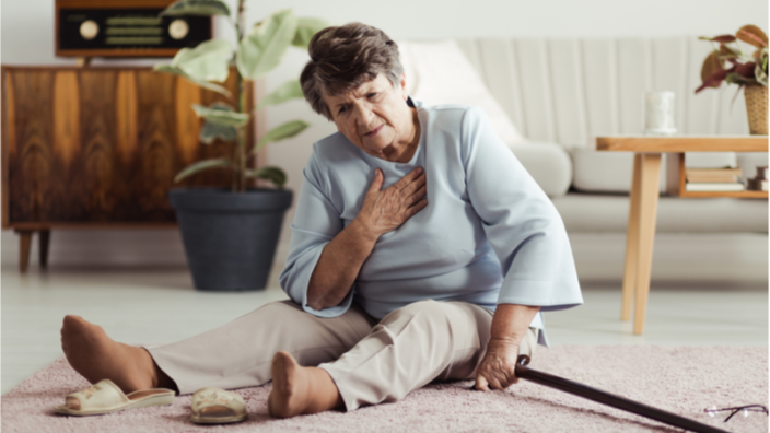
Pain Is Associated with Increased Risk of Falls in Adults

In a new longitudinal study published in European Geriatric Medicine, the researchers have shown that people experiencing moderate to severe pain in multiple sites have an increased risk of falls, particularly in middle age [1].
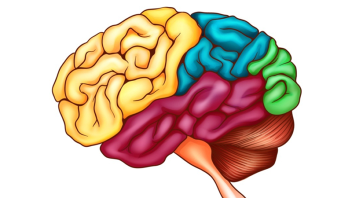
Aging and falls

In one’s youth, falling doesn’t seem dangerous and might be enjoyable; people talk about falling into fluffy snow and falling in love. However, falls often lead to serious injuries and can even be fatal for older adults. Older, inactive females are particularly vulnerable to falls and subsequent injuries.

Prevention of falls involves finding and reversing their risk factors. For example, a recent study has shown that older women with carotid plaques have a higher risk of fall-related hospitalization [2]. Therefore, lifestyle changes, including exercise and a healthier diet, might reduce the risk of falls while improving cardiovascular health.

Pain is highly prevalent in adults over the age of 65 years old [3] and has been associated with a higher risk of falls. Whether or not pain is a cause of falls in this age group or merely a consequence of age-related co-morbidities that can lead to falls, such as arthritis, is unclear.

In this study, the researchers sought to explore the association between pain and falls in more detail in two age groups: middle-aged and older adults. They investigated if pain intensity and the number of pain sites were predictive of the risk of falling.

A multicountry survey

The Survey of Health, Ageing and Retirement in Europe (SHARE) recruited 40,636 people over 50 years old from 14 European countries. As the name suggests, the participants answered a series of questions about their height, weight, medications, co-morbidities, physical inactivity, vision, hearing, self-rated health, living companions, pain, and falls.

Participants reported the presence and intensity of pain and specified where it occurred. The participants were then asked if they experienced falls in the last 6 months. Two interviews were conducted two years apart.

First, the researchers show that ~40% of all the participants reported pain of varying degrees (mild, moderate, or severe). Women experience more pain of higher intensity and in multiple sites than men. Not surprisingly, severe pain was reported by older, inactive participants who were in poorer health and had co-morbidities that required medications. These participants were also more likely to have pain in multiple sites. Of all the pain sites, back pain was the most frequent complaint.

No pain, no falls

The initial survey revealed that 4.3% of men and 7.7% women experienced falls in the last 6 months. These numbers increased to 5.0% and 8.4%, respectively, two years later. Participants with pain were more likely to fall in the preceding and subsequent months, regardless of sex.

After adjusting for co-variates, such as co-mobidities and BMI, moderate and severe pain were associated with an increased risk of falls. Interestingly, there were age-specific differences: the presence of moderate and severe pain was predictive of falls in participants between 50 and 79 years of age but not in people over 80 years old.

Importantly, participants with pain in multiple sites had a higher risk of falling than those with one-site pain. ~50% of participants suffering from pain of various severity reported pain in multiple sites.

There were some country-specific differences in terms of pain prevailance and fall frequency. For example, participants from Switzerland were among the most likely to be pain- and fall-free. Meanwhile the participants from France were among the most likely to experience pain and fall regardless of sex.

Abstract

Aim: To explore the longitudinal associations between pain characteristics at baseline and subsequent falls risks, at 2-year follow-up, in community-dwelling adults aged ≥ 50 years, in the Survey of Health, Ageing and Retirement in Europe (SHARE).

Findings: Higher intensity of pain and number of pain sites at baseline were associated with an increased risk of subsequent falls in community-dwelling adults, in a dose–response way, independent of socio-demographic and clinical characteristics. The strength of the association between pain intensity and falls risk varied by age, being greater in middle-aged adults.

Message: The association between pain intensity and falls risk is of greater clinical significance in middle-aged adults versus older adults.

Conclusion

This study revealed that a higher intensity of pain and multiple pain sites are predictive of falls. Although this study doesn’t prove any causal link between pain and falls, the authors suggest several mechanisms that might underlie the association, including physical inactivity. If pain is caused by a sedentary lifestyle [4], it could lead to activity avoidance, which deconditions the muscles, reduces their strength, and impairs balance, making one more prone to falling. Therefore, minimizing the risk of falls is yet another reason to lead an active lifestyle.

Literature

[1] ​​Ogliari, G., Ryg, J., Andersen-Ranberg, K., Scheel-Hincke, L. L., Collins, J. T., Cowley, A., … & Masud, T. (2022). Association of pain and risk of falls in community-dwelling adults: a prospective study in the Survey of Health, Ageing and Retirement in Europe (SHARE). European geriatric medicine, 1-14.

[2] Gebre, A. K., Sim, M., Dalla Via, J., Rodríguez, A. J., Hodgson, J. M., Bondonno, C. P., … & Lewis, J. R. (2022). Measures of carotid atherosclerosis and fall-related hospitalization risk: The Perth Longitudinal Study of Ageing Women. Nutrition, Metabolism and Cardiovascular Diseases.

[3] Domenichiello, A. F., & Ramsden, C. E. (2019). The silent epidemic of chronic pain in older adults. Progress in Neuro-Psychopharmacology and Biological Psychiatry, 93, 284-290.

[4] Hanna, F., Daas, R. N., El-Shareif, T. J., Al-Marridi, H. H., Al-Rojoub, Z. M., & Adegboye, O. A. (2019). The relationship between sedentary behavior, back pain, and psychosocial correlates among university employees. Frontiers in public health, 7, 80.

Mogling

Kizoo Invests in Rejuvenation Startup MoglingBio

Having received new funding from Kizoo Technology Capital in its first seed round, MoglingBio intends to further its research into the rejuvenation of aging stem cells, finding ways to stave off stem cell exhaustion.

Berlin/Ulm, Germany – October 26, 2022 – MoglingBio, a privately held biotech company, announces today that it has successfully completed its first seed investment round. Sole investor is venture capital firm Kizoo Technology Capital, a rejuvenation biotech investor focused on startups reversing age-related damage on a cellular and molecular level.

MoglingBio is developing new pharmacological approaches to rejuvenate old stem cells of the hematopoietic (blood cell formation) system. Aging causes stem cells to lose their normal structure by increased activity of the protein CDC42. This loss of structure leads to decreased production and quality of blood and immune cells. It can cause leukemia, various blood diseases, and severely weaken the immune system. Normalizing CDC42 activity can restore structure, order and functionality in those aged stem cells. Treated cells can perform their tasks again in a juvenile way, and thereby contribute to both, the rejuvenation of stem cells and the immune system.

The underlying technology was developed over the last 15 years by the two scientific co-founders, Prof. Yi Zheng, co-director of the Cincinnati Children’s Hospital Cancer and Blood Diseases Institute and leader of the Signaling and Drug Discovery Program at the Cincinnati Children’s Research Foundation, and Prof. Dr. Hartmut Geiger, director of the Institute of Molecular Medicine at Ulm University in Germany.

“We are really excited that Kizoo’s financial support will allow us to pursue our goal of rejuvenating aging stem cells, which are so important for blood cell production and the immune system. Our approach has particular potential in treating diseases of the blood system and improving the immune system,” said Dr. Jürgen Reess, CEO of MoglingBio. Dr. Reess previously held the position of Senior Vice President at Boehringer Ingelheim Pharma, where he supervised the development, approval, and launch of numerous blockbuster therapies for autoimmune diseases, interstitial lung diseases, central nervous system disorders, and cancer.

“We believe that MoglingBio’s technology is groundbreaking by truly rejuvenating old stem cells – not only alleviating age-related diseases, but reversing age-related decline in immune system function”, added Frank Schueler, Managing Director of Kizoo Technology Capital.

About Kizoo

Kizoo provides seed and follow-on financing with a focus on rejuvenation biotech. Having been entrepreneurs, VC, and mentors in both high-growth tech and biotech companies ourselves for many years with multiple exits and massive value created for the founders, Kizoo now brings this experience to the emerging field of rejuvenation biotech. We see it as a young industry that will eventually outgrow today’s largest technology markets.

As part of Michael Greve’s Forever Healthy Group, Kizoo directly supports the creation of startups turning research on the root causes of aging into therapies and services for human application. Investments include Cellvie, Cyclarity Therapeutics, Revel Pharmaceuticals, Elastrin Therapeutics, LIfT BioSciences and others.

For more information, please visit: www.kizoo.com.

About MoglingBio

MoglingBio Inc. is developing new pharmacological approaches to rejuvenate old stem cells of the hematopoietic (blood cell formation) system. Aging causes stem cells to lose their normal structure by increased activity of the protein CDC42. This loss of structure leads to decreased production and quality of blood and immune cells. It can cause leukemia, various blood diseases, and severely weaken the immune system. Normalizing CDC42 activity can restore structure, order and functionality in those aged stem cells. Treated cells can perform their tasks again in a juvenile way, and thereby contribute to both, the rejuvenation of stem cells and the immune system. The underlying technology was developed over the last 15 years by the two scientific co-founders, Prof. Yi Zheng, co-director of the Cincinnati Children’s Hospital Cancer and Blood Diseases Institute and leader of the Signaling and Drug Discovery Program at the Cincinnati Children’s Research Foundation, and Prof. Dr. Hartmut Geiger, director of the Institute of Molecular Medicine at Ulm University in Germany.

For more information, please visit: www.moglingbio.com.

We would like to ask you a small favor. We are a non-profit foundation, and unlike some other organizations, we have no shareholders and no products to sell you. All our news and educational content is free for everyone to read, but it does mean that we rely on the help of people like you. Every contribution, no matter if it’s big or small, supports independent journalism and sustains our future.

Squatting at desk

Intermittent Exercise Encourages Muscle Protein Uptake

A research paper published in the Journal of Applied Physiology has found that regular, intermittent exercise encourages amino acids to form proteins in muscle.

Anabolic resistance

Previous research has found that a lack of activity accelerates the age-related muscle loss known as sarcopenia [1]. This has been found to be strongly influenced by anabolic resistance, a phenomenon in which muscles fail to acquire sufficient proteins with which to rebuild tissue [2]. Therefore, suppressing anabolic resistance is a sensible strategy for slowing or preventing the deterioration of muscle tissue, particularly in the context of aging [3].

Previous research has found that physical activity encourages muscle protein development for up to 24 hours in young men [4], and other research has found that breaking up long sedentary periods with physical activity provides benefits to glucose and insulin use [5]. This paper takes this line of inquiry a step further, conducting biopsies to determine what happens in muscle fiber over the course of a day.

Marked proteins show the effects

In this study, all participants ate the same, standardized diet that approximates Western eating habits: prepackaged meals consisting of 55% carbohydrate, 30% fat, and 15% protein. A tracer was included to measure how much of this newly consumed protein was entering the muscle tissue. Participants were divided into three groups: an entirely sedentary control group (SIT), a group that performed 15 bodyweight squats every half hour (SQUAT), and a group that went for two-minute walks every half hour (WALK). The researchers refer to these exercise breaks as ‘activity snacks’.

While exercise caused the tracer to simply vary more in blood plasma while the average among participants remained approximately the same, the effects on muscle protein synthesis were clear within muscle tissue. While there were outliers, the SQUAT and particularly the WALK group enjoyed significantly more muscular protein synthesis than the SIT group did.

Interestingly, the SQUAT group, not the WALK group, showed an increase in a marker related to mTORC1, which, in this context, is suggestive of protein synthesis. The researchers hypothesize that the WALK group might have encouraged muscle growth through other means, such as increased blood flow.

A different look at regular exercise

The researchers note a previous study showing that older men who rested for 10 hours after acute physical activity did not achieve significant benefits in muscle mass [6]. While this study involved younger people, it is a credible hypothesis that interrupting sedentary periods might be a key factor in the continued uptake of muscle-building proteins, even among older people. More research will need to be done to determine if ‘activity snacks’, conducted throughout the day, are more effective than simple daily exercise in combating sarcopenia.

We would like to ask you a small favor. We are a non-profit foundation, and unlike some other organizations, we have no shareholders and no products to sell you. All our news and educational content is free for everyone to read, but it does mean that we rely on the help of people like you. Every contribution, no matter if it’s big or small, supports independent journalism and sustains our future.

Literature

[1] Oikawa, S. Y., Holloway, T. M., & Phillips, S. M. (2019). The impact of step reduction on muscle health in aging: protein and exercise as countermeasures. Frontiers in nutrition, 75.

[2] Wall, B. T., Dirks, M. L., Snijders, T., van Dijk, J. W., Fritsch, M., Verdijk, L. B., & van Loon, L. J. (2016). Short-term muscle disuse lowers myofibrillar protein synthesis rates and induces anabolic resistance to protein ingestion. American journal of physiology-endocrinology and metabolism, 310(2), E137-E147.

[3] Oikawa, S. Y., Holloway, T. M., & Phillips, S. M. (2019). The impact of step reduction on muscle health in aging: protein and exercise as countermeasures. Frontiers in nutrition, 75.

[4] Burd, N. A., West, D. W., Moore, D. R., Atherton, P. J., Staples, A. W., Prior, T., … & Phillips, S. M. (2011). Enhanced amino acid sensitivity of myofibrillar protein synthesis persists for up to 24 h after resistance exercise in young men. The Journal of nutrition, 141(4), 568-573.

[5] Loh, R., Stamatakis, E., Folkerts, D., Allgrove, J. E., & Moir, H. J. (2020). Effects of interrupting prolonged sitting with physical activity breaks on blood glucose, insulin and triacylglycerol measures: a systematic review and meta-analysis. Sports Medicine, 50(2), 295-330.

[6] Bülow, J., Agergaard, J., Kjaer, M., Holm, L., & Reitelseder, S. (2016). No additional effect of different types of physical activity on 10-hour muscle protein synthesis in elderly men on a controlled energy-and protein-sufficient diet. Experimental gerontology, 79, 16-25.

HIIT and Intermittent Fasting Show Cumulative Effect

Scientists have shown that both high-intensity interval training (HIIT) and time-restricted eating improve metabolism in healthy overweight women, with their combination providing the biggest effect [1].

Which one is better?

Good dietary habits and physical activity are currently our best options for living longer and healthier lives. In recent years, two interventions have been gaining popularity – intermittent fasting and HIIT. Some swear by one, and some by the other, and there is already considerable scientific evidence of their health benefits. However, how these two interventions compare to each other and to their combination, in terms of metabolic and cardiovascular outcomes, has remained an open question. The results of a new randomized, controlled trial published in Cell Metabolism seem to provide some answers.

Female-only design

The study was female-only and included 131 women of reproductive age (mean age of 36), randomly assigned to four study groups: time-restricted eating (TRE), HIIT, TRE+HIIT, and a control group. While sex bias might limit a study’s generalizability, we now know that men and women have somewhat different metabolisms. Moreover, women had been underrepresented in medical trials until recently, so a lot of our scientific knowledge might be skewed towards male biology. Obesity and insulin resistance in women also have some sex-specific implications, including accelerated reproductive aging and increased risk of adverse pregnancy outcomes.

In this study, TRE is basically intermittent fasting attuned to diurnal circadian rhythms, with eating confined to an 8- to 10-hour window during the day. While TRE does not limit the total number of calories or food composition, people on TRE often spontaneously reduce their energy intake, leading to some weight loss [2]. TRE is also known to improve insulin sensitivity in men with prediabetes [3] and even in healthy non-obese people [4].

HIIT is characterized by short, repeated bouts of high-intensity aerobic exercise separated by low-intensity breaks. It has been shown to improve cardiorespiratory fitness and insulin sensitivity in high-risk populations [5]. HIIT also seems to be much more effective in inducing weight loss than continuous moderate-intensity workouts [6]. A combination of TRE and HIIT has already been shown to attenuate fat mass gain and some adverse metabolic changes in mice on a high-fat diet [7], but human data is limited.

Combine them if you can

This new study brings mixed results. The combination treatment had no effect on the primary outcome, glucose area under curve (AUC), which shows how well someone tackles post-meal glucose spikes. However, participants in all study groups experienced significant weight loss, and both TRE and TRE+HIIT induced improvements in secondary glycemic control outcomes. While TRE alone was able to reduce levels of nocturnal glucose, TRE+HIIT improved levels of glycated hemoglobin (HbA1c), a primary metric of long-term blood glucose.

TREHIIT 1

TRE+HIIT also led to a significant improvement in peak glucose levels. This metric has been gaining in importance, including in the context of aging, as two of the drugs shown to reliably extend lifespan in mice, acarbose and canagliflozin, work by flattening post-meal glucose spikes.

As expected, both TRE and HIIT led to some weight loss, but the combination treatment clearly showed a cumulative effect. TRE induced mild muscle mass loss that was partially rescued in the TRE+HIIT group. HIIT, both alone and in combination with TRE, produced noticeable improvement in maximum oxygen capacity (VO2max), a metric of physical fitness.

TREHIIT 2

The researchers note that most previous studies have shown bigger benefits for already unhealthy people. In this study, most of the participants, despite being obese, showed normal glycemic values at baseline. This might be the reason why the primary outcome was not achieved.

While the study was mostly well-designed, one detail pops out: mean BMI at baseline was the lowest in the TRE+HIIT group (31.4) and the highest in the control group (33.1). Such discrepancies can potentially contaminate the results, though there is no evidence that this happened in this study.

Conclusion

This randomized, controlled trial confirms the benefits of both intermittent fasting and HIIT on certain metabolic outcomes in generally healthy overweight women. Both interventions are beneficial on their own, but women who practice them together, according to this study, are rewarded with a cumulative effect. Future research will confirm the likely possibility that this is just as true for men.

We would like to ask you a small favor. We are a non-profit foundation, and unlike some other organizations, we have no shareholders and no products to sell you. All our news and educational content is free for everyone to read, but it does mean that we rely on the help of people like you. Every contribution, no matter if it’s big or small, supports independent journalism and sustains our future.

Literature

[1] Haganes, K. L., Silva, C. P., Eyjólfsdóttir, S. K., Steen, S., Grindberg, M., Lydersen, S., … & Moholdt, T. (2022). Time-restricted eating and exercise training improve HbA1c and body composition in women with overweight/obesity: A randomized controlled trial. Cell Metabolism, 34(10), 1457-1471.

[2] Kang, J., Ratamess, N. A., Faigenbaum, A. D., Bush, J. A., Beller, N., Vargas, A., … & Andriopoulos, T. (2021). Effect of time-restricted feeding on anthropometric, metabolic, and fitness parameters: a systematic review. Journal of the American College of Nutrition, 1-16.

[3] Sutton, E. F., Beyl, R., Early, K. S., Cefalu, W. T., Ravussin, E., & Peterson, C. M. (2018). Early time-restricted feeding improves insulin sensitivity, blood pressure, and oxidative stress even without weight loss in men with prediabetes. Cell metabolism, 27(6), 1212-1221.

[4] Xie, Z., Sun, Y., Ye, Y., Hu, D., Zhang, H., He, Z., … & Mao, Y. (2022). Randomized controlled trial for time-restricted eating in healthy volunteers without obesity. Nature communications, 13(1), 1-10.

[5] Maturana, F. M., Martus, P., Zipfel, S., & Nieß, A. M. (2020). Effectiveness of HIIE versus MICT in improving cardiometabolic risk factors in health and disease: a meta-analysis. Med. Sci. Sport Exer, 53, 559-573.

[6] Viana, R. B., Naves, J. P. A., Coswig, V. S., De Lira, C. A. B., Steele, J., Fisher, J. P., & Gentil, P. (2019). Is interval training the magic bullet for fat loss? A systematic review and meta-analysis comparing moderate-intensity continuous training with high-intensity interval training (HIIT). British journal of sports medicine.

[7] Vieira, R. F. L., Muñoz, V. R., Junqueira, R. L., de Oliveira, F., Gaspar, R. C., Nakandakari, S. C. B. R., … & Pauli, J. R. (2022). Time‐restricted feeding combined with aerobic exercise training can prevent weight gain and improve metabolic disorders in mice fed a high‐fat diet. The Journal of Physiology, 600(4), 797-813.

Bowhead whale

Bowhead Whale’s Longevity May Be Due to Twin Genes

A preprint published in bioRxiv has partially explained the lifespan of the bowhead whale through its duplication of CDKN2C, which regulates how cells divide.

Peto’s paradox

It would be logical for longer-lived animals to be more susceptible to cancer than shorter-lived animals over time, as they have longer to develop the mutations that lead to cancer. However, this is not the case; there is no correlation between lifespan and cancer. This counterintuitive finding is known as Peto’s paradox, which was originally discovered in 1975 [1] and backed up by studies performed this year [2].

This fact can be explained by genes that increase resilience to genomic instability. Mice that overexpress the tumor suppressor p53 and the cell cycle regulator CDKN2A live longer and are more resistant to cancer [3], despite the fact that CDKN2A is associated with cellular senescence. Increasing the presence of SIRT1, which protects against telomere attrition, protects mice against some aging-associated diseases, including cancer [4].

A whale of a lifespan

Unlike most cetaceans, which normally live between 50 and 70 years, the bowhead whale is estimated to have a lifespan of over two centuries [5]. It is far longer-lived than the right whale, which it diverged from between four and five million years ago. To live this long, it must have some extra resistance to cancer, even beyond the formidable cancer protection of other cetaceans.

Comparing the genes of the bowhead and its nearest relatives, the researchers found that CDKN2C is duplicated in a way that does not occur in related whales. Retrotransposition, a form of genetic mutation, had copied this gene into the cetacean-specific LINE L1 genetic sequence, which drives its substantial expression. Previous research has shown that this gene suppresses cancerous tumors [6].

This abundance of CDKN2C also dramatically slows down the cellular cycle of division and replication. In a way, this is related to the maxim ‘live slow, die old’ in longevity research, but this extra slowdown also gives cells more time to prevent cancerous mutations from occurring. While the researchers have not fully explored the biochemistry involved, they hypothesize that this extra time allows two related genes to do more work: CDKN2A, which inhibits cellular death by apoptosis, and CDKN2D, which enhances DNA repair mechanisms. Apparently, the cells are not just dividing more slowly; they are being more careful in their division.

On the other hand, the researchers also hypothesize that this protection leads to trade-offs in other areas (antagonistic pleiotropy). While it has not been proven, they suggest that this mutation may lead to slow maturation and reduced male fertility in these whales.

Conclusion

It is, of course, completely infeasible to do direct lifespan experiments on whales, even moreso than on humans; any such experiment would take mutliple (current) human lifetimes to complete. However, the gene CDKN2C exists in mice and in people. If this approach can be proven to work through trials, genetic or RNA-based therapies that cause the overexpression of this gene may become part of a near-term approach towards extending human lifespan.

We would like to ask you a small favor. We are a non-profit foundation, and unlike some other organizations, we have no shareholders and no products to sell you. All our news and educational content is free for everyone to read, but it does mean that we rely on the help of people like you. Every contribution, no matter if it’s big or small, supports independent journalism and sustains our future.

Literature

[1] Peto, R., Roe, F. J., Lee, P. N., Levy, L., & Clack, J. (1975). Cancer and ageing in mice and men. British journal of cancer, 32(4), 411-426.

[2] Vincze, O., Colchero, F., Lemaître, J. F., Conde, D. A., Pavard, S., Bieuville, M., … & Giraudeau, M. (2022). Cancer risk across mammals. Nature, 601(7892), 263-267.

[3] Matheu, A., Maraver, A., Klatt, P., Flores, I., Garcia-Cao, I., Borras, C., … & Serrano, M. (2007). Delayed ageing through damage protection by the Arf/p53 pathway. Nature, 448(7151), 375-379.

[4] Palacios, J. A., Herranz, D., De Bonis, M. L., Velasco, S., Serrano, M., & Blasco, M. A. (2010). SIRT1 contributes to telomere maintenance and augments global homologous recombination. Journal of Cell Biology, 191(7), 1299-1313.

[5] George, J. C., Follmann, E., Zeh, J., Sousa, M., Tarpley, R., Suydam, R., & Horstmann-Dehn, L. (2011). A new way to estimate the age of bowhead whales (Balaena mysticetus) using ovarian corpora counts. Canadian Journal of Zoology, 89(9), 840-852.

[6] Franklin, D. S., Godfrey, V. L., O’Brien, D. A., Deng, C., & Xiong, Y. (2000). Functional collaboration between different cyclin-dependent kinase inhibitors suppresses tumor growth with distinct tissue specificity. Molecular and cellular biology, 20(16), 6147-6158.

Good and bad

Senescent Cells Can Be Good or Bad According to New Study

New research has scientists reconsidering the role of ‘zombie’ cells that drive some aspects of aging and whether they should be eliminated using senolytic drugs. It turns out that not all senescent cells are necessarily bad and that some may be helpful.

Introducing zombie cells

As we get older, more and more of our cells become senescent. Our cells should destroy themselves when they reach senescence, but, as we age, increasing numbers of them evade this process and become ‘zombie’ cells that refuse to die.

These cells do not divide, and they secrete an inflammatory cocktail of signals called the senescence-associated secretory phenotype (SASP). The SASP can contribute to age-related diseases and cause nearby healthy cells to become senescent, and their presence is thought to be one of the reasons we age.

One proposed solution to the problem is senolytics: drugs that specifically eliminate senescent cells. There has been a great deal of interest in developing senolytic drugs that can target senescent cells and destroy them.

Originally, it was thought that all senescent cells were harmful and supported the development of age-related diseases. Indeed, early senolytic experiments in mice that removed these cells delayed age-related diseases and increased the lifespan of the animals.

These initial results spurred great interest in developing senolytics with the hope that those beneficial effects might be translated to people. However, it turns out that not all senescent cells are harmful and that some could be beneficial. In other words, simply destroying them all with senolytics may not be the best approach.

Senescent cells healing or harmful

New research from UC San Francisco suggests that some senescent cells present in healthy tissue promote tissue repair following damage [1].

The researchers observed senescent cells in the tissues of the lungs, small intestine, colon and skin. When they used senolytic drugs to kill the senescent cells, it caused those tissues to heal injury slower.

The results of the study suggest that some senescent cells can act as ‘sentinels’ and sit in the tissue watching for signs of injury. When damage to the tissue occurs, these sentinels then respond by signaling nearby stem cells to engage their growth and repair systems.

The study authors caution that destroying senescent cells has some risks. On one hand, they suggest that senolytics have the potential to cause tissue dysfunction and hinder tissue repair. On the other hand, they also propose that senolytics may be able to address age-related diseases driven by senescent cells.

Identifying senescent cells by making them glow

One of the hurdles in studying senescent cells is being able to spot them among the thousands of regular cells in tissues. An ongoing challenge for the field has been to establish reliable biomarkers of senescence so that these cells can be identified.

For example, the p16 gene is often used as a biomarker of senescence, as it is typically very active in these cells, but this can be hard to detect, and other cells can also express it under certain conditions. What the researchers needed was a better way to identify senescent cells in tissue. Using a lab technique, they fused the p16 gene with green fluorescent protein (GFP) and amplified the fluorescence further. GFP glows under ultraviolet light, making it easier to see the senescent cells in tissues.

Using the GFP method, the researchers discovered that senescent cells are actually present in young tissues and in greater numbers than previously assumed. The research team found that they began to appear not long after birth, which is somewhat surprising given that they are typically associated with old age. The researchers also identified a number of growth factors that were being secreted by the senescent cells, signaling stem cells to activate and promote growth and repair.

Senescent cells play an important role in tissue repair 

While examining lung tissue, the team noticed that glowing senescent cells were located next to stem cells in the basement membrane. This membrane acts as a barrier to stop chemicals, bacteria, and other pathogens from entering the body. It also allows oxygen in the lungs to diffuse into the tissues below. The researchers observed that senescent cells were also occupying similar positions in other barrier tissues in other organs like the colon, small intestine, and skin.

This barrier can become damaged, and this is where senescent cells play an important role. The scientists found that if they killed the senescent cells in the lung tissue using senolytics, the stem cells were then unable to repair the barrier surface properly.

These results suggest that indiscriminate use of senolytics is a risky proposition and that research needs to focus on making them highly selective as to which cells it targets. Simply dumb-firing senolytics and killing all senescent cells is asking for trouble, as the results of this study support.

To be successful, not only must the harmful subsets of senescent cells be identified, senolytic drugs must be developed that only kill those cells and not ones serving a useful purpose. It should also serve as a cautionary tale for people engaging in self-experimentation with senolytics.

Abstract

We engineered an ultrasensitive reporter of p16INK4a, a biomarker of cellular senescence. Our reporter detected p16INK4a-expressing fibroblasts with certain senescent characteristics that appeared shortly after birth in the basement membrane adjacent to epithelial stem cells in the lung.

Furthermore, these p16INK4a+ fibroblasts had enhanced capacity to sense tissue inflammation and respond through their increased secretory capacity to promote epithelial regeneration.

In addition, p16INK4a expression was required in fibroblasts to enhance epithelial regeneration. This study highlights a role for p16INK4a+ fibroblasts as tissue-resident sentinels in the stem cell niche that monitor barrier integrity and rapidly respond to inflammation to promote tissue regeneration.

Conclusion

This study highlights that we currently do not have a full understanding of senescent cells and thier various roles in aging and repair. In any given tissue, there are various kinds of senescent cells, each using different pro-survival pathways.

Understanding these cell populations will help researchers determine which ones might be safely removed using senolytics and which should be left alone. The potential of senolytics to slow down aging and improve health is still on the cards, but it seems ever more likely that simply destroying all senescent cells is a bad idea.

The Campisi lab at the Buck institute for Research on Aging has been busy the last few years trying to work out the complexities of senescent cell populations. In 2020, they collaborated with other researchers to make a SASP Atlas, which is a step closer to understanding senescent cells [2].

Biology is never simple, and there is often more than one solution to a problem. Senolytics may yet prove their worth in the push to defeat age-related diseases; this study just serves to remind us that we have more to learn before we can do that.

We would like to ask you a small favor. We are a non-profit foundation, and unlike some other organizations, we have no shareholders and no products to sell you. All our news and educational content is free for everyone to read, but it does mean that we rely on the help of people like you. Every contribution, no matter if it’s big or small, supports independent journalism and sustains our future.

Literature

[1] Reyes, N. S., Krasilnikov, M., Allen, N. C., Lee, J. Y., Hyams, B., Zhou, M., Ravishankar, S., Cassandras, M., Wang, C., Khan, I., Matatia, P., Johmura, Y., Molofsky, A., Matthay, M., Nakanishi, M., Sheppard, D., Campisi, J., & Peng, T. (2022). Sentinel p16INK4a+ cells in the basement membrane form a reparative niche in the lung. Science (New York, N.Y.), 378(6616), 192–201. https://doi.org/10.1126/science.abf3326

[2] Basisty, N., Kale, A., Jeon, O. H., Kuehnemann, C., Payne, T., Rao, C., Holtz, A., Shah, S., Sharma, V., Ferrucci, L., Campisi, J., & Schilling, B. (2020). A proteomic atlas of senescence-associated secretomes for aging biomarker development. PLoS biology, 18(1), e3000599. https://doi.org/10.1371/journal.pbio.3000599

Air Pollution

Air Pollution Impairs Lung Function

In a new study published in Ecotoxicology and Environmental Safety, the researchers have shown that air pollution leads to lung function decline accompanied by lung and intestinal microbiome disbalances [1].

Air quality and longevity

It is widely accepted that both intrinsic and extrinsic factors influence lifespan. Extrinsic factors include both lifestyle choices and environmental factors such as air pollution, which can somewhat be controlled: theoretically speaking, one can choose to live in a place with cleaner air.

In reality, air quality is hardly on the top of the list of factors that people consider when deciding where to live. A disproportionally high number of job opportunities, more convenient infrastructure, and accessible healthcare make people move to big cities. As a result, more than half of the world population currently lives in urban areas, which are characterized by a high level of potentially harmful gaseous and particulate vehicular and industrial emissions.

Emitted particles with a diameter of ≤ 2.5 µm (PM2.5) are of particular concern, as they are associated with respiratory health issues. It was previously shown that PM2.5 contain trace metals capable of forming oxidants in the lungs, leading to lung injury. In addition, antibiotics and other drugs get dispersed into the air during the manufacturing process or get redistributed from soil and groundwater.

Although an adverse effect of air pollution on respiratory health is known, particularly in association with chronic obstructive pulmonary disease (COPD), the mechanisms are not clear. In this study, the researchers decided to investigate the effect of metals and several drugs on lung function in aging rats exposed to air pollution.

Air pollution, drugs, and metals

Thirty male 1.5-year-old rats were exposed for three months to either polluted air from a traffic-heavy urban area in Taiwan (experimental groups) or high-efficiency particulate air (HEPA)-filtered clean air (control group). The former were further subdivided into two groups: exposed to gaseous pollution (with HEPA filtration) or PM2.5 pollution (without HEPA filtration).

First, the lung function examination revealed a decline caused by air pollution in both experimental groups. Second, the concentrations of 12 drugs, including ketamine and ampicillin, became ~5-10 times higher in the HEPA group compared to controls, and ~3 times higher than that in the PM2.5 group as shown by liquid lung biopsies. Finally, there were differences in metal concentrations across the three groups, but none of them was significant.

Air pollution and microbiomes

The researchers then analyzed the lung microbiome diversity in the three groups of rats. Although all the groups contained three major bacterial phyla, with Protobacteria accounting for ~90% of all the bacteria, there were five bacterial groups significantly increased and one decreased in the PM2.5 group compared to controls.

Similar to the lung microbiome, the intestinal bacterial composition of all three groups contained three major bacterial phyla. There were differences in the bacterial abundances between all the three groups, with some bacteria increasing or decreasing depending on the exposure type.

Lastely, the researchers performed a series of correlational analyses. They showed that lung dysfunction caused by air pollution was correlated with lung and intestinal microbiome imbalances. Meanwhile, drugs and metals were correlated with lung functional decline as well as lung and intestinal dysbiosis.

Abstract

We investigated the effects of antibiotics, drugs, and metals on lung and intestinal microbiomes after sub-chronic exposure of low-level air pollution in ageing rats. Male 1.5-year-old Fischer 344 ageing rats were exposed to low-level traffic-related air pollution via whole-body exposure system for 3 months with/without high-efficiency particulate air (HEPA) filtration (gaseous vs. particulate matter with aerodynamic diameter of ≤2.5 µm (PM2.5) pollution). Lung functions, antibiotics, drugs, and metals in lungs were examined and linked to lung and fecal microbiome analyses by high-throughput sequencing analysis of 16 s ribosomal (r)DNA. Rats were exposed to 8.7 μg/m3 PM2.5, 10.1 ppb NO2, 1.6 ppb SO2, and 23.9 ppb O3 in average during the study period. Air pollution exposure decreased forced vital capacity (FVC), peak expiratory flow (PEF), forced expiratory volume in 20 ms (FEV20), and FEF at 25∼75% of FVC (FEF25–75). Air pollution exposure increased antibiotics and drugs (benzotriazole, methamphetamine, methyl-1 H-benzotriazole, ketamine, ampicillin, ciprofloxacin, pentoxifylline, erythromycin, clarithromycin, ceftriaxone, penicillin G, and penicillin V) and altered metals (V, Cr, Cu, Zn, and Ba) levels in lungs. Fusobacteria and Verrucomicrobia at phylum level were increased in lung microbiome by air pollution, whereas increased alpha diversity, Bacteroidetes and Proteobacteria and decreased Firmicutes at phylum level were occurred in intestinal microbiome. Lung function decline was correlated with increasing antibiotics, drugs, and metals in lungs as well as lung and intestinal microbiome dysbiosis. The antibiotics, drugs, and Cr, Co, Ca, and Cu levels in lung were correlated with lung and intestinal microbiome dysbiosis. The lung microbiome was correlated with intestinal microbiome at several phylum and family levels after air pollution exposure. Our results revealed that antibiotics, drugs, and metals in the lung caused lung and intestinal microbiome dysbiosis in ageing rats exposed to air pollution, which may lead to lung function decline.

Conclusion

This important study revealed that air pollution, particularly a relatively high level of PM2.5, impaires the lung function and increases the concentration of various drugs in the lungs leading to microbiomial dysbiosis in aging rats. The PM2.5 concentration used in this study was 8.7 ± 4.2 μg/m3, which is the level of northern Taiwan. Meanwhile, WHO recommends that the annual average PM2.5 shouldn’t exceed 5 µg/m3. It may be wise to check the PM2.5 level in your home city and make sure your air conditioner or air purifier is equipped with a HEPA filter.

We would like to ask you a small favor. We are a non-profit foundation, and unlike some other organizations, we have no shareholders and no products to sell you. All our news and educational content is free for everyone to read, but it does mean that we rely on the help of people like you. Every contribution, no matter if it’s big or small, supports independent journalism and sustains our future.

Literature

[1] ​​Laiman, V. et al. Effects of antibiotics and metals on lung and intestinal microbiome dysbiosis after sub-chronic lower-level exposure of air pollution in ageing rats. Ecotoxicol. Environ. Saf. 246, 114164 (2022)

Genetic mutation

The Impact of Genetic Risk Factors on Healthspan

A research paper published in Nature Medicine has shown the results of a broad investigation into gene variants associated with healthspan [1].

Genome-wide association studies

A genome-wide association study is an effort to use enormous databases and computational analysis to examine the genetic causes of disease [2]. Previous studies have evaluated the effects of genes on individual and multiple diseases [3,4] along with effects on lifespan [5]. This novel study builds upon that work with a focus on disability-adjusted life years (DALYs). This metric measures healthspan rather than lifespan, and it has used as part of the Global Burden of Disease Study to ascertain the impact of various diseases [6].

While previous studies have used this work to evaluate environmental and modifiable risk factors around the world, this research combines that study’s results with results from two biobank studies in order to measure the impact that genetic variations have on 80 of these diseases, thus measuring how much impact they have on DALYs.

The relationship of genetics and disease

This study derived its data from FinnGen’s database of 309,136 people and UK Biobank’s database of 426,612 people. Running this data through an algorithm that matched alleles to diseases, the researchers were able to identify 3,123 gene-disease relationships with very low P values. They then mathematically determined how many healthy years would have theoretically been preserved if those alleles would have been lower-risk alleles instead. The vast majority of the studied alleles had minimal effects; most of them were shown to be associated with less than a quarter of a DALY.

Some of the common mutations were associated with multiple problems, such as a lung-related gene that took away three months of healthspan on average by increasing the risks of lung cancer, COPD, and a handful of other diseases. One common mutation took away an average of half a year’s worth of healthspan through prostate cancer risk. Another common mutation of a heart gene took away an average of a healthy year through a substantially increased risk of ischemic heart disease. The infamous APOE4 allele, a well-known risk factor for Alzheimer’s disease, was found to take away an average of two healthy years through dementia and another half a year through heart disease.

Rarer variants were found to cause even more dramatic effects. While the margins of error were very high, one heart-related allele was found to take away, on average, roughly a dozen years of lifespan if it resulted in a loss of function; other pathogenic variants were found to take away approximately three years on average. Genes related to breast cancer, myocarditis, and colorectal cancers also appear to take away an average of five healthy years each.

There were also sex-specific differences. The allele that gave men a greater risk of prostate cancer was found to be associated with slightly longer healthspans in women. The allele that took an average of a healthy year due to heart disease took half a year from women and a year and a half from men. On the other hand, women were found to be more strongly affected by alleles related to Alzheimer’s disease.

Polygenic attributes

Some effects were found to be associated with multiple allele sets. Some of these sets were associated with three years of reduced lifespan, mostly attributable to heart disease. Other alleles were associated with decreased healthspan through chronic pain, largely in the lower back; this set was also associated with an increased risk of substance use disorders. Yet another set was found to be associated with three years of reduced healthspan through the intermediate risk factor of lower educational achievement.

Conclusion

These results are entirely statistical in nature and describe the effects of gene alleles in the aggregate, not how they influence any specific person. Someone who is at a genetically increased risk for ischemic heart disease is not necessarily going to get ischemic heart disease, particularly if that person is aware of the increased risk and takes steps to prevent it. A person whose alleles are associated with an increased susceptibility to substance use disorders can certainly prevent that from happening.

This highlights a potential for effective personalized medicine and perhaps gene therapy. While some mutations may have beneficial effects earlier in life (antagonistic pleiotropy), an older person with alleles that are statistically associated with shorter healthspans has every incentive to alter them. Further research will need to be conducted to determine if this can become an effective therapeutic strategy in the clinic.

Literature

[1] Jukarainen, S., Kiiskinen, T., Kuitunen, S., Havulinna, A. S., Karjalainen, J., Cordioli, M., … & Ganna, A. (2022). Genetic risk factors have a substantial impact on healthy life years. Nature medicine28(9), 1893-1901.

[2] Visscher, P. M., Wray, N. R., Zhang, Q., Sklar, P., McCarthy, M. I., Brown, M. A., & Yang, J. (2017). 10 years of GWAS discovery: biology, function, and translation. The American Journal of Human Genetics, 101(1), 5-22.

[3] Mars, N., Widén, E., Kerminen, S., Meretoja, T., Pirinen, M., della Briotta Parolo, P., … & Ripatti, S. (2020). The role of polygenic risk and susceptibility genes in breast cancer over the course of life. Nature communications, 11(1), 1-9.

[4] Mars, N., Koskela, J. T., Ripatti, P., Kiiskinen, T. T., Havulinna, A. S., Lindbohm, J. V., … & Ripatti, S. (2020). Polygenic and clinical risk scores and their impact on age at onset and prediction of cardiometabolic diseases and common cancers. Nature medicine, 26(4), 549-557.

[5] Sakaue, S., Kanai, M., Karjalainen, J., Akiyama, M., Kurki, M., Matoba, N., … & Okada, Y. (2020). Trans-biobank analysis with 676,000 individuals elucidates the association of polygenic risk scores of complex traits with human lifespan. Nature medicine, 26(4), 542-548.

[6] Murray, C. J., Aravkin, A. Y., Zheng, P., Abbafati, C., Abbas, K. M., Abbasi-Kangevari, M., … & Borzouei, S. (2020). Global burden of 87 risk factors in 204 countries and territories, 1990–2019: a systematic analysis for the Global Burden of Disease Study 2019. The Lancet, 396(10258), 1223-1249.

Boost

T Cells Acquire Telomeres from Antigen-Presenting Cells

Scientists have found that when T cells are activated by antigen-presenting cells, the activating cells donate chunks of their telomeres to the T cells, preventing senescence [1].

Imperfect protection

Telomeres are sequences of TTAGGG repeats at the ends of our chromosomes that protect their integrity. Because of the way in which our somatic cells copy DNA, with each cell division, their chromosomes become a tad shorter at the telomeres, which does not affect cellular function. However, when the telomeres are finally exhausted, the cell becomes senescent [2]. Some cells, such as stem and cancer cells, can extend their telomeres via the enzyme telomerase, but in most somatic cells, telomerase activity is turned off.

T cells, which are part of the immune system, are somewhat different: they can upregulate their telomerase activity following bouts of activation-induced proliferation, but this is insufficient to maintain their telomeres’ length. The ensuing replicative senescence contributes heavily to age-related pathologies.

Immune cells are activated by antigen-presenting cells (APCs), which communicate with them via immunological synapses. This synaptic stimulation is what leads to upregulation of telomerase in T cells.

Give me your telomeres!

In this study, the researchers uncovered another amazing mechanism of telomere preservation: they found that APCs can donate chunks of their telomeres, packed into tiny vesicles, to T cells via immunological synapses. T cells that acquire those extra chunks go on to become long-lived memory cells, while other T cells eventually end up being senescent.

In a series of elaborate experiments, the researchers observed telomere fragments clustering at the synapses of the APCs. Such clustering of molecules usually precedes their transfer to immune cells. Shortly afterwards, telomeres in some of the activated T cells were confirmed to grow by up to 3,000 base pairs. For comparison, post-activation telomerase activity can only extend telomeres by 100-200 base pairs. APCs’ telomeres were shortened as the T cells’ telomeres were lengthened.

To confirm that those extra chunks of telomeres were not native to the T cells, the researchers inhibited DNA polymerase activity in them. Without this enzyme, a cell cannot grow its own DNA. However, polymerase-deficient T cells experienced the same telomere growth.

The researchers also labeled APCs’ telomeres and then observed the labeled fragments taking their places at the ends of the T cells’ chromosomes:

Marked Telomeres

The researchers were able to link telomere transfer to the T cells’ proliferation activity: T cells cultured and activated in the presence of telomere-containing vesicles expanded (divided) over 30 to 40 days approximately thrice as much as when no vesicles were present or no telomeres were present in the vesicles.

Senescence prevented

Artificial telomerase overactivation by CRISPR/Cas9 was less effective in preserving the T cells’ telomeres than treatment with telomere vesicles. Ultra-short telomeres, a sign of imminent senescence, were almost eliminated by the telomere vesicle treatment but not by telomerase overexpression.

Biochemical markers of cellular senescence (β-galactosidase and sestrin) were greatly diminished when T cells were grown with telomere vesicles. This prevention of senescence led to enhanced generation of stem-like memory T cells.

The researchers also confirmed their findings in vivo. APCs carrying an antigen (ovalbumin) were injected in mice along with labeled T cells specific to this antigen. The researchers found that about half of all T cells had acquired telomeres from the antigen-loaded APCs. T cells that had acquired telomere chunks from APCs proliferated much more efficiently than those that had not.

The researchers suggest that telomere transfer from APCs protects the recipient T cells from replicative senescence and guides them toward long careers as memory T cells. However, failure to acquire telomeres from APCs, which is seen in a large proportion of T cells, shifts them towards a short-lived effector state and future senescence.

It is believed that T cells become senescent after repeated episodes of antigen stimulation, when highly differentiated effectors fail to further activate the telomerase, and proliferative activity ceases. However, our results support an alternative model for senescent T-cell generation whereby only one failed round of telomere transfer during antigen stimulation destines T cells towards senescence in the future.

In conclusion, our data describe fundamental ageing fate decisions of T cells being made immediately, during initial synaptic contacts with APCs, pending telomere transfer. It was suggested previously that as-yet-undefined signals must be responsible for terminal differentiation and senescence of T cells. We now propose telomere transfer to be that signal.

We suggest that senescent T cells, or their progenitors, may be short-lived cells that are continuously generated by episodes of activation that lack telomere transfer.

Conclusion

Immunosenescence is linked to various aspects of aging. This study elucidates a new mechanism of telomere elongation in T cells that prevents their senescence and can potentially be translated into anti-aging therapies. In particular, greatly accelerated senescence prevents immune cells from effectively fighting cancer [3]. It would be interesting to discover if telomere transfer can boost T cells’ anti-cancer potential.

We would like to ask you a small favor. We are a non-profit foundation, and unlike some other organizations, we have no shareholders and no products to sell you. All our news and educational content is free for everyone to read, but it does mean that we rely on the help of people like you. Every contribution, no matter if it’s big or small, supports independent journalism and sustains our future.

Literature

[1] Lanna, A., Vaz, B., D’Ambra, C., Valvo, S., Vuotto, C., Chiurchiù, V., … & Karin, M. (2022). An intercellular transfer of telomeres rescues T cells from senescence and promotes long-term immunological memory. Nature Cell Biology, 1-14.

[2] Shammas, M. A. (2011). Telomeres, lifestyle, cancer, and aging. Current opinion in clinical nutrition and metabolic care, 14(1), 28.

[3] Bellon, M., & Nicot, C. (2017). Telomere dynamics in immune senescence and exhaustion triggered by chronic viral infection. Viruses, 9(10), 289.

lifespan.io Editorial

Leading the Movement for Longer and Healthier Lives

As a non-profit organization, our goal is to engage and educate the public about the potential of rejuvenation and a world in which age-related diseases are a thing of the past. With that in mind, this is the ideal opportunity to tell you about some of the exciting things that we have done recently and about some of our future plans to support our goal.

Speaking at conferences advocating for longer, healthy lives

We have been very active lately at various conferences and events, sharing our vision of a future without aging:

  • Longevity Summer Camp
  • Aging Research and Drug Discovery 2022
  • Longevity Investors Conference
  • Ending Age-Related Diseases 2022
  • Longevity Summit Dublin
  • Rejuvenation Startup Summit

Longevity Summer Camp

Organized by LessDeath.org, the Longevity Summer Camp was a retreat-style inaugural event in July 2022 that focused on introducing young talent to the possibility of pivoting their careers and professional focus into the longevity biotech space. Stephanie Dainow, our new Executive Director, was at the event as a speaker, panelist and advisor.

This event is the ideal place to meet like-minded people, especially if you are interested in starting or advancing a career in longevity technology development. There will be another camp happening next year, so if you missed out this time, consider going in 2023.

Aging Research and Drug Discovery 2022

The 9th Aging Research and Drug Discovery conference happened at the end of August in Copenhagen. The event saw many experts and thought leaders from pharma and biotech companies working on aging come together.

We at lifespan.io were pleased to be an official media partner to the conference, and Stephanie was the moderator for the emerging tech panel, which explored DeSci blockchain funding technology.

Longevity Investors Conference

We were an official media partner for the Longevity Investors Conference, which took place in Gstaad in Switzerland recently. This event focused mainly on investment and the business of biotech. Stephanie was there representing lifespan.io and was the moderator for the Bridging business and Academia panel at the event.

Ending Age-Related Diseases 2022

On August 11-14, 2022, we held our fifth annual conference, Ending Age-Related Diseases 2022. The virtual event saw people gather to listen to the latest developments in the field from the leaders of rejuvenation biotechnology research and investment. We will be releasing more videos from the conference over the coming months.

We will be back in 2023, and our event will be returning to its traditional physical venue in New York City. If you would like to stay informed about the next conference and special offers on tickets, then register and tick the newsletter box.

Longevity Summit Dublin

The inaugural event of the Longevity Summit Dublin has recently finished. This new annual conference series has already established itself as an important event on the aging research calendar, and we were delighted to be a media partner this year.

Stephanie Dainow was at the event and gave a talk exploring how our organization works to connect various people working in different areas of our field and how that promotes our shared goal of increasing healthy human lifespan.

We were also delighted to learn that Stephanie was presented with the 2022 Rising Star Award by the Mayor of Dublin, Alison Gilliland, at the conference. This award was given in recognition of her contribution to the field.

Congratulations to Stephanie for all her hard work in the longevity space and for everything she is doing at lifespan.io. Thanks to her, lifespan.io continues to be the market leader in longevity and aging research news, education, advocacy and crowdfunding.

Arkadi Mazin, one of our journalists, also wrote a report on the Longevity Summit Dublin. Check that out to get an idea of what is happening and the interesting things going on in the field.

lifespan.io contributes to new book by Jay Olshansky

In 2015, James Kirkland, George Martin and Jay Olshansky edited a volume with Cold Spring Harbor Laboratory Press entitled “Aging: The Longevity Dividend”, in which they summarized the advances in aging research and the prospects for extending healthy human lives.

Jay Olshansky has been busy writing a new book which includes a chapter written by our President, Keith Comito. This is a follow-up to the 2015 book, is entitled “Geroscience: A New Frontier in Public Health”, and is due to be published by Cold Spring Harbor Laboratory Press this year or in early 2023.

This new volume aims to place geroscience as a major public health initiative and quite rightly so. Interventions against aging, and thus age-related diseases, have the potential to be truly game changing in the context of health and longevity. The book will explore the advances in aging research that have happened recently and the emerging research pathways of the near future.

As a taste of what to expect from the book, Cold Spring Harbor has made the chapter written by Keith available to enjoy ahead of print. If you would like to learn about crowdfunding and crowdsourcing of aging science, check it out!

Website improvements and knowledge hub

We also have plans to overhaul our website and give it an improved look and style. As part of this, we are continuing to develop our longevity knowledge hub, a curated content area for different interest groups, on the site.

These knowledge hubs will continue to grow thanks to our researchers’ hard work in creating them. It is our hope that our readers will find this free educational content to be helpful to them and will visit often as it grows. We want these hubs to become a valuable resource for the community and one that will always be free.

Sadly, our field has a problem with snake oil salesmen trying to sell bogus products without the rigorous scientific scrutiny and testing that they need. The field has a history of overpromising and underdelivering, and we hope to act as a counterbalance to that through transparent and ethical journalism.

Because we are a nonprofit organization, you can be confident in our content thanks to our commitment to responsible journalism. We do not sell products such as supplements, and we provide our educational materials to help you make informed choices. We would go as far to say that only a non-profit organization can reliably deliver trustworthy news and information because it has no motivation to sell you products or otherwise mislead.

Gitcoin funding round

Gitcoin is a platform focused on supporting the development of open-source Web3 software. This is a decentralized system based on blockchain technology with the goal of funding “public good” projects that are intended to benefit everyone.

Fighting Alzheimer’s disease with light and sound

We recently took part in the Gitcoin GR15 fundraising round, which included two projects from lifespan.io.

The first of these is the Mindset project, a project to create a headset that produces light and sound at variable frequencies and may combat Alzheimer’s disease. So far, the project has already built a hardware and software system to control the headset. Initial testing also confirms that it can affect the brain through light and sound stimulation.

The system has generated preclinical data that suggests effective neural entrainment in humans. Neural entrainment refers to the observation that brainwaves will naturally synchronize to the rhythm of periodic external stimuli, including flickering lights and sounds.

Web3 crowdsourced clinical trials

The second project is the lifespan.io Web3 program, which aims to improve how aging research and clinical trials are funded.

This project aims to develop a new form of blockchain-based crowdfunding. It is going to be designed in a way that allows optimal funding for large-scale initiatives such as human clinical trials for therapies that may slow down or even reverse age-related diseases.

It could also be used to help fund ambitious “moonshot” research projects, which typically receive no funding from regular channels. Traditional funding from grants suffers from intense competition and a culture of risk aversion, which favors projects with a higher chance of success but typically have limited impact if successful.

The organizers of the GR15 funding round are still doing their calculations to see how much each of the projects has raised, but we will keep you informed once we know more.

lifespan.io President Keith Comito was part of a small team helping to curate the funding round alongside the Gitcoin team and members of VitaDAO. Keith also played a key role in leading the Gitcoin twitter spaces for this funding round, you can listen to the recordings below:

Leading the movement for longer and healthier lives

If you love our work, then tell the world! Stories about us from people like you will help us make an even bigger impact and spread the word that a world without age-related diseases is a world worth striving for.

GreatNonprofits, the #1 source of nonprofit stories and feedback, is honoring highly regarded nonprofits with its top-rated list. Help us raise visibility for our work by posting a brief story of your experience, giving us a review, and telling the world why our work and what we do is important to you. To get started, please visit the lifespan.io page on GreatNonprofits.

We would like to ask you a small favor. We are a non-profit foundation, and unlike some other organizations, we have no shareholders and no products to sell you. All our news and educational content is free for everyone to read, but it does mean that we rely on the help of people like you. Every contribution, no matter if it’s big or small, supports independent journalism and sustains our future.
Confused researcher

Older Immune Cells Kill Cancer Cells More Quickly

In Aging Cell, researchers have published the surprising and counterintuitive finding that older immune cells are able to kill cancer cells more quickly than their younger counterparts.

The aging of CD8+ T cells

The researchers introduce their subject by discussing CD8+ T cells in the context of aging. These cells form a major part of the immune response; when presented with an antigen, they rapidly divide and differentiate into multiple types, notably central memory and effector memory cells [1]. These cells are heterogenous and highly complex, and they are known to deteriorate with age in multiple ways: they fail to proliferate as much [2], the way they produce inflammatory cytokines changes [3], and the populations of memory cells into which they proliferate change as well [4].

Bizarrely, though, previous research has shown that older CD8+ T cells are more effective in fighting cancer than younger ones [5]. This research took a deeper look into this discovery, using cells from one cohort of 12- to 24-week-old female mice and another cohort of 78- to 102-week-old mice to investigate why this is the case.

Quicker to begin their work

In the absence of activation, CD8+ cells from both younger and older mice did not effectively attack P815 tumor cells in vitro. Cells from older mice appeared to be slightly more effective, but the difference was not statistically significant.

When chemically activated, however, there was a clear difference in their immediate effectiveness against P815 cells. One hour after activation, older CD8+ T cells had killed thrice the cancer cells than their younger counterparts. At the three-hour mark,  the younger cells had finally caught up, being slightly more effective four hours into the experiment. Testing against a different tumor line, EL4, yielded similar results except that the older cells were slightly, but not significantly, more effective four hours later.

Ruling out explanations

The researchers then tested whether or not the regulatory cytokine IL-2 was responsible for any of these effects. This was not the case: increasing IL-2 had positive effects on the younger cells, making them more able to kill more cancer cells in the end, but it did little to speed up their activities; older cells were barely affected at all.

As aging affects the populations of central and effector memory cells, the researchers then tested to determine if these subtypes were responsible. This was also found not to be the case: the curves of both types of cells were very similar, and older cells had the rapid response that the younger cells did not.

Time-to-contact was also not responsible for this. Microscopic examination showed that younger cells were able to find cancer cells at almost exactly the same rate as their older counterparts. They simply did not begin destroying the cells as quickly.

A close look at the cellular weapons

CD8+ T cells contain granules full of compounds that are deadly to cells (cytotoxins), with one of the most notable being granzyme B. To kill cancer cells, these immune cells open these granules, and in the process, one of the released proteins is particularly easy for researchers to detect. However, these researchers found no differences between older and younger cells in their rates of degranulation.

There were differences, however, in the contents of these granules. Older cells had more granzyme B and engaged in more granule fusion, suggesting that their contents were more deadly to cancer cells. Further investigation showed that this difference in content was substantial: activated CD8+ T cells from older animals have five times the gene expression of granzyme B and almost 20 times the expressions of granzyme A and perforin, a compound that causes target cells’ membranes to rupture.

Conclusion

The authors of this paper show a simple and easy-to-understand reason for older T cells’ rapid effectiveness against cancer cells: they have stronger weapons on hand with which to kill them. However, as the authors readily admit, they do not know why this is the case, particularly in light of older cells’ weaknesses in many other areas, such as against non-cancerous pathogens. A substantial amount of investigation will need to be done in order to understand how and why older cells excel in this particular area even while deterioriating in many other ways.

We would like to ask you a small favor. We are a non-profit foundation, and unlike some other organizations, we have no shareholders and no products to sell you. All our news and educational content is free for everyone to read, but it does mean that we rely on the help of people like you. Every contribution, no matter if it’s big or small, supports independent journalism and sustains our future.

Literature

[1] Williams, M. A., & Bevan, M. J. (2007). Effector and memory CTL differentiation. Annual review of immunology, 25(1), 171-192.

[2] Jiang, J., Fisher, E. M., & Murasko, D. M. (2013). Intrinsic defects in CD8 T cells with aging contribute to impaired primary antiviral responses. Experimental gerontology, 48(6), 579-586.

[3] Mirza, N., Pollock, K., Hoelzinger, D. B., Dominguez, A. L., & Lustgarten, J. (2011). Comparative kinetic analyses of gene profiles of naive CD4+ and CD8+ T cells from young and old animals reveal novel age‐related alterations. Aging cell, 10(5), 853-867.

[4] Goronzy, J. J., & Weyand, C. M. (2017). Successful and maladaptive T cell aging. Immunity, 46(3), 364-378.

[5] Saxena, R. K., & Adler, W. H. (1999). Cytolytic activity of mitogen activated old and young mouse spleen cells against tumor target cells expressing high or low levels of Fas antigen. Experimental & Molecular Medicine, 31(3), 137-141.

Brain regions

Mitochondria in Different Brain Regions Age Differently

In a new study published in Free Radical Biology and Medicine, researchers have identified a link between mitochondrial function and the vulnerability of specific brain regions to age-associated neurodegeneration [1].

Brain region-specific vulnerability

It is well-known that the human brain is heterogeneous in terms of structure and function. The prefrontal cortex is responsible for complex cognitive behavior, the hippocampus is the major memory center, the cerebellum is the motor control center, and the brain stem controls automatic functions such as breathing.

Age-associated neurodegenerative diseases are characterized by neurodegeneration in specific brain regions. Thus, neuronal loss in the hippocampus seen in Alzheimer’s disease underlies the resulting memory loss, while basal ganglia neurodegeneration is responsible for the motor dysfunction in Parkinson’s disease and Huntington’s disease.

The mechanisms underlying aging-related brain region-specific neuronal vulnerability are unclear. If the physiological basis for such heterogeneity were known, it would be possible to selectively target these vulnerable regions to prevent neurodegeneration. For example, region-specific neuromodulation was recently shown to be a successful approach.

Previous studies have shown that mitochondrial dysfunction plays an important role in neurodegeneration. However, the connection between mitochondrial deficits and brain region-specific vulnerability to neuronal loss has not been investigated.

In this study, the researchers hypothesized that young age brain regions differ in their baseline mitochondrial function, which, when exaggerated with age, leads to disease. They analyzed post-mortem human brain specimens of different ages to verify that their conclusions are directly applicable to human physiology.

Mitochondrial activity differences 

The researchers analyzed the brains of neurological disease-free males of the following age groups: early young (1-19), adult (20-39), middle-aged (40–59), and old (60–90).

First, they assessed the mitochondrial activity (several protein complexes) of five brain regions: the frontal cortex, striatum (a basal ganglia structure), hippocampus, cerebellum, and medulla oblongata (a brainstem structure).

The analysis showed that the activity of the mitochondrial complexes differs significantly across the five brain regions at young ages, which is sustained at older ages. Interestingly, the medulla is characterized by the highest mitochondrial activity, hinting at a possible explanation of the relative neurodegeneration resistance of this region.

Next, the researchers compared the mitochondrial proteome across the five regions and ages. This analysis revealed a high degree of similarity between the frontal cortex, striatum, and hippocampus of both young and old people. The cerebellum and medulla were similar to each other but quite different from the former three. With age, the most dramatic protein expression change was observed in the striatum and cerebellum, and the least was in the medulla.

A functional analysis of the genes differentially expressed in the young brains showed that the medulla is characterized by the enrichment of mitochondrial and antioxidant proteins compared to other regions, which were enriched with proteins involved in synaptic processes instead. These differences were mostly sustained with age.

The results of the following analysis of the protein phosphorylation profile of the five brain regions were mostly in line with previously obtained data: the cerebellum and medulla were distinct from the other three regions. Microtubule-associated protein tau (MAPT) was of particular interest, as phosphorylated MAPT is associated with neurodegeneration and was found hyperphosphorylated and overexpressed in the frontal cortex and hippocampus with age.

“Last in, first out”

The results of this study are in line with the so-called “last in, first out” hypothesis of aging: the most recent to develop (both from the evolutionary and the developmental point of view) brain regions are the first to undergo neurodegeneration. Indeed, the cerebellum and medulla, more ancient parts, seem less vulnerable to neuronal loss compared to the more recent brain regions, such as the frontal cortex and hippocampus.

The vulnerability or resistance of certain brain regions to neurodegeneration is associated with the expression of neuroprotective proteins. The brain regions that are characterized by the enrichment of mitochondrial and antioxidant proteins as well as some other pro-longevity hub proteins are less vulnerable.

Abstract

Selective neuronal vulnerability (SNV) of specific neuroanatomical regions such as frontal cortex (FC) and hippocampus (HC) is characteristic of age-associated neurodegenerative diseases (NDDs), although its pathogenetic basis remains unresolved. We hypothesized that physiological differences in mitochondrial function in neuroanatomical regions could contribute to SNV. To investigate this, we evaluated mitochondrial function in human brains (age range:1–90 y) in FC, striatum (ST), HC, cerebellum (CB) and medulla oblongata (MD), using enzyme assays and quantitative proteomics. Striking differences were noted in resistant regions- MD and CB compared to vulnerable regions- FC, HC and ST. At younger age (25 ± 5 y), higher activity of electron transport chain enzymes and upregulation of metabolic and antioxidant proteins were noted in MD compared to FC and HC, that was sustained with increasing age (≥65 y). In contrast, the expression of synaptic proteins was higher in FC, HC and ST (vs. MD). In line with this, quantitative phospho-proteomics revealed activation of upstream regulators (ERS, PPARα) of mitochondrial metabolism and inhibition of synaptic pathways in MD. Microtubule Associated Protein Tau (MAPT) showed overexpression in FC, HC and ST both in young and older age (vs. MD). MAPT hyperphosphorylation and the activation of its kinases were noted in FC and HC with age. Our study demonstrates that regional heterogeneity in mitochondrial and other cellular functions contribute to SNV and protect regions such as MD, while rendering FC and HC vulnerable to NDDs. The findings also support the “last in, first out” hypothesis of ageing, wherein regions such as FC, that are the most recent to develop phylogenetically and ontogenetically, are the first to be affected in ageing and NDDs.

Conclusion

This study is the first comprehensive analysis of age-associated brain region-specific vulnerability to neurodegeneration in humans. Although it showed the differences in mitochondrial function across different brain regions, additional functional analyses are required to confirm its role in region-specific vulnerabilities to neurodegeneration. Moreover, only male brains were used in this study, calling for additional studies to explore if these results are applicable to the female brain.

We would like to ask you a small favor. We are a non-profit foundation, and unlike some other organizations, we have no shareholders and no products to sell you. All our news and educational content is free for everyone to read, but it does mean that we rely on the help of people like you. Every contribution, no matter if it’s big or small, supports independent journalism and sustains our future.

Literature

[1] Anusha-Kiran, Y. et al. Regional heterogeneity in mitochondrial function underlies region specific vulnerability in human brain ageing: Implications for neurodegeneration. Free Radical Biology and Medicine (2022) doi:10.1016/j.freeradbiomed.2022.09.027

Intestines

Heart Aging Associated With a Bad Gut Microbiome

In ESC Heart Failure, researchers have commented about the correlation between a poor gut microbiome and aging of the heart [1].

The microbiome is important in aging

Research increasingly suggests that the quality and composition of the gut microbiome may play a role as important as exercise on health. The gut microbiome is a living ecosystem that is made up of many types of bacteria, fungi, and other microbes. It plays an important role in health by aiding digestion, regulating the immune system, and supporting many other beneficial processes.

The relationship between the gut microbiome and immune system is a two-way street, with each supporting the other. The immune system ensures that helpful beneficial bacteria thrive while keeping populations of harmful bacteria down.

Unfortunately, as with many things during aging, the immune system goes into decline in a process called immunosenescence. This makes the immune system less able to respond to invading pathogens and maintain the health and diversity of the microbiome.

This is likely one of the reasons why microbiome health also tends to decline with age. Populations of beneficial bacteria dwindle, while harmful, pro-inflammatory bacteria begin to grow in numbers.

The case for the microbiome to influence aging of the heart

The commentary we want to highlight today sees researchers discussing the correlations between the presence and numbers of specific types of gut bacteria and the aging of the heart.

Changes in cardiac structure and function occur with ageing and may lead towards ageing-related cardiovascular disease. Recent explorations into intestinal microbiota have provided important insights into shifts in microbial composition that occur in response to cardiovascular disease pathogenesis. Several proposed mechanisms include altered gut permeability, endotoxemia, and the systemic effect of metabolites including trimethylamine (TMA), short-chain fatty acids (SCFA), and secondary bile acids. However, causal associations between gut microbes and left ventricular (LV) function have yet to be proven. We sought to determine whether gut microbial composition is associated with left ventricular myocardial relaxation, an early manifestation of myocardial ageing, among older adults.

Conclusion

While the size of this study is small, the data is still interesting and suggests that there is at least some correlation between gut microbiome health and heart aging.

The ideal situation would be to run a larger study of this kind to confirm the correlation and potentially look at interventions to address it. A crude but demonstrated way to improve the microbiome is the use of fecal transplantation, in which the microbiome from younger healthy individuals is transferred to older ones. Seeding the aged gut with beneficial bacteria via probiotics may be another approach worth exploring.

One thing is certain: the role of the gut microbiome in aging is becoming increasingly appreciated. Some researchers even suggest that it could be an additional reason why we age, though the jury is still out as to whether it is a cause or a consequence of one of the established aging processes.

We would like to ask you a small favor. We are a non-profit foundation, and unlike some other organizations, we have no shareholders and no products to sell you. All our news and educational content is free for everyone to read, but it does mean that we rely on the help of people like you. Every contribution, no matter if it’s big or small, supports independent journalism and sustains our future.

Literature

[1] Wong, J. J., Purbojati, R. W., Tan, R. S., Pettersson, S., & Koh, A. S. (2022). Distinct gut microbiota composition among older adults with myocardial ageing. ESC heart failure, 10.1002/ehf2.14139. Advance online publication. https://doi.org/10.1002/ehf2.14139

Mutant cell

How Cancers Handle Protein Misfolding

A preprint published in bioRxiv has shown the mechanisms by which cancer handles the protein dysfunction brought about by its own mutational load and how these mechanisms are different from ordinary proteostasis machinery.

A highly variable burden

Previous research has shown that tumors vary widely in their numbers of mutations. Some cells in a single tumor have a handful, while others have tens of thousands of individual mutations [1].

This paper largely treats cancer as its own organism, asexually reproducing in the way that bacteria do. The resulting lack of recombination causes beneficial (from the cancer’s point of view) and harmful mutations to become embedded in the same genomes. Combined with a lack of evolutionary selection pressure, this causes cancers to accumulate deleterious mutations over time [2].

Previous research has questioned how much damage these mutations actually do to cancers [3], and this paper builds upon that research by asking a related question: if cancers are accumulating mutations that cause aberrant, misfolded proteins to accumulate as well, how do they handle these misfolded proteins in a way that keeps them alive?

Proteostasis for cancer

While the loss of proteostasis is an issue for healthy cells, it must also be an issue for cancerous cells as well. Therefore, the researchers reasoned, the organelles responsible for handling misfolded proteins, the proteasome, must be particularly active in cancer.

They tested this hypothesis by examining the mutational load of cancer cells and using computer modeling to compare this load to the upregulation of proteostasis-related genes. Their results were as expected: an increase in deleterious mutations was accompanied by increased activity of proteostasis machinery. They also found that protein translation, DNA repair, and RNA splicing (the spliceosome) were upregulated, mirroring previous research suggesting that these aspects of cells are upregulated under stressful conditions [4].

With these results in hand, the researchers further hypothesized that the upregulated spliceosome was preventing mutations from misfolding proteins to begin with. Normally, DNA is transcribed into proteins through messenger RNA, but in this case, the message is being silenced before even being transcribed in a process called intron retention [5].

The researchers found that this was the case by examining gene expression and alternative splicing events. They found that the more mutations a cancerous cell had, the more likely it was that reads of its DNA were affected by intron retention rather than actually being expressed. Much of this silencing activity was related to genes related to cytoplasmic ribosomes, which are responsible for protein construction; the researchers found it likely that this prevents further protein misfolding.

Interestingly, the researchers also noted that while cancers may not be well-selected through evolutionary pressure to remove negative mutations, they are possibly selected to be able to deal with the results of these mutations, as cells without the ability to handle them would die off.

Conclusion

This research highlights two interesting points, both of which are critical for human health in opposing ways. Primarily, it highlights a potential attack vector against a wide variety of cancers; the researchers found that cancer type was not correlated with mutational load. It may be possible to attack protein folding machinery in a way that affects cancers far more than healthy cells, providing similar effects to chemotherapy or radiotherapy; right now, this approach is only being used against myeloma [6].

On the other hand, it provides an interesting avenue of research into genome-related proteostasis disorders in non-cancerous but aging cells, which deal with protein misfolding slightly differently; most healthy cells will undergo programmed death (apoptosis) instead of suffering under a heavy load. If cancerous cells can stay alive while experiencing so much genomic instability, this may be an ability that can be transferred to otherwise healthy somatic cells in a way that benefits the organism as a whole.

We would like to ask you a small favor. We are a non-profit foundation, and unlike some other organizations, we have no shareholders and no products to sell you. All our news and educational content is free for everyone to read, but it does mean that we rely on the help of people like you. Every contribution, no matter if it’s big or small, supports independent journalism and sustains our future.

Literature

[1] Lawrence, M. S., Stojanov, P., Polak, P., Kryukov, G. V., Cibulskis, K., Sivachenko, A., … & Getz, G. (2013). Mutational heterogeneity in cancer and the search for new cancer-associated genes. Nature, 499(7457), 214-218.

[2] Tilk, S., Tkachenko, S., Curtis, C., Petrov, D. A., & McFarland, C. D. (2022). Most cancers carry a substantial deleterious load due to Hill-Robertson interference. Elife, 11, e67790.

[3] McFarland, C. D., Yaglom, J. A., Wojtkowiak, J. W., Scott, J. G., Morse, D. L., Sherman, M. Y., & Mirny, L. A. (2017). The Damaging Effect of Passenger Mutations on Cancer ProgressionDamaging Passenger Mutations in Cancer. Cancer research, 77(18), 4763-4772.

[4] Dutertre, M., Sanchez, G., Barbier, J., Corcos, L., & Auboeuf, D. (2011). The emerging role of pre-messenger RNA splicing in stress responses: sending alternative messages and silent messengers. RNA biology, 8(5), 740-747.

[5] Lindeboom, R. G., Supek, F., & Lehner, B. (2016). The rules and impact of nonsense-mediated mRNA decay in human cancers. Nature genetics, 48(10), 1112-1118.

[6] Manasanch, E. E., & Orlowski, R. Z. (2017). Proteasome inhibitors in cancer therapy. Nature reviews Clinical oncology, 14(7), 417-433.

Morten Scheibye-Knudsen

The Longevity Molecule Project with Morten Scheibye-Knudsen

Dr. Morten Schiebye-Knudsen is the founder of the Longevity Molecule Project funded by VitaDAO. However, this is just one side of his research. After starting his career as a physician in Denmark, he studied at the National Institute on Aging before founding the Scheibye-Knudsen Lab.

LLP Vitadao
Visit the VitaDAO website to learn how you can get involved.

Thanks for joining us, Morten. Could you tell me a little bit about how you got into longevity research and your background?

I’m a medical doctor by background. I have an MD PhD from the University of Copenhagen, but even before medical school, I was interested in aging research and understanding why we get old and why we get sick when we get old.

I thought becoming a medical doctor would be a good angle on that, so I studied medicine, and then I worked as a physician for a little bit in Denmark and Greenland. After Greenland, I went to Baltimore in the United States, to the National Institute on Aging, where I really got into the research side of things. That’s where I really started to get into aging research.

After Baltimore, I went back to the University of Copenhagen and started my group. Now, I’m an Associate Professor at the University of Copenhagen, and I have a research group here, where we try to understand the molecular basis of aging and try to develop interventions for aging.

That is quite a career, to go from being a physician into the research side of things.

I think the good thing about my medical training was that I wanted to see my research proceed to human testing, and that’s not easy if you’re not a medical doctor. So, it’s been an almost 20-year dream to test interventions for humans, and now we’re finally doing clinical interventions. We have three clinical trials ongoing, and this is super exciting. Now we’re seeing if we can target aging. I think it is one of the most exciting developments in my career so far.

It’s very impressive. We touched on this a little before, but what was the motivation behind the research?

It is very similar to many people in the field. You see your parents and grandparents get old; my grandparents developed age-associated diseases. My grandma got Alzheimer’s. My granddad got Parkinson’s. That was tough, and later, my mom developed Alzheimer’s and my dad had Parkinson’s before he passed away. So, neurological aging has been very interesting for me.

I often wondered why we can’t treat these diseases. We can treat a lot of other diseases, but neurological diseases appear to be very, very difficult for us to treat. That’s a big challenge here, and the main issue here has been that we’ve maybe targeted the wrong processes.

The biggest risk factor for these diseases is aging, by far. If you get diabetes, you increase your risk of developing Alzheimer’s disease by 50%, but if you go from being 30 to being 80, you increase your risk of Alzheimer’s by 10,000%.

It’s different scales, and so understanding what happens in aging and why that drives diseases, I think is lacking. It’s something people take for granted when we age, but we don’t understand why.

Why do you think this is, and what can we do about it?

I think many people think that aging is a natural process, but I think it’s more. For me, I think the most important part to understand is that it’s a biological process. It occurs in different ways, in many different organisms.

We know that we can influence this biological process, and we can impact aging in most organisms where we tested it. It would be strange if we cannot do it in humans.

Will we be able to slow down or stop the aging process?

Yeah, I think so. For sure. I think it may take a long time before we can slow down the aging process. We are doing clinical tests (trials) right now using biomarkers of aging, but to completely stop aging, this will be a challenge. I don’t know when we’ll get to that stage, but there’s no law of nature that says that we have to age. There are organisms that don’t age. I think we’re going to be able to stop aging at some point, but maybe not in our lifetimes. That’s the unfortunate truth, but we’ll have to see, we’ll have to work hard, and maybe in a few generations.

Maybe. Could you tell me about the work of your lab and your team?

My lab uses different methodologies. We use computational approaches a lot and look at large data sets. This allows us to find potential interventions, then we can test these interventions in model organisms.

We use cells, fruit flies, mice, etc. We also started using fish for specific projects, and now we’re also testing humans. The whole purpose of the lab is to develop interventions, and sometimes you need to understand the process before you can develop an intervention.

Absolutely. How is the process to get to human clinical trials?

That depends on what it is. Because we can relatively easily go into clinical trials with behavioral interventions, like caloric restriction or using natural compounds. This is very easy to do in the sense that it’s feasible.

We also have a program where we are developing drugs, specific, completely new drugs. This takes a long time because you have to test them on mice, and then you have to make sure that they’re not toxic. Then you can test them in healthy humans, and then you can eventually get to a clinical trial with the target group.

Then we could also test FDA-approved drugs. So, we could refurbish or find a new indication for drugs that are already in the clinic. This is also quite feasible because there will already be the approvals to use it in humans, and there’s all the data on subjects and so forth.

How is the process going so far? Any significant steps? Milestones?

That depends on the project. We have projects in many phases right now. We have a drug in development with Insilico Medicine that looks really promising, particularly for age-associated neurodegeneration.

We also have some drugs in development with the DAO community that are crowd-funded, and I’ve been very fortunate to receive some funding from VitaDAO for the Longevity Molecule Project. We have potential drug candidates that could be used in clinical trials.

That’s impressive. Could you tell me a little more about that?

The VitaDAO project is very interesting. We’re fortunate to get access to the registries on pharmaceutical use; we have very good registries in Denmark. So, we have registries on people’s diagnoses, when they’re born, and when they die, which means that we can correlate the drugs that people are taking with their lifespans, for example.

We have a database of more than 1 billion prescriptions. It’s a huge database covering 5 million people. With the VitaDAO funding, we can test if drugs that are associated with longevity, based on the registries, affect signs of aging in cells.

We’ve tested a number of these compounds and found some potential hits that we are pursuing further, but this research always takes a little bit longer than you hope. You always want it to go very fast, and sometimes it takes a little bit more time, but I think we have some interesting possible candidates that could be pursued further.

This will, of course, also be up to the DAO community, which are the people that own the IP, the intellectual property from those compounds. This was sort of also an interesting adventure in itself.

Yes, it’s an interesting concept. How have you found working with DAO funding?

It’s been really interesting. I was contacted by the founders of Molecule, I think three, four years ago, when they were just starting up the Molecule platform with the idea that you could get a community and that community could get together and crowdfund the development of drugs so that individuals could then own a piece of the IP for that particular drug.

We had to talk a lot with my tech transfer office to persuade them that this was a good idea, but eventually we persuaded them and we set up a contract where the IP was owned by the VitaDAO (Molecule) team. We could then do the research, which was actually owned by Molecule, and then the rights of the IP were transferred via an non-fungible token.

There was an NFT created that contained the IP. I think this was the first example of an NFT that sort of contains the IP rights of biomedical research. That was interesting.

Yes, it’s a fascinating concept of how technology is getting involved with research and medicine.

Yes, it definitely is, I think allowing more flexibility of how research is funded and potentially how drug development takes place.

Would you say it’s more difficult to get funding in the traditional way or this way?

I think it’s very challenging in general to get funding. It’s a competitive landscape. There’s a lot of people that want funding and would like to get their research funded, so it’s always very competitive. I spend a lot of my time fundraising, writing grants, and trying to get more funding. I would say that working with the DAO is faster, whereas the traditional way usually takes a long time. With the DAO, you have a much more flexible process, which is good.

Absolutely. What’s the next steps for your project? Where’s it going?

The DAO project? I think the future is very exciting. In the next few weeks, we will decide on the next steps for the program. There are multiple directions that we could take. The exciting part is that these drugs, the FDA-approved ones, we could potentially very quickly go into a clinical trial with some of these compounds. This could be an exciting direction.

What kind of timeline would this be?

This could be within the next year.  I think we have quite good facilities to do it. Within the next nine to twelve months, we could have finished a clinical trial with these compounds and see if we can attenuate aging through these compounds.

Wow, that’s quick! Wrapping up, what can you tell us about the future of longevity research? Where is it heading?

I think one of the most interesting aspects of aging research right now is the transition into clinical trials. This is happening in many places around the globe. I was at the International Institute on Aging, and I started there 14, 15 years ago. 20 years ago, they created a program called the Interventions Testing Program, which tests compounds on mouse lifespan in three different locations. The original idea of that program was to transition into clinical trials for humans. This has taken us more than 20 years to get to this point, but now we are here. I think this is really, really an exciting prospect.

Right now, I’m also organizing  the next ARDD conference on aging research and drug discovery, ARDDX. At that conference, we’ll have a lot of speakers that are interested in what we now term as longevity medicine.

These are clinicians that are transitioning into the ultimate preventative medicine, which is not treating sickness but trying to maintain people’s health with age. I think this is a very exciting direction and something that will grow in the future.