The Blog

Building a Future Free of Age-Related Disease

B cells

Transplanting B Cells from Old to Young Mice Improves Them

Researchers have recently explored why B cells become less efficient as we age, analyzing how their behavior changes when they are transplanted from aged mice into young mice [1].

The immune system is a comprehensive defense system

Our immune systems protect us from disease and are vital to the healthy function of every organ and tissue we have. Our bodies contain many types of these cells, and each of them performs a different function. These include patrolling for bacteria, cancer cells, and invading viruses; removing damaged senescent cells that have reached replicative senescence; breaking down cellular waste; removing cell debris; and supporting healthy organ and tissue repair.

Unfortunately, as we age, our immune systems go into decline and become increasingly inefficient and disrupted. This gradual age-related failure of the immune system is known as immunosenescence.

The ultimate consequence of immunosenescence is that literally any infection or disease becomes a serious issue. Viruses, such as flu, that most young people shrug off become potentially life-threatening in the elderly. Understanding the complexities of immunosenescence is a challenge, but this research is a step towards finding out.

B cell function seems to be strongly influenced by their environment

B cells are white blood cells that form antibodies, making them an important part of the immune response to invading pathogens. These antibodies then bind to pathogens or foreign substances allowing the immune system to neutralize them.

Our B cells are no exception to age-related decline, and they too contribute to immunosenescence. Exactly how this happens is still somewhat of a mystery. Do the cells themselves become damaged and change how they function, or are they reacting to changes in the wider environment? The researchers set out to answer this question by transplanting B cells from old mice and transplanting them into young mice.

The data presented here suggests that old B cells work better when exposed to a youthful environment. While it is likely that intrinsic processes are occurring, just a change to a youthful environment is enough to improve old cell function.

Abstract

Vaccines typically protect against (re)infections by generating pathogen-neutralising antibodies. However, as we age, antibody-secreting cell formation and vaccine-induced antibody titres are reduced. Antibody-secreting plasma cells differentiate from B cells either early post-vaccination through the extrafollicular response or from the germinal centre (GC) reaction, which generates long-lived antibody-secreting cells. As the formation of both the extrafollicular antibody response and the GC requires the interaction of multiple cell types, the impaired antibody response in ageing could be caused by B cell intrinsic or extrinsic factors, or a combination of the two.

Here, we show that B cells from older people do not have intrinsic defects in their proliferation and differentiation into antibody-secreting cells in vitro compared to those from the younger donors. However, adoptive transfer of B cells from aged mice to young recipient mice showed that differentiation into extrafollicular plasma cells was favoured at the expense of B cells entering the GC during the early stages of GC formation. In contrast, by the peak of the GC response, GC B cells derived from the donor cells of aged mice had expanded to the same extent as those from the younger donors. This indicates that age-related intrinsic B cell changes delay the GC response but are not responsible for the impaired antibody-secreting response or smaller peak GC response in ageing.

Collectively, this study shows that B cells from aged individuals are not intrinsically defective in responding to stimulation and becoming antibody-secreting cells, implicating B cell-extrinsic factors as the primary cause of age-associated impairment in the humoral immunity.

Conclusion

These data suggest that B cells can work quite well given a more favorable environment. It also appears that environment has more influence on how B cells function than any intrinsic changes that happen in the cells.

This also begs the question if an approach such as therapeutic plasma exchange would improve B cell function. Given that plasma dilution appears to rejuvenate humans, including increasing B cell, macrophage, and natural killer (NK) cell counts, this seems quite plausible.

Therapeutic plasma exchange seems to shift a number of aging biomarkers towards more youthful levels. It would be interesting to learn more about how it affects immune cell populations. Understanding this may open the door to potential therapies that address immunosenescence in older adults.

Literature

[1] Lee, J. L., Fra‐Bido, S. C., Burton, A. R., Innocentin, S., Hill, D. L., & Linterman, M. A. (2022). B cell‐intrinsic changes with age do not impact antibody‐secreting cell formation but delay B cell participation in the germinal centre reaction. Aging Cell, e13692.

muscle and bone

One Drug to Fight Both Sarcopenia and Osteoporosis

In a new study published in Bone Research, Japanese researchers established a novel drug screening system and identified a promising compound to treat age-associated muscle and bone frailty [1].

Musculoskeletal system

Muscle health and bone health are tightly interconnected. Various muscular dystrophies are a great example: disease-caused muscle wasting leads to skeletal deformities, which prompts many patients to undergo orthopedic surgeries.

Aging is also characterized by muscle deterioration (sarcopenia) and bone deterioration (osteoporosis), which often co-exist. In both musculoskeletal diseases and aging, exercise is an important measure to prevent further deterioration, but it is hardly a solution on its own.

In this study, the researchers sought to identify drug candidates targeting both age-associated bone and muscle frailty. They note that most drugs are being developed for either one or the other, causing people to take multiple drugs at the same time (polypharmacy). This leads to poor adherence and might also increase the risk of adverse drug interactions.

Candidate screening

First, the researchers established a drug screening system using bone-building cells (osteoblasts), bone-degrading cells (osteoclasts), and muscle cells (myocytes).

It is believed that to be effective in fighting sarcopenia, a drug should promote myocyte production. Meanwhile, osteoporosis is accompanied by a high activity of osteoclasts, causing bone to be resorbed quickly, and a low activity of osteoblasts, causing bone to form slowly. Therefore, a successful drug should restore the balance between these two bone cell types.

An initial screening of 296 compounds using myocytes allowed the researchers to select eight that were the most effective at promoting the proliferation and differentiation of these cells. These eight compounds were then shown to promote osteoblast generation as well. Among these eight, one compound, locamidazole, had the most potent effect on both myocytes and osteoblasts in addition to suppressing the production of osteoclasts, so it was chosen for further experimentation.

In vivo experiments

To assess if locamidazole is effective in live animals, the researchers divided six-week-old male mice one of three substances: locamidazole, a locamidazole analog known as linifanib, or a control emulsion once a day for 14 days. Histological and behavior analysis confirmed the effectiveness of locamidazole for improving muscle and bone health. Mice in the locamidazole group not only ran a longer distance with fewer fatigue-like episodes than control mice, but also demonstrated an improved muscle strength, enlarged muscle fibers, and denser bones. Importantly, linifanib showed a similar effect.

Next, to uncover the molecular mechanism of locamidazole’s action, the researchers performed RNA-seq analysis and found that the locamidazole group expressed mitochondrial genes more strongly than controls. PGC-1α was then shown to be an important mediator of the beneficial effect of locamidazole on mitochondria synthesis. A follow-up series of experiments revealed that, similarly to exercise, locamidazole stimulates calcium influx into the muscle and bone cells, thus inducing the expression of PGC-1α and Mef2c.

In the final set of experiments, the researchers assessed the therapeutic potential of locamidazole using a mouse model of muscle and bone frailty. In this model, the mice are tail-suspended so that their hind limbs do not touch the ground, which leads to muscle and bone wasting due to disuse.

In line with previous experiments, oral locamidazole administration increased muscle fiber width, bone mass and density, and the expression of PGC-1α, thus ameliorating disuse-caused bone and muscle deterioration in mice. Moreover, delivering the drug via injections demonstrated the same effect, providing a proof-of-concept for multiple administration routes for patients with different limitations.

Abstract

Impaired locomotion has been extensively studied worldwide because those afflicted with it have a potential risk of becoming bedridden. Physical exercise at times can be an effective remedy for frailty, but exercise therapy cannot be applied in all clinical cases. Medication is safer than exercise, but there are no drugs that reinforce both muscle and bone when administered alone. Multiple medications increase the risk of adverse events; thus, there is a need for individual drugs targeting both tissues. To this end, we established a novel sequential drug screening system and identified an aminoindazole derivative, locamidazole (LAMZ), which promotes both myogenesis and osteoblastogenesis while suppressing osteoclastogenesis. Administration of this drug enhanced locomotor function, with muscle and bone significantly strengthened. Mechanistically, LAMZ induced Mef2c and PGC-1α in a calcium signaling–dependent manner. As this signaling is activated upon physical exercise, LAMZ mimics physical exercise. Thus, LAMZ is a promising therapeutic drug for locomotor diseases, including sarcopenia and osteoporosis.

Conclusion

This groundbreaking study underlines the importance of targeting multiple cell types and organs to combat age-associated frailty. Using a three-part drug screening system, the researchers identified locamidazole and its analogs as exercise mimetics, which improve both bone and muscle by increasing the synthesis of mitochondria and can be administered via several routes. While a combination of drugs targeting several pathways of aging is among the approaches being actively studied, one drug targeting several pathologies could be a more feasible solution in many cases.

We would like to ask you a small favor. We are a non-profit foundation, and unlike some other organizations, we have no shareholders and no products to sell you. All our news and educational content is free for everyone to read, but it does mean that we rely on the help of people like you. Every contribution, no matter if it’s big or small, supports independent journalism and sustains our future.

Literature

[1] Ono, T. et al. Simultaneous augmentation of muscle and bone by locomomimetism through calcium-PGC-1α signaling. Bone Res 10, 52 (2022)

sick patient

Early-Onset Cancer Cases Are Rising

In a review paper published in Nature, scientists discuss the problem of the rising burden of early-onset cancer [1].

Not just a disease of aging

We tend to think of cancer of a disease of aging, and for a good reason: cancer is indeed highly correlated with age. However, in recent decades, there has been a worrying uptick in cancer in people younger than 50: early-onset cancer. The problem has become so noticeable that the US National Cancer Institute named it a research priority. Its data suggests that cancer incidence in people aged 25-39 can increase by an additional 11%-12% by 2030, and we can only speculate about the causes. In this review, the authors tried to summarize what we know about this phenomenon and provide several ideas as to why it is happening and what can be done about it.

The usual suspects

Some possible reasons, like the obesity epidemic, immediately come to mind. Obesity is a risk factor for multiple types of cancer, including cancers with an increased number of early-onset cases (in their review, the authors mention breast, colorectal, pancreatic, esophageal, endometrial, head and neck, and kidney cancer as well as multiple myeloma). However, it must be noted that the relationship between obesity and breast cancer is more complicated: BMI is negatively associated with survival in early breast cancer, but positively in late breast cancer [2].

Another risk factor shared by most of these cancers is a sedentary lifestyle, which can also promote obesity. Just like obesity, a lack of physical activity is prevalent among children and young adults, with three out of four American teens, especially girls, not getting enough exercise [3].

Then, there are unhealthy dietary habits. The so-called Western diet based on heavily processed foods packed with empty calories significantly increases the risk of cancer, both by inducing obesity and on its own.

An unhealthy diet strongly and directly contributes to the risk of colorectal cancer [4]. In addition to obesity and type 2 diabetes, it also promotes inflammatory bowel disease, another risk factor for cancer [5]. Processed meat in particular has been implicated in cancer, and there is also some equivocal evidence about red meat. While meat production growth has almost ground to a halt in Europe and North America, it is skyrocketing in Asia.

This is a good moment to mention that the dynamics of early-onset cancer differ significantly between countries and regions, as do risk factors. For instance, while the West might be slowly weaning itself off its unhealthy diet, many developing countries seem to embrace it due to the decrease in poverty.

Less known factors

Other factors might not be as obvious. For instance, breast cancer has been shown to positively correlate with earlier menarche (first menstruation), use of oral contraceptives, nulliparity (never giving birth), older age at first birth, and never breastfeeding.

Lack of sleep is another risk factor with a high prevalence in school and college students [6]. While there is little hard data on whether sleep patterns have changed in recent decades, one Stanford publication discusses the possible effect of smartphones and tablets.

Not everything is bleak, though. First, early detection is definitely one of the factors behind the rise in cancer cases in people younger than 50; it catches slowly advancing cancers that previously could fly under the radar until older age. Second, smoking, a major risk factor for various types of cancer, is retreating. Same goes for alcohol consumption, although not in all countries and regions.

Paradigm shift needed

The authors advocate for a paradigm shift that includes more awareness of early-onset cancer. This echoes the current state of knowledge in the longevity field. As geroscientists know now, damage to our cells and organs starts accumulating very early, probably even before birth, and not even a young and vigorous organism can offset it completely. For instance, no matter when you quit smoking, previous smoking will remain a risk factor; of course, it’s still a good idea to stop smoking.

Conclusion

This thorough review raises awareness of the growing problem of early-onset cancer, catalogs possible causes, and suggests ways to combat this rising trend. This reinforces the position of the longevity field that fighting aging should start much earlier in life than previously thought.

We would like to ask you a small favor. We are a non-profit foundation, and unlike some other organizations, we have no shareholders and no products to sell you. All our news and educational content is free for everyone to read, but it does mean that we rely on the help of people like you. Every contribution, no matter if it’s big or small, supports independent journalism and sustains our future.

Literature

[1] Ugai, T., Sasamoto, N., Lee, H. Y., Ando, M., Song, M., Tamimi, R. M., … & Ogino, S. (2022). Is early-onset cancer an emerging global epidemic? Current evidence and future implications. Nature Reviews Clinical Oncology, 1-18.

[2] Modi, N. D., Tan, J. Q. E., Rowland, A., Koczwara, B., Abuhelwa, A. Y., Kichenadasse, G., … & Hopkins, A. M. (2021). The obesity paradox in early and advanced HER2 positive breast cancer: pooled analysis of clinical trial data. NPJ breast cancer, 7(1), 1-6.

[3] Rajbhandari‐Thapa, J., Metzger, I., Ingels, J., Thapa, K., & Chiang, K. (2022). School climate‐related determinants of physical activity among high school girls and boys. Journal of Adolescence.

[4] Mehta, R. S., Song, M., Nishihara, R., Drew, D. A., Wu, K., Qian, Z. R., … & Chan, A. T. (2017). Dietary patterns and risk of colorectal cancer: analysis by tumor location and molecular subtypes. Gastroenterology, 152(8), 1944-1953.

[5] Axelrad, J. E., Lichtiger, S., & Yajnik, V. (2016). Inflammatory bowel disease and cancer: the role of inflammation, immunosuppression, and cancer treatment. World journal of gastroenterology, 22(20), 4794.

[6] Leger, D., Beck, F., Richard, J. B., & Godeau, E. (2012). Total sleep time severely drops during adolescence.

Severe brain disease

Inflammation Linked To Alzheimer’s and Parkinson’s

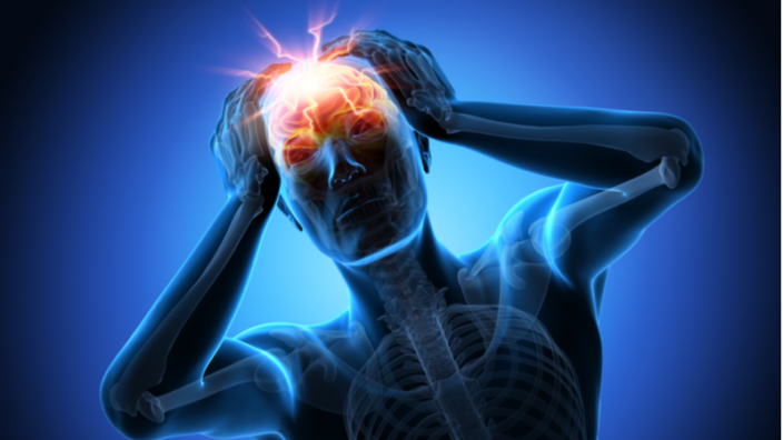
In the Journal of Inflammation, researchers from Johns Hopkins University have published a detailed review of the relationship between brain inflammation and the principal diseases of dementia.

A focus on genetics and environment

One out of twenty Americans over 85 have Parkinson’s disease [1], and seven out of twenty have Alzheimer’s [2]. While genes are obviously a factor in this prevalence, and environmental factors are another large factor, this review focuses on a combination of the two: gene-by-environment (GxE) interactions. This is the idea that the two are heavily intertwined; in other words, the way in which a biological process responds to its environment is dictated by its genes [3].

The researchers make it clear that it is impossible to reduce most instances of these neurodegenerative diseases down to specific environmental or genetic triggers; they can be associated with an increased likelihood of such diseases, such as the APOE4 allele, but there are only a handful of specific mutations that are known to consistently and directly result in neurodegeneration.

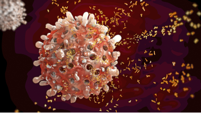
This review focuses on the GxE relationships involved in inflammation. This interaction between genes and the environment is very well known: immune cells, whose activation is often directly related to environmental insults such as bacteria, recognize pathogens and signs of damage and so begin the process of inflammation and later healing.

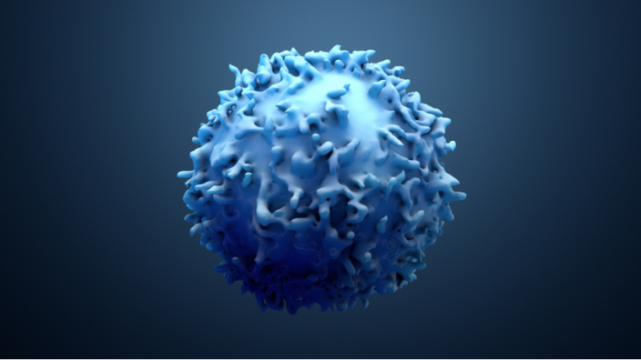
However, as the researchers note, the markers that are most commonly associated with tissue inflammation, such as redness and swelling, do not appear in the central nervous system, whether that inflammation is chronic or acute. Instead, the activation of the macrophages known as microglia send chemical messages to astrocytes, and the combination of these cells can send signals, some of which are neurotoxic and some of which are neuroprotective.

Microglia and Astrocytes

These environmental effects can also damage the genome, directly through mutation and indirectly through cellular stress, causing a wide variety of biological effects that become clear in -omics analysis [4]. Chronic inflammation is a well-known cause of other chronic ailments, to the point that this relationship can be found in well-studied encyclopedias [5]. As it is clear that the increase in neuroinflammation is associated with these diseases [6], the reviewers sought to determine precisely how this happens in two of the most well-known neurodegenerative disorders: Alzheimer’s and Parkinson’s.

Inflammation, genes, and Alzheimer’s

The researchers note that there are a few mutations to the amyloid precursor protein and presenilin genes that are very strongly associated with Alzheimer’s. However, people with these mutations are likely to get the disease before the age of 60, and these cases are fortunately very rare [7].

However, the APOE4 allele is rather common, and people with one copy of this variant have triple the Alzheimer’s risk of people without it; people with two copies have eight times the risk. The APOE2 allele confers some protection [8].

Other than these, there are more than 50 known genetic risk factors for Alzheimer’s disease, and 30 of them have been noted in more than one study [9]. While the exact relationship between all of these genes and the disease is hard to determine, neuroinflammation is a frequent contributor.

Inflammation, mediated by microglia, leads to tissue death and damage in Alzheimer’s, and while microglia can clear amyloid beta, doing so often causes more pro-inflammatory factors to be released, making the problem worse [10]. Reactive oxygen species (ROS) are also known to contribute to Alzheimer’s, and inflammation can lead to ROS in the brain, which can lead to more inflammation [11].

As expected, APOE4 is more associated with inflammation than other APOE allelles [12]; more neurotoxic factors were found in the plasma of people carrying it. An exact biochemical cause has been theorized: this particular form of inflammation has been reported to be driven by cholesterol, and the APOE4 mutation does not bind to cholesterol as well [13], thus leading to its accumulation in microglia. Mutations to TREM2, which is central to microglial function [14], and many other immunomodulatory genes have also been found to play roles in Alzheimer’s.

Inflammation, genes, and Parkinson’s

While Parkinson’s can run in families, only 15% of Parkinson’s patients have this sort of genetic transmission listed as their primary cause [15]. Often, the genetic variants are of the α-synuclein protein itself; if this protein is misfolded or overexpressed, the link to Parkinson’s is direct, without any immune system involvement.

However, with Parkinson’s later in life, the situation is similar to Alzheimer’s. A full 90 potential genetic factors have been found [16], and the researchers note that the specific neurons affected by Parkinson’s are uniquely vulnerable to inflammation; at least in mice, the area contains more microglia than anywhere else in the brain [17]. Under normal circumstances, the chemical neuromelanin is protective; however, as deteriorating neurons in the area release it into the extracellular environment, microglia treat it as a foreign chemical and release inflammatory markers when consuming it [18]; the iron in the neuromelanin also causes ROS through hydrogen peroxide [19].

The SNCA gene has been singled out as a potential cause. Mutations to this gene cause microglia to recognize the protein’s aggregates as foreign, activating them and causing further inflammation [20], just as amyloid beta causes microglial overactivation in Alzheimer’s. The LRRK2 gene was also identified, as it is associated with mitochondrial function and cytokine response.

Conclusion

Even though this review goes into exhaustive detail, finding every single combination of genetic predisposition and environmental insult is impossible. Rather, for any potential therapy to be developed and transmitted based on this research, it must modulate the effect of microglia and reduce inflammation while retaining these immune cells’ ability to protect the brain. Given the frequency and intensity of immune responses in these crippling and deadly diseases, it is not unreasonable to believe that a successful future combination therapy for Alzheimer’s or Parkinson’s will have an anti-inflammatory aspect.

We would like to ask you a small favor. We are a non-profit foundation, and unlike some other organizations, we have no shareholders and no products to sell you. All our news and educational content is free for everyone to read, but it does mean that we rely on the help of people like you. Every contribution, no matter if it’s big or small, supports independent journalism and sustains our future.

Literature

[1] Reeve, A., Simcox, E., & Turnbull, D. (2014). Ageing and Parkinson’s disease: why is advancing age the biggest risk factor?. Ageing research reviews, 14, 19-30.

[2] Alzheimer’s Association. (2019). 2019 Alzheimer’s disease facts and figures. Alzheimer’s & dementia, 15(3), 321-387.

[3] Hunter, D. J. (2005). Gene–environment interactions in human diseases. Nature reviews genetics, 6(4), 287-298.

[4] Hasin, Y., Seldin, M., & Lusis, A. (2017). Multi-omics approaches to disease. Genome biology, 18(1), 1-15.

[5] Fleit, H. B. (2014). Chronic inflammation.

[6] Sochocka, M., Diniz, B. S., & Leszek, J. (2017). Inflammatory response in the CNS: friend or foe?. Molecular neurobiology, 54(10), 8071-8089.

[7] Van Cauwenberghe, C., Van Broeckhoven, C., & Sleegers, K. (2016). The genetic landscape of Alzheimer disease: clinical implications and perspectives. Genetics in Medicine, 18(5), 421-430.

[8] Avramopoulos, D. (2009). Genetics of Alzheimer’s disease: recent advances. Genome medicine, 1(3), 1-7.

[9] Wightman, D. P., Jansen, I. E., Savage, J. E., Shadrin, A. A., Bahrami, S., Holland, D., … & Posthuma, D. (2021). A genome-wide association study with 1,126,563 individuals identifies new risk loci for Alzheimer’s disease. Nature genetics, 53(9), 1276-1282.

[10] Hickman, S. E., Allison, E. K., & El Khoury, J. (2008). Microglial dysfunction and defective β-amyloid clearance pathways in aging Alzheimer’s disease mice. Journal of Neuroscience, 28(33), 8354-8360.

[11] Popa-Wagner, A., Mitran, S., Sivanesan, S., Chang, E., & Buga, A. M. (2013). ROS and brain diseases: the good, the bad, and the ugly. Oxidative medicine and cellular longevity, 2013.

[12] Fan, Y. Y., Cai, Q. L., Gao, Z. Y., Lin, X., Huang, Q., Tang, W., & Liu, J. H. (2017). APOE ε4 allele elevates the expressions of inflammatory factors and promotes Alzheimer’s disease progression: a comparative study based on Han and She populations in the Wenzhou area. Brain research bulletin, 132, 39-43.

[13] Xu, Q., Brecht, W. J., Weisgraber, K. H., Mahley, R. W., & Huang, Y. (2004). Apolipoprotein E4 domain interaction occurs in living neuronal cells as determined by fluorescence resonance energy transfer. Journal of Biological Chemistry, 279(24), 25511-25516.

[14] Neumann, H., & Daly, M. J. (2013). Variant TREM2 as risk factor for Alzheimer’s disease. N Engl J Med, 368(2), 182-4.

[15] Tran, J., Anastacio, H., & Bardy, C. (2020). Genetic predispositions of Parkinson’s disease revealed in patient-derived brain cells. NPJ Parkinson’s disease, 6(1), 1-18.

[16] Nalls, M. A., Blauwendraat, C., Vallerga, C. L., Heilbron, K., Bandres-Ciga, S., Chang, D., … & Rizig, M. (2019). Identification of novel risk loci, causal insights, and heritable risk for Parkinson’s disease: a meta-analysis of genome-wide association studies. The Lancet Neurology, 18(12), 1091-1102.

[17] Lawson, L. J., Perry, V. H., Dri, P., & Gordon, S. (1990). Heterogeneity in the distribution and morphology of microglia in the normal adult mouse brain. Neuroscience, 39(1), 151-170.

[18] Hirsch, E. C., Vyas, S., & Hunot, S. (2012). Neuroinflammation in Parkinson’s disease. Parkinsonism & related disorders, 18, S210-S212.

[19] Zecca, L., Casella, L., Albertini, A., Bellei, C., Zucca, F. A., Engelen, M., … & Sarna, T. (2008). Neuromelanin can protect against iron‐mediated oxidative damage in system modeling iron overload of brain aging and Parkinson’s disease. Journal of neurochemistry, 106(4), 1866-1875.

[20] Hoenen, C., Gustin, A., Birck, C., Kirchmeyer, M., Beaume, N., Felten, P., … & Heurtaux, T. (2016). Alpha-synuclein proteins promote pro-inflammatory cascades in microglia: stronger effects of the A53T mutant. PloS one, 11(9), e0162717.

Multivitamins

Multivitamin Supplementation Shown to Improve Cognition

In a large randomized, placebo-controlled study, scientists hoped that cocoa extract would improve cognitive function in elderly patients, but instead it was a multivitamin supplement that shined [1].

A two-in-one study

Multivitamins are among the most popular dietary supplements, but their effectiveness is often questioned. While some studies did fail to find any significant benefits of multivitamin supplementation [2], it would be safe to say that the jury is still out.

This new study stands out in its size and robustness: it was a proper randomized, placebo-controlled trial rather than a population study, it lasted three years, and it encompassed more than 2,000 participants. However, the study wasn’t even about multivitamins in the first place. As their primary endpoint, the researchers wanted to investigate the effects of cocoa extract on cognitive function in elderly patients, while the effects of multivitamin supplementation were a secondary endpoint.

Cocoa extract contains a lot of catechins and epicatechins, which are flavonols: phytochemicals that are thought to possess many health benefits. It has been hypothesized that cocoa may help improve cognition by increasing blood flow and the production of new blood vessels (angiogenesis) [3], but the previous evidence was shaky, consisting of only a few population studies and small trials with somewhat contradictory results.

The underdog wins

This study, COcoa Supplement and Multivitamin Outcomes Study of the Mind (COSMOS-Mind) was ancillary to a larger study that studied the effects of cocoa extract and multivitamins on cardiovascular and cancer outcomes. That mother study showed that multivitamin supplementation significantly reduced mortality from lung cancer [4], and cocoa extract reduced mortality from cardiovascular disease [5].

The cocoa extract used in the study was produced by Mars Corporation, which is apparently trying to jump on the healthy lifestyle bandwagon. The participants had received 500 mg of cocoa-derived flavanols per day. This is equivalent to 300 grams of dark chocolate, a dose large enough to offset any health benefits from flavanols, so people who wish to enjoy those benefits should probably stick to cocoa extract.

In both the cocoa extract and multivitamin study arms, there were one study group and one placebo group. There was also a multivitamin plus cocoa extract group, in case the two products showed synergy (they did not).

Cognitive function was assessed yearly by a 50-minute-long phone interview, during which the participants were subjected to a battery of cognitive tests, and then a composite cognition score was computed. The bottom line is that this score did not significantly change vs placebo in the cocoa extract group, but it did in the multivitamin group. However, two caveats apply. First, the placebo effect was quite substantial. Second, both in the placebo groups and in drug groups, the effects peaked at two years.

cocoa and multivitamins 1

Stronger effects in people with cardiovascular problems

People with a history of cardiovascular disease (CVD) benefited from the multivitamin (but not from the cocoa extract) even more. Interestingly, in this subgroup, the placebo effect was much weaker and significantly declined after one year. In people with no CVD history, the effect of multivitamin supplementation was smaller but still statistically significant. It should be noted that people with CVD histories were more likely to be male; were generally older; had a higher BMI; had more hypertension, statin use, and depression; and were less physically active. They also scored worse on cognitive tests at baseline.

cocoa and multivitamins 2

The researchers calculated that by the end of the study, the participants who received multivitamins were on average 1.8 years cognitively “younger” than participants in the placebo group. In other words, multivitamin supplementation slowed cognitive aging by 60% and even more for people with histories of CVD. The researchers hypothesize that being a complex process, cognitive decline might be less affected by the levels of any single nutrient, such as flavanols, and more by the general balance of essential micronutrients in the body, something that multivitamin supplementation should provide, at least in theory.

Conclusion

This randomized, placebo-controlled, long-term study challenges two common opinions: that taking multivitamins is useless and that cacao products are good for your brain. This does not mean that the debate is settled, however: some previous studies beg to differ, and this one has its share of limitations. However, it might be a good idea to keep taking multivitamins just in case: there’s probably no harm in that, and future research might confirm their benefits.

We would like to ask you a small favor. We are a non-profit foundation, and unlike some other organizations, we have no shareholders and no products to sell you. All our news and educational content is free for everyone to read, but it does mean that we rely on the help of people like you. Every contribution, no matter if it’s big or small, supports independent journalism and sustains our future.

Literature

[1] Baker LD, Manson JE, Rapp SR, et al. Effects of cocoa extract and a multivitamin on cognitive function: A randomized clinical trial. Alzheimer’s Dement. 2022;1-12.

[2] Paranjpe, M. D., Chin, A. C., Paranjpe, I., Reid, N. J., Duy, P. Q., Wang, J. K., … & Nadkarni, G. N. (2020). Self-reported health without clinically measurable benefits among adult users of multivitamin and multimineral supplements: a cross-sectional study. BMJ open, 10(11), e039119.

[3] Barrera-Reyes, P. K., de Lara, J. C. F., González-Soto, M., & Tejero, M. E. (2020). Effects of cocoa-derived polyphenols on cognitive function in humans. Systematic review and analysis of methodological aspects. Plant Foods for Human Nutrition, 75(1), 1-11.

[4] Sesso, H. D., Rist, P. M., Aragaki, A. K., Rautiainen, S., Johnson, L. G., Friedenberg, G., … & Manson, J. E. (2022). Multivitamins in the prevention of cancer and cardiovascular disease: the COcoa Supplement and Multivitamin Outcomes Study (COSMOS) randomized clinical trial. The American Journal of Clinical Nutrition, 115(6), 1501-1510.

[5] Sesso, H. D., Rist, P. M., Aragaki, A. K., Rautiainen, S., Johnson, L. G., Friedenberg, G., … & Manson, J. E. (2022). Multivitamins in the prevention of cancer and cardiovascular disease: the COcoa Supplement and Multivitamin Outcomes Study (COSMOS) randomized clinical trial. The American Journal of Clinical Nutrition, 115(6), 1501-1510.

Dividing cell

Stem Cell Activity Regulated By Mitochondrial Protein

Researchers publishing in Cell Stem Cell have found that the mitochondrial protein OPA1 is an integral, regulatory part of muscle stem cell activation, the process by which muscle stem cells proliferate and become active muscle tissue (myogenesis) [1].

Another piece of the puzzle

We have recently reported on the effects of mitochondrial fusion and fission on muscle stem cell proliferation. This paper looks at the same issue in a different way, focusing on a different protein and a different link in the chain.

Here, the researchers begin with a discussion of the quiescent state of stem cells. Stem cells are not always active; in order to preserve function and longevity, they stay quiescent, saving energy and preserving longevity until they are called upon to divide [2]. The ability of these cells to wake up from this state, divide rapidly, and replenish themselves is central to maintenance and regeneration after injuries [3].

Progressing through the stages with OPA1

Muscle stem cells can be categorized into four states: quiescent, activated (measured by Pax7), committed (measured by MyoD), and differentiating (measured by MyoD and MyoG). During activation, mitochondria quickly fragment, substantially decreasinglength and volume; in the later stages, they regain their size.

However, this only applies in the presence of OPA1. Mice engineered not to express the OPA1 protein have much smaller mitochondria in their muscle stem cells than their wild-type counterparts, and these mitochondria do not follow the normal curve. While their Pax7+ stem cell quantity, before injury, was only slightly less than that of wild-type mice, the wild-type mice greatly increased their Pax7+ stem cell numbers 21 days after injury while the OPA1-knockout mice sustained a decrease, showing enormously impaired muscle regeneration.

These cells’ gene expression was found to match their behavior; genes related to the cellular cycle, myogenesis, proliferation, and chromosomal division were strongly, and seemingly directly, affected by OPA1.

Interestingly, these mice also suffered a loss of Pax7 cells in uninjured limbs as well. Four days after an injury in one limb, the OPA1-knockout mice had far more MyoD and MyoG cells in an uninjured limb than wild-type mice did, showing the propensity of these cells to commit to differentiation without being able to replenish themselves. The researchers hold that these cells, before injury, are behaving in a G-alert state halfway between deep quiescence (G0) and activation (G1).

The researchers also found the inverse to be true: during activation (two days after injury), normal, wild-type cells express much less OPA1 than their fully quiescent counterparts.

The role of mitochondrial division

The researchers then tested to see if directly interfering with mitochondrial division would affect stem cell fate. Dosing cells with the mitochondrial fission inhibitor Mdivi-1 steered those cells towards quiescence and self-renewal. Interestingly, this approach seemed to neutralize the OPA1 knockout in multiple respects, including reducing MyoD and MyoG, bringing cells of this type more in line with their wild-type counterparts.

This paper also identified the molecules involved in the natural mitochondrial shift upon injury. Specifically, hepatocyte growth factor activator (HGFA) is released after an injury, stimulating cells along a pathway that uses mTORC1 to induce the G-alert state. As would be expected, introducing active HGF or HGFA promotes entry into this cell cycle, while rapamycin, which impedes mTORC1, discourages it.

Redox signaling was also found to be involved. OPA1-knockout muscle stem cells had slightly but significantly higher reactive oxygen species (ROS) than controls, along with reduced regulation. Glutathione, which has already been shown to be linked to OPA1 [4], was upregulated in the OPA1-knockout cells. The anti-ROS compound mitoTEMPO caused the stem cells of OPA1-knockout mice to behave more like their wild-type counterparts.

A connection to aging

The researchers also took a look at the gene expression of aged mice, finding that OPA1-related genes, glutathione-related genes, and genes governing mitochondrial function were downregulated with aging. Mice that were engineered to overexpress OPA1 were, at older ages, more inclined to retain stem cell quiescence and replenishment than wild-type mice, suggesting that a gradual loss of OPA1 plays a role in age-related muscle decline.

Conclusion

This in-depth research shows a complicated, intertwined relationship between multiple aspects of mitochondrial function and stem cell activity. It also shows the impact of the mTORC1 pathway and its influence on this aspect of aging. While it is far too early to determine if directly targeting OPA1 can be effective as a therapy in human beings, or if rapamycin and/or rapalogs are a more effective solution, this research is a fundamental stepping stone to research in that direction.

We would like to ask you a small favor. We are a non-profit foundation, and unlike some other organizations, we have no shareholders and no products to sell you. All our news and educational content is free for everyone to read, but it does mean that we rely on the help of people like you. Every contribution, no matter if it’s big or small, supports independent journalism and sustains our future.

Literature

[1] Baker, N., Wade, S., Triolo, M., Girgis, J., Chwastek, D., Larrigan, S., … & Khacho, M. (2022). The mitochondrial protein OPA1 regulates the quiescent state of adult muscle stem cells. Cell Stem Cell.

[2] Cho, I. J., Lui, P. P., Obajdin, J., Riccio, F., Stroukov, W., Willis, T. L., … & Watt, F. M. (2019). Mechanisms, hallmarks, and implications of stem cell quiescence. Stem cell reports, 12(6), 1190-1200.

[3] van Velthoven, C. T., & Rando, T. A. (2019). Stem cell quiescence: dynamism, restraint, and cellular idling. Cell stem cell, 24(2), 213-225.

[4] Patten, D. A., McGuirk, S., Anilkumar, U., Antoun, G., Gandhi, K., Parmar, G., … & Harper, M. E. (2021). Altered mitochondrial fusion drives defensive glutathione synthesis in cells able to switch to glycolytic ATP production. Biochimica et Biophysica Acta (BBA)-Molecular Cell Research, 1868(1), 118854.

Duchenne

Removing Senescent Cells to Alleviate Muscular Dystrophy

A study published in Aging has shown that the removal of senescent cells through senolytics alleviates muscle degeneration in a mouse model of Duchenne muscular dystrophy.

A genetic disease with hallmarks of aging

Duchenne muscular dystrophy is caused by a mutation that gradually destroys the muscles, eventually becoming fatal as it destroys the diaphragm or heart. This is an X-linked genetic disease, so females can carry one copy of it without normally suffering from negative physiological effects; sufferers are almost always male. Previous research on a mouse model of severe Duchenne has found that this disease is also linked to RhoA signaling in senescent macrophages near the dystrophic muscles, triggering the senescence-associated secretory phenotype (SASP) and potentially making the problem worse [1].

The researchers also note that this is connected to normal aging: previous research has found that senescent immune cells contribute to other aspects of aging, including senescent cells in other organs [2].

However, a causal link between senescence and muscular dystrophy in this genetic disease had not previously been determined, and it was not clear if removing these senescent cells would offer any benefit against Duchenne. This research was conducted to fill that gap, testing if the well-known natural senolytic fisetin could improve the muscles of these mice.

Stem cells and senescent cells

Compared to their wild-type counterparts, the muscles of 8-week-old mice of this model have considerably less Pax7, a marker of muscle stem cells. Instead, they have much greater amounts of the senescence markers CD68 and SA-β-gal, which is nearly absent in wild-type mice of this age. CD68 and mTORC1 signaling, which is also a regulator of senescence, are also heavily present in the macrophages that reside in this skeletal muscle. As expected from senescent macrophages, the SASP was also present, including a variety of well-known inflammatory factors.

Culturing senescent macrophages next to healthy muscle stem cells was found to deplete the function of the stem cells, reducing their ability to form muscle somatic cells properly.

Fisetin to the rescue

Introducing fisetin into this cellular culture showed positive results. Fisetin effectively removed senescent macrophages, substantially decreasing the amount of CD68 and SA-β-gal in the cultures. The stem cells that had been negatively affected by these senescent macrophages recovered, and they were once again able to perform as normal.

Finally, the researchers tested the effects of fisetin on the Duchenne mouse model. Compared to the Duchenne mice in the control group, the treated mice had less fibrosis, substantially reduced biomarkers of senescence, and much more active stem cells. While these biomarkers were not improved to the levels of wild-type mice, they showed a marked improvement.

Conclusion

There were several limitations to this study. Like mouse models of Alzheimer’s, the mouse model of Duchenne may not perfectly mimic the actual disease. Murine, not human, cells were used for the cellular cultures, and senolytics that have shown enormous promise in mice are not always particularly effective in human beings. The physical performance of the mice was also not measured.

However, these results are notable from both an anti-aging perspective and the perspective of people interested in treating this particular genetic disease. If senescent macrophages are causing such harm in this case, they may also be doing the same in older people as well. Therefore, if these results hold true in human beings, strategies to destroy or convert senescent cells may be effective as a treatment against the age-related muscle degeneration known as sarcopenia.

We would like to ask you a small favor. We are a non-profit foundation, and unlike some other organizations, we have no shareholders and no products to sell you. All our news and educational content is free for everyone to read, but it does mean that we rely on the help of people like you. Every contribution, no matter if it’s big or small, supports independent journalism and sustains our future.
Literature

[1] Mu, X., Lin, C. Y., Hambright, W. S., Tang, Y., Ravuri, S., Lu, A., … & Huard, J. (2020). Aberrant RhoA activation in macrophages increases senescence-associated secretory phenotypes and ectopic calcification in muscular dystrophic mice. Aging (Albany NY), 12(24), 24853.

[2] Yousefzadeh, M. J., Flores, R. R., Zhu, Y. I., Schmiechen, Z. C., Brooks, R. W., Trussoni, C. E., … & Niedernhofer, L. J. (2021). An aged immune system drives senescence and ageing of solid organs. Nature, 594(7861), 100-105.

Brainwaves

Neuromodulation Improves Memory in Older Adults

A new study published in Nature Neuroscience has shown that neuromodulation of low-frequency neuronal activity in the parietal cortex improved working memory, while high-frequency modulation in the prefrontal cortex improved long-term memory in older adults [1].

As people age, their cognitive abilities get worse. This includes memory decline, which might dramatically impact quality of life. Although age-associated memory decline varies from person to person, in some people, it might lead to being unable to perform well at work or deal with daily tasks. As accelerated memory decline might also be indicative of dementia, it is critical to develop technologies for memory enhancement in the elderly.

Manipulating brain electricity

Neurons are electrically active cells. The electrical currents generated by multiple neurons can be measured by electrodes placed on the scalp using a non-invasive technique called electroencephalography (EEG). EEGs have long been used to assess mental states and diagnose various conditions such as epilepsy.

EEGs can detect distinct brain activity patterns that correspond to various mental states. These rhythmic neural activities are recorded with EEGs at different oscillation bands (frequencies): delta (≤ 3 Hz, seen in deep sleep), theta (3.5 – 7.5 Hz, seen in drowsy adults), alpha (7.5 – 13 Hz, seen in awake adults), beta (13 – 31 Hz, most frequently seen range in adults and children), and gamma (≥ 30 Hz, detected in a state of active information processing).

High-definition transcranial alternating current stimulation (HD-tACS) is the inverse of EEG: instead of recording, the brain activity is stimulated at a specific frequency using electrodes placed on the scalp.

Previous research has shown that working memory and long-term memory are stored differently, with distinct anatomical regions of the brain involved. It was also suggested that by modulating brain activity in specific locations at specific frequencies, it would be possible to selectively improve either working or long-term memory.

In this study, the researchers explored if HD-tACS modulation of low-frequency (theta rhythm) in the inferior parietal lobule and high frequency (gamma rhythm) in the dorsolateral prefrontal cortex could enhance working and long-term memory functions, respectively, in adults over 65 years of age.

Location and frequency matter

The study consisted of three experiments. The first two were randomized, double-blind studies consisting of three groups. The third experiment was conducted to test the reproducibility of the findings from the first two.

In the first experiment, the participants were assigned to the gamma prefrontal cortex neuromodulation group (group 1), the theta parietal lobule neuromodulation group (group 2), or a control group with no stimulation. In the second experiment, the experimental groups were switched, and the researchers performed gamma parietal lobule neuromodulation and theta prefrontal cortex neuromodulation.

The participants underwent neurostimulation using a BrainCap for 20 minutes every day over a four-day period. They were then asked to recall as many words as they could remember within two minutes from the 100 words that had been read to them previously. The task was repeated on each day of the experiment and a month later.

The results showed improved working and long-term memory only in the first experiment, suggesting that it’s crucial to stimulate specific regions of the brain at specific frequencies to enhance specific memory types.

In the third experiment, the neuromodulation was done for three consecutive days in an independent cohort of participants, and only experimental groups were included. Unlike the first two experiments, there was no long-term follow-up, but the overall results replicated the primary findings.

Individual responses

Not all the participants responded the same to the neuromodulation. During the first experiment, 85% of group 1 and 90% of group 2 showed memory improvement over the four days of neurostimulation. Moreover, how well a person’s memory improved in response to the neurostimulation predicted that person’s future memory enhancement one month after treatment. Therefore, personalization of the neuromodulation protocol is required to achieve the best results in each individual.

Abstract

The development of technologies to protect or enhance memory in older people is an enduring goal of translational medicine. Here we describe repetitive (4-day) transcranial alternating current stimulation (tACS) protocols for the selective, sustainable enhancement of auditory–verbal working memory and long-term memory in 65–88-year-old people. Modulation of synchronous low-frequency, but not high-frequency, activity in parietal cortex preferentially improved working memory on day 3 and day 4 and 1 month after intervention, whereas modulation of synchronous high-frequency, but not low-frequency, activity in prefrontal cortex preferentially improved long-term memory on days 2–4 and 1 month after intervention. The rate of memory improvements over 4 days predicted the size of memory benefits 1 month later. Individuals with lower baseline cognitive function experienced larger, more enduring memory improvements. Our findings demonstrate that the plasticity of the aging brain can be selectively and sustainably exploited using repetitive and highly focalized neuromodulation grounded in spatiospectral parameters of memory-specific cortical circuitry.

Conclusion

This study presents very promising results of non-invasive neurostimulation to improve both working and long-term memory in older adults. While showing the importance of using specific frequencies to improve different memory functions in different brain regions, it also demonstrates individual differences in response to neurostimulation. It is unclear if the positive effects achieved in this study would last longer than one month. Hopefully, future studies will clarify how long-lasting the effect of such neurostimulation is and how to develop personalized protocols for people to get the most out of this treatment.

We would like to ask you a small favor. We are a non-profit foundation, and unlike some other organizations, we have no shareholders and no products to sell you. All our news and educational content is free for everyone to read, but it does mean that we rely on the help of people like you. Every contribution, no matter if it’s big or small, supports independent journalism and sustains our future.

Literature

[1] Grover, S., Wen, W., Viswanathan, V. et al. Long-lasting, dissociable improvements in working memory and long-term memory in older adults with repetitive neuromodulation. Nat Neurosci 25, 1237–1246 (2022).

Lung cancer

Injectable Cytokine Factories Eradicate Cancer in Mice

Researchers have succeeded in beating a type of cancer previously considered incurable by using microcapsules that contained live cytokine-producing cells [1].

IL-2 is potent but toxic

Interleukin-2 (IL-2) therapy was the first anti-cancer immunotherapy approved decades ago. It is based on the ability of this inflammatory cytokine to ramp up the body’s immune response to cancer, mostly by increasing the proliferation of cytotoxic T cells [2].

However, systemic administration of IL-2 causes severe toxicity issues: for instance, it can trigger the dangerous overproduction of cytokines known as the cytokine storm, which can also be caused by viruses such as COVID-19. IL-2 also has a very short half-life, so it requires continuous intravenous administration in large doses.

Dozens of research group and biotech companies have been devising ingenious ways to overcome the limitations of IL-2 therapy. Notably, the start-up Neoleukin has introduced an engineered version of IL-2, “the world’s first computationally designed de novo protein therapeutic.” Yet, as of now, none of those attempts has resulted in a clinically approved therapy.

90% reduction in tumor burden

In this new study, the researchers used an intriguing new approach that involves injectable “cytokine factories”: tiny balls of gel that encapsulate retinal pigmented epithelial cells (RPEs). The cells were genetically modified using the PiggyBac transposone system to produce lots of IL-2. After the injection, the cells remain viable in the body for a long time, churning out the cytokine. Since this is happening in the vicinity of the tumor, toxicity issues are controlled, at least in theory.

The same group of researchers, publishing in Science in March this year, demonstrated the effectiveness of this approach against mouse models of ovarian and colorectal cancer [3]. This time, they employed their “cytokine factories” against mesothelioma, a rare but extremely aggressive and deadly cancer that affects the lungs, abdomen, or heart and is usually caused by asbestos exposure. Mesothelioma is considered incurable, except in very rare cases; the best available therapies, such as a PD-1 immune checkpoint blockade, usually give patients a mere few additional months of life.

However, the “cytokine factories” proved extremely effective in a mouse model of mesothelioma, with all mice in the high-dose group showing at least a 90% reduction in tumor burden after one week of treatment. Even at the lowest dose, most mice experienced at least a 45% decrease in tumor burden. Analysis of immune cell population showed that the treatment indeed induced its effect by increasing the proliferation of cytotoxic (CD8+) T cells.

Even better in combination

The results were even more impressive when the IL-2 therapy was combined with a PD-1 checkpoint blockade. In all seven mice that received the combination, tumors were eradicated after one week of treatment and did not recur, at least until the end of the study.

A subset of those animal participated in a rechallenge experiment. Two months after they had healed, those mice were again injected with mesothelioma cells. All previously treated mice did not develop new tumors, showing that the treatment induces “immunological memory”.

Low toxicity

Obviously, toxicity was a major concern, but it proved unfounded: the “cytokine factories” were well tolerated by the immune system and did not induce a significant immune response. The concentration of IL-2 at the tumor loci was more than 100 times higher than the systemic concentration, which was close to normal values. Intravenous administration of IL-2 usually takes a toll on several organs, including the kidneys, liver and lungs, but histologic analysis in this study did not reveal any pathologies.

Conclusion

Among dozens of attempts to harness the true anti-cancer potential of IL-2, this one stands out because of its unusual mode of delivery that seems to check all the boxes. The treatment showed stunning effectiveness against one of the most aggressive types of cancer that is still considered incurable in humans, and it did not cause any of the major toxicity issues that IL-2 is known for. The researchers are sure that their discovery has a great translation potential and plan to initiate a Phase 1 human clinical trial in the following months.

We would like to ask you a small favor. We are a non-profit foundation, and unlike some other organizations, we have no shareholders and no products to sell you. All our news and educational content is free for everyone to read, but it does mean that we rely on the help of people like you. Every contribution, no matter if it’s big or small, supports independent journalism and sustains our future.

Literature

[1] Nash, A. M., Aghlara-Fotovat, S., Castillio, B., Hernandez, A., Pugazenthi, A., Lee, H. S., … & Veiseh, O. (2022). Activation of adaptive and innate immune cells via localized Interleukin-2 cytokine factories eradicates mesothelioma tumors. Clinical Cancer Research: an Official Journal of the American Association for Cancer Research, CCR-22.

[2] Jiang, T., Zhou, C., & Ren, S. (2016). Role of IL-2 in cancer immunotherapy. Oncoimmunology, 5(6), e1163462.

[3] Nash, A. M., Jarvis, M. I., Aghlara-Fotovat, S., Mukherjee, S., Hernandez, A., Hecht, A. D., … & Veiseh, O. (2022). Clinically translatable cytokine delivery platform for eradication of intraperitoneal tumors. Science advances, 8(9), eabm1032.

Varied diet

Ketogenic and Mediterranean Diets Effective Against Diabetes

Pitting a ketogenic diet against a Mediterranean diet in pre-diabetes and Type 2 diabetes patients, scientists have learned that both confer metabolic benefits, but “keto” also has a darker side [1].

Diabetes is often reversible with diet

While many anti-diabetes drugs have been developed in recent decades, with some being repurposed as anti-aging drugs [2], there is a growing understanding that Type 2 diabetes can be prevented and even reversed with the help of a healthy diet [3].

However, there is an ongoing battle between diets, such the ketogenic diet and the Mediterranean diet. The latter is less restrictive, more balanced, and arguably has greater scientific evidence behind it [4], while the former touts inducing the state of ketosis, the metabolic switch from carbohydrates to fat as the body’s main energy source, as its main advantage.

“Keto” is thought to be quite effective when it comes to weight loss, but since it derives most calories from animal protein and fat (although some rather convoluted vegan variants exist), it is suspected of raising cholesterol levels, including LDL, a form of cholesterol with largely harmful effects.

Keto vs Med

In this new study, the researchers directly pitted those two diets against each other in people with pre-diabetes and Type 2 diabetes. The study consisted of two arms, numbering 16 and 17 participants, with a median age of 60 years and a median BMI of 30. Both arms started with one diet, either the ketogenic or the Mediterranean, and switched to the other diet after 12 weeks for another 12 weeks. At the end of the study, there were 12 additional weeks of follow-up.

With diets, the devil is in the details; for instance, foods packed with empty calories, such as French fries, can be part of a vegan diet. To account for this, the researchers tried to make the two diets as healthy as possible. They used a well-formulated ketogenic diet (WFKD) and encouraged the participants on the Mediterranean diet to enhance it by completely avoiding refined sugars and grains (Med-Plus).

According to the researchers, both diets incorporate three key nutrition principles endorsed by diabetes organizations worldwide: including non-starchy vegetables, restricting added sugars, and limiting refined grains. The main differences between the two involve legumes, fruits, and whole grains, which are avoided in the ketogenic diet but are more than welcome in the Mediterranean diet.

During the keto phase, participants had to sustain ketosis by limiting carbohydrates to 20–50 grams per day and keeping protein consumption at about five grams per one kilogram of body weight per day, with all other calories coming from fats. During the Mediterranean phase, participants followed a mostly plant-based diet consisting of vegetables (including starchy vegetables), legumes, fruits, whole grains, nuts, and seeds, with fish as the primary source of animal protein and olive oil as the primary source of fat. In both phases, the participants were strongly advised against consuming any processed foods.

Participants were also advised to eat ad libitum, that is, without limiting calorie intake. Despite that, on average, participants on both diets lost weight. The weight loss was stronger with the ketogenic diet.

Med vs Keto

The pros and the cons

The researchers hypothesized that after 12 weeks on each diet, glycated hemoglobin (HbA1c), a standard measure of blood sugar, would similarly improve from baseline due to shared dietary characteristics but that the keto diet would increase some health risks due to the lack of legumes, fruits, and whole grains.

The results matched the expectations. Both diets led to similar decreases in HbA1c levels, even though participants on WFKD had been consuming 50% fewer carbohydrates compared to participants on Med-Plus. However, the ketogenic diet led to a more significant decrease in triglyceride levels (16% vs 5% for the Mediterranean diet).

The ketogenic diet also resulted in a substantial increase in LDL cholesterol (+10% vs -5% for the Mediterranean diet). While some resent research suggests that not all LDL cholesterol is equally bad [5], the researchers interpreted the rise in LDL cholesterol as a health risk.

The nutrient balance was generally worse under the ketogenic diet. It led to greater decreases in essential nutrients than the Mediterranean diet, including in fiber, folate, vitamin C, and magnesium, which the researchers attribute to the exclusion of legumes, fruits, and whole grains.

There were no substantial differences in adherence to and in satisfaction from both diets during the study. Interestingly, participants tended to give higher marks to whatever diet they had started with. However, during the follow-up period, when all restrictions were off, the participants’ diet bore a greater resemblance to Med-Plus than to WFKD, suggesting that the former might be easier to adhere to.

Conclusion

The results of this comparative  study are generally in line with previous research. Both diets produced considerable metabolic benefits for people with pre-diabetes and Type 2 diabetes. However, there were obvious trade-offs: the ketogenic diet was more effective in decreasing weight and triglyceride levels, but it also raised LDL cholesterol and restricted essential nutrients such as fiber. Larger studies  are probably needed to supply more definitive answers.

We would like to ask you a small favor. We are a non-profit foundation, and unlike some other organizations, we have no shareholders and no products to sell you. All our news and educational content is free for everyone to read, but it does mean that we rely on the help of people like you. Every contribution, no matter if it’s big or small, supports independent journalism and sustains our future.

Literature

[1] Gardner, C. D., Landry, M. J., Perelman, D., Petlura, C., Durand, L. R., Aronica, L., … & Kim, S. H. (2022). Effect of a Ketogenic Diet versus Mediterranean Diet on HbA1c in Individuals with Prediabetes and Type 2 Diabetes Mellitus: the Interventional Keto-Med Randomized Crossover Trial. The American Journal of Clinical Nutrition.

[2] Harrison, D. E., Strong, R., Alavez, S., Astle, C. M., DiGiovanni, J., Fernandez, E., … & Miller, R. A. (2019). Acarbose improves health and lifespan in aging HET3 mice. Aging Cell, 18(2), e12898.

[3] Taheri, S., Zaghloul, H., Chagoury, O., Elhadad, S., Ahmed, S. H., El Khatib, N., … & Abou-Samra, A. B. (2020). Effect of intensive lifestyle intervention on bodyweight and glycaemia in early type 2 diabetes (DIADEM-I): an open-label, parallel-group, randomised controlled trial. The lancet Diabetes & endocrinology, 8(6), 477-489.

[4] Martínez-González, M. A., Gea, A., & Ruiz-Canela, M. (2019). The Mediterranean diet and cardiovascular health: A critical review. Circulation research, 124(5), 779-798.

[5] Ivanova, E. A., Myasoedova, V. A., Melnichenko, A. A., Grechko, A. V., & Orekhov, A. N. (2017). Small dense low-density lipoprotein as biomarker for atherosclerotic diseases. Oxidative medicine and cellular longevity, 2017.

Cancer cell

Lactate Inhibits Tumor Growth in Mice

Publishing in Nature, scientists have found that sodium lactate boosts the T cell-mediated anti-cancer immune response, leading to significant inhibition of tumor growth [1].

A double-edged sword

As a means of energy production, the Krebs cycle is preferred by most cells in most conditions. However, there is also a seemingly less efficient and more ancient method: anaerobic glycolysis. While it produces much less energy per one glucose molecule, it is fast and does not require oxygen, which is why it is turned on in muscles when we are working out. This leads to accumulation of lactic acid, a waste product and the cause of the familiar post-exercise muscle pain.

However, most cancer cells also prefer getting their energy via glycolysis even when oxygen is abundant. This is called aerobic glycolysis or the Warburg effect [2]. The reasons for this paradox are still poorly understood, but scientists suspect that, beside energy, aerobic glycolysis produces metabolites that enhance the tumor’s defenses. For example, lactic acid production leads to accumulation of lactate (lactic acid’s conjugate base) and an increase in the acidity of the tumor microenvironment – something that immune cells very much dislike [3].

However, a novel hypothesis that the researchers set out to test postulates that lactate by itself has a positive effect on anti-tumor immunity, which is simply overshadowed by the acidity. Interestingly, one earlier study found that lactate mediates immune protection induced by exercise [4].

Great by itself, greater in combination

The researchers subjected mice with M38 colon cancer tumors to subcutaneous administration of sodium lactate or glucose as control. Sodium lactate, the sodium salt of lactic acid, is used in the food industry, cosmetics, and in medicine to treat metabolic acidosis and some arrhythmias.

Amazingly, sodium lactate proved to be highly effective in suppressing tumor growth. However, it did not work in Rag1-knockout mice, which are unable to produce B or T cells. Of various types of immune cells, only depletion of CD8+ (cytotoxic) T cells abolished the effect of the sodium lactate treatment, pointing at those cells as the sole mediators of sodium lactate’s tumor-suppressing effects.

The researchers then wanted to see how sodium lactate stacks up against two existing drugs that enhance anti-tumor response by T cells – a PD1 checkpoint inhibitor and the PC7A nanovaccine [5] – and whether it would work synergistically with any of them. In the experiments with three different cancer types, both the PD1 inhibitor and the vaccine showed some anti-tumor effectiveness, although it was a far cry from that of sodium lactate. However, combining any of the drugs with sodium lactate indeed produced remarkable synergy, leading to an even more pronounced inhibition of tumor growth. As a result, half of the combination-treated mice survived and lived on cancer-free, while all others died within 40-50 days.

Lactate 1

Increased T cell stemness

Further analysis showed that sodium lactate facilitates infiltration of the tumor by T cells and significantly improves their function. The treatment also decreased the number of T cells in the tumor that had died by apoptosis.

The researchers also found that sodium lactate enhances the function and survivability of cytotoxic T cells by increasing their stemness. Stem-like T cells express the transcription factor TCF1 and are characterized by increased self-renewal, multipotency, and persistence. As a result, these cells do most of the heavy lifting in destroying the tumor.

Pre-treatment in vivo works too

In their next experiment, the researchers treated donor-derived human T cells with sodium lactate in vitro. Just like in vivo, the treatment increased the cells’ stemness by upregulating TCF1 and several other stemness-related proteins. The treatment also decreased the percentage of apoptotic cells.

Finally, the researchers re-introduced mouse T cells treated with sodium lactate into tumor-bearing mice, which produced spectacular dose-dependent results. While 500 thousand pre-treated T cells was enough to significantly impede tumor growth, 2 million cells actually reverted it:

Lactate 2

Conclusion

Lactate may seem an unlikely anti-cancer drug candidate, since lactic acid actually helps tumors evade immune defenses. However, this intriguing study shows that when taken out of the context of tumor acidity, sodium lactate can significantly impede, and even revert, tumor growth, especially when combined with some existing anti-cancer medications – a new paradigm that probably deserves additional research. One of its obvious advantages is the ability to induce the stem-like phenotype in T cells in vitro. The researchers suggest that sodium lactate can be used that way to increase the effectiveness of CAR-T cell therapy.

We would like to ask you a small favor. We are a non-profit foundation, and unlike some other organizations, we have no shareholders and no products to sell you. All our news and educational content is free for everyone to read, but it does mean that we rely on the help of people like you. Every contribution, no matter if it’s big or small, supports independent journalism and sustains our future.

Literature

[1] Feng, Q., Liu, Z., Yu, X. et al. Lactate increases stemness of CD8 + T cells to augment anti-tumor immunity. Nat Commun 13, 4981 (2022).

[2] Liberti, M. V., & Locasale, J. W. (2016). The Warburg effect: how does it benefit cancer cells?. Trends in biochemical sciences, 41(3), 211-218.

[3] Bellone, M., Calcinotto, A., Filipazzi, P., De Milito, A., Fais, S., & Rivoltini, L. (2013). The acidity of the tumor microenvironment is a mechanism of immune escape that can be overcome by proton pump inhibitors. Oncoimmunology, 2(1), e22058.

[4] Rundqvist, H., Veliça, P., Barbieri, L., Gameiro, P. A., Bargiela, D., Gojkovic, M., … & Johnson, R. S. (2020). Cytotoxic T-cells mediate exercise-induced reductions in tumor growth. Elife, 9, e59996.

[5] Luo, M., Wang, H., Wang, Z., Cai, H., Lu, Z., Li, Y., … & Gao, J. (2017). A STING-activating nanovaccine for cancer immunotherapy. Nature nanotechnology, 12(7), 648-654.

Skin fibroblast

Senescence-Associated Protein Promotes Skin Aging in Mice

Researchers publishing in Aging have identified an individual protein, secreted frizzled-related protein 4 (SFRP4), that is produced by senescent cells and contributes to skin aging in mice.

The spread of the SASP

The authors of this paper introduce this topic with a familiar discussion of the senescence-associated secretory phenotype (SASP) and how it can lead to a gradual cascade of cellular senescence. They cite a study showing how fibroblasts, the cells that produce collagen and maintain the extracellular matrix, are affected in this way [1].

It has also previously been found that SFRP4 is upregulated with aging and that this molecule promotes cells to become senescent [2]. This compound serves a normal function as a direct antagonist to Wnt, a critical signaling pathway that regulates multiple other tissues, including bone [3]. However, with aging, the extra SFRP4 seems to pose a hazard to tissue health, as it promotes bone loss in aging [3] and is linked to tissue fibrosis and poor outcomes in colon cancer patients [4].

This study delved further into this compound, with researchers examining its effects and whether or not it might be a suitable target for interventions.

Cellular analysis

This experiment began with an analysis of cellular senescence, comparing non-proliferating human skin fibroblasts to their proliferating counterparts. As expected, the normal markers of the SASP were found, including interleukins, MMP3, and TNF-α. However, senescent fibroblasts had roughly 3,000 times the SFRP4 of their proliferating counterparts.

The researchers then determined if SFRP4, as a part of the SASP, drove other cells senescent. Administering SFRP4 to a culture of proliferating human fibroblasts delivered stark results: the proliferating cells behaved more like senescent cells, producing the common SASP components at nearly the same rates as senescent cells.

Then, the researchers did the opposite: through the use of silencing RNA, the researchers prevented senescent fibroblasts from expressing SFRP4. The results were equally stark in the other direction: these fibroblasts expressed dramatically less of every measured component of the SASP

With the relationship between SFRP4 and the SASP confirmed in fibroblasts, the researchers then went on to test their findings in an animal model.

Knocking out SFRP4 promotes skin health in old mice

In this experiment, the researchers used four groups: young 15-week-old mice, a control group of 90-week-old mice, another control group of 90-week-old mice given an ineffective RNA treatment, and finally a group of 90-week-old mice given an effective RNA treatment against SFRP4 over four weeks.

As in the human cell tests, the reduced expression of the SASP in these mice’s skin was stark. Every portion of the SASP was decreased in the treatment group compared to the aged control groups. The skin of the treated group looked much more like the skin of the young mice under the microscope, with thicker, more youthful collagen structures.

Conclusion

While hardly unknown, SFRP4 seems to be a much stronger and much more prominent component of the SASP, at least in fibroblasts, than was previously known. If these results are found to hold true in human clinical trials, it may be possible to create a therapy that attacks excessive SFRP4 production at its source, discouraging the spread of the SASP, fighting cellular senescence, and potentially offering people an effective treatment for skin aging.

We would like to ask you a small favor. We are a non-profit foundation, and unlike some other organizations, we have no shareholders and no products to sell you. All our news and educational content is free for everyone to read, but it does mean that we rely on the help of people like you. Every contribution, no matter if it’s big or small, supports independent journalism and sustains our future.

Literature

[1] Nelson, G., Wordsworth, J., Wang, C., Jurk, D., Lawless, C., Martin‐Ruiz, C., & von Zglinicki, T. (2012). A senescent cell bystander effect: Senescence‐induced senescence. Aging cell, 11(2), 345-349.

[2] Marthandan, S., Baumgart, M., Priebe, S., Groth, M., Schaer, J., Kaether, C., … & Hemmerich, P. (2016). Conserved senescence associated genes and pathways in primary human fibroblasts detected by RNA-seq. PloS one, 11(5), e0154531.

[3] Haraguchi, R., Kitazawa, R., Mori, K., Tachibana, R., Kiyonari, H., Imai, Y., … & Kitazawa, S. (2016). sFRP4-dependent Wnt signal modulation is critical for bone remodeling during postnatal development and age-related bone loss. Scientific reports, 6(1), 1-14.

[4] Nfonsam, L. E., Jandova, J., Jecius, H. C., Omesiete, P. N., & Nfonsam, V. N. (2019). SFRP4 expression correlates with epithelial mesenchymal transition-linked genes and poor overall survival in colon cancer patients. World journal of gastrointestinal oncology, 11(8), 589.

Young and old mice

Brief Rapamycin Treatment in Early Adulthood Combats Aging

A study published in Nature Aging has shown that short-term rapamycin treatment in early adulthood extends lifespan in flies and improves gut health in both flies and mice [1].

A well-studied drug

Rapamycin, an inhibitor of mTOR signaling, is capable of extending the lifespan of several organisms and is thus believed to be one of the most promising anti-aging drugs. Researchers are actively studying the longevity-promoting effects of rapamycin when taken by itself and in combinations with other drugs.

Within the last few months, several studies demonstrating a synergetic rejuvenation effect of drug cocktails containing rapamycin have been published. Moreover, when taken with metformin, rapamycin seems less toxic, at least in pre-diabetic mice.

It was previously shown that chronic rapamycin treatment can have a detrimental effect. Therefore, in this study, the researchers sought to explore if a brief treatment of rapamycin alone would attenuate age-related decline in two model organisms without the unwanted toxicity.

One of the co-authors of the paper, Dr. Xu-Xuan Lu, a scientist at Max Planck Institute for Biology of Aging, was interviewed by one of our writers, Arkadi Mazin, during Ending Age-Related Diseases 2022. Dr. Lu shared his insights about the therapeutic potential of rapamycin as a geroprotector. We are excited to share the latest research results coming from his lab.

Rapamycin and flies

In the first set of experiments, the researchers treated female fruit flies with rapamycin at different ages. They show that 30-day rapamycin treatment (~⅓ of these flies’ lifespan) in early adulthood, but not later, extends the lifespan of flies as much as lifelong exposure. Moreover, flies treated with rapamycin in only the first 15 days of their adult lives enjoyed similarly extended lifespans.

In addition to the increased lifespan, these flies demonstrated preserved intestinal function. This was evidenced by the reduced turnover of the intestinal epithelium and apoptotic enterocyte cells along with fewer lesions and a more intact gut barrier. Age-related increased intestinal permeability (leaky gut) is a well-described phenomenon in flies and other organisms, and ways to fix it are actively being explored.

Next, the researchers confirmed that the beneficial effect of short rapamycin treatment early in life was achieved via the inhibition of TORC1 and prolonged upregulated autophagy. Indeed, blocking the increase of rapamycin-induced autophagy canceled out both the lifespan extension and the improved intestinal health of rapamycin-treated flies.

The researchers also showed that unlike chronic rapamycin treatment, short rapamycin exposure does not act through the increased expression of histones, structural proteins that DNA is wrapped around. Instead, rapamycin increases the levels of proteins involved in branched-chain amino acid (valine, leucine, isoleucine) and carbohydrate metabolism. It also seems that one of rapamycin’s key beneficial effects is to increase the expression of a gene involved in lysosomal activity, lysosomal alpha-mannosidase V (LManV).

Rapamycin and mice

In the next set of experiments, the researchers compared chronic and short-term rapamycin treatment in female mice. Both groups of mice were exposed to rapamycin starting from the age of 3 months, but the second group was only treated for 3 months. The tissues of all the mice were collected at the age of 12 months and evaluated.

Similarly to flies, gut integrity was preserved in short-term rapamycin-treated mice on par with the chronically treated mice. The researchers show that short-term rapamycin treatment improves the health of Paneth cells, specialized epithelial cells in the gut, by increasing autophagy, leading to improved regenerative capacity of the intestine.

Importantly, treating 15-month-old mice with rapamycin for 3 months resulted in improved regenerative capacity of the intestine, suggesting a beneficial effect of the drug even when taken later in life. Rapamycin seems to act in mice as it does in flies, by increasing lysosomal activity via LManV.

Abstract

The licensed drug rapamycin has potential to be repurposed for geroprotection. A key challenge is to avoid adverse side effects from continuous dosing. Here we show that geroprotective effects of chronic rapamycin treatment can be obtained with a brief pulse of the drug in early adulthood in female Drosophila and mice. In Drosophila, a brief, early rapamycin treatment of adults extended lifespan and attenuated age-related decline in the intestine to the same degree as lifelong dosing. Lasting memory of earlier treatment was mediated by elevated autophagy in intestinal enterocytes, accompanied by increased levels of intestinal LManV and lysozyme. Brief elevation of autophagy in early adulthood itself induced a long-term increase in autophagy. In mice, a 3-month, early treatment also induced a memory effect, with maintenance similar to chronic treatment, of lysozyme distribution, Man2B1 level in intestinal crypts, Paneth cell architecture and gut barrier function, even 6 months after rapamycin was withdrawn.

Conclusion

This enlightening study demonstrates that short-term rapamycin treatment in early adulthood has the same beneficial long-lasting effects as chronic rapamycin exposure without the adverse effects that the latter might bring. The researchers call this phenomenon “rapamycin memory”, which is achieved via upregulated autophagy. Unfortunately, the effect of short-term rapamycin treatment on murine lifespans was not assessed. In addition, only females of both flies and mice were used in this study.

Finally, the effect of this treatment on organs other than the intestine warrants further investigation. Therefore, there are still many questions regarding the effect of various rapamycin regimens even in animals, let alone in humans. Nevertheless, time and time again rapamycin shows its geroprotective effects conserved across species.

We would like to ask you a small favor. We are a non-profit foundation, and unlike some other organizations, we have no shareholders and no products to sell you. All our news and educational content is free for everyone to read, but it does mean that we rely on the help of people like you. Every contribution, no matter if it’s big or small, supports independent journalism and sustains our future.

Literature

[1] Juricic, P. et al. Long-lasting geroprotection from brief rapamycin treatment in early adulthood by persistently increased intestinal autophagy. Nature Aging 1–13 (2022)

Smooth muscle

Mitochondrial Division Promotes Muscle Regeneration

A paper published in Cell Stem Cell has detailed how mitochondrial division is critical to the ability of muscle stem cells to regenerate damaged tissue [1].

Waiting until needed

This paper begins with a discussion of quiescent muscle stem cells, which are held in reserve until injury causes them to form new somatic muscle cells, restoring fibers and tissue function. This shift from quiescence to proliferation deteriorates with aging [2].

Part of the maintenance of muscle stem cells involves these cells consuming their own components (organelles) in a process called autophagy [3]. In the absence of regular and consistent autophagy, cells begin to accumulate damaged organelles, which also occurs with aging and may be responsible for the deterioration of muscle stem cells [2].

Mitochondria naturally combine with one another (fusion) and divide (fission) within cells. Previous research has found that this process naturally contributes to autophagy by sequestering damaged mitochondria and making them easier to consume and destroy [4]. However, a causal link between this process and muscle stem cell function had not yet been determined, which is the purpose of this paper.

Examining gene expression

The researchers began their experiments by conducting gene expression analyses on muscle stem cells taken from uninjured and injured mice. Of course, pathways relating to cellular division were strongly upregulated in the cells taken one and three days after injury, as were pathways regulating the generation of energy.

What the researchers found particularly interesting, however, was the upregulation of Drp1, a gene that was even more upregulated than cellular growth. This gene is associated with mitochondrial fission. The physical transition matched this upregulation; while quiescent stem cells had only small mitochondria, these mitochondria grew to medium and large sizes one day after injury, and three days after injury, the mitochondria were small again, suggesting rapid growth and division.

Proving its necessity

To determine how much Drp1 and mitochondrial fission are necessary for stem cell proliferation, the researchers created a strain of mice that are impaired in Drp1 expression. While there were few differences and little evidence of dysfunction while their stem cells were quiescent, proliferation after injury led to abnormalities in these altered mice.

Compared to controls, the muscle stem cells of these mice had abnormally elongated, dysfunctional mitochondria that had more reactive oxygen species (ROS) and failed to transition towards oxidative phosphorlyation, an energetic mode of respiration. Their gene expression showed less proliferation as well. At the macro level, these mice had smaller muscle fibers, even two weeks after injury.

Mitochondrial recycling through autophagy (mitophagy) was also found to be impaired in the modified mice. While the Parkin signal for mitophagy was substantially upregulated, that signal was not being properly taken up by the impaired mitochondria, which was likely to be why Parkin was accumulating in the cytoplasm instead.

Developing treatment strategies

The researchers then experimented to see if hampering mitochondrial fusion through silencing the relevant RNA would have any effects on the mitochondrial fission-impaired cells. While this somewhat normalized mitochondria in these cells and increased their proliferative capacity and ability to form muscle fibers, it harmed cells taken from wild-type mice in this respect.

Restoring oxidative phosphorylation through dichloroacetate and promoting autophagy through rapamycin were also helpful in restoring these cells’ proliferative abilities. Most importantly, this approach was also found to be beneficial for muscle stem cells taken from aged wild-type mice, improving their proliferative and regenerative abilities. The researchers noted that old cells have more problems than the Drp1-impaired cells of modified young animals, such as inflammation.

Conclusion

This research puts together the two aging hallmarks of mitochondrial dysfunction and stem cell exhaustion, showing how they are tied together in a very specific way. This is the first research of its kind that seeks a solution to a generally unknown but significant problem. If mitochondria in muscle stem cells can be safely affected in human beings through small molecule treatments, this research might lead to at least a partial treatment for the age-related muscle loss known as sarcopenia.

We would like to ask you a small favor. We are a non-profit foundation, and unlike some other organizations, we have no shareholders and no products to sell you. All our news and educational content is free for everyone to read, but it does mean that we rely on the help of people like you. Every contribution, no matter if it’s big or small, supports independent journalism and sustains our future.

Literature

[1] Hong, X., Isern, J., Campanario, S., Perdiguero, E., Ramírez-Pardo, I., Segalés, J., … & Muñoz-Cánoves, P. (2022). Mitochondrial dynamics maintain muscle stem cell regenerative competence throughout adult life by regulating metabolism and mitophagy. Cell Stem Cell.

[2] Sousa-Victor, P., Neves, J., & Muñoz-Cánoves, P. (2020). Muscle stem cell aging: identifying ways to induce tissue rejuvenation. Mechanisms of Ageing and Development, 188, 111246.

[3] García-Prat, L., Martínez-Vicente, M., Perdiguero, E., Ortet, L., Rodríguez-Ubreva, J., Rebollo, E., … & Muñoz-Cánoves, P. (2016). Autophagy maintains stemness by preventing senescence. Nature, 529(7584), 37-42.

[4] Twig, G., Elorza, A., Molina, A. J., Mohamed, H., Wikstrom, J. D., Walzer, G., … & Shirihai, O. S. (2008). Fission and selective fusion govern mitochondrial segregation and elimination by autophagy. The EMBO journal, 27(2), 433-446.

Umbilical Cord

Umbilical Plasma Injections Reduce Biological Age in Humans

Researchers have found that intramuscular injections of plasma concentrate made from human umbilical cord blood improve various health biomarkers and decrease biological age in elderly people [1].

Old blood, young blood

Circulating proteins have a significant impact on our health, and blood plasma transfusion is increasingly used against inflammatory conditions as well as some autoimmune and genetic diseases [2]. Basically, plasma transfusion enables physicians to alter the concentration of interesting molecules in the blood.

In recent years, Michael and Irina Conboy, along with other researchers, have experimented with blood/plasma exchange in the context of longevity [3]. Their continuing research has shown that heterochronic parabiosis (blood exchange between old and young animals), as well as plasma transfusion and even dilution of old plasma [4], alleviate various aspects of aging and decrease biological age as measured by methylation clocks.

Plasma in a shot

This news study takes the concept one step further. First, it was done in humans, which is still a rare and welcome occurrence in the longevity field. Second, rather than blood or plasma transfusion, participants were given weekly shots of plasma concentrate made from umbilical cord blood. The list of authors is interesting as well, as it includes researchers from the University of Texas, Altos Labs, Epigenetic Clock Development Foundation, and the longevity-oriented/transhumanist non-profit Better Humans. The aim of the study was to assess the safety of the treatment and multiple health biomarkers, including epigenetic age.

The study was modestly sized, counting 18 participants with a mean age of 74 who were given a weekly intramuscular injection of 1 ml of a plasma concentrate for 10 weeks. Each shot was derived from 100 ml of plasma, meaning that every participant received an equivalent of 1 liter of umbilical cord plasma over the course of the study.

Umbilical cord blood is used in medicine mostly as a source of hematopoietic and mesenchymal stem cells. Plasma, of course, is the cell-free blood fraction, but umbilical cord plasma is thought to contain many beneficial factors, including factors secreted by stem cells. As the researchers note, umbilical cord plasma treatment can potentially recapitulate some of the benefits of stem cell therapy without actually transplanting living cells. One study showed that human umbilical cord plasma rejuvenates hippocampus in mice [5].

Younger by almost a year

Changes in 10 of the 78 measured biomarkers reached the level of statistical significance after adjusting for multiple measurements. Most notably, markers of kidney function (creatinine and glomerular filtration rate) were greatly improved. Since the foremost goal of the study was to assess safety, the researchers also measured immunoglobulin levels, a marker of immune response. Repeated injections of a protein-containing substance can elicit an immune reaction; however, the levels of most immunoglobulins were actually reduced by the treatment, and no significant changes in lymphocyte numbers were observed.

The researchers looked for changes in biological age using several well-established methylation clocks. However, only one of them, GrimAge, showed a significant reduction of biological age: 0.82 years on average. Although this is a bit disappointing, GrimAge, which predicts morbidity and mortality risk in humans, is known as one of the best clocks. While the study lacked the statistical power to reliably detect sex differences, the researchers did include this data, which indicated that the GrimAge estimates decreased in women only.

Among the elements of GrimAge, methylation of the genes producing the proteins cystatin C and GDF15 was altered the most. Cystatin C levels in plasma are used to assess kidney function, so this finding is consistent with the improvement in creatinine levels and glomerular filtration rate. However, higher levels of cystatin C have also been associated with other age-related conditions, including chronic obstructive pulmonary disease and cardiovascular disease. GDF15 blood levels are known to rise with age and are significantly associated with cardiovascular and all-cause mortality.

The treatment also decreased Shannon entropy of methylation. Since methylation governs gene transcription, an increase in DNA methylation entropy means that methylation and demethylation events occur more randomly – probably causing the wrong genes to be transcribed or silenced.

Conclusion

Despite its promise, plasma transfer is invasive since it requires intravenous administration and is otherwise logistically demanding. Shots of plasma concentrate might be a much more convenient way to deliver at least some of the same benefits. As the researchers note, human umbilical cord plasma is an ethical and relatively abundant source of rejuvenating factors, including the secretome of stem cells. However, to determine the extent of the benefits that plasma concentrate injections confer, more robust clinical trials are needed.

We would like to ask you a small favor. We are a non-profit foundation, and unlike some other organizations, we have no shareholders and no products to sell you. All our news and educational content is free for everyone to read, but it does mean that we rely on the help of people like you. Every contribution, no matter if it’s big or small, supports independent journalism and sustains our future.

Literature

[1] Clement, J., Yan, Q., Agrawal, M., Coronado, R. E., Sturges, J. A., Horvath, M., … & Horvath, S. Umbilical cord plasma concentrate has beneficial effects on DNA methylation GrimAge and human clinical biomarkers. Aging cell, e13696.

[2] Bobati, S. S., & Naik, K. R. (2017). Therapeutic plasma exchange-an emerging treatment modality in patients with neurologic and non-neurologic diseases. Journal of clinical and diagnostic research: JCDR, 11(8), EC35.

[3] Conboy, I. M., Conboy, M. J., Wagers, A. J., Girma, E. R., Weissman, I. L., & Rando, T. A. (2005). Rejuvenation of aged progenitor cells by exposure to a young systemic environment. Nature, 433(7027), 760-764.

[4] Mehdipour, M., Skinner, C., Wong, N., Lieb, M., Liu, C., Etienne, J., … & Conboy, I. M. (2020). Rejuvenation of three germ layers tissues by exchanging old blood plasma with saline-albumin. Aging (Albany NY), 12(10), 8790.

[5] Castellano, J. M., Mosher, K. I., Abbey, R. J., McBride, A. A., James, M. L., Berdnik, D., … & Wyss-Coray, T. (2017). Human umbilical cord plasma proteins revitalize hippocampal function in aged mice. Nature, 544(7651), 488-492.