The Blog

Building a Future Free of Age-Related Disease

handshake

Combining Senolytic Pathways Has Synergistic Effects

A team of researchers have explained in Aging how multiple compounds that target the BCL-2 protein family are considerably more effective against senescent cells than each compound by itself [1].

The limitations of existing senolytics

The researchers begin their paper with a familiar discussion of senescent cells and their dangers, citing a 2019 paper outlining their effects on a panoply of age-related diseases [2]. The paper then discusses methods of eliminating these cells, focusing on the BCL-2 family of proteins, which prevent senescent cells from eliminating themselves: they are anti-apoptotic. ABT-737 and its more orally bioavailable cousin, ABT-263 (navitoclax), target some, but not all, of these proteins [3] by mimicking proteins that promote apoptosis..

However, navitoclax diminishes the number of blood platelets (thrombocytopenia), making it dangerous to use at high doses [4]. It also does not target the full BCL-2 family of proteins, most notably MCL-1. Therefore, the researchers sought to determine if navitoclax could be made more effective when taken in combination with an MCL-1 inhibitor.

Many doses, many tests

To begin their experiment, the researchers used homoharringtonine (HHT), a non-specific MCL-1 inhibitor, in conjunction with navitoclax and ABT-737 on a wide variety of senescent cells. These included multiple quiescent and proliferating cells along with cells that were driven senescent through chemicals, radiation, and cancer-related gene expression.

HHT is a cellular killer (a cytotoxin). Worse, it preferentially kills proliferating, rather than senescent, cells. However, moderate doses of HHT in conjunction with navitoclax had a synergistic effect, significantly increasing the ability of navitoclax to preferentially kill senescent, rather than proliferating, cells.

With these results in hand, the researchers moved on to a less dangerous MCL-1 inhibitor: MIK665. This is a drug that has been studied as a potential cancer treatment [5] but, on its own, is not effective as a senolytic. However, when testing it in conjunction with the BCL-2 inhibitors, the researchers were able to find doses that significantly increased the ability of navitoclax to preferentially remove senescent cells, regardless of whether that senescence was induced by radiation or drugs.

The team then examined three other inhibitors: ABT-199 (venetoclax), which only targets BCL-2 itself, A1331852, which targets BCL-XL, and S63845, another inhibitor of MCL-1. Similar results were found here as well: each of the first two compounds was found to work in synergy with the third, providing much more senolytic power than either has by itself.

Further analysis confirmed the researchers’ results, showing that cells that are resistant to BCL-2 inhibitors exhibit increased amounts of MCL-1. The researchers note that senescent cells are naturally heterogenous in this way, showing that it is necessary to defeat all of the potential methods of self-preservation in order to completely remove a population of senescent cells.

Conclusion

This is a cellular in vitro study that was not conducted in an animal model; therefore, it has yet to be seen whether or not its findings will be found to hold true in mice, let alone people. However, these findings are particularly promising, and such experiments are certainly worth conducting. It may be that such a combination, or even a more detailed combination with a broader range of targets, will succeed where previous senolytic therapies have failed.

We would like to ask you a small favor. We are a non-profit foundation, and unlike some other organizations, we have no shareholders and no products to sell you. All our news and educational content is free for everyone to read, but it does mean that we rely on the help of people like you. Every contribution, no matter if it’s big or small, supports independent journalism and sustains our future.

Literature

[1] Rysanek, D., Vasicova, P., Kolla, J. N., Sedlak, D., Andera, L., Bartek, J., & Hodny, Z. Synergism of BCL-2 family inhibitors facilitates selective elimination of senescent cells. Aging, 14(undefined).

[2] Myrianthopoulos, V., Evangelou, K., Vasileiou, P. V., Cooks, T., Vassilakopoulos, T. P., Pangalis, G. A., … & Gorgoulis, V. G. (2019). Senescence and senotherapeutics: a new field in cancer therapy. Pharmacology & therapeutics, 193, 31-49.

[3] Tse, C., Shoemaker, A. R., Adickes, J., Anderson, M. G., Chen, J., Jin, S., … & Elmore, S. W. (2008). ABT-263: a potent and orally bioavailable Bcl-2 family inhibitor. Cancer research, 68(9), 3421-3428.

[4] Knight, T., Luedtke, D., Edwards, H., Taub, J. W., & Ge, Y. (2019). A delicate balance–The BCL-2 family and its role in apoptosis, oncogenesis, and cancer therapeutics. Biochemical pharmacology, 162, 250-261.

[5] Wei, A. H., Roberts, A. W., Spencer, A., Rosenberg, A. S., Siegel, D., Walter, R. B., … & Stein, A. (2020). Targeting MCL-1 in hematologic malignancies: Rationale and progress. Blood reviews, 44, 100672.

Mouse maze

Dietary Restrictions Do Not Help Cognitive Function in Mice

A new study published in Neurobiology of Aging has shown that neither caloric restriction nor intermittent fasting improve late-life cognition in genetically diverse mice, but the effect depends on genetic composition [1].

Eat less, think better?

Dietary interventions are known to have potential to extend lifespan and delay age-associated diseases, at least in animal models. Caloric restriction and intermittent fasting have become particularly popular among scientists inquiring about possible longevity-promoting mechanisms and longevity enthusiasts who want to experiment with their bodies.

There are some anecdotal reports of how caloric restriction or intermittent fasting regimens make people think clearer after the initial brain fog is overcome. On the other hand, scientific evidence of improved cognition following any of these regimens is conflicting. Moreover, people tend to share success stories and are vulnerable to the placebo effect. It is also possible that dietary restriction could be beneficial for some and detrimental for others.

This study sought to explore if caloric restriction and intermittent fasting improve cognitive function in the Diversity Outbred, a heterogeneous strain of mice that models the genetic diversity of humans. The experiments were conducted on 940 6-month old female mice divided into five groups: freely fed controls, 20% caloric restriction, 40% caloric restriction, 1-day intermittent fasting, and 2-day intermittent fasting.

Eat less, remember less, live more

First, the researchers assessed the working memory of the young (10-month-old) and middle-aged (22-month-old) mice following the diets using a behavioral task, in which the animals are free to explore a Y-shaped maze. Healthy young mice tend to prefer unexplored maze arms and therefore have a higher percentage of “spontaneous alterations.” The researchers did not observe any significant difference in young and old mice due to dietary changes. They hypothesize that because no spatial cues were provided to mice, the task was hippocampus-independent, and therefore might not be sensitive to age-related cognitive decline.

Next, to assess the effect of the diets on memory acquisition, consolidation, and recall, the researchers exposed the 24-month old mice to contextual fear conditioning. The mice were given four foot shock stimulations on day 1 (training), and time spent freezing after each shock was determined. The analysis showed that none of the diets affected the fear acquisition. Moreover, fear memory estimated by the time spent freezing on day 2 (testing, no shocks) revealed that 40% caloric restriction impaired memory recall, while other feeding regimes did not differ from controls.

Importantly, although the dietary restrictions used in this study did not promote late-life cognitive performance, they were effective in extending the lifespan of mice. The researchers showed that more mice following any of the diets survived till the age of 24 months old, compared to the freely fed controls. 40% caloric restriction was the most effective intervention for lifespan extension.

It’s all about genetics

Interestingly, the researchers noticed that the cognitive performance of Diversity Outbred mice varied more than that of genetically homogenous mice. Perhaps not surprisingly, this variance can be explained by the genetic diversity of the mice. In other words, how well a specific mouse on a certain diet performs on cognitive tests depends on its genetic background.

The researchers then identified that a specific region of chromosome 10 containing  Slc16a7 gene encoding a lactate and pyruvate transporter (monocarboxylate transporter 2) was responsible for the varied cognitive outcomes in response to the dietary restrictions. Previous research has already shown its involvement in cognitive performance.

Abstract

Several studies report that caloric restriction (CR) or intermittent fasting (IF) can improve cognition, while others report limited or no cognitive benefits. Here, we compare the effects of 20% CR, 40% CR, 1-day IF, and 2-day IF feeding paradigms to ad libitum controls (AL) on Y-maze working memory and contextual fear memory (CFM) in a large population of Diversity Outbred mice that model the genetic diversity of humans. While CR and IF interventions improve lifespan, we observed no enhancement of working memory or CFM in mice on these feeding paradigms, and report 40% CR to be damaging in the context of long-term memory. Using Quantitative Trait Loci mapping, we identified the gene Slc16a7 to be associated with late-life long-term memory outcomes in mice on lifespan promoting feeding paradigms. Limited utility of dieting and fasting on memory in mice that recapitulate genetic diversity in the human population highlights the need for anti-aging therapeutics that promote cognitive function, with a neuronal monocarboxylate transporter encoded by Slc16a7 highlighted as novel target.

Conclusion

This enlightening study attempts to model how dietary restriction regimes would affect cognitive performance in aging humans. Simply put, it depends on genes. If the results of the study are transferable to humans, it’s possible that the “right” sequence of Slc16a7 and other genes will confer benefits from caloric restriction and/or intermittent fasting, while incompatible individuals would not receive any such benefits. At the same time, none of the regimens described in the study improved cognitive functions in aged mice, with 40% caloric restriction bringing detrimental results despite being the most effective in extending lifespan. It may be that the question of whether someone wants to live longer in a sicker body is not completely pointless after all.

We would like to ask you a small favor. We are a non-profit foundation, and unlike some other organizations, we have no shareholders and no products to sell you. All our news and educational content is free for everyone to read, but it does mean that we rely on the help of people like you. Every contribution, no matter if it’s big or small, supports independent journalism and sustains our future.

Literature

[1]  Ouellette, A. R. et al. Life-long Dietary Restrictions have Negligible or Damaging Effects on Late-life Cognitive Performance: A Key Role for Genetics in Outcomes. bioRxiv 2022.04.09.487742 (2022) doi:10.1101/2022.04.09.487742.

Genes computer

Identifying Mitonuclear Genes for Longevity

Publishing in GeroScience, a team of researchers that included Nir Barzilai and Matt Kaeberlein examined genes that may affect both mitochondria and lifespan [1].

From the mitochondria to the nucleus

Over time, evolution has moved mitochondrial DNA from the individual mitochondria to the nucleus, where they are better protected. SENS Research Foundation, in conjunction with lifespan.io, has conducted research into moving more of these genes there.

However, the mitochondria-affecting genes that are already in the nucleus (mitonuclear genes) are also worthy of examination. Mitochondrial dysfunction is a hallmark of aging, and these researchers found it likely that gene variants in this area may have effects on lifespan. To that end, they examined a well-established cohort of approximately 660 mitonuclear genes in 496 centenarians and 572 controls, all of whom were of Ashkenazi Jewish descent.

A new push of research

The researchers begin their paper by discussing mitochondrial dysfunction and its roots before moving on to centenarians: people who have lived for at least 100 years. Extensive previous research has shown that there is a significant genetic component to this kind of longevity [2], and these researchers have previously enumerated several genes that affect it through signaling pathways [3]. This mitonuclear research builds upon that previous work.

To identify their candidate genes, the team fished through a substantial number of rich databases, including the Human Ageing Genomics Resources and the Digital Ageing Atlas, and the functions of these genes was categorized through other databases, such as Reactome and the Gene Ontology Resource. Variants of the potential genes were heavily analyzed: one algorithm was used to predict whether mutations would affect the resulting proteins, and another system determined how mitochondrially localized the genes were.

Mitonuclear Genes

The researchers report that about a hundred of the mitonuclear genes have variants that are associated with age-related diseases, mostly related to metabolism and the immune system. Only six candidates were found to be directly correlated with lifespan, and the researchers found only one gene variant, rs689454, that met their threshold of multiple testing correction significance, which drives the p value to a very low number. This is a variant of NQO1, a gene responsible for preventing cyclic molecules from forming free radicals.

The researchers found a total of 76 genes that they deemed likely to affect lifespan, even though they could not prove this to the multiple testing standard. These genes were responsible for such things as ketone metabolism, mitochondrial morphology, and roles in the metabolism of cholesterol and fatty acids.

The researchers also focused strongly on rare variants. They found particular interest in two extremely rare mutations, which have only been detected in centenarians, of the gene coding for the LRPPRC protein, whose crystal structure has never been analyzed and whose role in longevity has never been explained. The team believes that this protein is a good target for future research.

Conclusion

This is an extremely in-depth paper with a great amount of computational resources utilized and many interesting avenues earmarked for future analysis. However, many of its findings do not pass the desired statistical significance. It seems to be that the cohort of approximately a thousand people is simply too small to offer any certainty about the effects of truly rare gene variants.

While this paper advances the field, if we are to truly get anything useful out of this sort of analysis, we must understand in detail what these genes are and what biological effects their mutations are having – a task that is extremely difficult even for the researchers and computers of 2022.

We would like to ask you a small favor. We are a non-profit foundation, and unlike some other organizations, we have no shareholders and no products to sell you. All our news and educational content is free for everyone to read, but it does mean that we rely on the help of people like you. Every contribution, no matter if it’s big or small, supports independent journalism and sustains our future.

Literature

[1] Gonzalez, B., Tare, A., Ryu, S., Johnson, S. C., Atzmon, G., Barzilai, N., … & Suh, Y. (2022). High-throughput sequencing analysis of nuclear-encoded mitochondrial genes reveals a genetic signature of human longevity. GeroScience, 1-20.

[2] Schoenmaker, M., de Craen, A. J., de Meijer, P. H., Beekman, M., Blauw, G. J., Slagboom, P. E., & Westendorp, R. G. (2006). Evidence of genetic enrichment for exceptional survival using a family approach: the Leiden Longevity Study. European Journal of Human Genetics, 14(1), 79-84.

[3] Ryu, S., Han, J., Norden‐Krichmar, T. M., Zhang, Q., Lee, S., Zhang, Z., … & Suh, Y. (2021). Genetic signature of human longevity in PKC and NF‐κB signaling. Aging cell, 20(7), e13362.

Hairy Mouse

Rapamycin-Loaded Microneedles Reverse Hair Loss in Mice

Scientists have successfully regrown hair in a mouse model of hair loss using custom-made plastic microneedles loaded with rapamycin and epigallocatechin gallate (EGCG), an active ingredient in green tea [1].

A hairy problem

While not life-threatening or debilitating, age-related hair loss (senescent alopecia) is one of the most conspicuous and hated manifestations of aging. As such, it attracts a lot of attention from geroscientists. However, it’s not just about looks: understanding the mechanisms of senescent alopecia and ways to reverse it can provide insights into other aspects of aging.

The mechanisms of hair loss are complicated, but in a nutshell, follicles produce hair in a cyclic fashion, with the two main phases being anagen (the active growth phase), and telogen, the resting phase when hair does not grow but can shed. As more hair follicles go into the resting phase, hair gets thinner.

Although some mildly effective therapies for hair loss already exist, most of them require topical application, and their effectiveness is limited by the stratum corneum, the skin’s hard-to-penetrate outer layer. Another option is follicle transplantation, but high costs restrict its availability.

Strong and soluble

In this new study, the researchers used an emerging microneedle technology to deliver drugs directly to the inner layers of the skin, bypassing the stratum corneum altogether. Cone-like microneedles were formed in molds from biocompatible and water-soluble plastic polyvinylpyrrolidone (PVP). The needles were then loaded with nanoparticles containing rapamycin and/or EGCG.

Rapamycin, one of the most promising geroprotective drugs that was originally used as an immunosuppressant in transplantation patients, is now being tested for many uses, including hair regeneration. One study found that not only does rapamycin stimulate hair regrowth, it can also partially reverse hair graying [2]. EGCG is a polyphenol found in green tea and a potent antioxidant that has shown effectiveness against various conditions, including androgenic alopecia [3].

The scientists used C57BL/6J mice (also known simply as B6 mice), the most common inbred strain used in medical research. Incidentally, these mice are also a great model for alopecia studies, because they enter a prolonged telogen phase by six weeks of age. When shaved, such mice hardly regrow any hair, unless the experimental drug restarts the hair cycle, which would be the desired outcome.

More effective than topical application

The researchers divided the mice into several study and control groups with various combinations of dosage and drugs. The controls included untreated mice as well as mice treated with the same drugs, except topically and in various concentrations.

The microneedles, just 680 μm in height, were attached to patches that were then applied to the shaved parts of the mice’s bodies using pressure. The patches were removed after two hours, while the needles remained in place and slowly dissolved, releasing the drugs. The nanoparticles’ design slowed the release even further, ensuring sustained action. The researchers detected no inflammatory reaction to the penetration by microneedles, further proving the technique’s safety.

The researchers found the microneedle-based treatment substantially more effective than topical application, even though in the second case, much higher doses were used. Rapamycin groups demonstrated more rapid hair growth, while EGCG groups had higher follicle density. Consequently, the best results were achieved with a combination of rapamycin and EGCG.

The results were dose-dependent, with moderate doses of rapamycin being the most effective. By day 15 of the experiment, the mice on a rapamycin and EGCG combination had their thick black fur restored almost completely, while untreated controls hardly showed any hair growth at all. The researchers also confirmed that the treatment resulted in increased autophagy in follicular regions (promoting autophagy is currently thought to be rapamycin’s central mechanism of action).

In summary, a dissolvable PVP-based microneedle patch was prepared for the codelivery of RAPA and EGCG nanoparticles. Microneedles were constructed with high mechanical properties to break the barrier of the stratum corneum via punctuation and then dissolve rapidly, transporting RAPA and EGCG nanoparticles to the hair follicle niche. In vivo experiments demonstrated that the DMN can significantly improve hair regrowth with biocompatibility. Our findings indicate that it is expected to be utilized as a potential candidate to address hair loss in a minimally invasive manner.

Conclusion

This study pioneers an ingenious use of soluble plastic microneedles and nanoparticles to deliver drugs that promote hair regeneration directly into the inner skin layers. It also reiterates the potential health benefits of two molecules popular in the longevity field: rapamycin and EGCG. Microneedle-based drug delivery, of course, is not limited to hair regeneration, and it can be used for treating various other skin conditions.

We would like to ask you a small favor. We are a non-profit foundation, and unlike some other organizations, we have no shareholders and no products to sell you. All our news and educational content is free for everyone to read, but it does mean that we rely on the help of people like you. Every contribution, no matter if it’s big or small, supports independent journalism and sustains our future.

Literature

[1] Lin, Y., Shao, R., Xiao, T., & Sun, S. (2022). Promotion of Hair Regrowth by Transdermal Dissolvable Microneedles Loaded with Rapamycin and Epigallocatechin Gallate Nanoparticles. Pharmaceutics, 14(7), 1404.

[2] Cheret, J., Suzuki, T., Scala, F., O’Sullivan, J., Nicu, C., Gherardini, J., … & Paus, R. (2021). 602 mTORC1 activity controls human scalp hair follicle pigmentation and growth. Journal of Investigative Dermatology, 141(5), S104.

[3] Shin, S., Kim, K., Lee, M. J., Lee, J., Choi, S., Kim, K. S., … & Cha, H. J. (2016). Epigallocatechin gallate-mediated alteration of the MicroRNA expression profile in 5α-dihydrotestosterone-treated human dermal papilla cells. Annals of Dermatology, 28(3), 327-334.

Martin O'Dea Interview

Martin O’Dea Talks About the Longevity Summit

We recently had the opportunity to speak to Martin O’Dea about a new longevity-focused event happening in Ireland’s capital city on September 18th-20th. Martin holds an MBS and is a business lecturer at Dublin Business School in Dublin, Ireland. He is also the author of Beyond the Subjectivity Trap.

Martin, you are one of the organizers of the all-new Longevity Summit Dublin 2022. Can you tell us a little bit about yourself and how you got interested in the field? 

Thanks Steve, I first met Aubrey de Grey in 2011, in London. The logic of his broad thesis, particularly the link between the decline of old age and the chronic diseases we try so hard to treat (though not in a preventative manner), struck me very forcibly. My background is varied but included lecturing in business strategy, and I wanted to contribute what I could to this most urgent task of investing in preventing this decline.

What made you decide to start a new annual conference focused on rejuvenation biotechnology?

I really loved the events I attended; Berlin, New York, London, Brussels, etc. were all such wonderful experiences. There is nothing as great as watching people come together, collaborate, invest, socialize and put in motion efforts that can have such enormous health benefits.

What is the goal of the Summit, and how can it help drive the field forward?

There are a few.

There is an enormous industry emerging (perhaps the industry of the 21st century) from research that was conducted 10-15 years ago under the broad umbrella of Strategies for Engineered Negligible Senescence.

This type of pioneering investigation must continue so that the science for the industry of 2035 can be built now. This is a platform to explain and raise awareness of this essential non-profit research. We must expand the current thriving industry but also work towards the future.

Another goal is to bring investors, scientists, entrepreneurs, and people from all backgrounds together to build and work on tackling this enormously important work.

One more goal to highlight is, honestly, that people have fun. In a world where armaments companies can raise vast sums of money in palatial settings, I think this should be something that people can express and enjoy their passion in.

Ireland is a wonderful place in general, but the location you have chosen is not entirely by chance, is it? Why host the conference here?

The Mansion House has an amazing history and was the home to the very first Irish parliament. It is currently home to the city’s Lord Mayor and is also just in the perfect city center location. It is within 2 minutes walk from Grafton Street and St. Stephens Green for anyone looking to see a nice city center park or take in a moment of really top-class buskers and shopping. I do hope people will take in some of the history of the building itself at some point through the event.

What is it about Ireland and its economic situation that makes it so important for the aging research and rejuvenation field?

For the last few decades post the formation of the European Union, Ireland has been the hub of a lot of investment, particularly U.S. investment into Europe. This is attributable to many things, including language and cultural connections, tax rates, and levels and focus of education.

What has occurred from this is that Dublin is not only home to practically all European headquarters of the biggest companies in Pharma and Tech, among others, but also Ireland has grown supporting industries, including corporate law and finance, to the very highest standards. If there is a multi-million or multi-billion deal, it will happen with professionalism and assuredness. Because of this, I think Ireland is excellently positioned to immediately become a longevity hub.

Would you say that being part of the European Union is also helpful for the emerging rejuvenation biotechnology industry in Ireland and in what ways?

Yes, membership has been central to Ireland’s financial success over recent decades, but will also continue to be so into the future. One could make the argument that the unfortunate departure of the other English-speaking island may well enhance this position and also the unwavering commitment to that membership.

It has typically been a struggle to popularize the idea of rejuvenation biotechnology with the general public and even the authorities. Do you find the situation in Ireland to be similar, or are people more receptive to the idea?

Another ‘big’ question. I think, generally, over the last decade, this field has become drastically more accepted and embraced with each passing year.

Realistically speaking, though, it should be vastly ahead of where it is now in terms of popularity, and pursuant to this, the public should be pushing for progress from governments and authorities and not just investors.

There have been some ‘very big, small’ decisions recently, such as looking at multiple outcomes in a clinical trial with TAME that bodes well for future years. But, when it comes to people maintaining health, then things can almost never move fast enough.

Can you tell us a bit more about the Awards Presentation happening at the event?

I think scientists who tackle this area, which is mind-bogglingly complex, and find routes to progress and thus add millions of life years across very large populations are really heroic, and it is just a little effort on our part to shine a light on the work and the people. They richly deserve this and much more.

We will have a contribution award and a rising star award, which will be presented by a group you are very familiar with: lifespan.io.

A decade ago, it seemed to be that the biggest thing holding back progress in the field was funding, but that appears to have changed in the last couple of years, and there has been an influx of funding and investment coming in. Do you agree that the funding situation has improved, and what do you see as being the key moments in the last few years supporting this?

I think that investors had to see other investors invest and preferably some big hitters back around 2010. I recall 2013 and Google’s launching Calico as something that really changed the conversation.

I think in terms of the money involved and also the public awareness the Jeff Bezos investment in Altos Labs feels like another step-change.

There have been many others through the years who did the intelligent investment and also provided the support to the long term pre-investible work, such as Jim Mellon, who, needless to say, we are thrilled is coming to the event in Dublin.

So, if funding has improved, what do you think is holding back progress and support for the idea the most now?

Public awareness. There is just a remaining gap in the general public’s familiarity with what is possible and what is most effective.

So many people now wish to maintain health and well-being. They want to track their own health with devices and take as much control as they can of their continued good health.

People also want to invest heavily in fighting cardiovascular disease, neurodegenerative disease, frailty, and immunosenescence, but what is really needed is the most important joining-the-dots exercise perhaps ever. That this is all part of a biological continuum, and that we understand the underlying causes and have roadmaps, at various stages of implementation, to tackle those causes, needs rooftop shouting.

Anything else you would like to tell us?

I would like to thank Lifespan for this opportunity and also Executive Director Stephanie Dainow, who we are also delighted will speak in just a matter of weeks now at the conference. Other than that, I just hope as many people as possible can make it to Dublin and we can make this an historic and great fun event.

We would like to ask you a small favor. We are a non-profit foundation, and unlike some other organizations, we have no shareholders and no products to sell you. All our news and educational content is free for everyone to read, but it does mean that we rely on the help of people like you. Every contribution, no matter if it’s big or small, supports independent journalism and sustains our future.

Albert Barabasi

Prof. Albert-László Barabási on Network Medicine

Albert-László Barabási is the Robert Gray Dodge Professor of Network Science at Northeastern University, and he also holds an appointment in the Department of Medicine at Harvard Medical School. We talked about a revolutionary ‘network medicine’ approach that can greatly enhance our ability to understand biological processes and seek cures for disease, and we also discussed why ultra-processed food is really, really bad for you.

You’re a physicist by training, which is pretty cool if you ask me. How did you end up studying biology and specifically the biology of aging?

Almost by accident. I got interested in networks around 1994-95. I was living in Chicago at that time, and I met a medical researcher. We started talking, and he said he was studying cancer. I said, I study networks, and he said, you know, there are lots of networks in biology. So, he asked me to tell him more. This conversation continued, and he actually found us a network, the metabolic network, to work on. We wrote a great paper about it, sent it to Nature, and it was accepted.

Then, he brought us another network that he called the protein interaction network. We analyzed it, wrote another paper, sent it to Nature, and it was accepted again. After that second paper, I thought maybe I should actually learn a bit about cell biology, and that’s how my journey began. I got a big book, sort of the Bible of cell biology, started to read it, and I said, geez, if only I would have known all these details about how the metabolism network and the protein interaction network work, I would have probably never dared to write those papers. Ignorance is bliss, right?

Soon after, I came to Dana-Farber Cancer Institute at Harvard Medical School. I spent a year there on sabbatical, one thing led to another, and now I’m in the medicine department, among others, at Harvard.

What was your opinion of biology as science when you encountered it?

I first encountered biology in high school, and for me it felt like a phone book where you had to learn lots of terms that you didn’t really understand and you also didn’t understand how they all fit together. That’s not a criticism of biology, more like of how biology was taught in Transylvania, Romania, where I’m from, back when I was a student.

As a result, I had absolutely zero interest in biology, and I only reconnected with it in a meaningful way when I was at Dana-Farber. That was really exciting time because, by then, biology became digital. This was the time, around 2005-2006 and just a few years after the Human Genome Project, when everybody around me was discovering yet another gene connected to yet another disease. It became a digital story: there were units that were connected in a discreet way.

For me, it was a game changer in how to approach it. Before that, with my colleague Zoltan Oltvai, we’d be focusing on model organisms, but when I went to Dana-Farber, I realized there’s so much more interesting data on humans, so I switched my lab entirely away from model organisms and straight into human biology. It’s a journey that has never ended, as we’re continuing to focus mainly on humans.

Human biology is, of course, a giant field, and many people are contributing to it greatly. What we brought to this discussion, however, is the network perspective, and what had started at Dana-Farber eventually converged into the network medicine division at Harvard, where today, more than 200 medical doctors and researchers are focusing on the network paradigm.

I’m not sure it’s even doable in the format of an interview, but we have to try. Could you explain what network science is and why do you think it is a proper way to study living things?

Let’s start with network science in general. By the way, my lab is bigger than biology; it focuses on networks in general. Network science is important because most complex systems around us work as networks, and we cannot understand them without mapping the networks around.

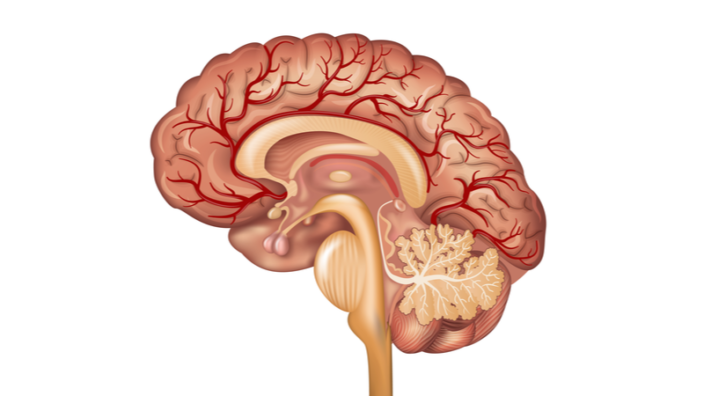
The most obvious example is the cell, which is a network of chemicals, proteins, and metabolites that are connected to each other in metabolic and protein interaction networks. Our brain is a network of neurons, and its function emerges through the network, not through the action of individual neurons. But, if you think about it in a wider sense, the social and professional networks that we are part of are a mainstay of our society. We can also talk about economic networks, technological networks, social media, and so on.

What we have understood in the last 20 years is that if we cannot map out the networks behind these systems, we have no chance to understand how they work, because the emerging properties of these systems, their functions are driven not by individual components, but by different components working together in a specific way to achieve certain function.

Network science itself is now a big discipline. It has emerged slightly more than 20 years ago, and now, you can get a PhD in it in Boston, in Vienna, in Budapest. There are thousands of researchers focusing on all kinds of networks. Within biology, however, networks offer a post-genomic path towards understanding disease, because what the human genome has really provided us is the phone book that shows you the components of the cell, but it does not tell you how those pieces interact with each other.

If you ask how the system behaves, the answer is never ‘here’s the list of components’. It’s always, ‘here’s the list of components, and here’s how they interact, which results in certain outcomes and behaviors’. So, if we really want to understand how the cell behaves, and what life is, we have to map the networks, the interactions between the pieces.

Network science aims to further this journey on many levels by creating accurate maps of the subcellular and intracellular environment and developing the mathematical and algorithmic tools that can make sense of these networks. You start from a big network perspective and zoom into a particular disease or a particular gene, you bring the knowledge that is eventually experimentally testable, and this can lead to therapies and cures for disease.

Things like machine learning are probably good instruments to study those networks.

Yes and no. Machine learning is obviously a fabulous tool if you have a giant amount of data, and you need to make sense of it. There are certain aspects of network medicine where machine learning is necessary, but network medicine does not deal with extremely large networks. In the end, we only have 20,000 proteins in our cell. Those, of course, have multiple isoforms, so you’re in a hundred thousand region. You don’t really need machine learning to make sense of that.

What you need machine learning for is to categorize, to classify, and this really is what machine learning does. It finds boxes, puts things into those boxes, and makes predictions based on all this, and yes, it is a useful tool. For example, we use machine learning along with other network medicine tools to predict drug repurposing candidates.

It’s interesting that the 20,000 proteins present in a cell don’t form a giant network. This begs the question, when will we finally have a computer model of a cell? 

The question is, what is a model of a cell? There are several models out there. People who’ve been trying to do this got very far away, creating really cool stuff. For instance, they try to take every single interaction and build it into a giant cell model. But when you try to create a model of a cell, it is not enough to have the interactions, you also need to have the dynamics of the interactions.

We have a pretty accurate knowledge of human and bacterial metabolism. Most of the reactions are known. So, we know the network, but we don’t know the kinetic constants. Without having all the kinetic constants, you cannot model the dynamics of metabolism. We have those kinetic constants only for selected pathways, like the cell cycle pathway and a couple of others, but a high-throughput methodology that can determine such kinetic constants in a systematic way is not even on the horizon.

So, do we have the data to map the networks out? Yes. Do we have a way of turning that knowledge into dynamic models? Not yet. Do we want to turn them into dynamical models? Sure. Can we go without that? Yes, and that’s the interesting part. One of the things that we have shown about three years ago, in a paper published in PNAS that was inspired by other people’s work, is that just knowing the network gives you about 70-80% predictive power about the behavior of the system. For the remaining 20-30%, you do need to know the accurate kinetic constants.

Since we don’t even have a hope of getting to know them all, we go as far as we can with what we have, which is the network map, and the idea is that the network maps that the cell has developed within itself are very much attuned by evolution to the function of the system. So, once you know the map, you can deduce most of the functions. Yes, it would be great to know the kinetic constants and make dynamical models, but you can get pretty far in understanding and eventually curing diseases even without that.

At a certain point, you became interested in nutrition. Most of our knowledge in this field comes from population studies, which frankly are not a good source. How do you apply network medicine to nutrition and why is it important? 

The Foodome Project, as we nicknamed it in the lab, began about five years ago, when I realized that every network that we had built and curated in the lab is really based on genomic and genetic information, and we know that having just the genetic information gives you only limited predictive power about the occurrence of disease and so on. If you look at heart disease, only about 20% of the occurrence is explained by genetic factors. Where does the rest come from? The environment. So, how could we integrate these environmental effects into the subcellular models that we’ve been building for 15 years?

We started with the food because food is one of the biggest environmental components. There are others, of course, such as stress, exercise, and so on, but the biggest single one is probably food. And we thought that it should be easy because in the end, food is molecules, and molecules get into the cell and interact with various cellular components. So, let’s just figure out what the molecules are and how they interact with the cell, right? That’s when I was in for a big surprise that started this big journey for us. I realized we have absolutely no clue what chemicals are in our food.

What about nutritional science? Sure, it has done a great job of identifying the energy sources and the vitamins that we need to survive. These are typically the chemicals that are processed by our metabolism, like sugars, fats, carbohydrates, and many other things. You must get them on a daily basis because if you don’t, you will not survive.

These are about 150 components, and they are indeed being tracked by the USDA in multiple foods, and we know a lot about them. But to my surprise, already early on, we realized that food has more than 20,000 components. That was four years ago. Today, in my lab, we have a very accurate database of about 135,000 chemicals that can be found in food.

What are these chemicals? We call them the nutritional dark matter because they are not systematically tracked by any agency. We mapped out the full scientific literature, and the knowledge is sporadic. That includes things we hear a lot about, like polyphenols.

Polyphenols can be considered nutritional dark matter because they are not processed by the human metabolism, although there’s some processing by the bacterial metabolism in our gut. Their primary role is not metabolic – they are not a source of energy or of any building blocks in the cell. Their function is rather regulatory. Once they get into the bloodstream and into the cells, they bind to human proteins and regulate their function.

In the end, we have learned that while the nutritional components are essential, they are like the gas and oil for your car, there is also this nutritional dark matter. Amazingly, for two thirds of the molecules that constitute it, we have known health effects. So, the big question is how can we even attempt to understand how our environment affects us, if we don’t have an accurate map and the list of these chemicals?

What the Foodome Project have been doing in the last five years is first, trying to curate an accurate list of all the chemicals that are potentially present in our food and to understand their characteristics. We work with mass spectrometry labs, we comb the literature, we develop tools that predict the presence and the concentration of those chemicals, and that’s one part of the project. The other part is, once you know that the chemical is there, how do you predict its effect on health? Or, if you put it the other way around, we want to pick a disease and find out which food chemicals may have a positive or negative impact on that disease.

This is probably a huge project. Do you have some interesting insights already that you’d like to share with us?

We do have several. First, in our work with polyphenols, we ended up identifying several polyphenols with previously unknown health effects, and connecting one of them to specific health outcomes. We showed that it has an impact on platelet function in the cell, which is relevant for cardiovascular diseases.

The key part is that not only we can determine its impact, we can also precisely predict the pathway, the chemical interactions involved, which makes it experimentally testable. My colleagues at Harvard added this chemical to cell lines and measured the expression patterns of multiple genes and proteins, validating our predictions at a cellular level. So, our methodology allows us to make experimental testable predictions about which chemicals are beneficial or deleterious.

We also predicted what chemicals could affect rheumatoid arthritis, and of the 30 chemicals that we predicted, I believe only one had no effect once it was tested by an independent lab. So, the combination of food and network medicine gives us a pipeline, the ability not only to make experimentally testable mechanistic predictions of how food affects health but also to make disease-specific predictions that may be directly actionable for patients. We are now trying to raise funds for a clinical trial so that we can prove that those chemicals indeed have the impact that we’ve predicted.

This is really interesting considering that nutritionists seem to disagree on some basic things, like the amount of protein that’s good or bad for us. Has your approach added something to this or other discussions that are going on in nutritional science?

Let’s be fair with nutritionists. We consume tens of thousands of chemicals daily, and the challenge that nutritionists have in front of them is to piece out the impact of one or a few chemicals on health. This is such a convoluted problem that it is virtually impossible to solve unless you have millions of patients in clinical trials. So, many of the disagreements in the literature are rooted in the uncertainty or the inability to make accurate measurements of outcomes.

They’re dealing with a massive network-based problem, trying to solve it with standard statistical tools that were developed for Gaussian distributions. There’s a giant gap between the complexity of the problem they’re trying to address and the toolset available to them currently. And I’m amazed at the clean results they were able to get in some cases despite of that, like the impact of sugars and salt.

Or of processed meat?

That’s right. So, if the signal is strong enough, they can discern it. Can we do better? I hope we can. I think an approach based on networks and machine learning, when applied to a large dataset, may give us a way to do this, but we must start designing trials that take advantage of those tools. With the availability of apps where people are tracking their eating pattern and the massive clinical data coming in, I think in the next few years, we’ll see a revolution in the way we understand the impact of food on our health.

I’m looking forward to this revolution. I was also fascinated by your research into processed food. You have developed a measure of just how processed various types of food are, and this measure seems to correlate with health effects. We knew that processed food was bad for you, but this is much deeper. But why actually any amount of processing seems to correlate with health?

We started to understand what processing does to food and how it affects our health only recently. So, let’s start with what is processing. We process food every day, and this isn’t a problem in itself. When you peel an orange, or when you cut a cucumber, salt it, cook it – all this is processing, but those are mechanical and simple chemical processes that do not fundamentally change the chemical composition of the food. Maybe cooking, frying, etc. have some impact, but nothing drastic.

And then there is ultra-processing. It is a precise process that can only be done in factories or laboratories. It’s when you take the food, decompose it, and then reassemble its components into some other product. You are effectively creating new food from food-based components.

Let me give you an example. So, you go to the supermarket, and you buy ‘natural orange juice’. Natural because it’s based on oranges, but it’s ultra-processed because once the oranges are collected, they’re squeezed out. Then, they’re decomposed into three different components, which is the juice itself, the pulp that is taken out, and the water.

Those components are stored separately, and only later, often hundreds of miles away from the collection point, they are reassembled. Whatever you get bears no resemblance to orange juice. It doesn’t taste like it, it doesn’t have the right consistency. So, you must add lots of stuff that give you the consistency, the taste, the smell of orange juice. This is ultra-processed orange juice. What you make at home by squeezing an orange is just processed orange juice.

About five-ten years ago, a Brazilian group has started to systematically classify food in terms of whether it’s ultra-processed or unprocessed. Thanks to their effort, people have started to reanalyze the health data they had on individuals, and to realize that one of the biggest negative health effects is coming from ultra-processed food.

Let me give you some rough numbers. If you move 10% of your calorie intake from unprocessed to ultra-processed food, your chances of diabetes increase by 12%, and your chances of cancer by 10%. There’s a whole list of diseases where the risk suddenly shoots up by 5% to 10%. All you did is replace food that your grandmother would cook with the ultra-processed food that you buy at the supermarket. Otherwise, you’re eating the same things. The reason for that is still a big mystery, but there is already very strong evidence that ultra-processed food has deleterious health effects.

It’s probably not necessarily true about each and every kind of ultra-processed food, it’s just an average, right?

We don’t know that because the clinical trials so far just say what percentage of your calories is coming from ultra-processed food and whether this correlates with health outcomes, but people can go deeper. It has been shown that when it comes to meat, most of the health effects that we previously assigned to red meat are limited to ultra-processed meat. What’s important is that in the US, calorie intake is dominated by processed food. Americans get 60% of their calories from ultra-processed food. That’s mind-blowing, and that’s where the problem is.

I wonder if this has something to do with the obesity epidemic.

Yes, of course. This is the other result that clearly comes out of the data: that moving 10% of your calories from less-processed to ultra-processed food increases your chances of developing obesity and metabolic syndrome by the same 10%. So, most of the bad health outcomes that are systemic in the American population can be linked to consumption of ultra-processed food.

Here’s one problem with ultra-processed food that we come in to solve. When you go to the supermarket, you have no idea whether the food that you’re about to buy is ultra-processed or merely processed. That information is not available anywhere on the boxing or in databases. We built an AI tool that looks at all the foods at the supermarket, and we have created a website called Truefood.tech where you can type in your favorite food, and it will tell you the degree of processing it was subjected to. It will also tell you what other items of the same type are sold in the same store, that are less processed, so it offers you an alternative.

That came out of our foodome research. We were curious about not only what chemicals are in the food but also what is the concentration of the chemicals that are in the food? We made a very surprising discovery that was just recently published in Nature Food. We realized that in natural foods, the variation in the concentration of specific chemicals is very narrowly bounded, and there’s a precise mathematical formula that describes, say, how much vitamin C and how much sugar is expected to be in your food.

When we apply the same formula to ultra-processed food, we see major deviations because when we reassemble food from components, we don’t respect the natural concentrations anymore. Rather, companies do whatever they think is right taste-wise or consistency-wise. So, we create a chimera that has the same ingredients as the original food and then some, but in different proportions.

Why is that a problem? Let’s say I give you 10 atoms of hydrogen and one atom of oxygen and ask you to make me a lot of water. You can’t because one atom of oxygen can only take up two atoms of hydrogen, and the rest is just floating around. Previously we thought that getting all those extra chemicals with food was no problem because they would just go in and out, the body doesn’t know what to do with them. But the body has been evolutionary engineered to deposit anything it gets because you never know when it’s going to become useful. In the end, much of this stuff gets deposited, like all the cholesterol in your arteries.

This stuff comes into your body through many different foods, and the body doesn’t know what to get rid of. Our hypothesis was that the problem with ultra-processed food it that it is chemically unbalanced. We normally eat ourselves, meaning that anything we eat has the same carbon-based chemical engine as the human metabolism. But ultra-processed food is different, the ratios of chemicals are different.

So, our AI model looks at the nutritional components that every food producer must disclose by law, and it detects these fine changes in the concentrations and determines how ultra-processed the food is. You can’t tell it by just looking at the label. Yes, maybe it has a little bit more sugar, maybe a little more salt or carbohydrates, but you don’t know what the proper ratio should be. The AI, on the other hand, learns and knows, and immediately detects if the food has been tampered with.

I think this online tool that you built is extremely useful, although I was mostly reassured by it that I’m doing okay. One food that is hard to replace is bread, and all bread is processed to some extent.

Bread does not need to be heavily processed. It’s a simple combination of yeast, grain, and a couple of other things. Why do we ultra-process bread? Because making bread at home, that is, by processing, and not ultra-processing, takes a day, but if you want to make large amounts of it fast, you must add new processes.

Even at the supermarket, as our database shows, you can find minimally processed bread beside ultra-processed bread, and the same goes for most foods. Yes, for some food categories, ultra-processing is necessary, but for most, there is a minimally processed alternative.

Why is this important? As we show in our paper, if you just take your normal eating pattern (we eat about 20 different items per day), and you replace one item with a less-processed version, this can already give you a considerable reduction in many of the outcomes associated with ultra-processed food, because typically, there’s always one item in your diet that is the biggest contributor to your ultra-processed food intake.

Just replace it. Keep eating the same. If it’s a burger, replace it with an unprocessed burger. If it’s soda, replace it with water, and so on. This single change that just replaces one item with another without changing your diet could have a huge impact on your health.

Yes, this particular parameter blew me away when I read your paper. You also mention the obvious fact that processed food is cheaper. So, basically, eating less processed food can be considered a privilege that may not be available to everyone.

Yes, of course. Ultra-processed food has clear advantages: it’s cheaper, it has a longer shelf time, which is also reflected in the price, it has higher caloric content, and so on. Do you know where processed food comes from? It turns out that the story goes back to a place about 50 miles from my home here in Boston. It was originally developed by the US military that needed non-perishing food with high energy content, light enough for the soldiers to carry it on the battlefield. In the 1960-70s, Congress allowed food companies to start utilizing the patents developed by the military, and that’s when ultra-processed food invaded American supermarkets.

I think that if we care about population health, we will find a way to get less processed food to people, but we also need to know what is processed and what is not, and there has to be awareness and demand. There’s a lot you can do with tax policy to encourage consumption of less processed food over ultra-processed food, and many European countries are already doing that. I think eventually, the solution will come not from simply banning processed food but from doing what we did with sugary food and cigarettes, which is to tax the hell out of it and use the money to promote and support the production of healthier food. It is possible, but it’s a long-term journey, and it requires legislation and leadership.

Pandemics are also networks, right? What can network science teach us about the COVID pandemic and pandemics as a whole?

Sure, epidemics are network-based problems. Viruses spread through social networks, and that’s why the current leader of my Network Science Institute in Boston, Alessandro Vespignani, has also been the White House modeler of COVID since March 2020. He has predicted the problems that we’re going to have with COVID already in December 2019, way before the pandemic really took off. Many other network scientists are involved in predicting COVID patterns and continue to study it. I would go as far as to say that there’s no way you can build good predictive tools for disease spreading without considering the network perspective.

Curing diseases is also fundamentally a network problem. My lab has been deeply involved in the fight against COVID from early on, in order to identify, using tools from network medicine, drugs that could be repurposed for COVID patients, because at that time, it wasn’t clear how long creating vaccines will take, and the only solution was to repurpose existing drugs. We ended up predicting and testing, together with colleagues at Boston university, 6,000 drugs and identifying about a dozen that could be, and were, repurposed for COVID patients.

Fundamentally, for any infectious disease, the first person you need to consult is a network epidemiologist. That’s what’s happening right now with the outbreak of monkeypox. You probably can google it and find out that many of my colleagues are already involved in modeling the spread of this disease. They were also involved in the modeling of Ebola and Zika, and so on. There’s no way to understand and stop infectious diseases without a network perspective.

How would you describe the general situation in the longevity field today? What are you excited or frustrated about, and what directions do you think are especially worth pursuing?

I think that longevity research is undergoing a fundamental transformation right now. The pieces have been assembled, and the will is there to really try to make an impact. However, one of the challenges with longevity is that everybody’s hoping for that simple pill, but also everybody who is really in the field understands that the aging is not a single mechanism; there are multiple components and mechanisms involved. Aging is the sum of many different diseases and conditions that come together, and they are always coupled with each other.

So, I believe, and that’s what we are focusing on together with Vadim Gladyshev, that we must bring the network toolbox to aging research. I think this is the only way to put those many pieces together and perhaps find some solutions, although we are far from actually seeking for solutions right now; we’re trying to map out the networks involved in longevity and aging.

If we are successful at that, then we can also think of potential interventions that may alter those pathways in the way we’re doing with other diseases. The bottom line is, based on what I see by reading the literature and going to conferences, every field has this moment when it totally rejuvenates itself, if we use our favorite term, and I feel like this moment for longevity research has arrived.

Ending Aging

SENS Research Foundation Announces Ending Aging Forum 2022

SENS Research Foundation has announced this year’s Ending Aging Forum, which will be held through a virtual conference platform with an immersive environment.

Come spend a wonderful and thought provoking time with the team at SENS Research Foundation. This virtual event is your opportunity to hear first-hand about the latest advances that our in-house researchers are making toward new rejuvenation biotechnologies, along with some of our young scientists-in-training and outside researchers whose research we fund. In addition to the formal presentations, you’ll have the opportunity to talk one-on-one with the scientists and other members of our team, as well as with citizens, donors, and activists who dream of and work for a future free of degenerative aging.

Breaking down the barriers to attendance, the Forum will be hosted virtually through EXVO, a strong platform for this VR experience. Registered attendees will receive instructions on how to join the conversation shortly before the event begins. The virtual event will have a Conference Hall, where feature presentations are made, along with project-specific Research Booths and booths for scientific posters presented by our students that break down different research projects. In the Expo Room, attendees can also meet and talk one-on-one or in small groups with the team and other supporters, or watch videos in which our team members and scientists-in-training introduce themselves and what drew them to this Mission. The platform dispenses with the tedium of navigating your avatar around the event space and also giving you the power to ‘teleport’ from one virtual location to another. Join us to learn and celebrate how far we’ve come, and to catch a glimpse of the future we’re building!

We would like to ask you a small favor. We are a non-profit foundation, and unlike some other organizations, we have no shareholders and no products to sell you. All our news and educational content is free for everyone to read, but it does mean that we rely on the help of people like you. Every contribution, no matter if it’s big or small, supports independent journalism and sustains our future.
LSN Longevity Prize

Announcing the Longevity Prize

The Longevity Prize is a series of prizes designed to honor the researchers who are helping to build a future in which age-related diseases are a thing of the past. This new initiative aims to accelerate progress in the rejuvenation biotechnology field and encourage innovation.

While great strides in our understanding of aging have been made in the last decade, there is still a lot we do not know and much work to be done. There has been some amazing progress made, but solving aging is the most challenging endeavor that humankind has ever faced.

There are more and more rejuvenation companies entering the arena to take on this challenge, but the clock is ticking for us all. The Longevity Prize aims to provide researchers with an incentive to develop novel approaches to bring aging under medical control, thus providing longer, healthier lives for all.

Longevity Prize is doing something different

Unlike traditional funding and prize models where researchers are given a fixed amount of money to focus on a specific goal, the Longevity Prize aims to do something different. By creating a series of prizes, the goal is to create a rising tide of research proposals, experiments, and collaborations.

Because the Longevity Prize is free from the traditional systems of funding, this means that more ambitious projects have the potential to receive attention. Risk aversion is a huge problem in normal funding, so this initiative helps to address that by supporting undervalued or overlooked projects.

The first round of prizes totals $180k and was fundraised through Gitcoin. Community donations were matched by VitaDAO, Vitalik Buterin, and Stefan George.

The first prize to be announced is the Hypothesis Prize.

Over a century of all the world’s biological knowledge is available to anyone taking the time to read the literature. There are cases where key discoveries are made in the past, but forgotten for long periods of time – only to be rediscovered. The hypothesis prize aims to resurface such discoveries and research areas, focusing our attention on the most promising directions.

Normally, researchers can only start experiments when they successfully get the funds to begin. The first round aims to combat this problem by giving out prizes based on hypothesis generation that will then help to shape the second and subsequent prize rounds that follow.

After more than a century of research, the literature is filled with hidden gems and forgotten knowledge. Most of this knowledge is available to anyone to read, and there are plenty of cases where discoveries are forgotten for decades only to be found again years later.

Longevity Prize would love to hear from you

Do you have a suggestion for an underappreciated area of aging research that deserves some attention? Perhaps you have read the scientific literature and can explain why a particular area of research needs more focus?

You can submit a proposal (1-3 pages max) for consideration and could win a prize of up to 20k. Finalists will be invited to present their proposal to the judges. Excellent proposals will be moved to the next phase, where they will be eligible for follow-on funding.

lifespan.io is proud to be part of this awesome initiative alongside VitaDAO, Foresight Institute, and the Methuselah Foundation.

We would like to ask you a small favor. We are a non-profit foundation, and unlike some other organizations, we have no shareholders and no products to sell you. All our news and educational content is free for everyone to read, but it does mean that we rely on the help of people like you. Every contribution, no matter if it’s big or small, supports independent journalism and sustains our future.
Blood vessels brain

Hypertension Is Associated with Brain Drainage Changes

Researchers publishing in Aging have found that enlarged perivascular spaces (ePVS) in the brain are correlated with vascular disorders [1]. These spaces, which are part of the brain’s glymphatic system, allow for the drainage of potentially dangerous metabolites such as beta amyloid [2].

A known biomarker of brain disorders

As they become enlarged, perivascular spaces become visible through MRI imaging. This enlargement represents microvascular damage and brain degeneration [3], and it is associated with increased cognitive impairment and risk of stroke [4]. Prior research has found that the location of ePVS reflects different types of disease: lobal ePVS signify issues with amyloid beta, while deeper ePVS signify hypertension [5].

A large, well-known cohort study

This research builds upon prior work, using the well-known Framingham Heart Study cohort to ascertain the relationships between known risk factors and ePVS. After excluding people for stroke, a total of 3,710 people with 4,101 MRI records became part of the study.

The determination of ePVS was made using rigorous criteria, including placement, size, and signal intensity. Both of the measured brain regions were given grades from I to IV based on ePVS prevalence. The study also measured other well-known biomarkers of vascular health, including blood pressure, BMI, cholesterol, and diabetes.

The researchers then correlated the prevalence of grades 3 and 4 of ePVS with these other biomarkers, and the results were as expected. Systolic and diastolic blood pressures were both correlated with ePVS in lobar and basal regions. Hypertension was very significantly correlated with ePVS in each of the regions, and prehypertension was correlated with basal ePVS. Smoking, age, and the use of hypertensive medication were also significantly correlated with ePVS in both regions.

Interestingly, while there were tendencies, cholesterol, lipids, and diabetes did not have significant effects on ePVS.

Conclusion

This was a relatively simple study with a straightforward conclusion, but that conclusion puts together vascular issues in the body and in the brain. While a causal link has not yet been elucidated, there is clearly a strong correlation between this particular aspect of brain dysfunction and greater cardiovascular issues.

We would like to ask you a small favor. We are a non-profit foundation, and unlike some other organizations, we have no shareholders and no products to sell you. All our news and educational content is free for everyone to read, but it does mean that we rely on the help of people like you. Every contribution, no matter if it’s big or small, supports independent journalism and sustains our future.

Literature

[1] Lara, F. R., Scruton, A. L., Pinheiro, A., Demissie, S., Parva, P., Charidimou, A., … & Romero, J. R. Aging, prevalence and risk factors of MRI-visible enlarged perivascular spaces. Aging, 14(undefined).

[2] Schley, D., Carare-Nnadi, R., Please, C. P., Perry, V. H., & Weller, R. O. (2006). Mechanisms to explain the reverse perivascular transport of solutes out of the brain. Journal of theoretical biology, 238(4), 962-974.

[3] Gouveia-Freitas, K., & Bastos-Leite, A. J. (2021). Perivascular spaces and brain waste clearance systems: relevance for neurodegenerative and cerebrovascular pathology. Neuroradiology, 63(10), 1581-1597.

[4] Francis, F., Ballerini, L., & Wardlaw, J. M. (2019). Perivascular spaces and their associations with risk factors, clinical disorders and neuroimaging features: a systematic review and meta-analysis. International Journal of Stroke, 14(4), 359-371.

[5] Wardlaw, J. M., Benveniste, H., Nedergaard, M., Zlokovic, B. V., Mestre, H., Lee, H., … & Black, S. E. (2020). Perivascular spaces in the brain: anatomy, physiology and pathology. Nature Reviews Neurology, 16(3), 137-153.

Skin aging

An In-Depth Review of Skin Aging Genes

In a new systematic review published in Scientific Reports, multiple genes driving skin aging were identified [1].

The authors start by explaining the intrinsic (genetic and chronological) and extrinsic (environmental) factors that drive skin aging. While the environmental factors that affect the rate of skin aging, such as sun exposure and smoking, can be controlled, the intrinsic factors are not yet amenable to manipulation. Therefore, the researchers focus their review on these intrinsic drivers of skin aging.

Genetics are hard to beat

Studies show that skin ages differently in different genders and ethnicities. For example, Korean men have a smaller risk of developing wrinkles than Korean women, while the reverse is true for Japanese people up to the age of 65. Overall, Asians have deeper wrinkles on the forehead and in the crow’s feet area compared to Caucasians. However, the latter develop more wrinkles under the eyes, and their skin sags more than other races. In addition, a lower level of melanin in Caucasian skin makes it less protected from sun-driven photoaging. Meanwhile, both Asians and people with black skin are more prone to abnormal pigmentation.

Interestingly, different ethnicities demonstrate a different pace of skin aging. It was shown that French women develop wrinkles in a linear fashion from 20 to 60 years of age, while Chinese women become wrinkled rapidly at the age of 40-50. This may explain why Europeans find it hard to accurately judge the age of Asians.

All these examples point to the importance of genetics dictating different skin aging phenotypes. Previous studies have already identified single-nucleotide polymorphisms (one “letter” differences at a specific genomic position in different people) associated with skin aging phenotypes. This review sought to explore the genetics of skin aging and uncover some of the biological processes underlying skin aging by employing bioinformatics techniques.

Skin aging phenotypes

Often, systematic reviews have to combine results from studies that differ greatly in terms of methodology. This review is not an exception: entirely different features (wrinkles, pigmentation, etc.) and assessment measures (image analysis, visual assessment by a dermatologist, etc.) are used in different studies that describe skin aging. Therefore, the authors first examine what skin aging phenotypes have already been described and use them as a proxy of overall skin aging.

Surprisingly, there is no complete list of skin aging phenotypes. Thus, the researchers mined various studies, databases, and two medical books to compile a comprehensive list of skin aging phenotypes. As a result, 56 phenotypes were identified with “eyebags”, “sagging of jawline”, “global facial photoaging” among others. All the phenotypes can be grouped into 4 categories: skin cancer-related, skin color-related, wrinkling and sagging-related, skin global impression. Skin aging can then be treated as a result of skin changes in these four aspects.

Skin aging: is it just about single “letters”?

Next, the authors looked into the connection between skin aging phenotypes and single-nucleotide polymorphisms. They included 44 observational studies conducted in 16 countries with participants of different ages, genders, and nationalities. They only analyzed single-nucleotide polymorphisms that had been reported in association with a skin aging phenotype in at least two independent studies.

Although thousands of single-nucleotide polymorphisms were reported to be associated with skin aging phenotypes, only 19 of them were significant in several studies. For example, single-nucleotide polymorphisms in genes PRDM16 and TANC2 are associated with the wrinkling phenotype category. This might indicate that instead of individual single-nucleotide polymorphisms, many of them in the same genomic region are involved in driving a skin aging phenotype

A few genes define your skin color and aging

After assembling a comprehensive list of genes associated with skin aging, the researchers were able to determine which genes are most responsible for it. They identified a specific region on chromosome 16, band 16q24.3, as hosting a particularly high number of pleiotropic genes, which are associated with two or more morphologically different skin aging phenotypes (different phenotype categories).

Many of the identified pleiotropic genes are known to be related to skin color, such as MC1R, which suggests that in addition to their pigmentation role, these genes are responsible for setting the pace of skin aging. On the other hand, some genes on chromosome 16 that are not known to be skin color genes are associated with skin color-related phenotypes.

Gene enrichment analysis confirmed the results: a handful of 44 pleiotropic genes, which belong to chromosomal band 16q24.3 or are related to skin color, encode highly interconnected proteins driving skin aging phenotypes.

In order to get more insight into the biology of skin aging, the researchers extracted and analyzed expression data of the 44 pleiotropic skin aging genes and 32 skin color genes (some of which are also pleiotropic genes) in young and aged skin. Among the pleiotropic genes, SPIRE2, BNC2, SHC4, SLC24A5, and TYR were found to be downregulated with aging. Meanwhile, aging is accompanied by the upregulation of several skin color genes, such as AGR3, DSTYK, and TPCN2.

Importantly, there are some genes, such as DBNDD1, that are pleiotropic genes, but their expression is affected by environmental factors (UV exposure) rather than chronological age. This is an important reminder that protecting skin from sun exposure is still the best method of keeping it as young as possible, as there are no interventions that directly manipulate skin aging genes.

Abstract

Skin ageing is the result of intrinsic genetic and extrinsic lifestyle factors. However, there is no consensus on skin ageing phenotypes and ways to quantify them. In this systematic review, we first carefully identified 56 skin ageing phenotypes from multiple literature sources and sought the best photo-numeric grading scales to evaluate them. Next, we conducted a systematic review on all 44 Genome-wide Association Studies (GWAS) on skin ageing published to date and identified genetic risk factors (2349 SNPs and 366 genes) associated with skin ageing. We identified 19 promising SNPs found to be significantly (p-Value < 1E−05) associated with skin ageing phenotypes in two or more independent studies. Here we show, using enrichment analyses strategies and gene expression data, that (1) pleiotropy is a recurring theme among skin ageing genes, (2) SNPs associated with skin ageing phenotypes are mostly located in a small handful of 44 pleiotropic and hub genes (mostly on the chromosome band 16q24.3) and 32 skin colour genes. Since numerous genes on the chromosome band 16q24.3 and skin colour genes show pleiotropy, we propose that (1) genes traditionally identified to contribute to skin colour have more than just skin pigmentation roles, and (2) further progress towards understand the development of skin pigmentation requires understanding the contributions of genes on the chromosomal band 16q24.3. We anticipate our systematic review to serve as a hub to locate primary literature sources pertaining to the genetics of skin ageing and to be a starting point for more sophisticated work examining pleiotropic genes, hub genes, and skin ageing phenotypes.

Conclusion

This comprehensive systematic review addressed several challenging issues: defining skin aging, compiling a list of skin aging phenotypes and skin aging genes, and identifying the pleiotropic genes that drive several skin aging phenotypes. Although skin aging seems to be manifested in several distinct phenotypes, the genes associated with them are interconnected, and most are involved in defining skin color. The results of this review could serve as a platform to explore skin aging genes in detail in order to develop effective skin rejuvenation approaches.

We would like to ask you a small favor. We are a non-profit foundation, and unlike some other organizations, we have no shareholders and no products to sell you. All our news and educational content is free for everyone to read, but it does mean that we rely on the help of people like you. Every contribution, no matter if it’s big or small, supports independent journalism and sustains our future.

Literature

[1] Ng, J. Y. & Chew, F. T. A systematic review of skin ageing genes: gene pleiotropy and genes on the chromosomal band 16q24.3 may drive skin ageing. Sci. Rep. 12, 13099 (2022).

Back from the dead

Scientists Move the Boundaries of Post-Mortem Recovery

Researchers have been able to achieve substantial recovery of cellular and organismal activity in pigs that had been dead for a full hour [1].

When is it too late?

Advances in resuscitation have already moved the boundaries of life and death, making it possible to revive a person several minutes after the heart stops beating. However, bringing someone back an hour or more after a fatal ischemic injury, such as a stroke or heart attack, has been considered utterly impossible. This new study by scientists from Yale, published in Nature, shows that it may be possible after all.

Without blood supply, cells begin to deteriorate quickly. Then, if blood flow is restored, it paradoxically brings even more destruction of cellular membranes and organelles in what is known as reperfusion injury [2]. However, cells are also quite resilient and capable of regeneration. What if we could remove the shock of reperfusion injury and help the cells recover?

An attempt at full-body preservation

The scientists built a system called OrganEx, which has a lot in common with a heart-lung machine, but it reperfuses the body with a special non-coagulative hemoglobin-based solution that contains nutrients, anti-inflammatory drugs, various restorative molecules, and also some original blood from the patient: a domestic pig in this case.

Back in 2019, the same group of scientists reported restoring some cellular functions in a pig brain four hours after an ischemic injury (although no network brain activity was detected) [3]. This time, the researchers attempted a full-body preservation.

The time after the ischemic injury was reduced to one hour, which is still way above the current resuscitation threshold. The researchers did everything to eliminate the possibility of animal suffering: the pigs were anesthetized, and then heart failure was induced by ventricular fibrillation. After an hour of being effectively dead at room temperature, the pigs’ bodies were connected to OrganEx machines and reperfused with OrganEx solution.

The researchers used two other groups as controls: pigs that were put on a regular extracorporeal membrane oxygenation (ECMO) machine, which uses autologous blood for reperfusion, and pigs that had their blood flow restored almost immediately after it was interrupted (the zero-hour group).

Multiple signs of recovery

Perfusion with the ECMO system one hour after the ischemic injury resulted in very low blood flow and, accordingly, limited filling of major arteries and organs. OrganEx machines, however, were able to restore normal flow that resulted in adequate oxygenation of the whole body.

Organs in the OrganEx group also had fewer signs of hemorrhage and tissue edema (on par with the zero-hour group), which reflects reduced cellular damage. Interestingly, post-mortem stiffening (rigor mortis), which was clearly observed in the ECMO group, was absent in the OrganEx group.

The researchers ran a battery of other tests to assess various aspects of cell and tissue viability. Markers of apoptosis and all other types of cellular death were greatly reduced in the OrganEx group compared to the ECMO group, and, again, on par with the zero-hour group.

There was also an assessment of glucose uptake by several organs in order to measure their metabolism. The OrganEx group easily beat the ECMO group and, by a small margin, the zero-hour group. Histological analysis demonstrated a substantial preservation of cells and tissues as well, and electrocardiography showed the spontaneous re-emergence of QRS complexes (heartbeat patterns) during OrganEx reperfusion.

Organ transplantation – and maybe cryopreservation?

While these results are extremely impressive, they do not mean that the pigs or even single organs were “revived”. As a sobering example, the primary metric of kidney function, urine output, remained low. Moreover, just like in the 2019 experiment, no global network activity was detected in the brain. The researchers name several possible reasons, including the presence in the OrganEx solution of nerve blockers that decrease neuronal activity to make sure the pigs stay completely unconscious.

According to the researchers, this technology could revolutionize the field of organ transplantation. Today, organs often do not make it to the patients who need them: for instance, when a terminally ill person gets disconnected from life support, it might take the doctors too much time, due to technical and regulatory reasons, to extract the organs.

However, if OrganEx-like technologies will ever be perfected to the point of brain function restoration, they could also bring back to life people who died from causes that do not involve major organ damage, such as suffocation. Moreover, the success of OrganEx gives more weight to the concept of cryopreservation, which is currently severely limited by the rapid accumulation of post-mortem damage.

Conclusion

This study pioneers the use of a non-blood solution to remove the limits put on resuscitation by both ischemic injury and reperfusion injury. The proposed technology might be a true gamechanger in organ transplantation, if validated by subsequent research. It would be especially interesting to see whether any kind of brain activity can ultimately be restored.

We would like to ask you a small favor. We are a non-profit foundation, and unlike some other organizations, we have no shareholders and no products to sell you. All our news and educational content is free for everyone to read, but it does mean that we rely on the help of people like you. Every contribution, no matter if it’s big or small, supports independent journalism and sustains our future.

Literature

[1] Andrijevic, D., Vrselja, Z., Lysyy, T., Zhang, S., Skarica, M., Spajic, A., … & Sestan, N. (2022). Cellular recovery after prolonged warm ischaemia of the whole body. Nature, 1-8.

[2] Verma, S., Fedak, P. W., Weisel, R. D., Butany, J., Rao, V., Maitland, A., … & Yau, T. M. (2002). Fundamentals of reperfusion injury for the clinical cardiologist. Circulation, 105(20), 2332-2336.

[3] Vrselja, Z., Daniele, S. G., Silbereis, J., Talpo, F., Morozov, Y. M., Sousa, A. M., … & Sestan, N. (2019). Restoration of brain circulation and cellular functions hours post-mortem. Nature, 568(7752), 336-343.

Lewy bodies

Developing Nanobodies to Fight Parkinson’s Disease

A team of researchers publishing in Nature Communications has described nanobodies that can destroy the α-synuclein aggregates that characterize Lewy bodies, which are associated with dementia and Parkinson’s disease [1].

What are nanobodies?

Traditional antibody therapies, while promising in some studies, are too large to enter cells in order to affect the aggregates there [2]. This is a possible reason why antibodies against amyloid beta aggregates, which characterize Alzheimer’s disease, often show promise in early clinical trials but later fail the clinical trial process [3].

Therefore, the researchers turned to nanobodies, which are tiny, stable antibodies that can be deployed through adeno-associated viruses (AAVs) in order to act within cells [4]. Previous research had singled out NbSyn87, which targets all forms of α-synuclein, as a potential method of combating Lewy bodies and Parkinson’s disease [5].

However, as these resarchers note, NbSyn87 targets not only the aggregates of α-synuclein, it targets the protein itself, which serves valid physiological functions. Therefore, they began their search for a nanobody that targets just the fibril aggregates.

A successful hunt for a specific variant

The first part of the researchers’ quest involved a specific point of chemistry. Under oxidizing conditions, a common disulfide bond enhances the stability of nanobodies, but under the opposite (reducing) conditions commonly found in cells, it can be destructive [6]. Therefore, the researchers first used mutagens to develop a platform that was free of this bond.

Once that was done, they then began using genetically engineered yeast that produces α-synuclein aggregates as their nanobody target. Out of 28 clones, the researchers identified a single nanobody, PFFNB2, that preferentially binds to α-synuclein aggregates instead of its healthy monomer form.

This nanobody was found to work in live cells, including immortalized human cells and mouse brain cells, both from genetically modified and wild-type mice. Interestingly, it did not work in cells that had been subject to chemical fixation. It was extremely effective against the fibrils themselves in vitro.

Finally, in a mouse model that produces human α-synuclein rather than its murine form, and then had additional α-synuclein aggregates directly introduced into it, an AAV-administered form of PFFNB2 was shown to dramatically reduce the quantity of these α-synuclein fibrils.

Conclusion

This is a proof-of-concept study that certainly proves the concept. The researchers hold that their findings should be used in a preclinical mouse model of α-synuclein aggregation. If it is successful there, the next steps could possibly involve human clinical trials. If this approach is as effective in people as it is in cells, it could mean a substantial reduction in the frequency and severity of Lewy body dementia and Parkinson’s disease.

We would like to ask you a small favor. We are a non-profit foundation, and unlike some other organizations, we have no shareholders and no products to sell you. All our news and educational content is free for everyone to read, but it does mean that we rely on the help of people like you. Every contribution, no matter if it’s big or small, supports independent journalism and sustains our future.

Literature

[1] Butler, Y. R., Liu, Y., Kumbhar, R., Zhao, P., Gadhave, K., Wang, N., … & Wang, W. (2022). α-Synuclein fibril-specific nanobody reduces prion-like α-synuclein spreading in mice. Nature Communications, 13(1), 1-13.

[2] Henderson, M. X., Covell, D. J., Chung, C. H. Y., Pitkin, R. M., Sandler, R. M., Decker, S. C., … & Luk, K. C. (2020). Characterization of novel conformation-selective α-synuclein antibodies as potential immunotherapeutic agents for Parkinson’s disease. Neurobiology of disease, 136, 104712.

[3] Mehta, D., Jackson, R., Paul, G., Shi, J., & Sabbagh, M. (2017). Why do trials for Alzheimer’s disease drugs keep failing? A discontinued drug perspective for 2010-2015. Expert opinion on investigational drugs, 26(6), 735-739.

[4] Zhang, C., Ötjengerdes, R. M., Roewe, J., Mejias, R., & Marschall, A. L. (2020). Applying antibodies inside cells: Principles and recent advances in neurobiology, virology and oncology. BioDrugs, 34(4), 435-462.

[5] Chatterjee, D., Bhatt, M., Butler, D., De Genst, E., Dobson, C. M., Messer, A., & Kordower, J. H. (2018). Proteasome-targeted nanobodies alleviate pathology and functional decline in an α-synuclein-based Parkinson’s disease model. npj Parkinson’s Disease, 4(1), 1-10.

[6] Kunz, P., Zinner, K., Mücke, N., Bartoschik, T., Muyldermans, S., & Hoheisel, J. D. (2018). The structural basis of nanobody unfolding reversibility and thermoresistance. Scientific reports, 8(1), 1-10.

Steve Horvath

Steve Horvath on the Present and Future of Epigenetic Clocks

Dr. Steve Horvath hardly needs an introduction, so we will be brief: he is the inventor of the epigenetic clock and, currently, principal investigator at Altos Labs. We talked about the recent developments in this immensely important field, including pan-mammalian clocks, two-species clocks, and single-cell clocks, along with the challenges the field faces.

You are a mathematician by training. How did you end up studying biology and the biology of aging in particular?

I was already interested in aging research as a teenager, but my first love was math. Over the years, I became more and more applied. After getting a PhD in mathematics, I retrained and got a second PhD in statistical genetics. While working at UCLA, I turned into a bioinformatician. These days, I view myself as a biologist and biogerontologist. My lab generates lots of data which we distribute via Gene Expression Omnibus to the public. As you see, every year, I become more applied.

Why? It’s a good question. I’ve always loved math, but you should know a lot about biology if you want to reverse aging. The ideal training would provide knowledge in molecular biology, genetics, medicine, computational biology, and computer science. The problem is all of this would probably require 20 years of training. Our lifespans are too short for that, so everybody has to find their own angle.

I have always wondered what people coming from mathematics or physics think of biology when they encounter it?

In the beginning, my angle was statistical genetics. When you think about it, Mendelian inheritance laws are quite mathematical and probabilistic. So, for me, it was a natural transition from biostatistics to genetics and ultimately epigenetics. But we need to use many additional data including gene expression, histone modifications, transcription factor binding information, proteomics, metabolomics.

My first reaction was that there is so much noise compared to physics. In physics, you have elegant formulas that encapsulate the laws of nature, whereas in biology, there’s so much noise. The large amount of noise probably explains why simple statistical models work well in biology. Only a few people attempt to use partial differential equations for modeling biological phenomena.

Let us get to what you are most famous for, which is epigenetic clocks. This field keeps developing at a stunning speed. I watch talks from a few months ago and I see that a lot of things have already changed since then. So, could you give us a quick update?

In my lab, we are interested in third-generation clocks. We are looking for clocks that apply to multiple species at the same time. For example, universal pan-mammalian clocks. Several groups, including mine, are working on single cell methylation clocks. Many researchers are building clocks that respond to lifestyle interventions, such as exercise. Moving away from methylation, it would be nice to build similar clocks for other ‘omics’ data. Many researchers build clocks on the basis of other omics data, such as for chromatin, proteomics, and gene expression.

Why are things like pan-mammalian clocks, two-species clocks, and single-cell clocks important? What can they give us? 

Single-cell clocks are important for understanding the mechanism underlying epigenetic clocks.

These clocks lend themselves for assessing whether each cell from a given person has the same age. Let’s take blood cells. Do all the blood cells of a 50-year-old man have the same age, or are some of them a hundred years old and some two years old? By the way, I think it’s the latter. I think that each cell has a separate age. Most current epigenetic clocks measure age in bulk tissue, which represents an average over thousands of cells.

It is a very important question: does each cell have its own age? It also relates to the question of how methylation relates to gene expression. This is a very difficult question, and most people believe that you do need to study the relationship between methylation and gene expression in single cells to avoid confounding by different cell types. So, that’s the case for single-cell studies.

Why do we need third-generation clocks that apply to multiple species? It’s all about enhancing translation. Imagine that you find an intervention that rejuvenates a mouse. There’s no guarantee that it rejuvenates a human being. You can enhance the chances of success by looking at highly conserved DNA. If you have an intervention that rejuvenates a mouse, a rat, a dog, and a cat according to the same clock, then chances are high that it will also work in humans.

But there’s a trade-off in terms of how precise the clocks are, right? 

Yes. In general, when you have a clock that applies to multiple species, it’s less accurate than a clock that only applies to one species.

On the other hand, if you have a rat and human or a mouse and human clock, it should be very useful in translation.

 Yes. And we have such a clock. We’ve developed it and even applied it. For example, we applied our human-rat clocks to a study of young plasma in rats in collaboration with Harold Katcher and Rudy Goya.

Are we getting any closer to understanding the mechanisms behind the correlation between methylation and age?

Yes, definitely. Fortunately, many people are working on it, including my group. We already know quite a lot. There was a paper from Ken Raj in Nature Aging where he describes the relationship between the hallmarks of aging and epigenetic clocks in vitro.

We have learned a lot from genetic studies in mice and humans, such as studies about developmental disorders, such as progeria. Just to give you some highlights, when you look at cytosines that relate to methylation aging, they are often close to polycomb repressive complex 2 binding sites. We also know that stem cell biology relates: often, stem cells are younger. We also know that there’s a connection to cellular identity. Epigenetic clocks relate to the loss of cellular identity that comes with aging. We have also a good understanding about which stress factors accelerate epigenetic aging: for example, metabolic stress and viral stress from HIV.

Epigenetic clocks come from a machine learning analysis, which means you start with a black box. To characterize this black box, you need to characterize perturbations that affect epigenetic age. For a biologist, black box predictors are not entirely satisfactory. They will, say, enumerate enzymes and pathways that play a role. Naturally, DNA methyltransferases and TET enzymes play a role. We know several other enzymes that play a direct role. Having said that, although we know a lot, the research is ongoing, so we learn more and more.

Considering aging in other species, which seems to be a hot topic now, how do you interpret the finding by Vadim Gladyshev that the naked mole rat ages epigenetically, even though it doesn’t age demographically?

There are actually three papers on epigenetic clocks in the naked mole rat. We published a paper in Nature Aging a few months ago. We have several clocks that apply to the naked mole rat including dual species clocks that apply to both humans and naked mole rats.

How to interpret it? Initially, I was puzzled because the naked mole rat appears to exhibit negligible senescence. But then I thought about it and realized that there’s another species that seems to have negligible senescence, and that’s humans. Humans live remarkably long lives now. If you measure human aging with clinical biomarkers, you will perhaps come to the conclusion that humans have negligible senescence in the first 30-40 years of life.

The question of why you can build an epigenetic clock for the naked mole rat is equivalent to the question of why you can build an epigenetic clock for humans. Both are very long-lived species, and this comes down to the question, what do epigenetic clocks measure? When I published the pan-tissue clock, I proposed that epigenetic clocks relate to the action of the epigenomic maintenance system. If this turns out to be true, then it stands to reason that all species have an epigenomic maintenance system. Therefore, you can build epigenetic clocks for all species, especially for long-lived ones. Our mammalian methylation project has shown that this is true. If anything, it’s easier to build clocks for long-lived species than for short-lived ones.

Another interesting topic is the germline reset, or the embryonic reset. Many geroscientists think this holds the key to solving aging. What can epigenetic clocks tell us about this reset event?

First of all, embryonic stem cells (ESCs) are perfectly young, as you said, as well as induced pluripotent stem cells (iPSCs), which is an interesting insight. Also, passaging of those cells doesn’t affect their epigenetic aging. If you have embryonic stem cells, and they divide, and you culture them in a dish, it doesn’t accelerate clocks. I showed this back in 2013 in the pan-tissue clock.

Recently, Vadim Gladyshev had this paper where he revealed a fascinating early rejuvenation event. I really liked the paper. People debate whether this is valid, because epigenetic clocks are usually trained on adults, or postnatal tissues, and so, it’s risky to apply these biomarkers to tissues collected during early development. But Vadim’s team evaluated many different epigenetic clocks in several data sets. They all pointed to the same early rejuvenation event.

The question is, which biomarkers are usable just a few days after conception?

Exactly, and this highlights the great advantage of epigenetic clocks. They are “life course clocks”. I don’t know any other biomarkers of aging that applies to fetal tissues as well, because most other biomarkers measure organ dysfunction.

I think it’s really interesting that we know for sure that such a rejuvenation event does happen, it just cannot be any other way, and it’s exciting that this has been confirmed by epigenetic clocks.

Yes, I agree with you. Back in 2012, when I first saw that epigenetic clocks reveal that iPSCs and ESCs are perfectly young, I was quite pleased with this finding. It makes intuitive sense.

I’d like to go back to that paper about the hallmarks of aging and epigenetic clocks. It covers a lot of ground, so can you give us just the gist of it, things that you find most important or surprising?

The most surprising to me was that cellular senescence doesn’t seem to be related to epigenetic clocks. But everyone knows that cellular senescence is important in aging, right? Senescent cells clearly increase with chronologic age. Before seeing our results, I would have said that a senescent cell must be epigenetically older than a non-senescent cell. The fact that epigenetic clocks are disconnected from cellular senescence is puzzling. I think more work is needed here, it’s an opportunity to understand the dichotomy between these hallmarks of aging.

Some say that cellular senescence is not really well-defined. Could that be the problem?

Maybe. I really admire the experts in cellular senescence for their honest communication. They are the first to point out that senescent cells are very difficult to define. To begin with, it depends on the cell type. There’s no consensus on what cellular markers to use for senescence. I expect that cellular senescence is very cell-type-specific. However, if we go back to the paper from Ken Raj, replicative senescence is very well-defined, as well as oncogene-induced senescence. Ken used these gold standard interventions for inducing senescence and found no relationship with epigenetic clocks in vitro. So, though senescence is an ill-defined term, we can still confidently say that all these different ways of inducing senescence don’t seem to relate to epigenetic clocks in vitro.

This reminds me: many people are saying that the field needs a unified, or at least an established, theory of aging. Do you think the lack of such theory is a problem? How can we proceed without it?

We should proceed even without having a theory because we cannot wait. To reach consensus, we’d have to wait decades. But yes, the lack of a theory of aging has consequences. If we had a good theory of aging, it would help the regulatory process at the FDA.

In one of your talks, you called it “Catch 22”, which I think is a great metaphor. So, what can be done about it, and when do you think we will see epigenetic aging clocks used in human clinical trials?

The good news is that epigenetic clocks are already being used in clinical trials. There’s this company called Intervene Immune, founded by Greg Fahy and Bobby Brook, and they are using GrimAge and other epigenetic clocks in clinical trials. I could name several other groups who are using epigenetic clocks in clinical trials.

Right, the thymus rejuvenation study, TRIIM.

Yes, and they use epigenetic aging as their primary endpoint, if I am not mistaken. Most researchers will agree that we need to use multiple biomarkers of aging at the same time for clinical trials. It is a bit frustrating that we haven’t come up with a consensus view of which biomarkers should be used. But we have come a long way.

Initially, there were good reasons to be skeptical about epigenetic clocks. I remember submitting a grant proposal promising to develop an epigenetic clock for mice. The grant was rejected two times because the peer reviewers had good reasons to doubt the utility of clocks. Since then, the field has changed. My impression: most scientists who study age-related molecular changes will measure methylation. So, epigenetic clocks are increasingly being used. I think it would be interesting if more people would measure epigenetic age in clinical trials in humans, at least as a secondary outcome, because there’s always an opportunity to make a discovery.

What do you think about the TRIIM study, and did we really see rejuvenation there? The results were quite remarkable.

I certainly liked the study. As a statistician on the study, I saw the data firsthand and was impressed by the results. The major limitation was the small sample size, only nine participants. This does give me pause. Fortunately, Greg Fahy and Bobby Brooke are doing a Phase II clinical trial. I think they’ve already enrolled about 30 people. By the way, I’m one of the participants. But we haven’t generated any methylation data yet. Only after all samples are collected, we’ll carry out the methylation study.

We know that epigenetic aging can be transiently affected by factors such as stress. How serious are the implications for collecting methylation data?

I think it only has a minor effect. You only detect an effect if there’s massive, prolonged stress, such as PTSD, or the most severe forms of depression. Short term stress probably doesn’t affect our current methylation clocks.

Still, as Morgan Levine has discovered, when you take blood from the same person several times over a short period of time, there can be a pretty big variability in the results.

Everything is relative and depends on the biomarker. The original version of PhenoAge was highly variable. By contrast, GrimAge is very robust to technical noise. It’s about 4% of technical noise. If you compare GrimAge to other biomarkers, such as cholesterol or glucose levels, you will see similar noise levels there. Epigenetic clocks are remarkably robust compared to what else is used in the clinic. We just released GrimAge version 2, which has even less technical noise than the original version of GrimAge. I would say that the issue with technical noise in epigenetic clocks has really been solved. Select clocks are ready for prime time. We are already using them in clinical trials.

Both you and Morgan are currently with Altos Labs. I know it’s kind of a secretive company, but maybe you can tell me something about what’s going on there?

I don’t speak for Altos, but I would say that we are not a secretive company. Quite the contrary, I would call us an open science company. Altos is very comfortable with sharing and publishing results. Many of those who have joined Altos are academics who believe in open science. Altos doesn’t view itself as an anti-aging company. Rather, Altos aims to promote cellular health and resilience. Clearly, cellular health declines with aging, but many stress factors impact health. Therefore, Altos is taking a broad view. Altos aims to boost the resilience of cells to various stresses. Altos is also very interested in the rejuvenation paradigm that started with Yamanaka’s OSKM factors.

That’s one of the few things we actually know about Altos, but we don’t know whether it pursues any other avenues.

You need to remember that Altos is just a few months old. Altos Labs aims to promote cellular health and resilience using the paradigm of reprogramming. The founders, including Rick Klausner, recruited several academics. You can just look at what these academics have been working on to extrapolate what they will be working on in the future. Several people are working on the integrated stress response, like Peter Walter. Juan Carlos Izpisua-Belmonte pioneered partial reprogramming. Wolf Reik also worked on reprogramming and the connection between aging and development. But others pursue other exciting avenues.

What is your opinion of cellular reprogramming? After all, it’s one of the hottest topics in geroscience.

I clearly like this approach, but I think of it as one strategy among many others. I’m really glad that different companies and researchers pursue different avenues, since it diversifies our risk. If one of these approaches works, it will change the world. Take senolytics, for example. I thought senolytics were a beautiful idea. Or exosomes, young blood – those are cool approaches too. I like caloric restriction mimetics, exercise mimetics. It’s good that many researchers and companies pursue this idea of using Yamanaka factors, because it will take a lot of work and a bit of luck to optimize this intervention.

What do you think about commercially available epigenetic clocks?

I never did a comparison of different vendors, so I cannot quite comment on what is out there. I give people the benefit of the doubt. Several companies were started by superb scientists. I started the nonprofit Epigenetic Clock Development Foundation. The CEO Bobby Brook collaborates with many groups all over the world to promote rigorous epigenetic clock testing in humans and other mammals.

The companies that work in the field can be distinguished by what kind of samples they collect. Some companies use saliva, which is easier to collect but might have certain drawbacks. Other companies focus on blood. Companies also use different technologies for measuring methylation levels.

Do you maybe think that it’s just too early for such products to hit the shelves and that this can even cast a shadow on the whole field of geroscience?

I wouldn’t go that far. I think we should definitely educate the public not to misinterpret epigenetic clock results. This is my fear. Let’s say that a 50-year-old man gets a result that puts his epigenetic age at 60. I fear that such a person may misinterpret this finding. There’s actually a highly complex, non-linear relationship between methylation age and lifespan.

Should we forbid commercially available clocks because of this concern? My answer would be absolutely not because I believe in giving people access to information. Here’s an analogy: people can also buy a blood pressure monitor or a glucose monitor, and those parameters are also highly predictive of how long you will live. Or even weight! Should we forbid measuring all those parameters because people may misinterpret the measurements? Of course not.

Do you feel like there’s sort of an arms race in the field of epigenetic clocks?

No, I don’t think we’re in an arms race. Let me use an analogy again. Take mobile phones. The are many providers, there’s Android and iOS, et cetera, and every technology has slightly different features, but on another level, there’s a convergence. I feel this is a very good metaphor for epigenetic clocks. There are many different platforms, but they all attempt to measure the same thing: biological age. This is probably frustrating to the reader of scientific literature because papers use different clocks. It would perhaps be desirable to standardize things to enhance “reader friendliness”. However, it will probably be impossible to get consensus.

In this context, do you think that some researchers maybe go shopping for a clock that will give them the desired results?

Yes, this represents a moral hazard. It takes inner strength to be honest with oneself and acknowledge that a finding is weak or nonexistent.

The best way to write a paper is to use several clocks or to explain why a particular clock was chosen. If you have space limitations, you should give a rationale why you used one clock but not the others, and then report the results of other clocks in a supplement.

I also want to make another critical point: not all clocks are equal. Clocks have different properties, so it shouldn’t be a plurality vote. For example, when I analyze fibroblasts, I use our skin and blood clock because it was tailored to fibroblasts. On the other hand, when it comes to mortality risk, I use GrimAge, because it was tailored to predict human mortality risk based on blood methylation data. I have a pretty good sense of what clock to use in what situation. I don’t want to confuse the reader with ten different clocks.

We would like to ask you a small favor. We are a non-profit foundation, and unlike some other organizations, we have no shareholders and no products to sell you. All our news and educational content is free for everyone to read, but it does mean that we rely on the help of people like you. Every contribution, no matter if it’s big or small, supports independent journalism and sustains our future.
Sauna

Sauna Combined with Exercise Improves Cardiovascular Health

In a randomized, controlled trial, scientists have shown that sauna and exercise, when taken together, might have a synergistic, beneficial effect on cardiovascular health and cholesterol levels [1].

Turn up the heat

Sauna bathing has been credited with many health benefits [2], predominantly for the cardiovascular system. However, the actual evidence has been limited to just a few studies, most of them populational. One non-randomized clinical trial found that a sauna session decreases blood pressure and improves several other aspects of cardiovascular function [3]. Another intervention that does that is exercise, and those two often go together, such as in a gym. This new study, like many other studies of its kind, was conducted in Finland, where the researchers set out to investigate whether exercise and sauna can work in synergy to improve cardiovascular health.

The design

This randomized, controlled trial included 47 participants aged 49 ± 9 years who, prior to the study, had low physical activity levels and at least one traditional cardiovascular risk factor. The participants were randomly assigned to three groups: the control group, the exercise group, and the exercise and sauna group. Importantly, there were no regular sauna users among the participants. The primary outcomes were blood pressure and cardiorespiratory fitness, and the secondary outcomes were fat mass, total cholesterol levels, and arterial stiffness.

The trial duration was eight weeks. The participants exercised three times a week for an hour (10 minutes warm-up, 20 minutes resistance training, and 30 minutes aerobic training), and in the exercise plus sauna group, these sessions were followed by a 15-minute visit to a sauna. The temperature started at 65 degrees Celsius, which is quite low for a sauna (the researchers probably wanted to be on the safe side) and was raised by 5 degrees every two weeks. Cardiorespiratory fitness was measured by maximal oxygen uptake (VO2 max), a common metric in exercise studies.

Possible synergy

As expected, the exercise group surpassed the control group in cardiovascular fitness and weight loss, but there were no significant improvements in blood pressure and total cholesterol. Things were slightly better in the exercise plus sauna group, which showed a statistically significant improvement over the exercise group in VO2 max, systolic blood pressure, and cholesterol levels.

However, baseline VO2 max levels in the exercise plus sauna group were substantially lower than in the exercise group. This imperfect randomization makes it harder to assess the real intervention effect: it is possible that participants in the exercise plus sauna group experienced bigger gains in cardiovascular fitness due to their lower baseline fitness, and if this effect were accounted for, the already borderline statistical significance of the result would have vanished.

The decline in cholesterol levels was only borderline statistically significant as well. However, the decrease in systolic blood pressure was highly significant. This result is important: as research shows, such major drops in systolic blood pressure are associated with noticeable improvements in cardiovascular health.

VO2max sauna

Yet another obvious limitation that makes interpreting the results harder is the absence of a sauna-only group. Since sauna use alone has been found to decrease blood pressure, it is actually impossible to say whether the effect in this trial was additive, especially given that the exercise-only group fared no better in terms of blood pressure than controls.

However, previous sauna-only trials have shown more modest effect on blood pressure, so some synergy might have been at play in this new trial. Finally, all three groups were predominantly female. As geroscientists have learned over the recent decades, female and male “aging signatures” can differ substantially in animal models and possibly in humans.

The design of this experiment allowed us to ascertain to a reasonable extent, the additive effect of regular sauna exposure to exercise on cardiovascular health outcomes. These beneficial changes seen are promising, given that the essential methodological parameters of sauna exposure, such as duration and frequency were not only relatively short and tolerable, but practically feasible and replicable as well. Taken into context with mechanistic studies from molecular physiology, this is indicative of the noteworthy potential that passive heat therapy has. In addition, this study opens up opportunities to investigate shorter bouts of regular exercise in conjunction with sauna use and lends support for regular sauna bathing to be a possible therapeutic alternative, particularly for those with compromised exercise capacities, and possibly other rehabilitation settings as well.

Conclusion

Despite its limitations, this randomized, controlled trial is one of the most robust studies on the health effects of sauna bathing. It mostly agrees with previous research and hints at the existence of a synergistic effect between sauna and exercise. While these results are not definitive, and more research is needed, they certainly give a good reason not to skip the sauna next time you visit your gym. Importantly, the study also confirmed the relative safety of saunas for people with cardiovascular risk factors.

We would like to ask you a small favor. We are a non-profit foundation, and unlike some other organizations, we have no shareholders and no products to sell you. All our news and educational content is free for everyone to read, but it does mean that we rely on the help of people like you. Every contribution, no matter if it’s big or small, supports independent journalism and sustains our future.

Literature

[1] Lee, E., Kolunsarka, I. A., Kostensalo, J., Ahtiainen, J. P., Haapala, E. A., Willeit, P., … & Laukkanen, J. A. (2022). The effects of regular sauna bathing in conjunction with exercise on cardiovascular function: A multi-arm randomized controlled trial. American Journal of Physiology-Regulatory, Integrative and Comparative Physiology.

[2] Laukkanen, J. A., Laukkanen, T., & Kunutsor, S. K. (2018, August). Cardiovascular and other health benefits of sauna bathing: a review of the evidence. In Mayo clinic proceedings (Vol. 93, No. 8, pp. 1111-1121). Elsevier.

[3] Lee, E., Laukkanen, T., Kunutsor, S. K., Khan, H., Willeit, P., Zaccardi, F., & Laukkanen, J. A. (2018). Sauna exposure leads to improved arterial compliance: findings from a non-randomised experimental study. European journal of preventive cardiology, 25(2), 130-138.

Mouse running

Ghrelin Is Associated with Worse Muscle Aging in Mice

A team of researchers publishing through Multidisciplinary Digital Publishing Institute has described an association between the peptide ghrelin and skeletal muscle aging in mice [1].

An appetite stimulant with potential side effects

Ghrelin is a peptide containing 28 amino acids. Its main function is to stimulate the appetite through receptors in the hypothalamus [2], but secondary functions on behavior have been suggested as well [3]. The receptor of ghrelin is growth hormone secretagogue receptor (GHS-R), and previous murine work has shown that knocking down GHS-R may be effective against diet-induced obesity [4].

Ghrelin also has studied effects on fat. Thermogenesis, the conversion of fat to heat, is regulated by GHS-R, and knocking out this receptor encourages thermogenesis in mice, according to the researchers’ previous work [5]. Irisin, a molecule released by muscle tissue (myokine), may have some opposing effects to ghrelin, most notably in the encouragement of metabolism [6]. However, the relationship between ghrelin and irisin, along with irisin’s effects on aging muscle, has not been previously elucidated.

The association between ghrelin and muscle aging

As their first step, the researchers examined the muscles of wild-type young, middle-aged, and old mice. As expected, insulin signaling and glucose transport markers dropped dramatically with age, as did mitochondrial transport markers such as SIRT1. However, GHS-R increased significantly in old animals.

The researchers then examined animals that had been genetically engineered not to express the GHS-R receptor. While metabolic markers were not restored to the levels seen in young animals, the differences between aged wild-type mice and mice without GHS-R were significant. The engineered mice also had fewer lipids in their muscle tissue.

With aging, animal muscle tissue converts from a fast-twitch to a slower type, reducing overall performance. This process did not occur to the same extent in mice without GHS-R, and this fact was reflected in superior treadmill performance.

Finally, the researchers found that irisin increased slightly but significantly in the GHS-R knockout mice. The gene FNDC5, which expresses irisin, was similarly increased in its expression.

Conclusion

While substantially more biochemical and genetic research is needed to establish the relationship, this research shows that ghrelin and irisin are connected, providing an interesting avenue of future investigations into potential therapies to alleviate sarcopenia and metabolic disorders. Notably, these mice were fed freely, and it remains to be seen whether or not these effects of ghrelin are seen in animals on more restrictive diets.

We would like to ask you a small favor. We are a non-profit foundation, and unlike some other organizations, we have no shareholders and no products to sell you. All our news and educational content is free for everyone to read, but it does mean that we rely on the help of people like you. Every contribution, no matter if it’s big or small, supports independent journalism and sustains our future.

Literature

[1] O’Reilly, C., Lin, L., Wang, H., Fluckey, J., & Sun, Y. (2022). Ablation of Ghrelin Receptor Mitigates the Metabolic Decline of Aging Skeletal Muscle. Genes13(8), 1368.

[2] Kojima, M., Hosoda, H., Nakazato, M., Matsuo, H., & Kangawa, K. (1999). Ghrelin is a growth-hormone-releasing acylated peptide from stomach. Nature, 402(6762), 656-660.

[3] Cornejo, M. P., Mustafá, E. R., Barrile, F., Cassano, D., De Francesco, P. N., Raingo, J., & Perello, M. (2021). The intriguing ligand-dependent and ligand-independent actions of the growth hormone secretagogue receptor on reward-related behaviors. Neuroscience & Biobehavioral Reviews, 120, 401-416.

[4] Zigman, J. M., Nakano, Y., Coppari, R., Balthasar, N., Marcus, J. N., Lee, C. E., … & Elmquist, J. K. (2005). Mice lacking ghrelin receptors resist the development of diet-induced obesity. The Journal of clinical investigation, 115(12), 3564-3572.

[5] Lin, L., Saha, P. K., Ma, X., Henshaw, I. O., Shao, L., Chang, B. H., … & Sun, Y. (2011). Ablation of ghrelin receptor reduces adiposity and improves insulin sensitivity during aging by regulating fat metabolism in white and brown adipose tissues. Aging cell, 10(6), 996-1010.

[6] Lee, H. J., Lee, J. O., Kim, N., Kim, J. K., Kim, H. I., Lee, Y. W., … & Kim, H. S. (2015). Irisin, a novel myokine, regulates glucose uptake in skeletal muscle cells via AMPK. Molecular endocrinology, 29(6), 873-881.