The Blog

Building a Future Free of Age-Related Disease

Periodontitis

VitaDAO Backs Research into Chronic Oral Disease

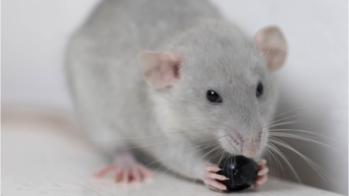
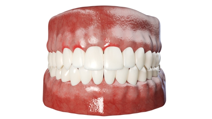
Periodontal disease affects more than 47% of adults aged 30 and over. For people over 65 years of age, that number rises to over 70%, making periodontitis one of the most commonly observed age-related illnesses.

LLP Vitadao
Visit the VitaDAO website to learn how you can get involved.

Jonathan An’s lab seeks to research inflammation-targeting compounds that can help treat periodontal disease and reduce its negative impact on human longevity. Recently, VitaDAO, a biotech DAO funding early-stage research projects, voted to fund An’s project to the sum of $300,000 via an IP-NFT.

Research focused on gum health

Led by Dr. Jonathan An, Assistant Professor at the University of Washington, Dr. Matt Kaeberlein, Professor at the University of Washington, and Dr. Simon Johnson, Assistant Professor at the University of Washington, this periodontal disease research project aims to discover a geroscience-based treatment for age-related periodontitis.

Current therapies in the field of periodontal disease, such as scaling and deep cleaning, involve treating the symptoms of the disease rather than the underlying causes of the illness. Although periodontitis occurs in more than 70% of the population over the age of 65, little research has been done to specifically target the disease.

Jonathan An’s team proposes a novel approach that places the low-grade inflammation associated with both periodontitis and aging at the center of the lab’s research and seeks to test and record a series of compounds to treat such inflammation.

How is periodontitis research connected to longevity?

The underlying factor of low-grade inflammation in the absence of infection has long been noted in longevity science. Known as inflammaging, it is commonly observed not only in periodontal disease but also in other age-related illnesses, such as Alzheimer’s, heart disease, and diabetes. Dr. An’s team supposes that by targeting such inflammation, the onset and worsening of periodontal disease could be prevented, and there could be an additional positive impact on overall human healthspan.

By testing small molecule inhibitors of the PI3K/NFkB/mTOR pathway, the research team aims to establish a novel therapy for periodontal disease. As a positive control, the researchers plan to use rapamycin, which has supposed oral rejuvenation effects when used in mice. Drawing from this research, An’s team believes that these small molecules could ameliorate the inflammation that leads to periodontal disease or even the disease itself.

What is the role of VitaDAO?

In April 2022, 99.87% of VitaDAO voted in favor of funding Dr. An’s research with $300,000 (USD). The funding will be delivered under the DAO’s innovative IP-NFT format, which covers various elements of the project, including animal ordering and facility costs, the research team, inflammatory panels and antibodies, lab consumables and drug costs, microCT costs, and personnel wages.

VitaDAO believes that the lab’s geroscience-based approach could have a potential positive impact on inflammaging with wider-reaching impacts in the longevity field, such as possible discoveries about other targets in the PI3K/NFkB/mTOR pathway. In spite of the lab’s inexperience in entrepreneurial ventures, VitaDAO has confidence in the project’s feasibility.

Backing the project via an IP-NFT, the DAO believes a number of therapeutics could result from the research, including localized oral cavity treatments, such as compound-based toothpaste, drect delivery solutions to periodontal pockets, and compound trays, such as whitening tray solutions.

Although this project is still in its early stages, if successful, it may lead to further research down the line and additional spin-off projects that are closely related to the longevity field.

Summary

Receiving a 99.87% favorable vote, Dr. Jonathan An’s team was awarded $300,000 to fund its novel periodontitis research project that seeks to reduce inflammaging. VitaDAO believes such geroscience-based treatments will have a positive impact not only on oral health but also on longevity.

We would like to ask you a small favor. We are a non-profit foundation, and unlike some other organizations, we have no shareholders and no products to sell you. All our news and educational content is free for everyone to read, but it does mean that we rely on the help of people like you. Every contribution, no matter if it’s big or small, supports independent journalism and sustains our future.
Small healthy meal

Caloric Restriction Improves Immune System Function

A new study published in Mechanisms of Aging and Development has shown that caloric restriction effectively restores T cell abundance in aged mice [1].

Is it really just about getting fewer calories?

Caloric restriction has become a well-known anti-aging intervention, as it can reverse several hallmarks of aging and extend lifespan in different animal models. Due to the positive effects achieved with caloric restriction and intermittent fasting in animals, many people already practice variations of these regimens. However, the two are distinctly different.

Caloric restriction is simply a reduction of overall calorie intake, while intermittent fasting is a general term for various time-restricted eating regimens that may not necessarily reduce calories. It is still unclear which, if any, of the two is more beneficial for extending human lifespan, particularly in non-obese people who already follow healthy diets. Moreover, there is often overlap between the two in both controlled settings and real situations: time-restricted eating leads to reduced calorie intake and vice versa.

14% caloric restriction was shown to be beneficial even for non-obese healthy people between 24 and 60 years old in the Comprehensive Assessment of Long-Term Effects of Reducing Intake of Energy (CALERIE) trials [2]. However, quality matters as much, if not more, than quantity in diet, and it was shown that the caloric restriction participants followed a nutritionally superior diet according to the diet quality index [3]. This could have contributed to the positive outcomes.

Caloric restriction and the immune system

Participants in one caloric restriction experiment had a greater mass and volume of the thymus, an organ where immune T cell production and maturation takes place [4]. Thymic involution is important in age-associated immune function decline, and efforts are being made to restore this organ in order to achieve some measure of rejuvenation. It would be excellent if simply restricting calories could boost the immune system in this way and ward off infections.

In addition, it was previously shown that caloric restriction attenuates immunosenescence in aged mice [5]. However, the exact mechanisms underlying this phenomenon are still unclear. In this study, the researchers sought to explore the regulation of immunosenescence in mice following caloric restriction in the short and long term.

The diets

Male mice were divided into four groups: a young group on a freely fed normal diet up to the age of 2 months, a control group on a freely fed normal diet up to the age of 6 months followed by a controlled diet (4.5 grams/day) for 12 months, a long-term caloric restriction group on a freely fed normal diet up to the age of 6 months followed by a calorically restricted diet (3.15 g/day), and a short-term caloric restriction group on a freely fed normal diet up to the age of 6 months followed by 11 months on a controlled diet and 1 month on caloric restriction.

Therefore, the experimental groups received 30% fewer calories compared to controls, either over long or short periods of time. Importantly, the control and restricted diets included the same amounts of fiber, vitamins, minerals, and protein. The first change that the researchers observed was an overall weight and fat mass reduction in both short- and long-term calorically restricted groups compared to controls.

The T cells

The cell composition of the spleen was analyzed. Compared to the young group, mice in the control (aged) group were characterized by a decreased number of CD4+ and CD8+ T cells, NK cells, and NKT cells. Long-term caloric restriction reversed these changes except for NK cells. The control group also demonstrated an increase in B cell proportion, but neither of the caloric restriction regimens changed that.

To get a deeper understanding of caloric restriction’s effects on various T cells, the researchers analyzed naive, effector, and memory T cells (both CD4+ and CD8+). The names of these cell types reflect their functions: naive T cells don’t recognize specific antigens and can therefore respond to novel pathogens, effector T cells can interact with specific antigens and give rise to memory T cells, and memory T cells recognize specific antigens from previous encounters.

The researchers show that aging leads to a reduction of naive cells and an increase of effector and memory T cells. Long-term caloric restriction reversed all these changes, while the short-term version only increased the proportion of naive CD8+ T cells.

A closer analysis of effector T cells revealed age-associated changes consistent with increased inflammation in the control group. Long-term caloric restriction somewhat improved the situation but did not reverse the changes completely.

Finally, the researchers assessed if caloric restriction would affect T cell exhaustion, which increases with age. The expression of exhaustion markers was measured, and reduced exhaustion was confirmed in both the short- and long-term groups.

The scientists also measured the expression of the specific transcription factors (TFs) NR4A1 and TOX, which control the exhaustion marker expression. As expected, the expression of these TFs was higher in the control compared to the young group. Long-term caloric restriction brought down the levels of both TFs, whereas the short-term variety reduced only NR4A1.

Abstract

Aging is associated with a decrease in the function of the immune system, a phenomenon known as immunosenescence, which results in reduced resistance to infection. Caloric restriction (CR) is known to prolong lifespan and to regulate immune function. However, whether and how CR affects immunosenescence remains unclear. Here, we evaluated the effect of long- and short-term CR on immunosenescence by subjecting wild-type mice to CR between 6 and 18 months of age or between 17 and 18 months of age, respectively. Compared with a normal diet or short-term CR, long-term CR induced marked or complete attenuation of age-related decreases in the frequency of spleen NK cells and NKT cells; naïve CD4+ and CD8+ T cells; and cytokine- and granzyme B-secreting T cells. In contrast, both long- and short-term CR significantly suppressed age-related upregulation of the T cell exhaustion markers PD-1, Tim-3, and KLRG1, as well as the transcription factors NR4A1 and TOX, which regulate the expression of genes associated with the T cell exhaustion phenotype. These results suggest that CR might suppress age-associated immunosenescence by regulating the expression of transcription factors and target genes that control T cell exhaustion.

Conclusion

The immune system is very complex as evidenced by the plethora of cell types and names used to identify them. It requires fine tuning in order to be efficient at fighting off pathogens while not burdening the organism with excessive inflammation. As demonstrated by this study, both short- and long-term caloric restriction might be a promising approach to attenuate age-accompanied T cell immunosenescence by suppressing cell exhaustion. Although it’s not clear if these results are transferable to humans, the study gives insights about the molecular mechanisms of caloric restriction and some differences between short- and long-term regimens.

We would like to ask you a small favor. We are a non-profit foundation, and unlike some other organizations, we have no shareholders and no products to sell you. All our news and educational content is free for everyone to read, but it does mean that we rely on the help of people like you. Every contribution, no matter if it’s big or small, supports independent journalism and sustains our future.

Literature

[1] Asami, T. et al. Long-term caloric restriction ameliorates T cell immunosenescence in mice. Mech. Ageing Dev. 206, 111710 (2022).

[2] Dorling, J. L. et al. Effects of caloric restriction on human physiological, psychological, and behavioral outcomes: highlights from CALERIE phase 2. Nutr. Rev. 79, 98–113 (2021).

[3] Racette, S. B. et al. Nutritional quality of calorie restricted diets in the CALERIETM 1 trial. Exp. Gerontol. 165, 111840 (2022).

[4] Spadaro, O. et al. Caloric restriction in humans reveals immunometabolic regulators of health span. Science 375, 671–677 (2022).

[5] Yan, X., Imano, N., Tamaki, K., Sano, M. & Shinmura, K. The effect of caloric restriction on the increase in senescence-associated T cells and metabolic disorders in aged mice. PLoS One 16, e0252547 (2021).

Sophora Japonica

Hesperetin Upregulates Metabolism and Longevity in Mice

Researchers publishing in Journal of Biomedical Science have concluded that hesperetin, a compound found in various herbs, improves longevity in mice by promoting the expression of the pro-longevity gene Cisd2.

Mitochondria and Cisd2

This paper begins with an explanation of mitochondrial dysfunction, one of the hallmarks of aging, and the function of Cisd2. Previous research has shown that Cisd2 expression is associated with longevity in mice as it aids in mitochondrial function [1] and that Cisd2 deficiency is associated with premature aging in mice [2].

These researchers have performed previous work showing that Cisd2’s effects on mitochondria are due to its effects on calcium homeostasis and that its deficiency causes problems in multiple organs [3], as mitochondria become overloaded with calcium ions. This led to a drug discovery process in which the researchers searched for compounds that might influence Cisd2.

An effective compound from an herbal dictionary

Using a transgenic mouse model and a cell line, the researchers screened 60 herbs derived from a book on Chinese traditional medicine. Careful analysis of the compounds present in these herbs singled out hesperetin, a structural analog of the sophricoside and genistein found in Sophora japonica, as a uniquely powerful Cisd2 activator.

The researchers chose to treat wild-type 21-month-old mice with an effective dose of 100 micrograms per gram of body weight. Hesperetin showed no significant toxicity at this dose. Cisd2, which is abundant in 3-month-old mice but significantly downregulated in untreated 26-month-old mice, was found to be upregulated in the treatment group in multiple tissues. This increase gave the treatment group even greater Cisd2 levels than those of young mice, although this difference was not statistically significant.

The median lifespan of the treatment group was 28.2 months, while a vehicle-treated group lived for 25.95 months and a different control group lived for 26.5 months. There were also statistically significant improvements in lean-to-fat mass ratio, muscle fibers and strength, and heart function. Blood glucose was also notably improved, and the researchers showed that this is due to hesperetin’s beneficial influences on multiple enzymes relating to glucose synthesis and use.

This compound seemed to upregulate metabolism in general. Mice are more active at night, and measurements related to oxygen use and energy expenditure showed that 3-month-old mice, like their older counterparts, are inactive during the day; however, unlike their older counterparts, they are much more active at night. While not all the differences were statistically significant, hesperetin seemed to increase metabolism at all hours of the day rather than restoring this circadian rhythm.

Finally, the researchers examined gene expression. Unsurprisingly, many of the transcriptomic changes brought about by aging were partially reversed by this treatment, particularly in areas related to metabolism, the processing of reactive oxygen species, and protein maintenance.

Conclusion

With many downstream effects that restore youthful abilities, hesperetin clearly influences mitochondrial function in a way that leads to significant and positive effects on metabolism and longevity in mice. Of course, this is still only a mouse study. If hesperetin can be shown to have such a good safety profile with similar effects in human clinical trials, it may be developed as a treatment against metabolic disorders and ultimately become part of a standard regimen to increase lifespan and healthspan.

We would like to ask you a small favor. We are a non-profit foundation, and unlike some other organizations, we have no shareholders and no products to sell you. All our news and educational content is free for everyone to read, but it does mean that we rely on the help of people like you. Every contribution, no matter if it’s big or small, supports independent journalism and sustains our future.

Literature

[1] Wu, C. Y., Chen, Y. F., Wang, C. H., Kao, C. H., Zhuang, H. W., Chen, C. C., … & Tsai, T. F. (2012). A persistent level of Cisd2 extends healthy lifespan and delays aging in mice. Human molecular genetics, 21(18), 3956-3968.

[2] Chen, Y. F., Kao, C. H., Chen, Y. T., Wang, C. H., Wu, C. Y., Tsai, C. Y., … & Tsai, T. F. (2009). Cisd2 deficiency drives premature aging and causes mitochondria-mediated defects in mice. Genes & development, 23(10), 1183-1194.

[3] Shen, Z. Q., Chen, Y. F., Chen, J. R., Jou, Y. S., Wu, P. C., Kao, C. H., … & Tsai, T. F. (2017). CISD2 haploinsufficiency disrupts calcium homeostasis, causes nonalcoholic fatty liver disease, and promotes hepatocellular carcinoma. Cell reports, 21(8), 2198-2211.

No results

Vitamin D Fails to Improve Bone Health in Mega-Study

A high-quality, randomized, controlled trial found no effect of vitamin D supplementation or blood levels on the incidence of fractures in an aging population [1].

The common perception doesn’t match the science

It’s a long-held aphorism that vitamin D is good for bone health. This has been popular since it was discovered to be a cure for rickets, the child-specific form of osteomalacia (bone softening). Today, many products aimed primarily at children, such as milk and breakfast cereals, are fortified with vitamin D, which has also become a very popular supplement among adults. This includes many people in the longevity field, as age-related osteoporosis is a genuine concern, especially among women [2]. Some enthusiasts even advocate for vitamin D “mega-dosing”.

As the authors of this new study note, between 1999 and 2012, the use of vitamin D supplements increased from 5.1% to 19% among U.S. adults. However, scientists have been unable to confirm any positive effects of vitamin D supplementation except for preventing rickets and osteomalacia in high-risk groups [3].

Robust design

Several years ago, an extensive VITAL trial was commenced to study the health effects of vitamin D supplementation on cancer and cardiovascular disease. Almost 26 thousand men older than 50 and women older than 55 were enrolled. The results, however, were disappointing: vitamin D showed no significant effects on any of the endpoints [4]. This latest ancillary study used VITAL’s database to look for possible correlations between vitamin D supplementation and bone health, which is exactly where vitamin D is supposed to shine.

The study design was considerably robust. For instance, it included a three-month placebo-only run-in to identify participants who were reliably taking their pills at home. Only compliant participants were included in the actual study. The researchers also controlled for confounding variables that could independently affect bone health. The participants were not stratified by their baseline vitamin D levels, meaning that both people with healthy levels of vitamin D and those with vitamin D deficiency were included.

Current recommended daily vitamin D consumption varies from 800 to 2000 IU (international units), depending on which organization you ask. Participants in the study group were receiving 2000 IU daily to achieve mean vitamin D blood levels of 40 micrograms per liter, which is on the higher end of the current recommendations. Incidence of bone fractures was chosen as the primary endpoint, and the median follow-up was 5.3 years.

D for disappointment

Mean vitamin D levels increased from 29.2 to 41.2 micrograms per liter in the study group while decreasing from 30.0 to 29.4 micrograms per liter in the placebo group. However, this did not lead to any statistically significant effect on most endpoints. Suprisingly, none of the vitamin D thresholds (<12, <20, <30, or ≥50 micrograms per liter) significantly influenced the effect – or, rather, the lack thereof.

Patients with abnormally low, abnormally high, or normal levels of vitamin D in blood serum were equally prone to first-time as well as recurrent fractures. There was also no effect modification by baseline age, sex, race or ethnic group, BMI, or personal use of supplemental calcium. Notably, the same group, also working on VITAL data, previously found that vitamin D supplementation did not affect incident fall risk or changes in bone mineral density or structure [5].

Vitamin D Bone Health

Among 25,871 participants (50.6% women [13,085 of 25,871] and 20.2% Black [5106 of 25,304]), we confirmed 1991 incident fractures in 1551 participants over a median follow-up of 5.3 years. Supplemental vitamin D3, as compared with placebo, did not have a significant effect on total fractures (which occurred in 769 of 12,927 participants in the vitamin D group and in 782 of 12,944 participants in the placebo group; hazard ratio, 0.98; 95% confidence interval [CI], 0.89 to 1.08; P=0.70), nonvertebral fractures (hazard ratio, 0.97; 95% CI, 0.87 to 1.07; P=0.50), or hip fractures (hazard ratio, 1.01; 95% CI, 0.70 to 1.47; P=0.96). There was no modification of the treatment effect according to baseline characteristics, including age, sex, race or ethnic group, body-mass index, or serum 25-hydroxyvitamin D levels. There were no substantial between-group differences in adverse events as assessed in the parent trial.

Conclusion

This meticulous study is a great illustration of how deceptive “common knowledge” can be. While many signs had pointed to vitamin D being essential for bone health, studies have repeatedly produced inconclusive results, and this new study, due to its robust design and large sample size, might be the last nail in the myth’s coffin. While it is still possible that vitamin D supplementation conveys certain health benefits (although benefits for CVD and cancer have also been all but ruled out), if you want to keep your bones strong as you age, you probably should look elswhere, such as at resistance training [6]. As for mega-dosing, some previous research suggests that it can actually be bad for you [7].

We would like to ask you a small favor. We are a non-profit foundation, and unlike some other organizations, we have no shareholders and no products to sell you. All our news and educational content is free for everyone to read, but it does mean that we rely on the help of people like you. Every contribution, no matter if it’s big or small, supports independent journalism and sustains our future.

Literature

[1] LeBoff, M. S., Chou, S. H., Ratliff, K. A., Cook, N. R., Khurana, B., Kim, E., … & Manson, J. E. (2022). Supplemental Vitamin D and Incident Fractures in Midlife and Older Adults. New England Journal of Medicine, 387(4), 299-309.

[2] Li, G., Thabane, L., Papaioannou, A., Ioannidis, G., Levine, M. A., & Adachi, J. D. (2017). An overview of osteoporosis and frailty in the elderly. BMC musculoskeletal disorders, 18(1), 1-5.

[3] Reid, I. R., Bolland, M. J., & Grey, A. (2014). Effects of vitamin D supplements on bone mineral density: a systematic review and meta-analysis. The Lancet, 383(9912), 146-155.

[4] Manson, J. E., Cook, N. R., Lee, I. M., Christen, W., Bassuk, S. S., Mora, S., … & Buring, J. E. (2019). Vitamin D supplements and prevention of cancer and cardiovascular disease. New England Journal of Medicine, 380(1), 33-44.

[5] LeBoff, M. S., Chou, S. H., Murata, E. M., Donlon, C. M., Cook, N. R., Mora, S., … & Manson, J. E. (2020). Effects of supplemental vitamin D on bone health outcomes in women and men in the VITamin D and OmegA‐3 TriaL (VITAL). Journal of Bone and Mineral Research, 35(5), 883-893.

[6] Howe, T. E., Shea, B., Dawson, L. J., Downie, F., Murray, A., Ross, C., … & Creed, G. (2011). Exercise for preventing and treating osteoporosis in postmenopausal women. Cochrane database of systematic reviews, (7).

[7] Sanders, K. M., Stuart, A. L., Williamson, E. J., Simpson, J. A., Kotowicz, M. A., Young, D., & Nicholson, G. C. (2010). Annual high-dose oral vitamin D and falls and fractures in older women: a randomized controlled trial. Jama, 303(18), 1815-1822.

Rejuvenation Roundup July

Rejuvenation Roundup July 2022

Ending Age-Related Diseases 2022 will occur in less than two weeks, but that’s far from the only thing happening this month, and there’s plenty that happened last month as well. Here’s the latest rejuvenation news from July.

LEAF News

Health Benefits of Cocoa: Ryan O’Shea explains how more evidence has emerged for the heart health benefits of cocoa; it appears that cocoa may lower the risk of death by cardiovascular disease in aging adults.

Age of Elected Officials: Ryan O’Shea jumps into the political arena by focusing on how rejuvenation biotechnology might make Elon Musk’s age-related comments not age well themselves.

Exercise, Metformin, and VO2max: This episode presents a recent study showing that metformin blunts the impact of exercise on performance. Metformin is a commonly prescribed diabetes drug, but it is also popular among biohackers and life extensionists who take it for what they believe to be its longevity benefits.

Interviews

Extracellular Matrix in Aging with Professor Collin Ewald: Dr. Collin Ewald is a molecular biologist and a professor at the Swiss Federal Institute of Technology in Zürich (ETH). At ETH, Collin leads the Laboratory of Extracellular Matrix (ECM) Regeneration. Here, his research focuses on age-related ECM remodeling and related longevity interventions.

Vadim GladyshevVadim Gladyshev on the Fundamentals of Aging: Vadim Gladyshev is a Professor of Medicine at Brigham and Women’s Hospital and Harvard Medical School, and he is one of the most prominent figures in geroscience today. We discussed various competing hypotheses on the nature of aging, the lack of the unified theory of aging and the need for one, and several fascinating papers.

Rejuvenation Roundup Podcast

Ryan O’Shea of Future Grind hosts this month’s podcast, showcasing the events and research discussed here.

Journal Club

Rejuvenating old human skin through VEGF-A: This month, Dr. Oliver Medvedik took a look at a recent paper in which researchers showed that old human skin was rejuvenated when implanted in young mice.

Advocacy and Analysis

Longevity Will Become a Mainstream Part of Drug Development: Joppe Nieuwenhuis, Innovation Scout at Rejuveron Life Sciences, discusses his views about the future of longevity and rejuvenation research and how it will merge with current medical frameworks and systems.

Research Roundup

Rejuvenated skinOld Human Skin Rejuvenated When Implanted in Young Mice: Publishing in Science, researchers have transplanted old human skin to young mice and witnessed a robust rejuvenation cascade that is probably triggered by vascular growth.

Rejuvenating the Brain with Transplanted Astrocytes: A new study published in Stem Cell Research and Therapy has shown that rejuvenating the astrocyte niche in the aged brains of mice leads to improved nervous system function at both the cellular and organismal levels.

Vascular brainMetformin, Rapamycin, NMN, and Vascular Cognitive Impairment: A study published in Frontiers in Neurology has detailed how three well-known longevity-associated compounds affect vascular cognitive impairment (VCI) in a rat model that mimics arterial occlusion in human beings.

Fruits and Vegetables Linked to Fewer Cognitive Disorders: A meta-analysis published in Frontiers in Nutrition has found that intake of fruits and vegetables is negatively correlated with cognitive decline in older adults. Sixteen different studies were used for this meta-analysis, and not all of them measured fruit and vegetable comsuption the same way or measured the same endpoints.

Red grapesShort-Term Resveratrol Reverses Ovarian Aging in Mice: Researchers have shown that resveratrol reverses several features of ovarian aging in mice while activating most sirtuins and boosting mitochondrial health. In many mammals, including humans, ovarian aging is premature compared to other systems.

AI-Powered Novel Therapeutic Target Discovery: A new study conducted by Insilico Medicine in collaboration with leading aging research institutions has identified novel therapeutic targets for amyotrophic lateral sclerosis, a devastating age-associated neurodegenerative disease.

OrgansDifferent Tissues’ Proteins Age in Different Ways: An in-depth preprint published in bioRxiv has thoroughly described how separate tissues lose proteostasis in different ways. The researchers compared protein accumulation between old and young killifish, examining the brain, gut, heart, liver, muscle, skin, and testis.

Mild COVID Can Cause Protracted Neurological Problems: Researchers have shown that even mild SARS-CoV-2 infection can cause lingering neurological symptoms. These findings might shed light on the infamous “long COVID”. COVID survivors sometimes experience cognitive symptoms, such as “brain fog”, that can linger for months.

Standing mouseThe Importance of Mitochondria in Joint Health: Publishing in Aging, a team of Spanish researchers has found that changing the mitochondria of standard Black 6 mice, without any other modifications, leads to marked improvements in joint health.

Bioreactor-Grown Mitochondria for Potential Anti-Aging: Researchers from the University of Connecticut, Stanford University, Université Laval Quebec, and NSU Florida have helped test large-scale exogenous mitochondrial transplants in animal disease models and in-vitro human cells. The results suggest broad efficacy for targeting the immune system, brain, retina, liver, skin, and systemic anti-aging.

Diet and exerciseExercise and Healthy Diet Cannot Replace Each Other: In a large-scale study, scientists have shown that exercise and good diet affect mortality independently, showing that you need both to stay healthy. Exercise is good for you, and so is a healthy diet. Moreover, those are probably the only currently available interventions with a proven effect on healthspan and lifespan.

Fighting Muscle Atrophy with Gene Editing: In a new study published in Journal of Nanobiotechnology, researchers prevented muscle atrophy both in vitro and in vivo by delivering a CRISPR-Cas9 gene editing system packaged in engineered extracellular vesicles.

Not what you wantVitamin D Supplements May Make Alzheimer’s Worse: In a study with data from about 16,000 people, researchers publishing in Aging Cell have found that administering Vitamin D to Alzheimer’s patients may actually make the problem worse.

Oocytes Age Slowly by Remodeling Electron Transport: Scientists have discovered that immature oocytes maintain their youth by shutting down one of the protein complexes that produce energy in mitochondria.

NK cellNAD+ Helps Natural Killer Cells to Fight Cancer: Scientists have shown that the effectiveness of natural killer cells, a crucial component of the immune system, is directly linked to NAD+ levels. Discovering and fighting cancer cells is energetically expensive.

Blueberry Extract Reduces Aging Biomarkers in Rat Livers: A study published in Oxidative Medicine and Cellular Longevity has shown how blueberry extract moderately improves several age-related biomarkers in the livers of aging male rats.

Old politicianPoliticians Live Longer Than Most People: By analyzing historical data, scientists have shown that politicians, an example of an elite group, have enjoyed longer than average life expectancy since about 1950.

Social Frailty Is Correlated with All-Cause Mortality: Scientists have shown that social frailty – a composite index based on income level, household status, and social activity – significantly affects all-cause mortality in the elderly, with poverty being the predominant factor.

GlutamineGlutamine Supplementation Has Anti-Aging Potential: A new study published in Frontiers of Pharmacology has shown that glutamine deprivation might speed up aging while glutamine supplementation reduces oxidative stress-induced senescence in mice.

Loss of IGF-1 May Contribute to Neurovascular Aging: Researchers publishing in GeroScience have reported that the age-related decline in IGF-1 levels is associated with neurovascular aging. However, given the weak correlations, even if there is a causal relationship, it is likely to be only one of many factors.

Future ClockEpigenetic Age Acceleration Is Correlated with Mortality: In an important step for the longevity field, scientists have found that epigenetic age acceleration, as measured by four popular methylation clocks, is correlated with aging and mortality in humans.

Acetyl-cholinesterase-inhibitors slow cognitive decline and decrease overall mortality in older patients with dementia: Among older people with dementia, treatment with these drugs was associated with a slower cognitive decline and with reduced mortality, after a mean follow-up of almost eight years.

Targeted Plasmalogen Supplementation in Cognitively Impaired Persons: Changes in cognition and mobility were statistically significant versus a random outcome. Baseline DHA-containing plasmalogen levels were not predictive of clinical response. DHA-plasmalogens were well tolerated at all dosages, and no adverse reactions were observed.

Age estimation from sleep studies using deep learning predicts life expectancy: Sleep disturbances increase with age and are predictors of mortality. In this study, the researchers present deep neural networks that estimate age and mortality risk through polysomnograms.

Mitochondrial Transfusion Improves Mitochondrial Function in the Hippocampus of Aged Mice: Methods of mitochondrial transfusion should be further tested to treat a variety of human diseases or disorders and to slow down or reverse processes of aging.

Anti-Aging Effect of the Stromal Vascular Fraction/Adipose-Derived Stem Cells in a Mouse Model of UVB-Induced Skin Aging: Changes in the epidermal cell layer were clearly observed after 7 days of treatment. After 28 days of treatment, the dermal tissue was thickened, and the collagen content and proportion were improved.

Pharmacological senolysis reduces doxorubicin-induced cardiotoxicity and improves cardiac function in mice: The researchers hold that a senolytic approach might help to overcome the clinical limitations of current chemotherapy treatments.

Elimination of Senescent Cells by Senolytics Facilitates Bony Endplate Microvessel Formation and Mitigates Disc Degeneration in Aged Mice: These findings may provide evidence that senolytics can eliminate the senescent cells and facilitate microvascular formation in the marrow space of the bony endplate.

The importance of elders: Extending Hamilton’s force of selection to include intergenerational transfers: These results formalize the theory that longevity can be favored under socioecological conditions characterized by parental and alloparental care funded through transfers of mid- to late-life production surpluses.

The damage-independent evolution of aging by selective destruction: Selective destruction could provide a proximal cause of aging that is both consistent with the gene expression hallmarks of aging and independent of accumulating damage.

News Nuggets

Rejuveron Press ReleaseSecond Rejuveron Company Transitions into Clinical Stage: Rejuveron Life Sciences AG (‘Rejuveron’), a Zürich-based biotechnology company developing therapies to promote healthy aging, is pleased to announce that the first participant has been treated in Endogena Therapeutics’ phase 1/2a clinical study of its lead product, EA-2353, a photoreceptor regeneration treatment for retinitis pigmentosa (RP).

The Evandro Fang Lab Receives VitaDAO’s Third IP-NFT Funding: On June 15, 2022, VitaDAO hosted an online IP-NFT Transfer Ceremony honoring its third project funded via an IP-NFT: Evandro Fang Lab. The finances delivered to this lab will go toward further research into identifying novel mitophagy inducers that contribute to healthy brain aging and longevity.

We would like to ask you a small favor. We are a non-profit foundation, and unlike some other organizations, we have no shareholders and no products to sell you. All our news and educational content is free for everyone to read, but it does mean that we rely on the help of people like you. Every contribution, no matter if it’s big or small, supports independent journalism and sustains our future.
Mebots

Mebots – Merging Gaming with Fitness

The pandemic has pushed many people indoors, and for many people, this has meant more time watching TV or using the computer. Screen time is now an average of 7 hours per day, which has led to all-time highs of obesity, worsening health, anxiety, and depression.

There’s a global health crisis, and Forever Today is hoping to merge the world of gaming with fitness and health to combat this problem.

Forever builds Web3, social games for healthy habits

Forever’s first game was a steps competition between NYC and Miami: 20,000 people signed up in less than a month and took more than 500 million steps. This was a great initial effort for gamifying fitness and healthy lifestyles, but the company wanted to do more.

Therefore, Forever is launching a character-based game called Mebots. In a way, it works like Pokemon Go. It measures how many steps you walk, health data, and other actions in the physical world to determine information about digital avatar. The more you walk and stay healthy, the more your digital twin improves and changes in its appearance.

Me + Bot = Mebot is your digital twin, or bio-avatar, a term coined by our President Keith Comito as part of our work in decentralized science. It’s the first web3 character that becomes healthier as you exercise IRL. It’s an NFT fitness collectible that is made more unique and valuable with each step you take in real life.

Once you have your Gen-0 Mebot, you can earn heart tokens through walking and exercise. These tokens can then be used to create new baby Gen-1 Mebots. These baby bots then need to be cared for and raised to maturity like Pokemon or Tamagotchi pets. Once they become adults, their unique next-generation characteristics are revealed.

To encourage people to be more healthy, taking more steps than other players also increases the chance to create a more rare Gen-0 Mebot.

To be most successful in the game, players can partner up with other players and create mutual contracts. Working together in a contract boosts the points you earn. In this way, the game encourages social interaction and cooperation towards staying healthy.

Mebots is the first social fitness game on Web3, and Forever hopes to help players live healthier, happier lives and become their best selves.

Forever is also supporting lifespan.io

Forever is also supporting the work of lifespan.io by donating a portion of its proceeds to help our nonprofit continue to publish news and educational materials, fundraise for research, and do the many other things we do.

We would like to ask you a small favor. We are a non-profit foundation, and unlike some other organizations, we have no shareholders and no products to sell you. All our news and educational content is free for everyone to read, but it does mean that we rely on the help of people like you. Every contribution, no matter if it’s big or small, supports independent journalism and sustains our future.
Future Clock

Epigenetic Age Acceleration Is Correlated with Mortality

In an important step for the longevity field, scientists have found that epigenetic age acceleration, as measured by four popular methylation clocks, is correlated with aging and mortality in humans [1].

Lack of human data

Studying aging obviously requires having reliable biomarkers of aging. Epigenetic clocks that utilize the correlation between aging and genome methylation have emerged as one of the best and most versatile biomarkers of aging, despite the fact that we still don’t really know what causes this correlation. These clocks are widely used in preclinical studies where they successfully predict mortality in animal models, but human data is still very limited, not least because of our species’ long lifespan. While epigenetic clocks have shown correlation with various health conditions in humans [2], mortality would be an important test.

In some human population studies, blood samples are collected and stored, which makes possible applying methylation clocks to those samples years and decades later. This, in turn, enables scientists to search for correlations between epigenetic age and mortality. While several attempts to do so have already been made [3], this new study is one of the most ambitious to date.

All-female cohort

The researchers used data from the Women’s Health Initiative, an important population study, and the sample size was a respectable 1813. Needless to say, all the subjects were women, which was one of the study’s limitations. As is well known, women on average live longer than men. This is likely to go beyond environmental and lifestyle factors, reflecting yet unknown sexual differences in aging.

The researchers analyzed whether or not the participants survived to the age of 90 and whether or not they had impairments in mobility and cognition. The researchers make an interesting argument that while surviving to the age of 90 was considered unusual in the not-so-distant past, today, it is a suitable longevity threshold (i.e., more or less the norm), especially in women. Samples were analyzed using four popular methylation clocks: the Horvath pan-tissue clock, the Hannum clock, PhenoAge, and GrimAge.

Among the participants, 464 women experienced healthy longevity (i.e., survived to the age of 90 with intact mobility and cognitive function); 420 women survived to the age of 90 years without intact mobility, cognitive functioning, or either; and 929 women did not survive. The mean age at baseline was almost identical across all categories (71.6, 71.3, and 70.2 respectively).

The profile of this aging female population contains few surprises. Women categorized as healthy at 90 (with both mobility and cognitive function intact) were more likely to be college graduates and white, had no history of smoking, had a normal or slightly elevated BMI, and had a habit of walking 2 to 6 times a week. This last parameter can be deceptive: while it would be reasonable to suggest that those women stayed healthy due to their regular walks, they also might have been able to perform regular walks due to being healthy in the first place.

A win for the clocks

The researchers looked at epigenetic age acceleration (EAA), which is the difference between a person’s chronological and biological age. If biological age as measured by an epigenetic clock is higher, the person is considered to be suffering from accelerated aging. In a major win for epigenetic clocks, all four of them showed strong correlation between EAA and the study’s outcomes. For every 1 standard deviation increase in EAA, the odds of surviving to the age of 90 with intact mobility and cognitive function vs not surviving at all were lower by 20 to 40 percent, depending on the clock. The odd ratios were almost similar when only mobility was taken into account. Correlations were also evident for “unhealthy aging” vs death.

This cohort study’s findings suggest that EAA may be a valid biomarker associated with healthy longevity among older women. Our results suggest that EAA may be used for risk stratification and risk estimation for future survival with intact mobility and cognitive functioning within populations. Future studies could usefully focus on the potential for public health interventions to reduce EAA and associated disease burden while increasing longevity.

Conclusion

This study, one of the first to link epigenetic aging acceleration to human mortality, is an important step in establishing epigenetic clocks as reliable biomarkers of aging that can be used in human clinical trials to test new anti-aging interventions. Interestingly, it can also eventually make epigenetic age a metric of choice for insurance companies (a couple of start-ups, such as FOXO Technologies, are already operating in this field). The study provides a solid proof of concept, though additional research on more heterogenic populations is needed.

We would like to ask you a small favor. We are a non-profit foundation, and unlike some other organizations, we have no shareholders and no products to sell you. All our news and educational content is free for everyone to read, but it does mean that we rely on the help of people like you. Every contribution, no matter if it’s big or small, supports independent journalism and sustains our future.

Literature

[1] Jain P, Binder AM, Chen B, et al. Analysis of Epigenetic Age Acceleration and Healthy Longevity Among Older US Women. JAMA Netw Open. 2022;5(7):e2223285.

[2] Lo, Y. H., & Lin, W. Y. (2022). Cardiovascular health and four epigenetic clocks. Clinical Epigenetics14(1), 1-10.

[3] Lu, A. T., Quach, A., Wilson, J. G., Reiner, A. P., Aviv, A., Raj, K., … & Horvath, S. (2019). DNA methylation GrimAge strongly predicts lifespan and healthspan. Aging (Albany NY)11(2), 303.

brain vasculature

Loss of IGF-1 May Contribute to Neurovascular Aging

Researchers publishing in GeroScience have reported that the age-related decline in IGF-1 levels is associated with neurovascular aging [1].

IGF-1 and brain vasculature

This paper begins with a discussion of neurovascular coupling (NVC) and its relationship to vascular dementia. Blood flow to the brain is extremely important, both to provide active brain regions with oxygen and nutrients and to wash out toxic metabolites. Murine studies have shown that oxidative stress is connected with the disruption of this vascular system [2].

Separate murine studies have also shown that IGF-1 deficiency in mice results in impaired NVC [3] and that specifically disrupting this signaling results in an impaired NVC response [4]. However, these are still studies involving transgenic mice. These researchers, therefore, sought to determine if a relationship between IGF-1 and NVC in human beings could be determined.

A human analysis

For this study, the younger group consisted of people with an average age of nearly 30 years, and the older group consisted of 32 people with an average age of nearly 70 years. People with a wide variety of medical conditions, including any condition that could affect IGF-1, were excluded from participation.

There was no measured difference between the sexes in IGF-1 levels. However, there was a gulf between some participants and others of the same age; some younger people had substantial amounts of this molecule, while others had levels that were more similar to members of the older group.

As expected, the older group suffered from deficiencies in neurovascular coupling, as measured by the cerebrovascular conductance index. Cerebral vasculature normally significantly responds when the brain is being put to a task, but this response was significantly diminished in the older group compared to the younger group. The basal level of cerebral blood flow was also significantly decreased in the older group.

IGF-1 and neurovascular response were found to be weakly but significantly correlated, although many (but not all) younger participants with low IGF-1 levels still enjoyed much stronger task responses than their older counterparts with similar IGF-1 levels, in both halves of the brain.

This was not true for basal cerebral bloodflow, however. The correlation between IGF-1 and this measurement was somewhat stronger, and older and younger people with similar levels of IGF-1 often, but not always, had similar amounts of cerebral bloodflow.

Conclusion

While the animal studies might have pointed towards a causal relationship, this is still only a longitudinal study, so causation in human beings cannot be proved. Given the weak correlations, it is very likely that, even if there is a causal relationship, it is only one of many factors. However, in light of the extensive preclinical evidence, IGF-1 is clearly worth exploring as a druggable, therapeutic target, particularly as part of a combination therapy. Vascular dementia is a serious issue among the elderly, and if it can be slowed or halted, it might improve the quality and length of life for a great many people.

We would like to ask you a small favor. We are a non-profit foundation, and unlike some other organizations, we have no shareholders and no products to sell you. All our news and educational content is free for everyone to read, but it does mean that we rely on the help of people like you. Every contribution, no matter if it’s big or small, supports independent journalism and sustains our future.

Literature

[1] Toth, L., Czigler, A., Hegedus, E. et al. Age-related decline in circulating IGF-1 associates with impaired neurovascular coupling responses in older adults. GeroScience (2022). https://doi.org/10.1007/s11357-022-00623-2

[2] Tarantini, S., Tran, C. H. T., Gordon, G. R., Ungvari, Z., & Csiszar, A. (2017). Impaired neurovascular coupling in aging and Alzheimer’s disease: contribution of astrocyte dysfunction and endothelial impairment to cognitive decline. Experimental gerontology, 94, 52-58.

[3] Toth, P., Tarantini, S., Ashpole, N. M., Tucsek, Z., Milne, G. L., Valcarcel‐Ares, N. M., … & Ungvari, Z. (2015). IGF‐1 deficiency impairs neurovascular coupling in mice: implications for cerebromicrovascular aging. Aging cell, 14(6), 1034-1044.

[4] Tarantini, S., Balasubramanian, P., Yabluchanskiy, A., Ashpole, N. M., Logan, S., Kiss, T., … & Ungvari, Z. (2021). IGF1R signaling regulates astrocyte-mediated neurovascular coupling in mice: implications for brain aging. Geroscience, 43(2), 901-911.

Glutamine

Glutamine Supplementation Has Anti-Aging Potential

A new study published in Frontiers of Pharmacology has shown that glutamine deprivation might speed up aging while glutamine supplementation reduces oxidative stress-induced senescence in mice [1].

Glutamine is an amino acid of paramount importance for many cellular functions. It is involved in the synthesis of various crucial molecules, such as the neurotransmitter glutamate and the energy molecule ATP. Recently, there has been an increased interest in the link between glutamine metabolism and aging.

Glutamine-glutamate levels are reduced in the brains of patients with Alzheimer’s disease [2], although studies examining this difference between Alzheimer’s patients and healthy controls have shown controversial results [3]. Nevertheless, therapeutic approaches aimed at restoring the imbalance between the glutamate (excitatory) and GABA (inhibitory) systems observed in Alzheimer’s disease are already being investigated in clinical trials.

Glutamine deprivation has been shown to stimulate senescence, while glutamate supplementation has demonstrated a neuroprotective effect in mouse models of some neurodegenerative diseases [4].

Glutamine was also shown to be vital for the regulation of mTOR, thus involved in autophagy, which plays a critical role in the context of aging. However, the exact role that glutamine plays in autophagy is unknown.

In this study, the researchers sought to investigate the role that glutamine plays in senescence and aging by uncovering the pathways activated by glutamine deprivation. They also explored if glutamine supplementation would be beneficial in a mouse model of accelerated aging.

First clues

In their first series of experiments, the researchers examined the effects of glutamine deprivation in vitro. They placed two cell lines in either glutamine-containing (control) or glutamine-free (experimental) mediums. Glutamine deprivation resulted in increased cell death, reduced cell proliferation, and induced expression of senescence-associated genes.

The researchers then exposed fruit flies to a glutamine-deficient diet to check if they could get similar results in vivo. Indeed, flies following the diet had a reduced lifespan and an increased level of senescence, as evidenced by accumulation of the senescence marker SA-β-gal in the gut.

Getting deeper

Next, the researchers assessed how glutamine deprivation affects autophagy using cell cultures. They showed that a glutamine-free medium impaired autophagy via activation of mTOR and disrupted lysosomal function, as evidenced by p62 accumulation and reduced expression of the autolysosomal gene TFEB and its targets, respectively, particularly in the long term.

Meanwhile, inhibiting mTOR signaling with either rapamycin or the PI3K/Akt inhibitor LY294002 mitigated the autophagy impairment and senescence in glutamine-deprived cells. Therefore, this set of experiments confirmed that low glutamine levels inhibit autophagy by activating mTOR and inducing senescence.

Aging context

In the final set of experiments, the researchers explored if glutamine supplementation could fight senescence induced by oxidative stress. First, they exposed a cell culture to hydrogen peroxide (H2O2) to induce oxidative stress. Glutamine supplementation, as expected, reduced senescence in this model.

Then, the scientists used a senescence-induced progeric mouse model to assess the effect of glutamine supplementation in vivo. In this case, D-galactose-treated mice were used as a model of accelerated aging caused by an increased oxidative stress. The mice were given 3% glutamine in drinking water for two months. As a result, the mice in the glutamine supplementation group showed improved hair gloss and density, restored muscle strength, reduced senescence, and increased autophagy compared to control D-galactose mice.

Abstract

Glutamine is a conditionally essential amino acid involved in energy production and redox homeostasis. Aging is commonly characterized by energy generation reduction and redox homeostasis dysfunction. Various aging-related diseases have been reported to be accompanied by glutamine exhaustion. Glutamine supplementation has been used as a nutritional therapy for patients and the elderly, although the mechanism by which glutamine availability affects aging remains elusive. Here, we show that chronic glutamine deprivation induces senescence in fibroblasts and aging in Drosophila melanogaster, while glutamine supplementation protects against oxidative stress-induced cellular senescence and rescues the D-galactose-prompted progeria phenotype in mice. Intriguingly, we found that long-term glutamine deprivation activates the Akt-mTOR pathway, together with the suppression of autolysosome function. However, the inhibition of the Akt-mTOR pathway effectively rescued the autophagy impairment and cellular senescence caused by glutamine deprivation. Collectively, our study demonstrates a novel interplay between glutamine availability and the aging process. Mechanistically, long-term glutamine deprivation could evoke mammalian target of rapamycin (mTOR) pathway activation and autophagy impairment. These findings provide new insights into the connection between glutamine availability and the aging process.

Conclusion

Although this study mostly explored the effect of glutamine deprivation in cell cultures and fruit flies, it provides some mechanistic insights regarding the role of glutamine in aging. Low levels of glutamine might impair autophagy via activating mTOR signaling, while glutamine supplementation could be a potential therapeutic approach for age-associated pathologies characterized by reduced levels of glutamine. This certainly requires additional research, especially to understand why glutamine levels go down in the first place.

We would like to ask you a small favor. We are a non-profit foundation, and unlike some other organizations, we have no shareholders and no products to sell you. All our news and educational content is free for everyone to read, but it does mean that we rely on the help of people like you. Every contribution, no matter if it’s big or small, supports independent journalism and sustains our future.

Literature

[1] Zhou, J. et al. Glutamine Availability Regulates the Development of Aging Mediated by mTOR Signaling and Autophagy. Front. Pharmacol. 13, 924081 (2022).

[2] Huang, D. et al. Glutamate-glutamine and GABA in brain of normal aged and patients with cognitive impairment. Eur. Radiol. 27, 2698–2705 (2017).

[3] Madeira, C. et al. Elevated Glutamate and Glutamine Levels in the Cerebrospinal Fluid of Patients With Probable Alzheimer’s Disease and Depression. Front. Psychiatry 9, 561 (2018).

[4] Chen, J. et al. The impact of glutamine supplementation on the symptoms of ataxia-telangiectasia: a preclinical assessment. Mol. Neurodegener. 11, 60 (2016).

Elderly poverty

Social Frailty Is Correlated with All-Cause Mortality

Scientists have shown that social frailty – a composite index based on income level, household status, and social activity – significantly affects all-cause mortality in the elderly, with poverty being the predominant factor [1].

There is more than one way to be frail

Physical frailty is a known companion of aging and a major risk factor for several age-related diseases [2]. As such, it gets a lot of attention from the longevity community. However, there is another type of frailty that usually evades the spotlight: social frailty. This rather novel concept is defined as “the absence of social resources, social activities, and self-management abilities that are important for fulfilling basic social needs” [3]. In other words, it is mostly about poverty, disability, and loneliness. In this new study, a group of European scientists argues that social frailty is a potent predictor of all-cause mortality in the elderly.

While it has been long known that poverty is significantly inversely correlated with health and longevity [4], the COVID pandemic has highlighted the adverse effects of a lack of social interaction. Several studies have shown that older people who were socially isolated during the lockdowns were also significantly more likely to develop cognitive impairment than those who were able to maintain social activity.

Some previous studies have already documented high prevalence of social frailty among older adults suffering from dementia, memory decline, and cognitive impairment, but those studies suffered from limitations such as small sample sizes [5]. This time, the researchers used data from the English Longitudinal Study of Ageing (ELSA) regarding over 4,000 elderly people, with a relatively long follow-up of 10 years.

Money or friends? Better have both

The social frailty index used by the researchers was based on income (UK poverty threshold of £20,346 per household per year), household status (living alone or not), participation in social activities, and contacts with friends (at least once a week in person, over the phone, or by email). For each risk factor a participant got a score of either 1 or 0. Social frailty was defined as 2 or more points, pre-frailty as 1 point, and a total absence of risk factors (robustness) as 0 points.

Of course, in such studies, everything revolves around the chicken and egg dilemma. It could be, for instance, that health is negatively influenced by the lack of social interactions, the other way around, or a bi-directional relationship. In addition to the long follow-up period, the researchers tried to increase the robustness of the study by controlling for several important variables: age, sex, education, marital status, body mass index, smoking status, disability, levels of physical activity, the presence of comorbidities, race, and depression symptoms.

Social frailty: highly prevalent

As it turns out, social frailty is highly prevalent among English elderly: it affected 43% of the participants. 39% were categorized as pre-frail, and only 18% as robust. The most frequent problem was poverty, experienced by 64.5% of the participants, and the least frequent was living alone (15.4%).

Unsurprisingly, social frailty was found to be positively correlated with age, lack of physical activity, obesity, smoking, and multimorbidity (numerous health problems). Interestingly, women were much more susceptible to social frailty. We can only speculate about the reasons, but it might be an echo of the gender income gap, which was much wider when the participants were in their productive years than it is today.

In a model fully adjusted for covariates, social frailty significantly correlated with all-cause mortality with a hazard ratio of 1.31 (that is, socially frail people were 31% more likely to die of any cause). When standalone elements of the social frailty index were analyzed, poverty emerged as an even more powerful predictor of mortality, with a hazard ratio of 1.60, while living alone came in second with 1.46.

Our study shows the importance of the issue of social frailty for geriatrics and general practitioners. Research on social frailty could be useful to prevent the onset of pathologies that can cause death in older people. We may suggest that as measures to early detect and prevent social frailty, the implementation of easy accessibility and limited costs by the national health systems may be of help also allowing better use of resources. Therefore, every government should have in its welfare program a scheme to avoid social frailty, and in particular, poverty and living alone for older people.

Conclusion

Despite all their drawbacks, population studies can raise important questions and draw attention to consequential issues. For instance, we can see that even in a country with universally accessible healthcare, poverty remains a major risk factor for all-cause mortality. The COVID pandemic has highlighted the importance of social interactions for our health and the vulnerability of the elderly, as this study confirms. More research is needed to further investigate the correlations revealed in this and similar studies, such as the role of gender.

We would like to ask you a small favor. We are a non-profit foundation, and unlike some other organizations, we have no shareholders and no products to sell you. All our news and educational content is free for everyone to read, but it does mean that we rely on the help of people like you. Every contribution, no matter if it’s big or small, supports independent journalism and sustains our future.

Literature

[1] Ragusa, F. S., Veronese, N., Smith, L., Koyanagi, A., Dominguez, L. J., & Barbagallo, M. (2022). Social frailty increases the risk of all-cause mortality: A longitudinal analysis of the English Longitudinal Study of Ageing. Experimental Gerontology, 111901.

[2] Thillainadesan, J., Scott, I. A., & Le Couteur, D. G. (2020). Frailty, a multisystem ageing syndrome. Age and ageing49(5), 758-763.

[3] Ma, L., Sun, F., & Tang, Z. (2018). Social frailty is associated with physical functioning, cognition, and depression, and predicts mortality. The journal of nutrition, health & aging22(8), 989-995.

[4] Chetty, R., Stepner, M., Abraham, S., Lin, S., Scuderi, B., Turner, N., … & Cutler, D. (2016). The association between income and life expectancy in the United States, 2001-2014. Jama315(16), 1750-1766.

[5] Ong, M., Pek, K., Tan, C. N., Chew, J., Lim, J. P., Yew, S., … & Lim, W. S. (2022). Social frailty and executive function: Association with geriatric syndromes, life space and quality of life in healthy community-dwelling older adults. The Journal of Frailty & Aging11(2), 206-213.

Rat eating blueberry

Blueberry Extract Reduces Aging Biomarkers in Rat Livers

A study published in Oxidative Medicine and Cellular Longevity has shown how blueberry extract moderately improves several age-related biomarkers in the livers of aging male rats.

Rich in polyphenols

In their introduction, the researchers describe polyphenols, a broad category of plant compounds that includes flavonoids. Specifically, blueberries are rich in anthocyanins, a group of polyphenols that has been reported to have positive effects in previous studies, including a rat study that focused on the brain [1]. However, the authors note that blueberry extract’s effects on the liver not been thoroughly analyzed.

Interestingly, this study does not attempt to identify the individual compounds responsible for any effects. Instead, the researchers simply used well-known extraction processes to refine blueberries, administering this extract at 200 milligrams per kilogram of bodyweight. The young rats were 6 months old, and the aged rats were 24 months old. Blueberry extract was orally administered for four weeks, after which the rats were euthanized and their livers examined.

Multiple positive effects

Blueberry extract had a statistically significant but very limited effect on bodyweight. Rats tend to grow larger as they age, but rats given blueberry extract lost a slight amount of weight by the end of the study rather than the gain of the aged control group.

Two enzymes commonly found in the liver, alanine transaminase (ALT), and aspartate transaminase (AST), are often used to indicate liver damage. Both of these enzymes were significantly decreased in the blueberry extract group compared to other aged rats, with AST being more affected than ALT.

In the serum of rats, insulin, cholesterol, and triglycerides increase with advancing age, while Vitamin D and blood glucose decrease. Blueberry extract restored all of these, approximately halfway to youthful levels. Three measurements of inflammation, NF-κB, TNF-α, and IL-6, were also restored halfway to youthful levels.

Measurements of antioxidant enzyme activity were analyzed. For every antioxidant in which aged and young rats significantly differed, blueberry extract significantly increased its level. In many cases, the level of an antioxidant was increased almost exactly to that of young rats.

Conclusion

While this is a preliminary rat study that makes no effort to determine any biochemical effects of the various polyphenols and other compounds in blueberry extract, its significant results warrant further exploration. Finding and analyzing the exact root causes of the myriad of positive effects seen in this study may offer potential breakthroughs in ameliorating liver-related diseases.

We would like to ask you a small favor. We are a non-profit foundation, and unlike some other organizations, we have no shareholders and no products to sell you. All our news and educational content is free for everyone to read, but it does mean that we rely on the help of people like you. Every contribution, no matter if it’s big or small, supports independent journalism and sustains our future.

Literature

[1] Papandreou, M. A., Dimakopoulou, A., Linardaki, Z. I., Cordopatis, P., Klimis-Zacas, D., Margarity, M., & Lamari, F. N. (2009). Effect of a polyphenol-rich wild blueberry extract on cognitive performance of mice, brain antioxidant markers and acetylcholinesterase activity. Behavioural brain research, 198(2), 352-358.

NK cell

NAD+ Helps Natural Killer Cells to Fight Cancer

Scientists have shown that the effectiveness of natural killer cells, a crucial component of the immune system, is directly linked to NAD+ levels [1].

Naturally deadly

Natural killer cells (NK) are an important part of the innate immune system. They got this designation, in arguably the best act of naming in the history of cellular biology, due to their ability to attack damaged or infected cells without requiring activation by antigen-presenting cells. This makes them one of the “first responders”, mostly to viral infections and nascent tumors.

Constantly on patrol, they “shake hands” with cells using various receptors, and if these handshakes are not to their liking, they unleash an arsenal of deadly molecules, such as membrane-piercing perforins and apoptosis-inducing granzymes. NK cells also release cytokines and chemokines that signal to other immune cells to join the fight.

Aging negatively affects many of our immune defenses, including the ability of NK cells to kill bad cells and summon reinforcements [2]. This immunosenescence has been linked to age-related decline in vaccine and anti-tumor response. However, its mechanisms are still not well understood.

NAD+ gives the energy to fight

Cellular warfare requires a lot of energy. Activated NK cells are known to increase their mitochondrial energy production, which is heavily reliant on the coenzyme NAD+. NAD+, a ubiquitous molecule that also facilitates several other important processes, sharply declines with age, probably contributing to many age-related deficiencies [3].

In this new study, the researchers hypothesized that NK’s killing potential is linked to NAD+. First, they established that NK activation by the pro-inflammatory cytokines IL-12 and IL-15 led to an increase in NAD+ and in the expression of enzymes related to NAD+ production. Supplementation of NAD+ in NK cells resulted in the enhanced production of several NK cellular weapons, such as perforins. The cytotoxicity of the treated NK cells increased accordingly, as did their proliferation potential and viability.

NAD+ is produced in cells via three different pathways, and the researchers were able to determine that the increase in NAD+ production in activated NK cells happens mostly due to the salvage pathway. When NAD+ is being used by the cell, it is turned into niacinamide (NAM). The salvage pathway uses the NAD+ precursors nicotinamide riboside (NR) and nicotinamide mononucleotide (NMN) to recycle NAM back to NAD+. The whole process is facilitated by the enzyme NAMPT. The researchers found that NMN supplementation produced the same positive effects as NAD+ supplementation, while inhibiting NAMPT created the opposite effect.

Outsmarting the tumor

If NK cells are supposed to kill cancer cells, why do humans still develop cancer? On top of age-related immunosenescence, the tumor microenvironment is known to impair the cytotoxic and cytokine-producing abilities of immune cells, NK cells included [4]. The researchers assumed that NAD+ might play a role here as well. They isolated NK cells from murine tumors and studied their transcriptomes in order to find significant downregulation of genes related to NAD+ metabolism. In another experiment, media from cancer cells reduced NAD+ levels and NAMPT expression when applied to NK cells.

The researchers also analyzed NAMPT expression in NK cells using 50 human samples of hepatocellular carcinoma (HCC), and they found that it positively correlates with patient survival. In a mouse model of HCC, NAMPT knockout significantly accelerated tumor growth and decreased survival, while supplementation with NMN enhanced the anti-tumor activity of NK cells. In a mouse model of melanoma, the researchers implanted cancer cells together with either NMN-treated or control NK cells and found that the treated cells were much better in suppressing tumor growth.

The researchers were also able to come up with a possible culprit for the decline in fitness that NK cells suffer in a tumor environment. Lactate, the conjugate base of lactic acid and a byproduct of tumor metabolism, is abundant in tumors. Experiments showed that treating NK cells with lactate leads to NAMPT downregulation in a dose-dependent manner, while blocking lactate uptake exerts the opposite effect.

NK cells NMN

Conclusion

This important study joins the growing body of evidence demonstrating that age-related NAD+ deficiency is responsible for various manifestations of aging, including the decline in the immune system’s ability to fight cancer. If this is indeed the case, boosting NAD+ levels, which looks like a solvable problem, can be a potent anti-aging intervention, although we should probably wait for more data before buying a pack of NMN pills online.

We would like to ask you a small favor. We are a non-profit foundation, and unlike some other organizations, we have no shareholders and no products to sell you. All our news and educational content is free for everyone to read, but it does mean that we rely on the help of people like you. Every contribution, no matter if it’s big or small, supports independent journalism and sustains our future.

Literature

[1] Guo, X., Tan, S., Wang, T., Sun, R., Li, S., Tian, P., … & Liang, X. NAD+ salvage governs mitochondrial metabolism invigorating NK cell anti‐tumor immunity. Hepatology.

[2] Hazeldine, J., & Lord, J. M. (2013). The impact of ageing on natural killer cell function and potential consequences for health in older adults. Ageing research reviews12(4), 1069-1078.

[3] Covarrubias, A. J., Perrone, R., Grozio, A., & Verdin, E. (2021). NAD+ metabolism and its roles in cellular processes during ageing. Nature Reviews Molecular Cell Biology22(2), 119-141.

[4] Cong, J., Wang, X., Zheng, X., Wang, D., Fu, B., Sun, R., … & Wei, H. (2018). Dysfunction of natural killer cells by FBP1-induced inhibition of glycolysis during lung cancer progression. Cell metabolism28(2), 243-255.

Early embryo

Oocytes Age Slowly by Remodeling Electron Transport

Scientists have discovered that immature oocytes maintain their youth by shutting down one of the protein complexes that produce energy in mitochondria [1].

Live slow, stay young

Every female is endowed with a finite number of oocytes that form before birth, then reside in the ovaries in a dormant state until maturation and release. Oocytes are faced with a tricky task: they must remain in good shape throughout the body’s reproductive span in order to be able to produce healthy offspring. Interestingly, in addition to oocytes aging very slowly, early embryos undergo an enigmatic rejuvenation event so that the offspring produced by the adult organism starts from age zero [2].

However, as time passes, oocyte quality begins to dwindle, contributing to reproductive dysfunction, which occurs  earlier than many other manifestations of aging. Researchers have been trying to understand the mechanisms involved, but with limited success. In this new groundbreaking study published in Nature, scientists from Spain made a huge leap forward, showing that oocytes modify one of the most important cellular processes so that it produces less damage.

Less energy, less byproducts

Cells acquire energy mostly via the intricate, multi-stage process known as the electron transport chain (ETC), which uses nutrients that we get from food to produce adenosine triphosphate (ATP), the ‘energy currency’ of the cell. Sadly, the system is not perfect. As professor Vadim Gladyshev (who was not involved in this study) explained in his recent interview with lifespan.io, all biological processes produce damage as a byproduct, and the indispensable ones can produce a lot of it.

The byproducts of the ETC are the infamous reactive oxygen species (ROS). While they also do some useful work such as cellular signaling, ROS mostly just wreak havoc, inducing DNA damage and disrupting various cellular processes [3]. Mitochondria are the main, though not the only, source of endogenous ROS.

Since ROS are linked to cellular aging [4], the researchers began with analyzing their levels in human and frog early-stage (immature) oocytes. Amazingly, neither produced any detectable ROS signal. The researchers then determined that the absence of ROS resulted not from their fast degradation by molecules called ROS scavengers, but from decreased ROS production. They also measured mitochondrial membrane potential which is higher when ETC is hard at work. In early-stage oocytes, membrane potential was remarkably low. Together with the low levels of mitochondrial ROS, this indicated reduced ETC activity.

Which one of the five?

The ETC is powered by five protein complexes (large molecules constructed from several different proteins), numbered from I to V. To pinpoint the particular complex that was responsible for the strange behavior of the ETC, the researchers tried shutting down the complexes one by one. Blocking any complex other than Complex I resulted in the oocytes’ death: in other words, only Complex I was not involved in any essential activity. Importantly, Complex I is also the foremost producer of ROS.

Proteomic analysis revealed that in early-stage oocytes, the levels of all ETC subunits (complex-forming proteins) were lower than in mature oocytes, with Complex I subunits almost completely absent. Somehow, oocytes managed to remodel their ETC to exclude Complex I! To date, in no other animal cell type that contains functioning mitochondria, absence of Complex I has ever been detected, and in the plant realm, the only known exception is mistletoe.

Oocytes apparently pay for this trick with diminished overall ETC activity, which is probably just enough to support immature oocytes in their dormant mode. As the researchers discovered, the ETC goes back on track – completely with Complex I – in mature oocytes that must do the heavy lifting of forming a new organism. Predictably, ROS levels also shot up in mature vs early-stage oocytes.

To sum things up, early-stage oocytes reside in a sort of hibernation: they are not really doing anything other than simply surviving, but they also accumulate very little damage. As the researchers note, stem cells, particularly neuronal and hematopoietic stem cells, also exhibit low ETC activity and ROS levels [5], and they are known to age slowly. The next step would be to determine whether stem cells also shut down their Complex I.

Conclusion

This groundbreaking study uncovers a previously unknown mechanism by which oocytes, and maybe also stem cells, greatly reduce their rate of aging. As mature organisms, we need our cells to do stuff (while accumulating damage) rather than lie dormant, so this discovery, as fascinating as it is, might not be immediately translatable into anti-aging therapies. However, it can teach us a lot about aging in general and reproductive aging in particular and, hence, about possible ways to counter it.

We would like to ask you a small favor. We are a non-profit foundation, and unlike some other organizations, we have no shareholders and no products to sell you. All our news and educational content is free for everyone to read, but it does mean that we rely on the help of people like you. Every contribution, no matter if it’s big or small, supports independent journalism and sustains our future.

Literature

[1] Rodríguez-Nuevo, A., Torres-Sanchez, A., Duran, J.M. et al. Oocytes maintain ROS-free mitochondrial metabolism by suppressing complex I. Nature (2022).

[2] Kerepesi, C., Zhang, B., Lee, S. G., Trapp, A., & Gladyshev, V. N. (2021). Epigenetic clocks reveal a rejuvenation event during embryogenesis followed by aging. Science Advances7(26), eabg6082.

[3] Pizzino, G., Irrera, N., Cucinotta, M., Pallio, G., Mannino, F., Arcoraci, V., … & Bitto, A. (2017). Oxidative stress: harms and benefits for human health. Oxidative medicine and cellular longevity2017.

[4] Shields, H. J., Traa, A., & Van Raamsdonk, J. M. (2021). Beneficial and detrimental effects of reactive oxygen species on lifespan: A comprehensive review of comparative and experimental studies. Frontiers in Cell and Developmental Biology9, 628157.

[5] Khacho, M., Harris, R., & Slack, R. S. (2019). Mitochondria as central regulators of neural stem cell fate and cognitive function. Nature Reviews Neuroscience20(1), 34-48.

Not what you want

Vitamin D Supplements May Make Alzheimer’s Worse

Researchers publishing in Aging Cell have found that administering Vitamin D to Alzheimer’s patients may actually make the problem worse.

A matter of cause and effect

Vitamin D deficiency is strongly linked to Alzheimer’s disease [1], and previous research has suggested that addressing this deficiency might be useful in treating the disease [2]. These researchers, however, believe that this conclusion represents a misunderstanding of cause and effect. Their previous work has shown that Alzheimer’s disease causes Vitamin D to be badly processed [3] and that supplementing Vitamin D at the earliest stages of progression in mice does not match human treatment plans.

This study, therefore, involves treating a mouse model of Alzheimer’s in its middle stages with Vitamin D and then comparing these murine results to human longitudinal studies.

A mouse model of Alzheimer’s uses Vitamin D badly

The researchers compared mice bred to have Alzheimer’s-like symptoms to wild-type mice, feeding both groups very specific amounts of Vitamin D. Within the first four months of life, the Alzheimer’s-prone group had significantly less Vitamin D in their plasma and cerebrospinal fluid than the wild-type mice did.

This was followed up with an analysis of Vitamin D in cells. Neurons exposed to amyloid beta, and then dosed with Vitamin D, showed much stronger cellular death (apoptosis) and self-consuming (autophagy) markers than amyloid beta-exposed neurons that were not given Vitamin D.

Encouraged, the researchers then went on to examine the effects of Vitamin D in their Alzheimer’s mouse model. Their findings were as they suspected: Alzheimer’s-prone mice given Vitamin D performed much worse on the cognitive Morris water maze test over time and had larger amyloid beta plaques along with more effects on the microglia.

Further research showed that, instead of the normal VDR/RXR interaction that characterizes normal Vitamin D processing, the presence of amyloid beta caused VDR to interact with p53 instead, which was in line with this team’s previous work [3]. Inhibition of p53 ameliorated many of these symptoms.

A broad source of human data

Taiwan’s national health database is extremely robust. With it, the researchers were able to obtain very specific data for a great many individuals to form longitudinal cohorts, excluding people with many comorbidities while matching Vitamin D takers to non-takers on a 1:1 basis for their study of incident dementia, which involved data from nearly 15,000 people. Additionally, data from nearly 1,000 people was used to study dementia-related mortality.

The data from these cohorts was clear. While low Vitamin D doses were not linked to a significant increase in dementia, adults taking medium or high doses of Vitamin D were much more likely to suffer from dementia over time. In the examination of people with pre-existing dementia, low and medium doses of vitamin D did not have a statistically significant effect, but a high dose was significantly associated with increased mortality.

Conclusion

This study is a sharp reminder as to why correlation and causation are different. Rather than low Vitamin D being a cause of Alzheimer’s disease, this research suggests that Alzheimer’s causes Vitamin D depletion in a way that promotes the accumulation of more amyloid beta. This thesis has not been conclusively proved in human beings, and it is clear that more direct human studies need to be done to determine the true relationship between Vitamin D and Alzheimer’s disease.

We would like to ask you a small favor. We are a non-profit foundation, and unlike some other organizations, we have no shareholders and no products to sell you. All our news and educational content is free for everyone to read, but it does mean that we rely on the help of people like you. Every contribution, no matter if it’s big or small, supports independent journalism and sustains our future.

Literature

[1] Sommer, I., Griebler, U., Kien, C., Auer, S., Klerings, I., Hammer, R., … & Gartlehner, G. (2017). Vitamin D deficiency as a risk factor for dementia: a systematic review and meta-analysis. BMC geriatrics, 17(1), 1-13.

[2] Banerjee, A., Khemka, V. K., Ganguly, A., Roy, D., Ganguly, U., & Chakrabarti, S. (2015). Vitamin D and Alzheimer’s disease: neurocognition to therapeutics. International Journal of Alzheimer’s Disease, 2015.

[3] Lai, R. H., Hsu, Y. Y., Shie, F. S., Huang, C. C., Chen, M. H., & Juang, J. L. (2021). Non‐genomic rewiring of vitamin D receptor to p53 as a key to Alzheimer’s disease. Aging cell, 20(12), e13509.

Muscle DNA

Fighting Muscle Atrophy with Gene Editing

In a new study published in Journal of Nanobiotechnology, researchers prevented muscle atrophy both in vitro and in vivo by delivering a CRISPR-Cas9 gene editing system packaged in engineered extracellular vesicles [1].

When just using it doesn’t quite cut it

Aging is accompanied by the loss of muscle mass, also known as sarcopenia, which deteriorates up to 50% of muscle tissue by the age of 80 [2]. Age-related muscle atrophy is associated with a number of pathological conditions, including osteoporosis, and ultimately leads to frailty.

Bones adapt to muscle load: in other words, the bigger the muscles, the stronger the bones. Integrating strength training into a workout routine is currently the only effective way to offset sarcopenia. Therefore, other therapeutic approaches are being explored, including gene therapy that targets microRNAs.

MicroRNAs are non-coding RNAs regulating the expression of their target messenger RNAs. One such microRNA gaining attention in aging research is miR-29b, a regulator of extracellular matrix proteins that is also involved in the differentiation of osteoblasts, bone-building cells. This microRNA was shown to be upregulated in a variety of aging-associated processes [3]. Therefore, reducing miR-29b is being investigated as an anti-aging therapeutic target.

miR-29b inhibitors are already commercially available, although their use is limited due to their unpredictable side effects and toxicity. Previously, a CRISPR/Cas9 gene editing system targeting miR-29b packaged into lentivirus and adeno-associated viruses (AAV) was shown to suppress this microRNA and protect against muscle atrophy in a mouse model [4].

In this study, the researchers packaged miR-29b-targeting CRISPR-Cas9 into extracellular vesicles instead of viral particles. Extracellular vesicles are lipid-bilayered particles naturally secreted by cells to deliver various molecules to recipient cells. They are considered a superior option compared to viral delivery in terms of immune response and packaging capacity.

Constructing the delivery system

First, the researchers constructed the delivery system by packaging the Cas9 protein, which is the molecular ‘scissors’, along with a small guide RNA that recognizes miR-29b and guides Cas9 to target it, into engineered extracellular vesicles (EVs). In addition to their gene editing cargo, the EVs contained a glycoprotein derived from a virus to make them more efficient at entering the target tissue’s cells.

These EVs were produced by introducing several plasmids encoding the sequences required for the production of EVs into a cell culture, followed by several centrifugation steps to get rid of other cell components and isolate the EVs. Various techniques were then used to confirm that the EVs contained the expected cargo and demonstrated the characteristic morphology.

The researchers then checked if the engineered EVs could achieve their goal of reducing the expression of miR-29b. They treated muscle cells in culture with the EVs and confirmed that the EVs disrupt the expression of miR-29b through gene editing. Importantly, the silencing effect was achieved in a short period of time (24 hours) and in almost all of the cells.

The delivery system in action

Next, the researchers induced muscle atrophy in a cell culture by applying two different drugs, dexamethasone and angiotensin II, and the cytokine TNF-α. They confirmed that the application of the engineered EVs reversed muscle atrophy in all these in vitro models as evidenced by increased muscle fiber diameter and reduced expression of muscle atrophy markers.

Finally, the engineered EVs were injected intramuscularly into eight-week-old male mice with muscle atrophy. The researchers used two mouse models of muscle atrophy: immobilization-induced and denervation-induced. For the former, the hind limbs of mice were fixed with screws to achieve immobilization. For the latter, the mice had their sciatic nerves cut. This prevented these mice from sending nerve signals to their legs, leading to muscle atrophy.

The EV injections were then performed into the gastrocnemius leg muscle seven times, spread over a five-week period following either the immobilization or the sciatic nerve surgery.

In both models, the EVs were successfully delivered into the muscle tissue of the mice, inhibiting the expression of miR-29b without any side effects. Most importantly, the EVs increased the leg muscle weight and the muscle fiber size in these in vivo models of muscle atrophy. In addition, the expression of muscle atrophy markers was reduced while the expression of IGF1 and PI3K was restored in EV-treated mice.

Abstract

Muscle atrophy is a frequently observed complication, characterized by the loss of muscle mass and strength, which diminishes the quality of life and survival. No effective therapy except exercise is currently available. In our previous study, repressing miR-29b has been shown to reduce muscle atrophy. In our current study, we have constructed artificially engineered extracellular vesicles for the delivery of CRISPR/Cas9 to target miR-29b (EVs-Cas9-29b). EVs-Cas9-29b has shown a favorable functional effect with respect to miR-29b repression in a specific and rapid manner by gene editing. In in vitro conditions, EVs-Cas9-29b could protect against muscle atrophy induced by dexamethasone (Dex), angiotensin II (AngII), and tumor necrosis factor-alpha (TNF-α). And EVs-Cas9-29b introduced in vivo preserved muscle function in the well-established immobilization and denervation-induced muscle atrophy mice model. Our work demonstrates an engineered extracellular vesicles delivery of the miR-29b editing system, which could be potentially used for muscle atrophy therapy.

Conclusion

This study shows that targeting miR-29b using an EV-packaged gene editing system is an effective and safe approach to treat muscle atrophy in mouse models. Importantly, the EVs were derived from muscle cells, so they did not trigger an immune response when injected into skeletal muscles. The authors acknowledge that some molecules from the cells used to produce the EVs could have been packaged into the EVs; therefore, the assumption of safety might be false, which requires further investigation.

Currently, one of the biggest challenges of gene therapy is tissue-specific delivery. By engineering EVs to contain surface proteins that recognize specific cells, it may be possible to achieve targeted delivery. Hopefully, future studies will explore this venue of translational research.

We would like to ask you a small favor. We are a non-profit foundation, and unlike some other organizations, we have no shareholders and no products to sell you. All our news and educational content is free for everyone to read, but it does mean that we rely on the help of people like you. Every contribution, no matter if it’s big or small, supports independent journalism and sustains our future.

Literature

[1] Chen, R. et al. Delivery of engineered extracellular vesicles with miR-29b editing system for muscle atrophy therapy. J. Nanobiotechnology 20, 1–16 (2022).

[2] Walston, J. D. Sarcopenia in older adults. Curr. Opin. Rheumatol.24, 623–627 (2012).

[3] Zhao, W., Cheng, L., Quek, C., Bellingham, S. A. & Hill, A. F. Novel miR-29b target regulation patterns are revealed in two different cell lines. Sci. Rep. 9, 17449 (2019).

[4] Li, J. et al. CRISPR/Cas9-Mediated miR-29b Editing as a Treatment of Different Types of Muscle Atrophy in Mice. Mol. Ther. 28, 1359–1372 (2020).