The Blog

Building a Future Free of Age-Related Disease

Journal Club

Rejuvenating old human skin through VEGF-A

Journal Club, hosted by Dr. Oliver Medvedik, will return on July 26, 2022 12:00 PM Eastern Time. This month, we are going to be taking a look at a recent paper where researchers showed that old human skin rejuvenated when implanted in young mice [1]. The study helps to pave the way for therapies that may one day rejuvenate aged organs and tissues.

Abstract

Transplanting aged human skin onto young SCID/beige mice morphologically rejuvenates the xenotransplants. This is accompanied by angiogenesis, epidermal repigmentation, and substantial improvements in key aging-associated biomarkers, including ß-galactosidase, p16ink4a, SIRT1, PGC1α, collagen 17A, and MMP1. Angiogenesis- and hypoxia-related pathways, namely, vascular endothelial growth factor A (VEGF-A) and HIF1A, are most up-regulated in rejuvenated human skin. This rejuvenation cascade, which can be prevented by VEGF-A-neutralizing antibodies, appears to be initiated by murine VEGF-A, which then up-regulates VEGF-A expression/secretion within aged human skin. While intradermally injected VEGF-loaded nanoparticles suffice to induce a molecular rejuvenation signature in aged human skin on old mice, VEGF-A treatment improves key aging parameters also in isolated, organ-cultured aged human skin, i.e., in the absence of functional skin vasculature, neural, or murine host inputs. This identifies VEGF-A as the first pharmacologically pliable master pathway for human organ rejuvenation in vivo and demonstrates the potential of our humanized mouse model for clinically relevant aging research.

Literature

[1] Keren, A., Bertolini, M., Keren, Y., Ullmann, Y., Paus, R., & Gilhar, A. (2022). Human organ rejuvenation by VEGF-A: Lessons from the skin. Science Advances, 8(25), eabm6756.

Journal Club

Journal Club July 2022

Journal Club, hosted by Dr. Oliver Medvedik, will return on July 26th at 12:00 Eastern. This month, we are going to be taking a look at a recent paper where researchers showed that old human skin rejuvenated when implanted in young mice [1]. The study helps to pave the way for therapies that may one day rejuvenate aged organs and tissues.

Abstract

Transplanting aged human skin onto young SCID/beige mice morphologically rejuvenates the xenotransplants. This is accompanied by angiogenesis, epidermal repigmentation, and substantial improvements in key aging-associated biomarkers, including ß-galactosidase, p16ink4a, SIRT1, PGC1α, collagen 17A, and MMP1. Angiogenesis- and hypoxia-related pathways, namely, vascular endothelial growth factor A (VEGF-A) and HIF1A, are most up-regulated in rejuvenated human skin. This rejuvenation cascade, which can be prevented by VEGF-A-neutralizing antibodies, appears to be initiated by murine VEGF-A, which then up-regulates VEGF-A expression/secretion within aged human skin. While intradermally injected VEGF-loaded nanoparticles suffice to induce a molecular rejuvenation signature in aged human skin on old mice, VEGF-A treatment improves key aging parameters also in isolated, organ-cultured aged human skin, i.e., in the absence of functional skin vasculature, neural, or murine host inputs. This identifies VEGF-A as the first pharmacologically pliable master pathway for human organ rejuvenation in vivo and demonstrates the potential of our humanized mouse model for clinically relevant aging research.

Join this session of Journal Club live!

Time: Jul 26, 2022 12:00 PM Eastern Time.

Join Zoom Meeting

https://lifespan-io.zoom.us/j/83879391388?pwd=dHo5REYzTWYvdFp4NjZVZFhTNi9QZz09

Meeting ID: 838 7939 1388 Passcode: 191276

Literature

[1] Keren, A., Bertolini, M., Keren, Y., Ullmann, Y., Paus, R., & Gilhar, A. (2022). Human organ rejuvenation by VEGF-A: Lessons from the skin. Science Advances, 8(25), eabm6756.

Evandro Fang Lab

The Evandro Fang Lab Receives VitaDAO’s Third IP-NFT Funding

On June 15, 2022, VitaDAO hosted an online IP-NFT Transfer Ceremony honoring its third project funded via an IP-NFT: Evandro Fang Lab. The finances delivered to this lab will go toward further research into identifying novel mitophagy inducers that contribute to healthy brain aging and longevity.

LLP Vitadao
Visit the VitaDAO website to learn how you can get involved.

The Evandro Fang Lab and its connection to longevity research

Since its opening on October 2nd, 2017, the Evandro Fang Lab, located at the Akershus University Hospital and the Faculty of Medicine at the University of Oslo, Norway, has focused on longevity-related research.

The head of the lab, Evandro Fei Fang, is a leader in the Alzheimer’s disease field. Having published 70+ peer-reviewed papers on cell metabolism, and neuroscience, among others, his current approach explores mitophagy dysfunction. The lab itself is dedicated to investigating the molecular mechanisms of aging and predisposition to Alzheimer’s disease, with the intention of discovering intervention strategies to enhance human lifespan and healthspan.

In February 2022, the Fang Lab applied to VitaDAO for funding to realize a research project into the identification of novel mitophagy inducers for the healthy aging of the brain, furthering its previous research.

Discovering novel mitophagy activators

Coming from a background of being the first team to propose defective mitophagy as a factor in Alzheimer’s disease initiation, the Fang Lab confirmed the effectiveness of mitophagy activators utilizing animal models. Seeking to further these studies, the team established a proprietary AI solution, known as Fang AI, utilized in combination with its wet lab validation platform.

Prior to its application to VitaDAO, the Fang Lab had already identified two key compounds: AD8 and AD9. Having established these, the team noted that supplementary studies are required to identify additional inhibitors and their therapeutic levels while avoiding unnecessary mitochondrial damage.

Springboarding off this, the Fang Lab team now seeks to continue the research and identify further mitophagy inducers. The team believes that the findings of this project will be essential for research into Alzheimer’s disease, which impacts 50+ million people around the world, among other age-related illnesses.

The Fang Lab team suggests that in understanding the underlying causes of Alzheimer’s disease and the role of mitophagy inhibitors, it will be possible to discover new drugs that will affect the onset and progression of the disease.

Previous pharmaceutical research in this field, undertaken since 2003, specifically research targeting pathological proteins such as amyloid-β (Aβ) and pathological tau, has failed to produce results in clinical trials. The Fang Lab team seeks to take an innovative approach, moving away from this theory to instead focus on data gathered using the wet lab and AI.

VitaDAO and IP-NFT funding

This project marks the third successfully funded IP-NFT project for the decentralized organization VitaDAO, with the Fang Lab joining the ranks of Korolchuk Lab and Molecule. The DAO (decentralized autonomous organization) was initially founded with the intention of funding early-stage longevity research. Traditionally, scientific research is funded by a combination of private investors along with governmental and international governmental bodies or companies.

These organizations are normally more risk-adverse in that they are more likely to invest into projects that they see as more likely to succeed, leaving more innovative but potentially risky projects on the sidelines.

Funds such as VitaDAO and other biotech DAOs deliver a lifeline to early-stage research. These biotech DAOs seek to tap into the power of community funding and collaboration to develop a sustainable solution, such as funding from a DAO.

One such approach is to utilize IP-NFTs (intellectual property non-fungible tokens). This allows organizations to transform research results into NFTs. These can be collected and monetized as part of the DAO’s financial ecosystem.

Although still in their early phases, IP-NFTs could potentially be employed to develop global solutions to healthcare problems, moving away from top-down centralized decision-making and opening up a new line of funding for more innovative research.

Summary

With DAO members voting 99.92% to 0.08% in favor of funding Fang Lab, the Lab’s project was awarded $300,000 to undertake research investigating mitophagy activators that could have potential impacts on healthy brain aging. The IP-NFT transfer ceremony took place, marking the decision to make it the third project utilizing IP-NFTs.

We would like to ask you a small favor. We are a non-profit foundation, and unlike some other organizations, we have no shareholders and no products to sell you. All our news and educational content is free for everyone to read, but it does mean that we rely on the help of people like you. Every contribution, no matter if it’s big or small, supports independent journalism and sustains our future.
Diet and exercise

Exercise and Healthy Diet Cannot Replace Each Other

In a large-scale study, scientists have shown that exercise and good diet affect mortality independently, showing that you need both to stay healthy [1].

Is vigorous exercise special?

Exercise is good for you, and so is a healthy diet. Moreover, those are probably the only currently available interventions with a proven effect on healthspan and mortality. Do you really need both, when some people suggest that you can “outrun the bad diet” [2]?

While a couple of recent studies have shown that vigorous exercise can offset at least some of the detrimental effects of overfeeding [3], these studies were small-scale and had short durations, thus ignoring the slowly accumulating effects of a bad diet. Still, vigorous exercise, which is defined as exceeding 6 metabolic equivalents of task (METs) and includes such types of physical activity as running, mountain biking, and very quick walking, has been shown to convey unique health benefits.

You can calculate your personal MET numbers with this simple formula if you know the MET value of an activity. On the other hand, if you know how many calories per hour you are actually burning, you can divide that by your weight in kilograms to get your activity’s MET value. Most calories-per-hour estimates available online are only estimates, particularly if they do not include body weight.

Large sample and robust design

In this new study, a group of scientists, using UK Biobank data from more than 300,000 Brits, attempted to settle the question of whether vigorous exercise can replace a healthy diet. The researchers analyzed self-reported exercise and eating patterns and controlled for an impressive array of possible confounding variables, including age, sex, smoking status, and type of job. The participants were divided into quartiles according to the overall amount of physical activity (0–210, 211–450, 451–920, 921–5040 min/week) and according to the amount of vigorous physical activity (VPA) (0-9, 10–74, 75–149 and 150+ min/week).

The researchers employed several more safeguards for robustness. For instance, they tried to minimize the risk of “reverse causation”, when a lifestyle change is caused by an already existing health condition. Hence, people with baseline diagnoses of cardiovascular disease (CVD), cancer, or degenerative neurological disorder, as well as people who had abnormally low BMIs or chronic pain, were excluded.

The participants were also assigned a diet quality score from 0 (the worst) to 3 (the best), although the researchers then bundled the data into tertiles and not quartiles: low diet quality, medium diet quality, and high diet quality. Unfortunately, this score was very basic, only accounting for meeting or missing the recommended daily consumption targets for three food categories: fruits and vegetables, fish, and red and processed meat.

Independent correlations

The researchers looked at all-cause mortality, cardiovascular mortality, and mortality from a subset or cancers related to physical activity, diet, and adiposity, collectively known as PDAR cancers. This subset includes bladder, breast, colorectal, stomach, kidney, liver, endometrial, esophageal, and pancreatic cancers that collectively account for almost 60% of all cancer mortality.

The results showed that over the relatively long follow-up period (a median of 11.2 years), higher levels of overall exercise at baseline were significantly inversely associated with all three mortality outcomes. Interestingly, while for all-cause and cancer mortality, the highest exercise quartile emerged as a winner, for CVD mortality, it was the second quartile, corresponding to moderate physical activity. VPA levels were also inversely correlated with CVD mortality, but not with cancer mortality, and here too, the lowest risk was observed for the two middle quartiles.

The correlation of diet quality index with all-cause and CVD mortality was statistically insignificant, but the people with the highest diet quality had 14% less risk of dying from PDAR cancers. The researchers suggest that both the oversimplified index and their attempts to control for a large number of variables might have moderated the correlation, and that the real correlation between diet quality and mortality is probably stronger, which would be in line with abundant previous research [4].

Importantly, physical activity and diet emerged as independent factors. The researchers found no additive effect and concluded that “overall, there is no evidence for high levels of physical activity, measured as MVPA (medium and vigorous physical activity) or VPA, fully offsetting low diet quality in any of the analyses; neither was there evidence for a higher diet quality index fully offsetting a lack of physical activity.”

Conclusion

Like any population study, this one has its strengths and weaknesses. While the enormous sample size increases robustness, the simplistic diet score and a lack of longitudinal data (measurements were only taken at baseline) work in the opposite direction. However, the results align with our best knowledge about healthy diet and exercise – that is, you probably need both. Another interesting question arising from the results is whether, as a few additional studies seem to suggest, too much vigorous exercise can be detrimental [5].

We would like to ask you a small favor. We are a non-profit foundation, and unlike some other organizations, we have no shareholders and no products to sell you. All our news and educational content is free for everyone to read, but it does mean that we rely on the help of people like you. Every contribution, no matter if it’s big or small, supports independent journalism and sustains our future.

Literature

[1] Ding, D., Van Buskirk, J., Nguyen, B., Stamatakis, E., Elbarbary, M., Veronese, N., Clare, P. J., Lee, I. M., Ekelund, U., & Fontana, L. (2022). Physical activity, diet quality and all-cause cardiovascular disease and cancer mortality: a prospective study of 346 627 UK Biobank participants. British journal of sports medicine, bjsports-2021-105195. Advance online publication.

[2] Phillips, S. M., & Joyner, M. J. (2019). Out-running ‘bad’diets: beyond weight loss there is clear evidence of the benefits of physical activity. British journal of sports medicine, 53(14), 854-855.

[3] Walhin, J. P., Richardson, J. D., Betts, J. A., & Thompson, D. (2013). Exercise counteracts the effects of short‐term overfeeding and reduced physical activity independent of energy imbalance in healthy young men. The Journal of physiology, 591(24), 6231-6243.

[4] English, L. K., Ard, J. D., Bailey, R. L., Bates, M., Bazzano, L. A., Boushey, C. J., … & Heymsfield, S. B. (2021). Evaluation of dietary patterns and all-cause mortality: a systematic review. JAMA network open, 4(8), e2122277-e2122277.

[5] Perez-Lasierra, J. L., Casajús, J. A., González-Agüero, A., & Moreno-Franco, B. (2021). Association of physical activity levels and prevalence of major degenerative diseases: Evidence from the national health and nutrition examination survey (NHANES) 1999–2018. Experimental Gerontology, 111656.

Bioreactor-Grown Mitochondria for Potential Anti-Aging

Researchers from the University of Connecticut, Stanford University, Université Laval Quebec, and NSU Florida have helped test large-scale exogenous mitochondrial transplants in animal disease models and in-vitro human cells. The results suggest broad efficacy for targeting the immune system, brain, retina, liver, skin, and systemic anti-aging. This is the first time that a mitochondrial transplant has been applied to adult age-related diseases in a practical way.

Palo Alto, CA – July 19, 2022 – Investment funds have been making billion-dollar investments in longevity research, but so far, no breakthroughs are in sight. Now, that may be changing.

Today, Mitrix Bio announced early results of an 18-month project which involved top experts and contractors from the University of Connecticut Technology Incubation Program, Stanford University, Université Laval Quebec, and Nova Southeastern University. A series of mitochondrial transplants were performed in brain, eye, liver, immune system, and skin tests. In these tests, “young” highly functional mitochondria are grown in prototype bioreactors and transfused into the bloodstream. Cells absorb them to help supplement old, dysfunctional mitochondria and reverse energetic decline. These tests showed apparent age reversal in multiple endpoints in animal disease models in-vivo and human cells in-vitro. The results indicate potential future therapies for diseases such as Alzheimer’s, macular degeneration, cardiovascular disease, frailty, and immune senescence. A series of peer-reviewed papers will be released in coming months.

Experiments not only point toward specific diseases but suggest anti-aging effects on test animals’ strength, cognition, and appearance.

“These are still early results, and more work remains to be done in human tests, but we think the data points to a potential breakthrough in longevity…in a shorter timeframe compared to other longevity treatments,” says CEO Tom Benson.

“There are plenty of commonsense ways to protect mitochondrial function, like exercise and good nutrition, but none of those are likely to stop mitochondrial decline at 80 years old,” Benson adds, “but with mitochondrial transplant, we’re not just tinkering around the edges, this replaces mitochondria entirely, like replacing the engine in your car to get another 100,000 miles.”

First time in adult disease

For the past decade, researchers have been testing exogenous mitochondrial transplants. But these tests have been confined mainly to rare pediatric diseases and surgery, not the larger world of adult diseases and longevity, due to scarce supplies of donor mitochondria. Just as liver or kidney organ transplants are limited by the availability of donors, mitochondrial “organelle transplants” are limited by scarce supplies of donor mitochondria.

The Mitrix Bio project was launched to overcome this limitation for adult diseases. In the Mitrix process, the first step is to grow mitochondria in prototype bioreactors. Next, those mitochondria are given a special coating to protect against immune reactions along with molecular receptors to target specific tissue types. These coated mitochondria are infused into the body, where they travel to desired tissues and take up residence in cells.

Mitrix Bio has also tested naturally occurring mitochondria in vesicles, called “Mitlets,” as announced in 2021.

What are mitochondria, and how do they lead to age-related disease?

Mitochondria are “the powerplants of the cell” – tiny organelles that generate power for cellular replication, waste removal, movement, and functioning. Research in the past decade has shown mitochondria are highly mobile, transferring from cell to cell or via the bloodstream. It is known that mitochondrial dysfunction, due to injury, age, or mutation, can cause disease.

Parts of the body currently being tested:

  • Skin. Potentially reversing the photoaging process on the face and hands.
  • Neurodegenerative (Alzheimer’s, Parkinson’s, ALS). Testing regeneration of mitochondrial energy in neural tissues.
  • Vision (AMD, Glaucoma). Potentially regenerating the retina, one of the body’s most energy-intensive organs.
  • Immune system. (Sepsis, infectious disease such as Covid). Testing reversal of immune system senescence.
  • General anti-aging. Potential anti-aging therapy to improve strength, cognition, and vitality.

A foundation beneath other longevity treatments

The longevity field often refers to the “7 pillars” of longevity. But according to Mitrix, mitochondrial treatment should be viewed not just as a pillar but as a foundation, or a floor, supporting the other six longevity pillars. They see energetic regeneration at a cellular level as a prerequisite for other treatments. Benson says: “As people age, their tissues experience chronic energy depletion – there’s not enough energy for cells to function, DNA becomes damaged, and stem cells lose their stemness. Our goal with mitochondrial transplant is to raise the energetics of the entire system so it’s ready for other types of longevity treatments.” Dr. Benedict Albensi, Ph.D., a noted expert in mitochondria and Principal Investigator for Mitrix Bio, says: “All things considered, having improved bioenergetics trumps many of the negative impacts of aging. Even if improvement from mitochondrial transplant is indirect, it buys time, and that is what longevity is all about.”

Mitochondrial Foundation

Is mitochondrial transplant FDA-approved or available for use?

No. Mitochondrial transplant is a purely experimental concept with significant risks, not yet FDA-approved for human use.

About Mitrix Bio Inc.

Mitrix Bio (www.mitrix.bio) is a Pleasanton, CA-based preclinical biotech startup developing mitochondrial transfusion therapeutics. It was founded by Tom Benson, a former manager at Stanford Linear Accelerator National Lab, with advisors such as Dr. Michael Snyder, Chair of the Genetics Department at Stanford University, Dr. Thomas Rando, Director of the UCLA Broad Stem Cell Research Center and former director of the Glenn Center for the Biology of Aging at Stanford University, and Dr. Ben Albensi, Chair of the Department of Pharmaceutical Sciences and Co-Director of the BRAIN Center at Nova Southeastern University in Florida. Mitrix Bio is funded by R42 Group, Longevity Tech Fund, and QuadraScope Health Ventures.

About UConn’s Technology Incubation Program (TIP):

TIP falls under the umbrella of UConn’s Office of the Vice President for Research, Technology Commercialization Services (TCS), which works with innovators, entrepreneurs, investors, and industry partners to transform UConn’s discoveries into products, companies, and jobs that benefit society and fuel economic development. Through a coordinated approach between tech transfer, licensing, and startup teams, TCS provides service that enables success for faculty, business, Connecticut, and beyond.

Visit www.mitrix.bio for more information.

Contact:

Tom Benson

CEO, Mitrix Bio, Inc.

tom@mitrix.bio

(650) 539-8950

We would like to ask you a small favor. We are a non-profit foundation, and unlike some other organizations, we have no shareholders and no products to sell you. All our news and educational content is free for everyone to read, but it does mean that we rely on the help of people like you. Every contribution, no matter if it’s big or small, supports independent journalism and sustains our future.
Standing mouse

The Importance of Mitochondria in Joint Health

Publishing in Aging, a team of Spanish researchers has found that changing the mitochondria of standard Black 6 mice leads to marked improvements in joint health.

Conplastic mice

The Black 6 mouse is well-known in research as a standard in the industry. However, it is not nearly a perfect mouse in terms of ability or longevity. In fact, many of the researchers on this team had previously extended this mouse’s lifespan by replacing its mitochondria [1], similar to what was done for this paper. They also note that certain mitochondrial variants are associated with longer lifespans in human populations [2].

Here, the researchers refer to the control group as the C57 strain and the mitochondrially replaced group as the NZB conplastic strain of the Black 6 mouse.

A better start, a better finish

This research is specifically focused on knee joints as its target for osteoarthritis analysis. A modified Mankin score, which is often used in this research, was used to analyze how the cartilage cells appear under a microscope. All four quadrants of the knees were analyzed. The researchers also took a look at markers of cellular maintenance (autophagy) and senescence along with the effects of reactive oxygen species (ROS) on these cells.

The NZB mice outperformed the C57 mice in Mankin scores at all three of the ages studied: 25, 75, and 90 weeks. While the NZB and C57 mice aged in largely similar way, this starting benefit kept the joints of the NZB mice healthier, particularly in the medial compartment.

This health extended to the other metrics as well. LC3, Beclin-1, and the inverse marker P62 were chosen to measure autophagy, and they all showed that more autophagy was taking place in the cells of the NZB mice at 90 weeks. Most notably, these markers were strongest in the medial compartment, where the difference in Mankin scores was also the strongest.

NZB mice were also found to be better than C57 mice on markers of senescence. MMP13, β-galactosidase, and p16 were all lower in the cartilage of NZB mice at 90 weeks, while the proliferation marker Ki67 was higher. C57 mice also had more 8-oxo-dG, which represents more oxidative stress, along with more cellular death (apoptosis), as they had more cleaved caspase-3.

Conclusion

While this research seems to be illustrative of better mitochondria providing a better start for mice that lasts them into old age, it also strongly suggests that altering mitochondria might be a valid approach for treating age-related diseases. Mitochondrial dysfunction is one of the hallmarks of aging. If future research shows that mitochondria themselves continue to have a strong effect on human cartilage as we age, replacing them might be a near-future possibility for the treatment of osteoarthritis.

We would like to ask you a small favor. We are a non-profit foundation, and unlike some other organizations, we have no shareholders and no products to sell you. All our news and educational content is free for everyone to read, but it does mean that we rely on the help of people like you. Every contribution, no matter if it’s big or small, supports independent journalism and sustains our future.

Literature

[1] Latorre-Pellicer, A., Moreno-Loshuertos, R., Lechuga-Vieco, A. V., Sánchez-Cabo, F., Torroja, C., Acín-Pérez, R., … & Enríquez, J. A. (2016). Mitochondrial and nuclear DNA matching shapes metabolism and healthy ageing. Nature, 535(7613), 561-565.

[2] Rea, I. M., McNerlan, S. E., Archbold, G. P., Middleton, D., Curran, M. D., Young, I. S., & Ross, O. A. (2013). Mitochondrial J haplogroup is associated with lower blood pressure and anti-oxidant status: findings in octo/nonagenarians from the BELFAST Study. Age, 35(4), 1445-1456.

Joppe Nieuwenhuis

Longevity Will Become a Mainstream Part of Drug Development

Joppe Nieuwenhuis, Innovation Scout at Rejuveron Life Sciences, discusses his views about the future of longevity and rejuvenation research and how it will merge with current medical frameworks and systems.

About Rejuveron

Rejuveron is a clinical-stage biotechnology company creating therapies to improve healthy aging. Its experienced drug discovery and development team applies a deep understanding of the biology of aging, alongside technological advances in biopharmaceutical R&D, to progress a new generation of medicines that will help people to age better and live longer.

Through its programs, each uniquely focused on preventing, repairing, or reversing the hallmarks of aging, Rejuveron is advancing a therapeutic pipeline that ranges from drug discovery to the clinic. Rejuveron’s business model is to create or acquire innovative programs, each being held within a wholly-owned or majority-owned company.

Co-founded in 2019 by entrepreneurial scientist, Matthias Steger, and visionary investor, Christian Angermayer, Rejuveron has state-of-the-art incubator laboratory facilities and offices at its headquarters in Zürich’s Bio-Technopark (Switzerland), New York (USA), as well as program teams located in Spain and Belgium.

An interview with Joppe Nieuwenhuis

After obtaining his PhD in functional genetics with publications in renowned journals, Joppe gained international dealmaking experience as Director Business Development at a bio-pharmaceutical CDMO. Currently, he focuses on identifying innovative programs to support the growth of Rejuveron and its programs.

Can you tell us a bit about yourself and your background?

I’m a business developer with a strong scientific background. I’ve always been interested in translating scientific breakthroughs to make a big impact on society. Biotechnology is shaped to do exactly that, and by combining my business development experience with my scientific background, I can add a piece to this very challenging puzzle.

Healthy aging and longevity are a particularly interesting space, as it is aimed to transform our thinking on the human aging process from something that has been accepted as a fact of life to something that we control, and potentially reverse.

What advice would you offer someone entering the field?

Longevity is still a young field, and a lot is happening simultaneously. Follow the science closely, but do not restrict yourself to just the healthy-aging and longevity companies. The longevity field will ride the wave of technical innovations that propel biotech advancement, just like any other medical advancement.

What are the major challenges as a business developer within the rejuvenation sector?

In my view, the main challenge for healthy aging is to improve our perception of preventative medicine and its development. This issue is shared by the general audience all the way to drug developers and regulatory agencies.

Our healthcare system is simply not optimally designed to prevent diseases, with an exception for communicable diseases, but it is likely that preventing age-related diseases would have a bigger impact than trying to revert a hallmark of aging.

As a business developer, I often see projects that aim to prevent diseases, and as much as I share the vision behind these projects, it is challenging to transform them into opportunities.

Which new approaches would you like to see emerge?

I believe, and hope, we will see a lot of innovative approaches for low-grade systemic inflammation. Its importance has been recognized for a very long time and impacts many key disease areas such as age-related cardiovascular and neurodegenerative diseases.

How do you see the future of the longevity industry?

I expect to see an accelerated maturation process in the longevity industry in the near future. There is a lot of momentum in the field right now, with great support from investors and academic groups identifying as healthy-aging or longevity-inspired organizations.

This will broaden the pyramid of academic inventions toward pharmaceutical products. We will probably learn what business model suits this industry best and the successful companies might get a lot of attention, with IPOs, clinical development milestones, and overcoming regulatory hurdles in the (near) future.

In the long term, longevity and healthy aging will become a mainstream part of drug development.

We would like to ask you a small favor. We are a non-profit foundation, and unlike some other organizations, we have no shareholders and no products to sell you. All our news and educational content is free for everyone to read, but it does mean that we rely on the help of people like you. Every contribution, no matter if it’s big or small, supports independent journalism and sustains our future.
COVID on the brain

Mild COVID Can Cause Protracted Neurological Problems

Researchers have shown that even mild SARS-CoV-2 infection can cause lingering neurological symptoms. These findings might shed light on the infamous “long COVID” [1].

Mild doesn’t mean harmless

COVID survivors sometimes experience cognitive symptoms, such as “brain fog”, that can linger for months. Those symptoms can be debilitating, affecting work performance and learning, and they are not limited to severe cases of COVID. This was especially relevant in the early days of the pandemic before vaccines and anti-COVID medicines were introduced and before less destructive (though more contagious) variants took over.

The cognitive symptoms of COVID resemble those of cancer-therapy-related cognitive impairment (CRCI), also known as “chemo fog” [2]. Scientists know that chemo fog’s foremost culprit is neuroinflammation, which is caused by the overactivation of microglia, the resident macrophages of the brain. Reactive microglia produce pro-inflammatory cytokines that impair generation of neurons and oligodendrocytes, the cells that produce the myelin sheaths that neurons need for insulation.

Could the same be happening during COVID, including in mild cases? In this study, published in the journal Cell, the researchers used a mouse model of mild SARS-CoV-2 and examined brain tissue. Infection in such mice is limited to the respiratory system and largely subsides in one week. To imitate COVID, human ACE-2 receptors, which are targeted by SARS-CoV-2, were introduced via adenoviral vectors.

Although the model produced mild, mostly asymptomatic COVID that did not directly affect the nervous system, the researchers found that pro-inflammatory cytokines and chemokines, such as IFN-γ, IL6, TNF-α, and CCL-11, were substantially elevated compared to healthy controls both in blood serum and in cerebrospinal fluid (CSF) seven days after infection. Most notably, CSF levels of CCL-11, a chemokine associated with age-related cognitive impairment [3], continued to increase. After seven weeks, CCL-11 was much higher than it was seven days after infection.

Just as the researchers suspected, microglia reactivity in subcortical white matter, but not in cortical grey matter, was also elevated seven days after infection and persisted at least until the end of the follow-up period.

Confirmed in human brains

The researchers were able to analyze several white matter samples from humans who died from various causes while having COVID. Lack of indication that those people had severe COVID symptoms (for instance, they were not admitted to ICU) suggested that their COVID was probably mild at the time of death. In most cases, this was also confirmed by lung examination. The researchers found that just like in the mouse model, microglial reactivity in the samples was considerably elevated compared to healthy controls.

In line with previous research showing that microglial overactivation can inhibit neurogenesis [4], the researchers detected a stark decrease in the production of new neurons in the hippocampus, which was inversely correlated with the actual load of reactive microglia.

The peculiar case of CCL-11

The unusual behavior of CCL-11 demanded further investigation in humans. The researchers examined circulating CCL-11 levels in 48 human subjects suffering from long COVID, some with and some without cognitive symptoms. CCL-11 levels were considerably elevated in patients with “brain fog” compared to patients who had not reported cognitive symptoms.

Two variables seemed to affect the severity of cognitive symptoms: sex and history of autoimmune disease. People with autoimmune disease had higher levels of CCL-11, and men had higher levels than women even though female COVID survivors report lingering cognitive symptoms more frequently. Other factors, such as body mass index (BMI) and the duration of long COVID, could not account for the variability in CCL-11 levels.

To further elucidate the role of CCL-11, the researchers injected four doses of it over ten days it into healthy mice. The treatment resulted in increased microglial reactivity specifically in hippocampal white matter, and hippocampal neurogenesis was decreased.

While the spotlight here on COVID, other respiratory viral infections such as the flu can also cause cognitive and other neurological symptoms [5]. The researchers looked at a mouse model of H1N1, better known as the swine flu. The behavior of pro-inflammatory cytokines and chemokines bore striking resemblance to their behavior in COVID. Just like in COVID, of all chemokines, only CCL-11 levels remained high beyond the seven-day mark.

Microglial reactivity shot up following the infection, but it was normalized in subcortical white matter, though not in the hippocampus during the follow-up period. This differentiates H1N1 from COVID, which affected those two brain regions equally.

Conclusion

This study shows that even mild infection with SARS-CoV-2 can lead to a long-term increase in neuroinflammation, which correlates with the experience of “brain fog” in long COVID. While it is possible that neuroinflammation levels go back to normal in the long run, its consequences might linger. COVID probably still holds some unpleasant surprises and should be avoided at all reasonable costs.

We would like to ask you a small favor. We are a non-profit foundation, and unlike some other organizations, we have no shareholders and no products to sell you. All our news and educational content is free for everyone to read, but it does mean that we rely on the help of people like you. Every contribution, no matter if it’s big or small, supports independent journalism and sustains our future.

Literature

[1] Fernández-Castañeda, A., Lu, P., Geraghty, A. C., Song, E., Lee, M. H., Wood, J., … & Monje, M. (2022). Mild respiratory COVID can cause multi-lineage neural cell and myelin dysregulation. Cell.

[2] Janelsins, M. C., Kesler, S. R., Ahles, T. A., & Morrow, G. R. (2014). Prevalence, mechanisms, and management of cancer-related cognitive impairment. International review of psychiatry26(1), 102-113.

[3] Butcher, L., Peres, K., André, P., Morris, R. H., Walter, S., Dartigues, J. F., … & Erusalimsky, J. D. (2018). Association between plasma CCL11 (eotaxin-1) and cognitive status in older adults: Differences between rural and urban dwellers. Experimental Gerontology113, 173-179.

[4] Monje, M. L., Toda, H., & Palmer, T. D. (2003). Inflammatory blockade restores adult hippocampal neurogenesis. Science302(5651), 1760-1765.

[5] Jurgens, H. A., Amancherla, K., & Johnson, R. W. (2012). Influenza infection induces neuroinflammation, alters hippocampal neuron morphology, and impairs cognition in adult mice. Journal of Neuroscience32(12), 3958-3968.

Organs

Different Tissues’ Proteins Age in Different Ways

An in-depth preprint published in bioRxiv has thoroughly described how separate tissues lose proteostasis in different ways.

Segmental progeria

As a model of accelerated aging, the researchers used an already short-lived vertebrate, the killifish, with a mutation that impairs its ability to create telomerase. This results in telomere attrition and genomic instability [1], two other hallmarks of aging, just as it does in humans. The affected tissues are the ones with the most proliferative capacity, such as the gut, blood, skin, and testes.

This research compares these sorts of tissues in old wild-type, young wild-type, and old telomerase-deficient killifish, focusing on specific protein aggregates.

Aggregated proteins were tissue-specific

For the first part of their experiment, the researchers compared protein accumulation between old and young killifish, examining the brain, gut, heart, liver, muscle, skin, and testis. They found that there were many more differences than similarities between tissues in protein accumulation (as determined by tissue lysation), aggregation (determined by molecular weight), and propensity for aggregation, the latter two of which were increasingly tissue-specific. Some of these aggregated proteins are chaperones that are responsible for proteostasis, and this is not the first time that we’ve seen broken proteostasis machinery making the problem worse. Many of the increased proteins are also linked to specific diseases, including myopathies, mitochondrial dysfunction, mental retardation, and cirrhosis.

One key surprising finding is that the proteins that accumulated in the telomerase mutants and their naturally aging counterparts were nearly all completely different. Similarly to the first experiment, very few of the proteins that were differentially regulated in progeria and normal aging were the same between tissues. The researchers enumerated the very few proteins that formed aggregates in both progeria and normal aging, and none of these proteins were the same between tissues.

Interestingly, in many critical tissues and protein aggregates that were heavily upregulated in telomerase-deficient mutants, there was no significant difference between naturally aged and young killifish. As expected, the lamin protein LMNA, which is a factor in human progeria, was increased in the skin of the telomerase mutants, both in amount and in aggregation.

Conclusion

While extremely in-depth, this research also showed multiple interesting facts that are fundamental to how aging works on the molecular level.

The first is that it reinforced well-known information about the interplay between the hallmarks of aging. The loss of proteostasis does not occur in a vacuum, unrelated to other hallmarks. Rather, the aggregation of proteins is strongly dependent on the dysregulation of the machinery that keeps these proteins from aggregating.

The second is that models of accelerated aging often do not resemble actual aging, even in such models as killifish. If this fact holds true for more humanlike animals, such as mice, it brings into question the value of using such progeric mutants for research.

The third is the importance of telomerase and telomere attrition. While no single hallmark of aging is ultimately responsible for all of its aspects, it is clear that telomerase deficiency leads to a significant number of downstream problems, including problems related to entirely different hallmarks of aging.

Finally, the differences between protein accumulation in tissues point to the true complexity of aging. If we want to stop the aggregation of hundreds of different proteins, the only practical way to do this is to repair the fundamental mechanisms at their source.

We would like to ask you a small favor. We are a non-profit foundation, and unlike some other organizations, we have no shareholders and no products to sell you. All our news and educational content is free for everyone to read, but it does mean that we rely on the help of people like you. Every contribution, no matter if it’s big or small, supports independent journalism and sustains our future.

Literature

[1] O’sullivan, R. J., & Karlseder, J. (2010). Telomeres: protecting chromosomes against genome instability. Nature reviews Molecular cell biology, 11(3), 171-181.

[2] Kirwan, M., & Dokal, I. (2009). Dyskeratosis congenita, stem cells and telomeres. Biochimica Et Biophysica Acta (BBA)-Molecular Basis of Disease, 1792(4), 371-379.

AI Drug Discovery

AI-Powered Novel Therapeutic Target Discovery

A new study conducted by Insilico Medicine in collaboration with leading aging research institutions has identified novel therapeutic targets for amyotrophic lateral sclerosis, a devastating age-associated neurodegenerative disease [1].

Disease and aging: not that different

Amyotrophic lateral sclerosis (ALS) is a rare neuromuscular disease. It is most famous for afflicting the well-known physicist Stephen Hawking, and the 2014 ALS Ice Bucket Challenge raised millions of dollars for ALS research. The disease is characterized by motor neuron degeneration, which leads to progressive muscle atrophy, paralysis, and death, typically within 3-10 years from symptom onset. Importantly, aging is the most prevalent risk factor for developing ALS.

There are familial and sporadic cases of ALS. The former are associated with mutations in a number of genes, of which SOD1, TARDBP, C9orf72, and FUS are implicated in ~50% of all ALS cases. Overall, however, the genetic basis and the pathophysiological mechanisms of ALS are still far from being understood.

Nevertheless, similarly to so many other neurodegenerative diseases, both age-related and not, ALS is characterized by several processes that also underlie aging, such as loss of proteostasis, mitochondrial dysfunction, DNA repair deficiency, and neuroinflammation. Therefore, ALS therapeutic target identification might also be beneficial in addressing the hallmarks of aging.

In this study, the researchers applied the AI-driven discovery platform PandaOmics to identify novel potential therapeutic targets based on the altered molecular pathways in ALS.

Big data to answer big questions

The researchers utilized two types of datasets collected from ALS patients and healthy controls: post-mortem CNS tissues and motor neurons that were differentiated from induced pluripotent stem cells that had been generated from blood cells. In both types of datasets, the ALS patients were classified into either familial or sporadic subcategories, based on their genotype.

The proteomic and transcriptomic data retrieved from these datasets were then used to identify therapeutic targets by employing the PandaOmics platform. This cloud-based platform utilizes deep learning models and AI approaches to predict genes associated with diseases based on a combination of scores. Interestingly, the scores did not just include the raw molecular and -omics data, they included such parameters as grant sizes and the impact factors of the journals in which the relevant papers were published.

Various built-in PandaOmics filters were then applied to refine the target search. For example, druggability filters assess how safe and easy it would be to manipulate the identified target. By restricting certain filters and disabling certain scores, the researchers came up with 50 target genes that are dysregulated in ALS compared to controls.

Precious few

Further analysis yielded 28 potential candidates, with 17 high-confidence targets and 11 novel therapeutic targets. Among these, the researchers then verified 8 previously unreported genes as promising targets in vivo: the suppression of their homologs rescued eye neurodegeneration in a Drosophila model of ALS.

These genes were found to be upregulated in ALS, and their dysfunction reflects possible pathological mechanisms of the disease, including loss of proteostasis (ADRA2B, PPP3CB), inflammation (NR3C1, P2RY14, PTPRC), excitotoxicity (KCNB2, KCNS3), and impaired neural differentiation (RARA). Importantly, some of these genes, e.g. NR3C1, were also shown to be age-related.

In addition to individual genes, the researchers analyzed the dysregulated molecular pathways in ALS patients. Pathways relating to the innate immune system, programmed cell death, and the unfolded protein response were all activated in ALS tissue samples. Notably, the dysregulated pathways identified in post-mortem tissue samples differed from those identified in the motor neurons. The authors explain that the latter purely reflect the disease pathology without the aging context present in the former.

There were also differences in the dysregulated pathways between familial and sporadic ALS groups, with a higher intra-group variability in the latter. This is in line with the complex genetic basis of sporadic ALS, which probably explains the symptom variability in ALS individuals that is so typical for many neurological diseases. This calls for further investigation and will probably lead to personalized treatment options in the future.

Abstract

Amyotrophic lateral sclerosis (ALS) is a severe neurodegenerative disease with ill-defined pathogenesis, calling for urgent developments of new therapeutic regimens. Herein, we applied PandaOmics, an AI-driven target discovery platform, to analyze the expression profiles of central nervous system (CNS) samples (237 cases; 91 controls) from public datasets, and direct iPSC-derived motor neurons (diMNs) (135 cases; 31 controls) from Answer ALS. Seventeen high-confidence and eleven novel therapeutic targets were identified and will be released onto ALS.AI (https://als.ai/). Among the proposed targets screened in the c9ALS Drosophila model, we verified 8 unreported genes (KCNB2, KCNS3, ADRA2B, NR3C1, P2RY14, PPP3CB, PTPRC, and RARA) whose suppression strongly rescues eye neurodegeneration. Dysregulated pathways identified from CNS and diMN data characterize different stages of disease development. Altogether, our study provides new insights into ALS pathophysiology and demonstrates how AI speeds up the target discovery process, and opens up new opportunities for therapeutic interventions.

Conclusion

This exciting study gives hope not only to people with a devastating rare neurological disease but to everyone looking to fight back against aging. Indeed, identifying targets that are implicated in both aging and disease is a promising approach that is being actively explored by Insilico Medicine [2]. It demonstrates the possibility of speeding up the therapeutic target discovery process by mining available data and applying sophisticated algorithms to focus on specific genes and pathways. Although the downstream analysis certainly requires in vivo validation, PandaOmics’ predictions regarding various clinically significant properties of its targets minimize the risk of failure.

We would like to ask you a small favor. We are a non-profit foundation, and unlike some other organizations, we have no shareholders and no products to sell you. All our news and educational content is free for everyone to read, but it does mean that we rely on the help of people like you. Every contribution, no matter if it’s big or small, supports independent journalism and sustains our future.

Literature

[1] Pun, F. W. et al. Identification of Therapeutic Targets for Amyotrophic Lateral Sclerosis Using PandaOmics – An AI-Enabled Biological Target Discovery Platform. Front. Aging Neurosci. 14, (2022).

[2] Pun, F. W. et al. Hallmarks of aging-based dual-purpose disease and age-associated targets predicted using PandaOmics AI-powered discovery engine. Aging  14, (2022).

Red grapes

Short-Term Resveratrol Reverses Ovarian Aging in Mice

Researchers have shown that resveratrol reverses several features of ovarian aging in mice while activating most sirtuins and boosting mitochondrial health [1].

Ovaries age first

In many mammals, including humans, ovarian aging is premature compared to other systems. The reasons for this accelerated loss of function are not entirely clear, but they are known to include chromosomal abnormalities, accumulation of DNA damage, decrease in mitochondrial activity, and problems with ATP production. Reproductive aging is a pressing problem for women in today’s society (read our interview with Dina Radenkovic, the founder of Gameto, a startup dedicated to reproductive longevity).

Resveratrol, a molecule well-known in the longevity field, is a polyphenol contained in red wine, grapes, and peanuts. Like many other polyphenols, it has antioxidant and anti-inflammatory properties. Resveratrol has been shown to activate the production of the beneficial proteins of the sirtuin family and to improve mitochondrial function [2].

Previous research has already demonstrated the positive effect of resveratrol supplementation on ovarian function in mice [3]. However, the treatment duration (6 or 12 months) was very long in murine terms, which makes it potentially less translatable. Moreover, the 6-month treatment was found to be mostly ineffective, probably because it had ended when the mice were still too young to experience a marked decline in ovarian function.

Shorter treatment is still effective

In this new study, the researchers searched for a shorter protocol that would still be effective. The mice were treated with resveratrol for either 1, 12, or 22 weeks until the age of 47 weeks (around 40 human years). Just like humans, by this time, mice usually have impaired ovarian function.

First, the researchers confirmed that the treatment had not changed the animals’ body weight, since such a change by itself could have affected ovarian function and contaminated the results. The mice’s estrous cycle remained normal as well.

In all study and control groups, IVF was conducted upon the completion of treatment, and a battery of tests was run. Old untreated controls had many fewer ovulated oocytes compared to young mice, but in all treatment groups, the decline was attenuated, even if not fully reversed. Interestingly, the most drastic reversal happened in the 12-week group, while the one-week treatment showed only modest improvement. Fertilization rate improved only in the 22-week group, but this probably is not a major problem, since even in old untreated mice, it didn’t fall below 60%.

Implantation rate, a basic measure of IVF success, was higher than 80% in young mice, but it fell below 20% in old untreated controls. However, it was reversed almost to youthful levels in all study groups, including, importantly, the one-week group.

The live offspring rate measured about 60% in young mice, dropped below 10% in aged untreated animals, and was significantly restored in all three study groups. Here, the results were somewhat counterintuitive as well, with the 12-week treatment being the least effective of all three. The 22-week treatment was the most effective and boosted the live offspring rate basically to youthful levels.

Resveratrol ovaries

Confirming the treatment’s safety, no abnormalities were detected in offspring or placentas derived from the mice after the 22-week treatment. The pups then underwent normal development and, after reaching adulthood and mating, delivered healthy offspring.

Sirtuin activation and healthier mitochondria

In a welcome contrast with previous research, the researchers analyzed serum levels of resveratrol in all study groups, since those can differ between individual animals. They found a strong correlation (r=0.58) between resveratrol levels and implantation rate and a borderline-strong correlation (r=0.49) between resveratrol levels and live offspring rate.

An even stronger correlation was detected between resveratrol serum levels and the expression of sirtuins, except SIRT2 and SIRT6. Since resveratrol and some sirtuins are known to improve mitochondrial function, the researchers assessed mitochondrial membrane potential, an indicator of mitochondrial health that drastically declines with age. Amazingly, resveratrol-treated mice had the same mitochondrial potential as young controls. Resveratrol also significantly boosted ATP production, although not back to youthful levels.

Interestingly, the treatment did not change mitochondrial DNA copy number, an indicator of the abundance of mitochondria in the cell, which also significantly decreases with age. This probably means that the entire improvement in mitochondrial function was due to an increase in mitochondrial health rather than in the number of mitochondria.

Conclusion

This study shows that even a relatively short-term treatment with resveratrol started later in life can significantly reverse ovarian aging in mice. The study’s design makes it more relevant to humans than some previous research, and the results confirm again that resveratrol does serve as a sirtuin activator and is beneficial to mitochondrial function, even if it does not affect mitochondrial biogenesis.

We would like to ask you a small favor. We are a non-profit foundation, and unlike some other organizations, we have no shareholders and no products to sell you. All our news and educational content is free for everyone to read, but it does mean that we rely on the help of people like you. Every contribution, no matter if it’s big or small, supports independent journalism and sustains our future.

Literature

[1] Okamoto, N., Sato, Y., Kawagoe, Y., Shimizu, T., & Kawamura, K. Short-term resveratrol treatment restored the quality of oocytes in aging mice. Aging14(undefined).

[2] Zhou, J., Yang, Z., Shen, R., Zhong, W., Zheng, H., Chen, Z., … & Zhu, J. (2021). Resveratrol improves mitochondrial biogenesis function and activates PGC-1α pathway in a preclinical model of early brain injury following subarachnoid hemorrhage. Frontiers in Molecular Biosciences8, 620683.

[3] Liu, M., Yin, Y., Ye, X., Zeng, M., Zhao, Q., Keefe, D. L., & Liu, L. (2013). Resveratrol protects against age-associated infertility in mice. Human reproduction28(3), 707-717.

Rejuveron Press Release

Second Rejuveron Company Transitions into Clinical Stage

Rejuveron Life Sciences AG (‘Rejuveron’), a Zürich-based biotechnology company developing therapies to promote healthy aging, is pleased to announce that the first participant has been treated in Endogena Therapeutics’ phase 1/2a clinical study of its lead product, EA-2353, a photoreceptor regeneration treatment for retinitis pigmentosa (RP).

See Endogena’s full press release here.

Rejuveron has a near-majority holding in Endogena, an endogenous regenerative therapeutics company, announcing in late 2021 that it had invested a further $20m in Endogena’s $29 million series A financing round, which has enabled it to progress treatments targeting degenerative diseases of the eye.

This is the second program in the Rejuveron pipeline to enter clinical trials and follows the announcement in March that Rejuvenate BioMed, a Belgium-based biomedical company developing novel combination drugs for age-related diseases had commenced its first Phase 1 clinical trial assessing the company’s lead candidate RJx-01 for the treatment of sarcopenia.

Endogena’s EA-2353 takes a novel, small-molecule approach and selectively activates endogenous retinal stem and progenitor cells, which differentiate into photoreceptors and can hence potentially preserve or restore visual function. This gene-independent treatment approach has significant advantages in RP, which has multiple genetic causes. EA-2353 has been granted orphan drug designation by the U.S. Food and Drug Administration in May 2021.

For more information on the clinical trials, please visit www.clinicaltrials.gov (Reference: NCT05392751).

We would like to ask you a small favor. We are a non-profit foundation, and unlike some other organizations, we have no shareholders and no products to sell you. All our news and educational content is free for everyone to read, but it does mean that we rely on the help of people like you. Every contribution, no matter if it’s big or small, supports independent journalism and sustains our future.

Fruits and vegetables

Fruits and Vegetables Linked to Fewer Cognitive Disorders

A meta-analysis published in Frontiers in Nutrition has found that intake of fruits and vegetables is negatively correlated with cognitive decline in older adults.

A study of studies

Sixteen different studies were used for this meta-analysis, and not all of them measured fruit and vegetable comsuption the same way or measured the same endpoints. Some of these studies were based on Alzheimer’s disease and dementia, and others were more focused on cognitive impairment. Some studies reported fruit intake, others reported vegetable intake, and a few reported both. Everyone studied was at least 60 years old, and most of the studies had higher minimum ages than that. This meta-analysis contained studies from all over the world.

On average, people who consumed more fruits and vegetables were less likely to have cognitive decline and less likely to have dementia. When the two categories were separated, fruit consumers were about a sixth less likely to have cognitive decline, and vegetable consumers were about a quarter less likely to have it.

While there was evidence that fruit and vegetable consumption could be linked to a reduced risk of Alzheimer’s disease, the data in this respect did not meet the level of statistical significance. Whether or not the well-known Food Frequency Questionnaire or other analyses were used did not seem to matter much.

Most importantly, a dose-response relationship was reported. According to this meta-analysis, there is a direct, linear correlation between consuming more fruits and vegetables and having a reduced likelihood of suffering from cognitive decline.

Stronger in some subgroups

The correlation was stronger in women than men, and studies that focused on Chinese populations found stronger associations than studies involving Westerners. The researchers elaborated on this second point, hypothesizing that the normal Western diet contains sugars and fats that have been linked to increased cognitive decline [1] and that Chinese people who consume more fruits and vegetables may be following something closer to the Mediterranean diet, which has been linked to decreased cognitive decline [2].

Conclusion

There are some potential issues with this meta-analysis. While the data themselves show a strong correlation, they were based on self-reporting, which may have led to recall bias, particularly given the nature of cognitive decline studies. The cited studies attempted to control for confounding factors, most of which are based on the fact that people who eat healthy diets are also likely to have healthy lifestyles in other ways, but these factors are inherent to the studies. Most importantly, these sorts of cohort studies, as always, show correlation but cannot prove causation.

Even with these caveats, this meta-analysis shows that the available evidence offers a strong link between healthy dietary practices and the retention of cognitive abilities in older individuals. People seeking to preserve their cognitive abilities, especially people who want to hold out until stronger rejuvenation biotechnology techniques are developed, are strongly encouraged to remember the importance of diet in maintaining brain health.

We would like to ask you a small favor. We are a non-profit foundation, and unlike some other organizations, we have no shareholders and no products to sell you. All our news and educational content is free for everyone to read, but it does mean that we rely on the help of people like you. Every contribution, no matter if it’s big or small, supports independent journalism and sustains our future.

Literature

[1] Yu, F. N., Hu, N. Q., Huang, X. L., Shi, Y. X., Zhao, H. Z., & Cheng, H. Y. (2018). Dietary patterns derived by factor analysis are associated with cognitive function among a middle-aged and elder Chinese population. Psychiatry Research, 269, 640-645.

[2] Miranda, A., Gomez-Gaete, C., & Mennickent, S. (2017). Role of Mediterranean diet on the prevention of Alzheimer disease. Revista medica de Chile, 145(4), 501-507.

Vadim Gladyshev

Vadim Gladyshev on the Fundamentals of Aging

Vadim Gladyshev is a Professor of Medicine at Brigham and Women’s Hospital and Harvard Medical School, and he is one of the most prominent figures in geroscience today. We discussed various competing hypotheses on the nature of aging, the lack of the unified theory of aging and the need for one, and several fascinating papers that he has recently co-authored.

How did you end up researching aging?

It happened accidentally. We initially studied selenium, which is an essential trace element. Our lab is known for the discovery of a full set of selenium-containing proteins in humans. As we tried to understand their functions, we realized these proteins are oxidoreductases with selenocysteine in their active sites, so we ended up in the redox biology area. There have been many studies involving redox biology in the aging field, so I got exposed to that area.

As I learned more about aging, I realized just how interesting it was. There are many fundamentally important, unanswered questions in the field, and still very little is known about aging, which makes it a very attractive area for any curious scientist.

The topic of selenoproteins is a really interesting one, and you talked about it in your previous interview with us. The field of aging is exciting, no doubt, but many people think it’s also too young and under-researched. Would you agree? 

Things have changed recently, and in fact, we see a huge influx of talent in the field. Even five, ten years ago, many graduate students wouldn’t consider studying aging; they would go to a more solid field, like immunology or cancer. But not anymore. Today, many students, including really strong ones, are attracted to this field, they see a potential. This is coupled with an influx of funds.

It’s both talent and resources, and this is making a huge difference. Many quantitative tools are now available. It’s not low-level science anymore. I would say we are much stronger as a field now, although many issues remain.

I think there’s a growing concern about geroscience lacking a unified theory, or even agreement on some basic, fundamental facts, like what aging is.

To me, the nature of aging is the most important question. There is no agreement whatsoever on what we study. Apparently, there is a huge diversity of opinion, which is amazing. We talk to each other, and it feels like we’re discussing the same thing, but once the question of the essence of aging comes up, suddenly we realize that we’ve been discussing completely different things.

Some think about aging in terms of damage accumulation, with our metabolism producing byproducts and other deleterious changes, which accumulate and eventually result in dysfunction, diseases, and, ultimately, death. Another school of thought is more into an evolutionary explanation. For example, it is thought that the strength of natural selection is high during development, and then at the onset of reproduction, it begins to drop, because the organism can produce offspring and therefore become less important. When the force of selection subsides, damage starts accumulating faster, organisms get diseases and die.

A third group of people think that aging is a sort of continuation of development, a trajectory that was set up during development. Yet another idea is that aging is a loss of function. I’m not fully in line with that thinking, because what exactly is function in this sense? How do we measure it? Other people think about aging in terms of mortality. They say that organisms age if they are more likely to die tomorrow than today. And yet another point of view is that aging strictly equals age-related changes. When something changes with age – that’s aging. It may be bad or good, but it’s aging.

How would you reconcile all this thinking? Yet, we need to know what aging is because we need to know our target. What is this enemy that we are trying to defeat? What should we interfere with – is it mortality, loss of function, changes in gene expression, changes in information that is transmitted from the genome, changes in damage, or something else?

Of course, these features are related to one another, especially later in life, when everything goes downhill, but they are different manifestations of aging. To develop the best approaches, strategies and interventions against aging, we need to know what is this thing that we are targeting.

This is also related to the definition and the essence of rejuvenation. If we want to make an organism younger, what does that mean exactly? Is an organism younger when it has less damage, when it’s less likely to die, or when it has improved some function? There is a complete lack of agreement.

What does this say about the field? Personally, I’ve always thought that when you have multiple “schools of thought”, this shows that the field is immature.

Certain things in biology are just hard to define. For example, what is life? It’s not an easy question. Maybe aging is just a complex phenomenon, something that is really hard to define. In fact, some people think that we shouldn’t even define aging, we don’t need a consensus on that, we just need to study it.

I’m against that idea, I think it’s important to address this fundamental question. Even if there are two, three, or several schools of thought, it’s still useful because this enables us to ask questions, design experiments according to these ideas, test them, and ultimately support some ideas and reject others.

But you do have your own favorite theory of aging, you think it’s mostly damage accumulation, right? 

You may call it a theory, or a concept, or maybe a model. Yes, like many, I have certain thinking about aging, which I think is logical. I’ll try to explain it in very simple terms. Basically, to live, we have functions. We need them so that we can develop and successfully compete with other organisms. These functions depend on genes. Each gene supports a particular function, or more than one function, but as those functions are being performed, damage is also produced. Biology is not perfect because chemistry is not perfect.

Each function produces some kind of a byproduct, not necessarily in the molecular sense, but in a broader sense as a negative consequence of having that function. Those are not just byproducts of metabolism, post-translational modifications, or mutations. This could be any kind of damage, that’s why we often call it “age-related deleterious changes” or the deleteriome rather than damage. There is so much diversity in these changes, and this is due to the way organisms, mammals in particular, are set up; we just cannot possibly deal with all forms of damage produced as we live.

For example, we have non-renewable, non-dividing cells like cardiomyocytes or neurons. Therefore, they necessarily age; they just cannot get rid of all the damage that accumulates. In that sense, we cannot stop aging; at least this is true for mammals, and this damage just expands and accumulates. It is important to relate this to evolutionary thinking particular to the aging theory called antagonistic pleiotropy.

This theory posits that, during evolution, species acquire certain alleles that offer advantages when the organism is young, but they lead to damage later in life. For example, there’s a gene that gives a certain advantage, such as a better visual function that helps to spot food. As a result, we may outcompete other organisms with poorer vision. That same gene, while functioning, would produce damage. Initially, there’s not a lot of that damage, so it won’t have overly serious negative consequences. What is important at that young age is improved vision, but over time, the damage accumulates, and the organism eventually develops diseases and dies.

Producing a certain amount of damage is intrinsic to having those functional alleles and genes. In a way, both the function and the production of damage are built-in in the use of genes from the very start. In other words, there is a dark side to every gene’s beneficial function. That’s antagonistic pleiotropy in a nutshell: every gene has this duality, and not just every gene, but every component that is purposely used by organisms.

For instance, we need iron for blood function, but the use of iron also contributes to the generation of reactive oxygen species. That’s antagonistic pleiotropy. Iron has not evolved; it was there already. Likewise, we do not need to invoke evolution of genes to explain whether organisms age. This means that antagonistic pleiotropy cannot really explain whether organisms age or not. This evolutionary mechanism can only contribute to changes in lifespan.

At the same time, clearly there are organisms that do not age, despite the fact that they have those antagonistically pleiotropic genes. My guess is that they are simply able to dilute and replace the damage. This can be accomplished by cell division, or asymmetric division, or replacement of cells. However, maintaining a constant level of damage isn’t possible in mammals simply because the way they are set up, biologically, as they have non-renewable cells and structures.

That’s the price we’re paying for our complexity?

You may call it complexity, but I would say it’s overall organization of the organism. For example, a decision was made some time during evolution to have a brain. We need it, among other functions, to have memories, but this demands a constant set of neurons. Eventually, neurons die one by one, and because we cannot renew them, neuronal function deteriorates, and the organism dies. This notion applies not just to neurons but to other non-renewable cells and all kinds of irreplaceable structures within the organism.

Some other organisms can replace all cells. For instance, the hydra is not a very simple organism, yet it seems to not age, as it can replace all its cells. Such a strategy would not work in mammals. That’s why when somebody discusses, for example, the naked mole rat and says, oh, its mortality doesn’t increase with age, so it’s a non-aging animal, I don’t understand that. It’s a mammal. There’s no way it does not age. It must age, as it has neurons, cardiomyocytes, and a skeleton.

This model that I’ve just described is quite logical. Until it’s validated, we cannot be sure it’s 100% correct, but it’s logical. Of course, because there is a huge diversity of opinion in the field, we have to be open-minded. We, the scientists, need to be critical first of our own models and must consider other explanations, and design experiments to test them. Then, we can ask other questions, such as when does aging begin? It’s a very instructive question.

This actually was my next question. Although the idea of aging as damage accumulation makes a lot of sense, it begs the question, when does aging begin, and how does germline reset fit into this picture? 

Yes, if we consider that aging is damage accumulation, we need to ask the question, when does it begin to accumulate? We know it accumulates late in life, but what about earlier times? Does it accumulate at the age of 30? Yes. At the age of 20? Yes. At the age of 10? Again, yes. At birth? Yes! And even before birth.

That’s the topic of one of your recent papers, where you also show that a rejuvenation event occurs early into development, right?

First, we published a paper where we demonstrated that damage begins to accumulate already early in life. At that point, we didn’t know exactly if it happens at conception or a bit later. Later, I published a conceptual paper predicting that biological age decreases during early development, and the reason for that is that the germline in parents is a set of live cells. Those cells also accumulate damage over time. They age, and therefore, for the next generation to start aging at age zero, there must be a rejuvenation event. The question is when exactly it happens. Logically, we should expect it to happen after conception. As the aged parental germlines form a zygote, it must have some damage, and this damage must be cleared for the new organism to begin aging at the same low biological age.

It has to be a complete reset.

Yes, a complete reset. Then, we consider what we know about early development, for example, about the length of telomeres. After conception, telomeres begin to extend, and only later, they begin to shorten again. So, there’s a reset there. Also, during early embryogenesis, DNA methylation is stripped, and the DNA is subsequently remethylated, that’s another reset. That’s when you realize that there’s something special about a certain phase of development.

It seems like there are many different lines of evidence pointing to a possible rejuvenation effect during embryogenesis. Again, we have to be critical of our own models, and it’s important for other labs to validate this concept before it is accepted. We just report what we find. From the perspective of damage accumulation, it makes sense that aging would start at that developmental stage – approximately at the stage of gastrulation.

How does this reset even happen? Is there any hope that eventually, we may be able to use the mechanisms of germline reset to reverse organismal aging?

I don’t know yet whether it will be possible, but I sure hope so. We also know little about the mechanisms involved. We need to better understand the timing, and what exactly happens. What does it mean to be rejuvenated? What happens to the cells? Is it activation of particular genes, or maybe cells divide so actively during this time that the damage gets diluted, or maybe they export damage by asymmetric division?

There are various possibilities here, and if it does involve activation of certain genes, then maybe we could activate the expression of these genes in somatic cells later in life. But, at this point, it’s too early to say. We simply don’t know.

Do you think cellular reprogramming has anything to do with this germline reset?

We know very little about reprogramming as well. Clearly, when cells are reprogrammed, they become younger, but the exact mechanism is not known. In the same way, it could be activation of certain genes, or it could be dilution of damage by massive cell division, or maybe some other mechanisms. Therefore, we cannot really compare it directly with embryonic rejuvenation. We don’t know whether they represent the same process or not.

Intuitively, I think they are not identical. Maybe there are some common features, but they seem to be different. I think it’s important to study both and also identify other examples of rejuvenation so that we can uncover common as well as unique features of different rejuvenation strategies. Perhaps, just like we consider different models of aging, we need to consider different models of rejuvenation.

We obviously have mechanisms that repair damage, so why do they fail us?

It’s impossible to deal with the entirety of damage. Of course, we have many protective systems, but they cannot remove damage in its entirety. The diversity of damage is too high. Nature solved this problem by building new organisms from within the old ones rather than by maintaining organisms indefinitely. There are various damage control systems: for example, the germline seems to accumulate damage very slowly.

When somatic cells are quite old, germline cells are older than they were in the beginning, but not as old as somatic cells, which makes it easier to reset their age during embryonic development. We also have many repair systems that deal with damage in somatic cells, like the renal system or the hepatic detoxification system, or systems that remove damage from the brain.

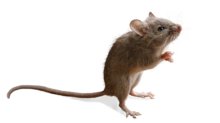
Let’s go back to the naked mole rat and this decoupling of mortality and aging that you demonstrated. Naked mole rats are thought not to age demographically, but your paper proves that they still age epigenetically, as measured by methylation clocks. How would you interpret these important findings?

In this particular project, we wanted to address the relationship between aging and mortality, because for many people, mortality is the manifestation of aging, a key feature of aging. According to this view, an organism ages if it’s more likely to die tomorrow than today. All demographic aging models are based on mortality.

The Gompertz equation is the most famous quantitative model in the aging field, which has been used for many years. However, it does not explain certain features of mortality. For example, starting from about 40 years old, we see an exponential increase in the mortality rate in human populations. But when we look earlier – for example, between the ages of 20 and 25 – there’s no increase in mortality. Do humans age during that period?

Moreover, during early childhood, mortality actually decreases. That means that people who think aging is measured by mortality do not even consider childhood, or the period between the ages of 20 and 25.

The constant mortality of the naked mole rat is another observation that they must somehow explain. They say that since mortality doesn’t increase during adult ages, the naked mole rat doesn’t age, but this is in clear contradiction with what we know about mammals. Naked mole rats have neurons, they have cardiomyocytes. We predict that those cells must age.

To address this issue, we built an epigenetic aging clock, and we proved that at least epigenetically, these animals age, even if demographically, they do not. So, do they age after all? Our interpretation is that they do, it’s just that mortality is not a good measure of aging in this species, it’s simply not representative of aging.

In certain species of fish and some other animals, mortality actually decreases with age, which puzzles evolutionary biologists. They say it’s negative senescence! I am not sure what negative senescence is.

I think those organisms still age, they accumulate damage over time, it’s just that their mortality does not increase with age because mortality is an integrative measure of not just organismal aging but also of organismal development and interaction of the organism with the environment. This includes features like predation, how well an organism can withstand certain stresses, and so on. Or simply becoming bigger, which happens in turtles and fishes. Maybe these organisms are less likely to be eaten as they grow, which is why we observe this pattern of reduced mortality, even though they age at the molecular level.

This issue directly relates to the essence of aging. What is it? We have tried to expose these contradictions. I’m not claiming that I’m correct, I’m just saying that there are contradictions. Let’s discuss them, debate, plan experiments, and solve this puzzle.

If we could only solve mortality and not aging, I’d take that. Looks like a good goal.

Yes, but that would be a slightly different goal, in my mind. This is what advances in medicine have done, especially in the last 150 years.

Your lab works on several directions of research. Could you tell us about them? 

We are interested broadly in aging and rejuvenation; we try to understand the nature of these processes. We also would like to better quantify them, so we develop tools to do that. Our specific projects are designed to address these big questions from multiple angles. For example, we have projects on cross-species analysis. We examine many species, trying to understand how nature changes lifespan. This is a way for us to identify new interventions and develop approaches to both extend lifespan and rejuvenate organisms in response to those interventions.

It’s a bit hard to discuss specific projects because some of them are in progress, and for some we don’t have much data yet. I guess I could mention some random projects. For example, we are studying parabiosis in collaboration with Jim White. We find that the young mouse parabiont gets older and the old parabiont gets younger when they get connected, but what does it mean? Do certain organs get rejuvenated or become older? Is it reversible or not?

In another example, if we transplant a young organ to an old mouse, or an old organ to a young mouse, how is aging of the animal affected and of the transplanted organ too? If you have an organism of a certain age, and it has an organ with a widely different age, what is the age of that organism? Those are some of the questions that we address.

We are also interested to know how stress is related to aging. We find that severe stress seems to increase the epigenetic age of organisms, but once the stress is relieved, the age is decreased too. We’re trying to understand, is it just an epigenetic age, or is it “real” aging, meaning that an animal, when placed under severe stress, literally becomes old, and when the stress is relieved, it actually becomes younger? This would mean that the biological age is not a feature that only increases but that it can fluctuate.

This would also mean that some kind of a local rejuvenation event happens. Yes, it’s a great paper.

It also opens many other questions, because if we look at the organism under severe stress, we see more damage, a higher epigenetic age, and also an increased risk of death and disease. And when the stress is relieved, we observe a drop in all those features. Some might say that it’s just inflammation, or just stress or another transient feature that we cannot call aging.

Then again, what is aging? How is it affected by this extra stress? Perhaps severe stress brings extra damage. This would make the organism slightly older, and once the damage is cleared, whether it was due to inflammation or some other challenge, perhaps the organism becomes younger. Therefore, it may also be possible to consider rejuvenation on short time scales.

Yet, some scientists might consider that going back to a normal level of damage shouldn’t be called rejuvenation, that rejuvenation is when an organism becomes younger than in the absence of stress. This discussion brings us again back to the core definitions: how do we define aging and rejuvenation? I hope our studies can help address, consider, and debate these issues, which ultimately will bring clarity to the field.

Going back to heterochronic parabiosis, I was wondering if you have any new insights on this. There are still many things we don’t know.

We have a couple of papers on parabiosis in BioRxiv in collaboration with Jim White and Steve Horvath. We attach animals for three months and then detach and follow them. For example, we found that old animals, after being attached to young animals, live longer and are younger than those attached to old animals. After separation of the animals, the rejuvenation effect was sustained. We also have another BioRxiv paper where we subjected mice to rapamycin only during early development, and this treatment extended lifespan. Apparently, it may be possible to interfere with early development of mice, affecting aging for the rest of the animal’s life.

Yes, this is another very interesting paper, but there seems to be a certain trade-off in terms of growth, right? 

Yes, growth is definitely suppressed in these animals. They are smaller, and they never reach the full body weight of untreated mice.

Interestingly, rapamycin is also the only molecule that was found to extend lifespan similarly when the treatment started in early adulthood or much later in life.

Yes, it’s a nice discovery, and rapamycin is a very promising molecule, for sure.

As one of the central figures in the field of geroscience, how would you describe the current state of affairs?

I think, in general, the field is in a better shape than ever before. We may discuss the fact that, unfortunately, there’s no consensus on the nature of aging or rejuvenation, on causes of aging and on the best approaches to target it, we may disagree on many issues, but at the same time, there’s clearly an improved quality of research and more and more recognition of the field. This research is now taken very seriously, which brings more resources and talent to the field.

These resources and talent will be instrumental in moving the field forward in the next 5, 10, 15 years. I expect a lot of progress. The field may be completely different by then. It may reject the current models or support certain ideas; it’s hard to predict. It’s very exciting to be in this field today and hopefully will be even more exciting in the coming years.

We would like to ask you a small favor. We are a non-profit foundation, and unlike some other organizations, we have no shareholders and no products to sell you. All our news and educational content is free for everyone to read, but it does mean that we rely on the help of people like you. Every contribution, no matter if it’s big or small, supports independent journalism and sustains our future.
Vascular brain

Metformin, Rapamycin, NMN, and Vascular Cognitive Impairment

A study published in Frontiers in Neurology has detailed how three well-known longevity-associated compounds affect vascular cognitive impairment (VCI) in a rat model.

A rat model of a common human disease

In bilateral common carotid artery occlusion (BCCAO), the arteries of rats are sutured to reduce blood flow. This mimics arterial occlusion in human beings [1], which frequently occurs in the elderly and leads to VCI. This disease is only slightly less common than Alzheimer’s disease, and its incidence is gradually increasing [2].

The researchers go into detail regarding nicotinamide mononucleotide (NMN), metformin, and rapamycin, citing previous studies that have shown positive effects. Metformin has been reported to potentially have effects against Alzheimer’s, Parkinson’s, and Huntington’s [3]. Rapamycin was reported to have effectiveness in a rat model of Alzheimer’s [4]. NMN was reported to have positive effects on Alzheimer’s by mediating one of the important activators involved [5].

With this prior research and well-known methodology in hand, the researchers began their own rat study, comparing these three compounds against one another along with an undrugged BCCAO group and a group given a sham surgery.

Pretreatment and behavior analysis

Instead of administering any of these compounds to the rats after their arteries were occluded, the researchers chose to administer them every day for two weeks beforehand. Rats in the BCCAO-only group and the rapamycin group had notably less body mass than the sham group, and rats given metformin and NMN had notably more.

The researchers performed two major cognitive tests on the rats. The novel object recognition test, which judges a rat’s ability to determine whether or not it has seen an object before, was conducted three weeks after the surgery. The BCCAO rats performed poorly on this test compared to the sham group. However, rats given BCCAO along with any of the three tested compounds performed almost as well as rats given the sham surgery.

The Morris water maze test focuses on rats’ ability to find and stay on a platform located underwater, and it was conducted a month after the surgery. Here, the story was similar; rats given BCCAO performed much more poorly than the sham group, although rapamycin seemed to be slightly but not significantly less effective than the other compounds.

Analysis under the microscope

After the cognitive tests, the researchers sacrificed the animals in order to examinine various regions of their brains. While none of the compounds yielded much benefit in the corpus callosum, BCCAO had much less effect on the internal capsule and the striatum in all three treatment groups. The effect of BCCAO on biomarkers for myelin were also greatly ameliorated in all three treatment groups as well.

The microglia were also substantially affected. Iba1+ microglia were dramatically increased after BCCAO but slightly reduced by any treatment. Iba1+ microglia that were also positive for CD68, a marker of the inflammatory M1 macrophage type, were also increased by BCCAO but halved by any treatment. Interestingly, Iba1+ and CD206+ proteins, markers for M2 healing-type macrophages, were not directly increased by BCCAO but were substantially increased by any of the treatments, with NMN doing the most good and rapamycin doing slightly less in this regard.

Conclusion

While strongly conclusive, this study leaves many questions unanswered. All three treatments had very similar effects despite having putatively different mechanisms of action. There was no non-BCCAO group given any of these treatments, which would have been potentially useful data. No combination therapy was given, so we do not know if multiple treatments might have been antagonistic, additive, or even synergistic.

Still, this research is strong enough to raise the question of whether it might be time to engage in human clinical trials of any or all of these compounds. If such trials are successful, the resulting treatment regimen could potentially reduce the incidence or severity of an all-too-common brain disease.

We would like to ask you a small favor. We are a non-profit foundation, and unlike some other organizations, we have no shareholders and no products to sell you. All our news and educational content is free for everyone to read, but it does mean that we rely on the help of people like you. Every contribution, no matter if it’s big or small, supports independent journalism and sustains our future.

Literature

[1] Farkas, E., Luiten, P. G., & Bari, F. (2007). Permanent, bilateral common carotid artery occlusion in the rat: a model for chronic cerebral hypoperfusion-related neurodegenerative diseases. Brain research reviews, 54(1), 162-180.

[2] Gorelick, P. B., Scuteri, A., Black, S. E., DeCarli, C., Greenberg, S. M., Iadecola, C., … & Seshadri, S. (2011). Vascular contributions to cognitive impairment and dementia: a statement for healthcare professionals from the American Heart Association/American Stroke Association. stroke, 42(9), 2672-2713.

[3] Aliper, A., Jellen, L., Cortese, F., Artemov, A., Karpinsky-Semper, D., Moskalev, A., … & Zhavoronkov, A. (2017). Towards natural mimetics of metformin and rapamycin. Aging (Albany NY), 9(11), 2245.

[4] Saunders, R. N., Metcalfe, M. S., & Nicholson, M. L. (2001). Rapamycin in transplantation: a review of the evidence. Kidney international, 59(1), 3-16.

[5] Yao, Z., Yang, W., Gao, Z., & Jia, P. (2017). Nicotinamide mononucleotide inhibits JNK activation to reverse Alzheimer disease. Neuroscience letters, 647, 133-140.