The Blog

Building a Future Free of Age-Related Disease

Rejuvenation Roundup April

Rejuvenation Roundup April 2022

April showers bring May flowers, and we’ve got a shower of interviews, new research, and press releases this month. Here’s what’s happened in rejuvenation in April.

LEAF News

Lifespan Docs: Doug Vakoch of METI: Created by Tim Maupin and sponsored by lifespan.io, this Lifespan Docs documentary features Doug Vakoch of METI, who wishes to live longer to have any hope of receiving responses to interstellar messages.

Lifespan News

Metformin Birth Defects: On this week’s episode of Lifespan News, Ryan O’Shea discusses some alarming research suggesting that metformin is linked to birth defects.

Avatars for Longevity: This week’s episode of Lifespan News is on the idea of using a virtual body in place of a physical one, which can help to mitigate risks. These include digital avatars that will represent us in virtual worlds where we’re increasingly likely to be working, shopping, socializing, and exploring.

The Branding Problem: Altos Labs doesn’t want to be known as a rejuvenation company, and this episode focuses on why.

Interviews

Raiany Romanni on the Ethical Aspects of Life Extension: Raiany Romanni is a Harvard Kennedy Fellow in Effective Altruism, an A360 Scholar, a Stanford Existential Risk Fellow, and a bioethicist. She is currently working on a non-fiction book aimed at catalyzing the ethics of longevity research, while suggesting that aging is the costliest of all human diseases.

Steven AustadDr. Steven Austad on Aging in Wild and Lab Animals: Dr. Steven Austad of the University of Alabama at Birmingham is not a typical geroscientist, or at least, he did not become one in a typical way.

Discussing Low-Dose Naltrexone with Sajad Zalzala: Dr. Sajad Zalzala, co-founder of AgelessRx, prescribes naltrexone for a variety of conditions and is currently enrolling patients in clinical trials for its effectiveness against the lingering effects of COVID-19 as well as for longevity. We caught up with him to discuss the progress in this area.

Rejuvenation Roundup Podcast

Ryan O’Shea of Future Grind hosts this month’s podcast, showcasing the events and research discussed here.

Journal Club

Aging Theory: The Hyperfunction Theory of Aging: Journal Club returned on Tuesday, April 26th at noon Eastern time on our Facebook page. Dr. Oliver Medvedik took a look at the recent paper ‘The hyperfunction theory: an emerging paradigm for the biology of aging’. This episode saw us taking a dive into the world of aging theory.

Research Roundup

Two running mice
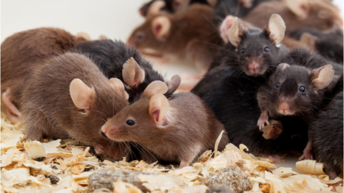
New Candidate Drug Extends Lifespan in Male Mice: Scientists from Mayo Clinic have significantly extended lifespan in male mice by inhibiting the enzyme CD38, which lowers NAD levels. The age-related decline of nicotinamide dinucleotide (NAD) has been associated with various metabolic abnormalities, age-related diseases, and fitness loss.

Senolytics Restore a-Klotho in Mice and Humans: New senolytics data was released from Dr. James L. Kirkland’s Mayo Clinic lab and published in The Lancet. Prior studies have shown that the a-Klotho protein decreases with age in mice and humans.

Assisted livingA Phase 1 Clinical Trial of Stem Cells for Alzheimer’s: A paper published in the journal of the Alzheimer’s Association has revealed the results of a Phase 1 clinical trial of stem cells for Alzheimer’s disease.

Ginger Extract Shows Strong Senolytic Effect: A paper supported by the National Institute on Aging has shown that gingerenone A, a component of ginger extract, is a natural senolytic that is potentially more powerful and less toxic than the combination of dasatinib and quercetin.

Weightlifting MouseSenolytics Improve Resistance Training in Old Mice: A paper published in GeroScience has reported that older mice taking the well-known senolytic combination of dasatinib and quercetin (D+Q) are able to build muscle more like young mice.

One Cycle of Partial Reprogramming for Tissue Rejuvenation: Scientists have shown that a single cycle of partial cellular reprogramming leads to various changes in the tissues, but about half of them gradually disappear after the treatment.

Fly eyesFundamental Protein Regulator Increases Lifespan in Flies: Increasing an important protein regulator improves the lifespan of Drosophila flies, according to a new paper published in Aging.

Mutation Burden Explains Species’ Differences in Lifespan: In a study published in Nature, scientists have found an extremely strong correlation between somatic mutation rate and lifespan across several species, highlighting the importance of mutation burden for aging.

Egg cell
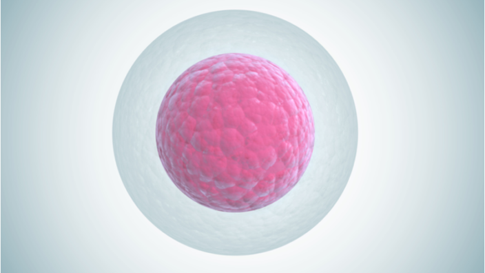
Small Molecule Protects Against Oxidative Aging in Egg Cells: A group of researchers has documented in Aging that Epitalon, a synthetic peptide made of four amino acids, slows the aging of egg cells (oocytes) after ovulation. Oocyte aging is well-known to lead to fertilization deficiencies and a host of other problems that are visible in the cells themselves.

Toll-like Receptor Deletion Improves Memory in Aged Mice: In a new study, genetic deletion of the TLR4 receptor ameliorated aspects of age-related cognitive decline in naturally aging mice, probably due to decreased inflammation. Toll-like receptor 4 (TLR4) is a member of an ancient, highly evolutionary conserved family of proteins.

Many lab miceCombining Caloric Restriction and Rapamycin: A new study published in Nature Communications has found that rapamycin, which is often considered to be a calorie restriction mimetic, has different and additive effects to caloric restriction in muscle tissue.

Plastic Nanoparticles in Cellular Senescence and Dysfunction: Scientists have found that exposure to plastic nanoparticles, a worldwide pollutant, drives cellular senescence and dysfunction in endothelial cells taken from a pig’s coronary artery.

Manhattan plotA Genetic Analysis of Chronic Inflammation: Nature Communications has recently published a paper discussing the genetic sources of C-reactive protein, a well-known biomarker of chronic inflammation. This is an extremely broad study of a wide variety of genes.

Early Life Rapamycin Treatment Effective in Flies and Mice: In a preprint paper, scientists have shown that treating drosophila flies and mice with rapamycin for a relatively brief period in early life mostly recapitulates the effects of a lifelong treatment, including lifespan extension in flies.

Alzheimer's MRIsAn Epigenetic Clock for Brain Age and Alzheimer’s Disease: The risk of Alzheimer’s disease goes up with age, and the number of people living with Alzheimer’s is growing. While it is known to be associated with the loss of proteostasis, it has also been found to be associated with epigenetic alterations.

Synergy Between Stem Cell Rejuvenation and Senolytics: In a preprint published in bioRxiv, a team of Singaporean researchers, including Jan Gruber, has found that a combination of stem cell rejuvenation and senescent cell removal is synergistically more effective than either alone.

Mouse TumorCellular Reprogramming Boosts Liver Regeneration in Mice: Scientists have shown that partial cellular reprogramming can significantly increase the already impressive regenerative capacity of the liver and protect this crucial organ from a potentially lethal injury.

Corylin Shown to Affect Multiple Processes of Aging: A new publication in Nature Communications has revealed that corylin, which is derived from the Chinese herb Psoralea corylifolia, increases the lifespan of model organisms through well-known pathways of aging.

Unhappy SpermMetformin in Fathers Linked to Birth Defects: A large-cohort population study from Denmark has linked metformin to a 40% increase in the risk of birth defects when taken by fathers during the spermatozoa development period.

Nutrition, longevity and disease: From molecular mechanisms to interventions: The researchers analyze aging and nutrition studies in simple organisms, rodents, monkeys, and humans to link longevity to conserved growth and metabolic pathways and outline their role in aging and age-related disease.

Oral Administration of Nicotinamide Mononucleotide Is Safe and Efficiently Increases Blood NAD in Healthy Subjects: These results suggest that oral administration of NMN is a safe and practical strategy to boost NAD+ levels in humans.

Combining adoptive NK cell infusion with a dopamine-releasing peptide reduces senescent cells in aged mice: The researchers observed the downregulation of senescence-related genes by adoptive infusion of natural killer (NK) cells in 26 cases in peripheral blood CD3+ T cells.

Association of Cumulative Blood Pressure With Cognitive Decline, Dementia, and Mortality: Long-term cumulative BP was associated with subsequent cognitive decline, dementia risk, and all-cause mortality in cognitively healthy adults aged at least 50 years.

The brain structure and genetic mechanisms underlying the nonlinear association between sleep duration, cognition and mental health: Genetic mechanisms and brain structural changes may underlie the nonlinear relationship between sleep duration and cognition and mental health.

Cannabidiol induces autophagy and improves neuronal health associated with SIRT1 mediated longevity: These findings collectively indicate the anti-aging benefits of CBD treatment, in both in vitro and in vivo models, and its potential to improve neuronal health and longevity.

Single Ascending Dose Study of a Short Interfering RNA Targeting Lipoprotein(a) Production: The siRNA SLN360 was well tolerated, and a dose-dependent lowering of plasma concentrations was observed.

Comprehensive analysis of epigenetic clocks reveals associations between disproportionate biological ageing and hippocampal volume: This study presents a link between age acceleration, as determined using DNA methylation, and hippocampal volume that was statistically significant across two highly characterised cohorts.

Combined Vitamin D, Omega-3 Fatty Acids, and a Simple Home Exercise Program May Reduce Cancer Risk Among Active Adults Aged 70 and Older: In this pre-defined exploratory analysis, time-to-development of any verified invasive cancer was the primary outcome in an adjusted, intent-to-treat analysis.

Resveratrol Inhibits Metabolism and Affects Blood Platelet Function in Type 2 Diabetes: Resveratrol may be beneficial to prevent vascular complications as a future complementary treatment in aspirin-resistant diabetic patients.

News Nuggets

ElastrinKizoo Company Elastrin Closes $10M Funding Round: The well-known biotechnology holding company Kizoo has engaged in another funding round, this time for Elastrin Therapeutics, a company that focuses on returning stiff tissues to their natural state.

1st VitaDAO Crypto Meets Longevity Symposium: Wednesday, April 13, 2022 from 2:30 PM to 9:30 PM BST will see the first VitaDAO Crypto meets Longevity Symposium. Hosted by Max Unfried and Eleanor Sheekey, the event will be focusing on where the worlds of Web3 and aging research meet.

BioAge logoFirst Cohort Dosed in Phase 1b Trial of BioAge’s BGE-105: BioAge, a biotechnology company that intends to target aging on the molecular level, has completed a Phase 1b clinical trial of BGE-105, a small molecule that influences muscular metabolism.

We would like to ask you a small favor. We are a non-profit foundation, and unlike some other organizations, we have no shareholders and no products to sell you. All our news and educational content is free for everyone to read, but it does mean that we rely on the help of people like you. Every contribution, no matter if it’s big or small, supports independent journalism and sustains our future.
Mouse Tumor

Cellular Reprogramming Boosts Liver Regeneration in Mice

Scientists have shown that partial cellular reprogramming can significantly increase the already impressive regenerative capacity of the liver and protect this crucial organ from a potentially lethal injury [1].

Why can’t we regrow limbs?

Cellular reprogramming induces de-differentiation of somatic cells back into pluripotent stem cells: a ‘factory reset’ that erases cell-specific software and hence cellular identity. In nature, this happens when germ cells are created, but it also occurs in some species during regeneration. For instance, de-differentiation contributes to heart regeneration in zebrafish [2] and limb regeneration in salamanders [3], though there is still a lot we do not know about these amazing repair mechanisms.

In mammals, de-differentiation barely happens, and this might be the reason why mammalian tissues are so bad at regeneration. One notable exception is the liver, which has some regenerative capabilities. In this new study, the researchers performed cellular reprogramming in the livers of mice to investigate how this would affect the organ’s regeneration following injury.

Reprogramming without cancer

For their study, the researchers used partial reprogramming, as full reprogramming in vivo is known to induce cancer. Partial reprogramming uses the same reprogramming factors (usually the Yamanaka factors) but induces them for a brief period of time in one or more “pulses”. By balancing the duration of the factors’ expression, it is possible to achieve various degrees of reprogramming (and also some rejuvenation) without taking the cells all the way back to pluripotency.

First, the researchers created a transgenic mouse model that enables liver-specific induction of the Yamanaka factors. Highlighting the dangers of cellular reprogramming in vivo, in the first experiment that induced the factors for 48 hours, all of the mice died of liver failure within days. The problem was solved after the researchers limited the factors’ expression to one day, and in subsequent experiments, the reprogramming treatment seemed to do much more good than harm.

It is hard to pinpoint the exact extent to which a partial reprogramming treatment changes cellular identity. The researchers usually can only tell that some markers associated with differentiated cells get downregulated and that others associated with pluripotency get upregulated, which is what happened in this study as well. The effect, though, appeared to be transient: the pluripotency markers went back to their normal levels after a few days. Importantly, no carcinogenesis was detected in the liver during the 9-month follow-up, showing that the final protocol is safe in this model.

The treatment also boosted the proliferation of hepatocytes, which is required for liver regeneration [4]. Importantly, increased proliferation following de-differentiation is also one of the mechanisms behind limb regrowth in animals.

Improved survival

To determine whether the treatment actually increased regeneration capacity in the liver, the researchers induced lethal chemical liver injury. While all the mice in the control group died two days later, in the group that had received the reprogramming treatment immediately before the injury, half of the mice survived and recovered, showing greatly increased levels of liver regeneration. Unfortunately, reprogramming was not as effective when induced after the injury, although it did improve liver function.

Cellular reprogramming largely remains a black box, as its exact mechanisms haven’t yet been elucidated. Discovering these mechanisms is extremely important, as this must be done before actual reprogramming-based therapies can be devised. In this study, the researchers performed single-cell transcriptomic analysis of both regular and reprogrammed hepatocytes and found that the treatment had significantly upregulated the enzyme topoisomerase2a (Top2a), which is associated with cellular development. Blocking Top2a led to a drastic reduction in reprogramming efficiency and in the mice’s survival following liver injury.

In summary, here, we have developed a mouse model that enables hepatocyte-specific 4F [Yamanaka factors] induction and subsequent lineage tracing of 4F-expressing cells. We demonstrate that liver-specific 4F expression rapidly and transiently induced partial reprogramming and that this enhanced liver regeneration. This study, the first to perform lineage tracing and single-cell transcriptome analyses for 4F-expressing cells in vivo, shows that 4F-mediated cellular partial reprogramming is a potential avenue for inducing a proliferative, plastic progenitor state.

Conclusion

This interesting study shows that partial cellular reprogramming in vivo can greatly increase hepatic regenerative capacity and even protect the liver from otherwise lethal chemical injury without causing cancer. It is also one of the few studies to identify a downstream target of reprogramming. This is an important piece of knowledge about the inner workings of cellular reprogramming, and it might lead to the creation of more safe and effective reprogramming protocols.

We would like to ask you a small favor. We are a non-profit foundation, and unlike some other organizations, we have no shareholders and no products to sell you. All our news and educational content is free for everyone to read, but it does mean that we rely on the help of people like you. Every contribution, no matter if it’s big or small, supports independent journalism and sustains our future.

Literature

[1] Hishida, T., Yamamoto, M., Hishida-Nozaki, Y., Shao, C., Huang, L., Wang, C., Shojima, K., Xue, Y., Hang, Y., Shokhirev, M., Memczak, S., Sahu, S. K., Hatanaka, F., Ros, R. R., Maxwell, M. B., Chavez, J., Shao, Y., Liao, H. K., Martinez-Redondo, P., Guillen-Guillen, I., … Izpisua Belmonte, J. C. (2022). In vivo partial cellular reprogramming enhances liver plasticity and regeneration. Cell reports, 39(4), 110730.

[2] Jopling, C., Sleep, E., Raya, M., Martí, M., Raya, A., & Belmonte, J. C. I. (2010). Zebrafish heart regeneration occurs by cardiomyocyte dedifferentiation and proliferation. Nature464(7288), 606-609.

[3] Wang, H., & Simon, A. (2016). Skeletal muscle dedifferentiation during salamander limb regeneration. Current Opinion in Genetics & Development40, 108-112.

[4] Ozaki, M. (2020, April). Cellular and molecular mechanisms of liver regeneration: Proliferation, growth, death and protection of hepatocytes. In Seminars in cell & developmental biology (Vol. 100, pp. 62-73). Academic Press.

Two Directions Infinity

Synergy Between Stem Cell Rejuvenation and Senolytics

In a preprint published in bioRxiv, a team of Singaporean researchers, including Jan Gruber, has found that a combination of stem cell rejuvenation and senescent cell removal is synergistically more effective than either alone.

Different but related aspects of aging

The researchers begin this paper by first discussing senescent cells and the SASP, whose inflammatory effects are well-known to contribute to age-related diseases, and they point out that senolytics have been shown to extend lifespan in mice [1]. They also discuss stem cell exhaustion, focusing on the Yamanaka factors (OSKM) and how short-term expression of these factors has also been shown to lead to lifespan extension in mice [2].

They then point out how these two aspects of aging are intertwined. The inflammatory signals of the SASP activate mTOR and create an environment that suppresses stem cell growth [3]. Building upon their previous nematode study showing drug-drug interactions [4], the researchers created this study to see if OSKM and senolytics would work synergistically in flies.

Gut cells and gene expression

The researchers’ first examination, which focused on intestinal stem cells (ISCs), had mixed results. Senolytics by themselves were actually shown to have more positive effects under some circumstances than combining them with OSKM expression, which may be because constant, rather than transitory, OSKM expression has negative effects.

The researchers also examined gene expression, attempting to discover if OSKM expression and senolytics affect the same pathways. This research shows that they do not: while both of these interventions are associated with increased amino acid metabolism and decreased fatty acid biosynthesis on the genomic level, the transcriptional programs they influence are entirely different.

Synergistic effects in lifespan extension

To overcome the known problems with continuous expression of OSKM and senolytics, the researchers genetically engineered flies to express OSKM, senolytics, or both when their environment reaches 25 degrees Celsius; the flies were normally kept at 18 degrees Celsius. In this way, the researchers could give the flies a dosing regimen directly to their cells.

As expected, such flies that were constantly exposed to a higher temperature, and thus constantly expressed these proteins, died more rapidly than wild-type flies in the same environment. Exposing these flies to these factors for 24 hours twice a week showed some positive results, but starker, more significant results were found in the 12-hour dosing group.

Among these flies, senolytics promoted the compression of morbidity: while it did not increase maximum lifespan, more flies were able to live that long than in the wild-type group. OSKM had a different effect: it simply extended the flies’ average lifespan, although many of the OSKM-only flies still died early, similar to the wild-type flies.

Combining the two gave the results the researchers were hoping for: synergy. Not only did the flies receive a benefit of lifespan extension from the OSKM and compression of morbidity from the senolytics, their lifespans benefited more than a theoretical combination in which the effects were merely additive.

Conclusion

This is a fly study, and even the temperature changes used in this study affect the lifespan of wild-type flies. Some of the intestinal stem cell data suggests that the combination of senolytics and OSKM may not always be positive. However, this study provides conclusive data showing the synergistic lifespan effects of combining the two interventions under a proper dosing regimen, and we hope that this data can be recapitulated in studies involving mice and, ultimately, human beings.

We would like to ask you a small favor. We are a non-profit foundation, and unlike some other organizations, we have no shareholders and no products to sell you. All our news and educational content is free for everyone to read, but it does mean that we rely on the help of people like you. Every contribution, no matter if it’s big or small, supports independent journalism and sustains our future.

Literature

[1] Xu, M., Pirtskhalava, T., Farr, J. N., Weigand, B. M., Palmer, A. K., Weivoda, M. M., … & Kirkland, J. L. (2018). Senolytics improve physical function and increase lifespan in old age. Nature medicine, 24(8), 1246-1256.

[2] Ocampo, A., Reddy, P., Martinez-Redondo, P., Platero-Luengo, A., Hatanaka, F., Hishida, T., … & Belmonte, J. C. I. (2016). In vivo amelioration of age-associated hallmarks by partial reprogramming. Cell, 167(7), 1719-1733.

[3] He, S., & Sharpless, N. E. (2017). Senescence in health and disease. Cell, 169(6), 1000-1011.

[4] Admasu, T. D., Batchu, K. C., Barardo, D., Ng, L. F., Lam, V. Y. M., Xiao, L., … & Gruber, J. (2018). Drug synergy slows aging and improves healthspan through IGF and SREBP lipid signaling. Developmental Cell, 47(1), 67-79.

Alzheimer's MRIs

An Epigenetic Clock for Brain Age and Alzheimer’s Disease

The risk of Alzheimer’s disease goes up with age, and the number of people living with Alzheimer’s is growing. While it is known to be associated with the loss of proteostasis, it has also been found to be associated with epigenetic alterations. An advanced online preprint in bioRxiv was published by Dr. Morgan E. Levine and colleagues, who created a methylation clock called PCBrainAge. They wanted to determine if PCBrainAge is predictive of Alzheimer’s disease [1].

PCBrainAge is associated with Alzheimer’s disease in the dorsal prefrontal cortex

The prefrontal cortex, a part of the brain, is where much of our executive function takes place. Executive function aids in functions such as decision making and cognitive control.

Using data from the dorsal prefrontal cortices of 700 participants in the Religious Orders Study and the Memory and Aging Project, the researchers generated linear models comparing PCBrainAge to the participants’ neuronal composition and true ages at death. As part of their analysis, the researchers determined these participants’ CERAD scores.

The first result indicates that the post-mortem stage of Alzheimer’s disease, according to CERAD scores, was significantly associated with increased brain aging and BRAAK staging, which is used to score the progression of Alzheimer’s disease and Parkinson’s disease.

PCBrainAge was shown to be significantly associated with Alzheimer’s disease when the disease had reached neocortical tissue, showing that it had reached its final stages. PCBrainAge acceleration was also positively associated with the pre-mortem clinical diagnosis of Alzheimer’s disease dementia. Additionally, carriers of one or two APOE e4 alleles, which are associated with Alzheimer’s disease, were significantly more likely to experience PCBrainAge acceleration.

This study also used a methylation clock known as DNAmClockCortical [2], which, like PCBrainAge, is used to measure both age and age acceleration. DNAmClockCortical is more strongly correlated with chronological age than PCBrainAge, but PCBrainAge takes into account the biological heterogeneity of aging, and its link to Alzheimer’s disease may give it more clinical value.

Additional results comparing these two clocks demonstrate that DNAmClockCortical showed less significant association with the pathological and clinical phenotypes of Alzheimer’s disease and APOE e4 carrier status than PCBrainAge. This suggests that DNAmClockCortical may not be sensitive enough to detect the genetic and aging signals associated with Alzheimer’s disease.

Furthermore, large increases in DNAmClockCortical acceleration were not correlated with increased pathological Alzheimer’s disease in an examination of amyloid and neuritic plaques. PCBrainAge demonstrated a closer correlation to Alzheimer’s disease pathology and was more balanced across post-mortem metrics of this pathology.

Unlike DNAmClockCortical, significantly increased PCBrainAge acceleration also showed an increase in the probability of dementia. The authors hypothesize that this result is due to the reduction in the noise from CpGs and the improved resolution of the PCBrainAge clock.

Alzheimer’s disease is correlated with PCBrainAge across many brain regions

The study then went on to use PCBrainAge to measure aging trends across brain regions and specific associations with Alzheimer’s disease.

This analysis examined 333 individuals from the APOE e4 carrier subcohort of this study. This data examined novel DNA methylation of three specific brain regions: the dorsolateral prefrontal cortex, the striatum and the cerebellum.

Age acceleration in the prefrontal cortex and the striatum were both associated with Alzheimer’s disease neuropathology and pre-mortem clinical diagnosis. A weaker association showed that age acceleration in the striatum was increased in APOE4 e4 carriers. The researchers believe that this weaker association was due to this dataset being smaller than the overall study.

In the cerebellum, PCBrainAge acceleration was not significantly correlated with Alzheimer’s disease or APOE e4 carrier status. Prior research also agrees that epigenetic clocks do not show correlations with cerebellum age acceleration and Alzheimer’s disease neuropathology [3].

Conclusion

While this is a preprint that has not yet been peer reviewed, its results show a link between DNA methylation patterns and advanced Alzheimer’s disease. Along with the PhenoAge clock, this research has unlocked epigenetic clues to the complex nature of Alzheimer’s disease.

We would like to ask you a small favor. We are a non-profit foundation, and unlike some other organizations, we have no shareholders and no products to sell you. All our news and educational content is free for everyone to read, but it does mean that we rely on the help of people like you. Every contribution, no matter if it’s big or small, supports independent journalism and sustains our future.

Literature

[1] Kyra L. Thrush, David A. Bennett, Christopher Gaiteri, Steve Horvath, Christopher H. van Dyck, Albert T. Higgins-Chen, Morgan E. Levine. bioRxiv (2022) bioRxiv preprint. doi: https://doi.org/10.1101/2022.02.28.481849

[2] Sierra, F. Geroscience and the Role of Aging in the Etiology and Management of Alzheimer’s Disease. J Prev Alzheimers Dis 7, 2–3 (2020). https://doi.org/10.14283/jpad.2019.49

[3] Horvath, S., Mah, V., Lu, A. T., Woo, J. S., Choi, O. W., Jasinska, A. J., Riancho, J. A., Tung, S., Coles, N. S., Braun, J., Vinters, H. V., & Coles, L. S. (2015). The cerebellum ages slowly according to the epigenetic clock. Aging, 7(5), 294–306. https://doi.org/10.18632/aging.100742

Fruit flies

Early Life Rapamycin Treatment Effective in Flies and Mice

In a preprint paper, scientists have shown that treating drosophila flies and mice with rapamycin for a relatively brief period in early life mostly recapitulates the effects of a lifelong treatment, including lifespan extension in flies [1].

Rapamycin is considered one of the most promising geroprotective drugs. It belongs to a handful of molecules that have shown reproducible lifespan extension in mice, and it is currently undergoing trials in humans and dogs.

When rapamycin was first tested by the Intervention Testing Program (ITP), which is the current gold standard for testing life-prolonging interventions in mice, the researchers were amazed to find that mice who started receiving rapamycin at the respectable age of 20 months benefited from virtually the same increase in lifespan as mice who had been fed rapamycin their entire life [2]. This finding made waves, since it demonstrated that it is possible to intervene late in life and still achieve a considerable lifespan extension.

How much rapamycin is just enough?

Unfortunately, rapamycin also has toxicity issues, so researchers have been searching for the minimum dose required to prolong life. That includes this study, in which the researchers started with drosophila flies and then moved on to mice. Although they are not as close to humans as mice are, drosophila flies, with a maximum lifespan of just over three months, are considered a cheap and effective model for proof-of-concept longevity studies.

The researchers divided the flies into several groups. In one group, the flies were fed rapamycin for their entire lives, which led to an expected increase in lifespan. What was less expected though, is that another group, which contained flies that had been fed rapamycin only during the first 15 days of their lives, enjoyed the same increase in lifespan. The researchers immediately dubbed this phenomenon “rapamycin memory”.

Early Life Rapamycin

Rapamycin affects nutrient sensing by interacting with the protein complex TOR (target of rapamycin). Basically, rapamycin nudges the organism towards maintenance and away from growth, which is why rapamycin treatment is known do decrease the number of actively dividing cells.

Drosophila guts are characterized by high rates of cellular division and turnover (the replacement of damaged cells by intestinal stem cells). This drives intestinal dysplasia (the presence of abnormal cells, which is considered a pre-cancerous condition) and shortens lifespan. In both groups, the rapamycin treatment resulted in reduced turnover, as cells remained healthy for longer, and there was less need to replace them.

This translated to fewer intestinal pathologies as well. While in the controls, aging resulted in widespread dysplasia, flies in both treatment groups had significantly fewer dysplastic lesions throughout their lifespans. Rapamycin also helped preserve gut barrier function; it protected the flies from leaky gut, a major cause of aging in both flies and humans [3].

It’s the autophagy

How exactly does “rapamycin memory” work? While the activity of target of rapamycin complex 1 (TORC1) was reduced by the treatment, it bounced back just 48 hours after withdrawal. This means that the repression of TORC1 by rapamycin must have activated other mechanisms that remained in place to maintain “rapamycin memory”.

One such downstream consequence of TORC1 repression is increased autophagy, which is required for rapamycin-induced lifespan extension. If autophagy remained upregulated even after the withdrawal of rapamycin, this could explain the “rapamycin memory” effect, and this is exactly what the researchers found. Even after the brief early-life rapamycin treatment, autophagy levels remained high throughout the flies’ lifespan, although not in the head, which hints at a tissue-specific response.

When the researchers downregulated autophagy by other means, it mostly blocked both rapamycin-induced lifespan extension and improvement in gut health, completely abolishing the “rapamycin memory” effect. On the contrary, when researchers upregulated autophagy for the same short period of time early in life, this caused roughly the same lifespan extension as rapamycin. Notably, the combination of rapamycin and autophagy inhibition fared no better than any single treatment.

Translatable to mammals

To see if “rapamycin memory” translates to mammals, the researchers assessed the effects of early-life short-term rapamycin treatment in mice. The levels of lipopolysaccharide-binding protein (LBP), a marker of bacterial translocation from the intestine into the bloodstream, were kept equally low by both the lifelong and the short-term rapamycin treatments, showing that the latter leads to a lasting improvement in gut health. This was confirmed by analyzing intestinal structure and cellular health in the mice’s guts. The researchers conclude:

Our findings suggest that the full geroprotective effects of rapamycin can be achieved without the adverse effects sometimes seen with chronic, long-term dosing.

Interestingly, one recent study did find that early-life treatment with rapamycin extends lifespan in mice.

Conclusion

Years ago, the scientific community was stunned by the discovery that rapamycin treatment gives the same boost to murine lifespan when started later in life. In this new study, the researchers show that rapamycin works just as well in a brief early-life intervention and suggest a mechanism behind this effect. Given rapamycin’s toxicity record, this finding could help devise better treatment regimens. On the other hand, this study suggests that we might not need rapamycin or similar compounds at all, as upregulating autophagy by other means could potentially recapitulate its effect.

We would like to ask you a small favor. We are a non-profit foundation, and unlike some other organizations, we have no shareholders and no products to sell you. All our news and educational content is free for everyone to read, but it does mean that we rely on the help of people like you. Every contribution, no matter if it’s big or small, supports independent journalism and sustains our future.

Literature

[1] Juricic, P., Lu, Y. X., Leech, T., Drews, L. F., Paulitz, J., Lu, J., … & Partridge, L. (2022). Full geroprotection from brief rapamycin treatment by persistently increased intestinal autophagy. bioRxiv.

[2] Harrison, D. E., Strong, R., Sharp, Z. D., Nelson, J. F., Astle, C. M., Flurkey, K., … & Miller, R. A. (2009). Rapamycin fed late in life extends lifespan in genetically heterogeneous mice. nature460(7253), 392-395.

[3] Mishra, S. P., Wang, B., Wang, S., Nagpal, R., Miller, B., Jain, S., … & Yadav, H. (2021). Microbiota induces aging-related leaky gut and inflammation by dampening mucin barriers and butyrate-FFAR2/3 signaling. bioRxiv.

Manhattan plot

A Genetic Analysis of Chronic Inflammation

Nature Communications has recently published a paper discussing the genetic sources of C-reactive protein, a well-known biomarker of chronic inflammation.

An extremely broad study with a wide variety of genes

Pulling genomic data from 427,367 participants in the UK Biobank and 148,164 people from the Cohorts for Heart and Aging Research in Genomic Epidemiology (CHARGE) consortia, which has been previously used in similar but less thorough research [1], these researchers have identified 266 genetic loci associated with C-reactive protein production, expanding upon the previous research with 211 previously unknown findings.

Many of these loci had hundreds of genetic variants, which is why such an enormous group of people was required and why such a study could not have possibly been completed without modern genomics and data analysis. Such an analysis uses Bayesian, probability-based techniques, and when so many potential associations are examined, sheer chance can easily result in false positives, so the standard p-value of 0.05 is insufficient.

Therefore, the researchers used Bonferroni thresholds instead, which take the number of tests into account and replace the p-value of 0.05 with far smaller numbers. Throughout this analysis, the amount of data led to some p-values that were much, much smaller than the Bonferroni threshold – a sign that the finding is, assuming that the study was properly conducted, nearly certain to be correct.

Basic functions and C-reactive protein production are interconnected

The researchers identified five genes that are practically certain (p-values between 8.40 x 10-162 and 5.32 x 10-147) to be associated with C-reactive protein and systemic inflammation: NECTIN2, PDE4B, OASL, IL6R, and APOE. IL6R is the interleukin-6 receptor, which is associated with inflammation [2], and APOE is well-known for its link to Alzheimer’s disease.

Links between many other genes and C-reactive protein were also plotted out, offering some useful and perhaps actionable information with which to develop therapies. Genes related to lipid (fat) transport, signaling pathways, and immune response were significant, and genes related to the regulation of gene expression were even more significant. The liver, as expected, and the blood were the most significantly affected by the expression of these genes.

Diseases linked to C-reactive protein

Many age-related diseases, including heart disease, atherosclerosis, and osteoarthritis, were found to be strongly related to the overexpression of C-reactive protein. By far, the most significant results involved the lungs: chronic obstructive pulmonary disease (COPD), bronchitis, and chronic airway obstruction were all found to be related to this systemic inflammation, according to a weighted risk score derived from the UK Biobank results.

Increased expression did seem to have some positive effects, however. Most notably, it seemed to be protective against schizophrenia, which prior research suggests is due to a reduced risk of childhood infections, which are linked with the disease [3].

Conclusion

While this study did not directly tackle aging or age-related diseases, systemic inflammation is well known to be related to aging (inflammaging), and gene expression changes with age. Studies such as this one offer useful information in determining which genes are associated with systemic inflammation, potentially paving the way for genetic or epigenetic therapies that directly target the overexpression of C-reactive protein and other inflammatory compounds.

There were, of course, a few limitations and caveats. This study focused on people of European descent, did not take BMI (which is linked to inflammation) into account as most of these genes are independent of BMI, and did not investigate rare variants of any gene. Further analysis would need to be done to see if there are any rare variants of specific genes that are linked to positive outcomes, and, if so, the biochemical pathways of such links.

We would like to ask you a small favor. We are a non-profit foundation, and unlike some other organizations, we have no shareholders and no products to sell you. All our news and educational content is free for everyone to read, but it does mean that we rely on the help of people like you. Every contribution, no matter if it’s big or small, supports independent journalism and sustains our future.

Literature

[1] Ligthart, S., Vaez, A., Võsa, U., Stathopoulou, M. G., De Vries, P. S., Prins, B. P., … & Saba, Y. (2018). Genome analyses of> 200,000 individuals identify 58 loci for chronic inflammation and highlight pathways that link inflammation and complex disorders. The American Journal of Human Genetics, 103(5), 691-706.

[2] Tanaka, T., Narazaki, M., & Kishimoto, T. (2014). IL-6 in inflammation, immunity, and disease. Cold Spring Harbor perspectives in biology, 6(10), a016295.

[3] Hartwig, F. P., Borges, M. C., Horta, B. L., Bowden, J., & Smith, G. D. (2017). Inflammatory biomarkers and risk of schizophrenia: a 2-sample mendelian randomization study. JAMA psychiatry, 74(12), 1226-1233.

Plastic for Dinner

Plastic Nanoparticles in Cellular Senescence and Dysfunction

Scientists have found that exposure to plastic nanoparticles drives cellular senescence and dysfunction in endothelial cells taken from a pig’s coronary artery [1].

A widespread pollutant

Plastic has changed the face of civilization, but the convenience of plastic comes at a price – mostly in terms of environmental pollution from plastic manufacturing and waste. There has also been a growing understanding that plastic pollutes us as well. Plastic can disintegrate into microparticles and nanoparticles, the latter of which are 1-100 nanometers in size and can be suspended in the atmosphere for prolonged periods of time and enter the body through breathing. Exposure can also occur via ingestion or even absorption by the skin [2] – for instance, following contact with plastic food packaging.

The research into the effects of plastic micro- and nanoparticles on human health is still in its infancy. There have been indications that thee particles in the blood drive pulmonary hypertension and vascular occlusions [3]. Still, the impact of plastic nanoparticles on human health remains mostly unclear.

Increased senescence

In this new study, the authors investigated the effect of plastic nanoparticles on cellular senescence, a major driver of aging. In particular, senescence in vascular endothelial cells, which compose the inner layer of blood vessels, is known to promote cardiovascular dysfunction [4].

The researchers used endothelial cells taken from a pig’s coronary artery, subjecting them to various concentrations of 25-nanometer particles of polystyrene, one of the most ubiquitous types of plastic. In the first experiment, the nanoparticle solution induced senescence in the cells in a concentration-dependent manner. The effect, as measured by ß-galactosidase levels, was small and statistically insignificant for the lowest concentration of 0.1 µg/mL but much larger for 1 and 10 µg/mL.

The scientists then checked for three other popular markers of senescence, the proteins p53, p21, and p16. Those were also upregulated by the treatment, with p53 being significantly elevated even by the 0.1 µg/mL solution.

Cellular senescence basically does not affect the viability of cells: they stop proliferating but continue to exist, producing harmful molecules that promote inflammation and induce senescence in neighboring cells. Subsequent experiments indeed showed that while nanoparticle exposure did not affect the cells’ viability, it significantly reduced their proliferation.

Nanoparticles cause functional decline

Higher levels of cellular senescence after the nanoparticle exposure manifested in functional decline as well. Exposing specimens of arterial tissue to nanoparticles for 24 hours significantly hampered their reactivity – a measure of how well an artery responds to stimuli by contracting and relaxing.

Senescent vascular cells are characterized by reduced production of nitric oxide (NO), a vasodilator crucial for vascular health. Sure enough, nanoparticle-exposed cells had much lower levels of the NO-producing enzyme epithelial nitric oxide synthase (eNOS).

Nanoparticles also significantly increased the levels of oxidative stress, even in the lowest concentration. Several pathways can contribute to oxidative stress, but experiments showed that nanoparticle exposure significantly affected one of them, the NADPH/Sirt1 pathway, which includes downregulation of the Sirt1 protein. Treatment with a NADPH inhibitor ameliorated the nanoparticle-induced increase in oxidative stress, as did treatment with a Sirt1 activator. The antioxidant drug NAC had a similar effect and restored proliferation levels in the exposed cells. This is not the first time that plastic nanoparticles have been implicated in increasing oxidative stress, both directly and by downregulating antioxidant enzymes.

Close to real-life concentrations

A major question is whether the concentrations that were used in this study bear any resemblance to real-life concentrations of plastic nanoparticles in our blood? Unfortunately, they do. While we still don’t have a lot of data on the subject, one recent study identified the mean concentration of plastic particles in the blood of healthy volunteers as 1.6 µg/ml – 1.6 times higher than the medium concentration used in this paper [5]. While that study measured the concentration of somewhat bigger particles (=700 nanometers), there is no reason to assume that the concentration of nanoparticles in the blood is any lower.

Conclusion

This paper is one of the first to directly study the harmful effects of plastic particles on the cardiovascular system. While it is too early to sound the alarm, this is an important research topic that warrants further investigation. Meanwhile, it seems possible that reasonable avoidance of plastics can be good not only for the planet but for your health as well.

We would like to ask you a small favor. We are a non-profit foundation, and unlike some other organizations, we have no shareholders and no products to sell you. All our news and educational content is free for everyone to read, but it does mean that we rely on the help of people like you. Every contribution, no matter if it’s big or small, supports independent journalism and sustains our future.

Literature

[1] Shiwakoti, S., Ko, J. Y., Gong, D., Dhakal, B., Lee, J. H., Adhikari, R., … & Oak, M. H. (2022). Effects of polystyrene nanoplastics on endothelium senescence and its underlying mechanism. Environment International, 107248.

[2] Prata, J. C., da Costa, J. P., Lopes, I., Duarte, A. C., & Rocha-Santos, T. (2020). Environmental exposure to microplastics: An overview on possible human health effects. Science of the total environment702, 134455.

[3] Zagorski, J., Debelak, J., Gellar, M., Watts, J. A., & Kline, J. A. (2003). Chemokines accumulate in the lungs of rats with severe pulmonary embolism induced by polystyrene microspheres. The Journal of Immunology171(10), 5529-5536.

[4] Mehdizadeh, M., Aguilar, M., Thorin, E., Ferbeyre, G., & Nattel, S. (2021). The role of cellular senescence in cardiac disease: Basic biology and clinical relevance. Nature Reviews Cardiology, 1-15.

[5] Leslie, H. A., Van Velzen, M. J., Brandsma, S. H., Vethaak, D., Garcia-Vallejo, J. J., & Lamoree, M. H. (2022). Discovery and quantification of plastic particle pollution in human blood. Environment International, 107199.

Many lab mice

Combining Caloric Restriction and Rapamycin

A new study published in Nature Communications has found that rapamycin, which is often considered to be a calorie restriction mimetic, has different and additive effects to caloric restriction in muscle tissue.

The role of mTORC1

Mammalian target of rapamycin complex 1 (mTORC1) is a well-known component of fundamental nutrient sensing pathways whose dysregulation is associated with aging. As its name suggests, mTORC1 is influenced by the well-known compound rapamycin, which has been, and is currently being, heavily explored as a life-extending intervention [1]. As a nutrient sensing pathway, it is also influenced by caloric restriction, which is also a well-known intervention for extending lifespan, although this is difficult to achieve in people [2].

However, as the researchers point out, the biochemical effects in mice treated with rapamycin and caloric restriction are different [3], and previously combining the two has shown an increased effect in liver tissue [4].

The researchers posed this fundamental question as follows:

But is this a case of ‘all roads lead to Rome’, where CR and RM travel different paths to mTORC1 suppression, or do these two quintessential life-prolonging interventions travel different roads with distinct destinations?

To answer it, they turned to an examination of skeletal muscle with a focus on sarcopenia, the age-related loss of muscle function.

Caloric restriction reduces weight and glucose but does not prevent sarcopenia

Restricting the calories of male wild-type mice at 15 months or 20 months of age, the researchers discovered that caloric restriction helped in some physical ways. Both fat and lean mass were reduced in the caloric restriction groups with a minimum influence on strength. With less weight to carry, the restricted mice were much more able to hang on to an upside-down grid than their freely fed counterparts; in fact, 30-month-old restricted mice soundly outperformed 10-month-old freely fed mice in this respect.

Blood glucose, which declines with aging, further declined with caloric restriction. Calorically restricted mice followed a circadian (day/night) cycle more than their unrestricted counterparts, and they chose to run more when possible.

While all the groups lost muscle strength with aging at roughly the same rate, caloric restriction promoted a fast-to-slow transition in muscle tissue. The researchers explain that slow-twitch muscle fibers are more resistant to age-related changes, and after an analysis of the muscle fibers, this study found that caloric restriction may have at least partially protected against sarcopenia in these mice.

Rapamycin helped to combat sarcopenia

Combining caloric restriction and rapamycin had many effects, many of which were additive. Rapamycin, by itself, decreased lean mass, but combining it with caloric restriction had no extra effects in this respect.

The combination decreased the mice’s upside-down hang time but substantially increased both daily running speed and running distance. Mice on both interventions ate more slowly than those just on caloric restriction. Interestingly, rapamycin increased blood glucose as well, both in the presence and absence of caloric restriction, a fact that the researchers believe is due to rapamycin’s effects on mTORC2.

Probably most importantly, rapamycin reduced the rate at which grip strength declines. 24- to 28-month-old mice on rapamycin, calorically restricted or not, had significantly stronger all-limb grip strength than their counterparts. The researchers analyzed specific muscles and found that many of the muscles that benefited from caloric restriction also benefited from rapamycin.

Gene expression signatures were almost entirely different

The researchers also compared the effects of these two interventions on gene expression, finding wildly different results. Interestingly, the majority of the effects of caloric restriction were in the same direction as aging, while rapamycin almost entirely influenced gene expression in the opposite direction. It is unsurprising, then, that many of the gene expression effects of these two interventions were in opposite directions. However, with the exception of IFN-γ, which only decreased with rapamycin, inflammatory markers were similarly decreased with both interventions.

Conclusion

These data lend strong evidence to the idea that rapamycin and caloric restriction have completely different effects on muscle tissue and offer different benefits for different reasons. While even a combination of both did not wholly prevent sarcopenia in mice, it seemed to have a strong influence on these animals’ muscle tissue and physical abilities, suggesting a possible low-hanging fruit in treating human frailty.

We would like to ask you a small favor. We are a non-profit foundation, and unlike some other organizations, we have no shareholders and no products to sell you. All our news and educational content is free for everyone to read, but it does mean that we rely on the help of people like you. Every contribution, no matter if it’s big or small, supports independent journalism and sustains our future.

Literature

[1] Harrison, D. E., Strong, R., Sharp, Z. D., Nelson, J. F., Astle, C. M., Flurkey, K., … & Miller, R. A. (2009). Rapamycin fed late in life extends lifespan in genetically heterogeneous mice. nature, 460(7253), 392-395.

[2] Most, J., Tosti, V., Redman, L. M., & Fontana, L. (2017). Calorie restriction in humans: an update. Ageing research reviews, 39, 36-45.

[3] Karunadharma, P. P., Basisty, N., Dai, D. F., Chiao, Y. A., Quarles, E. K., Hsieh, E. J., … & Rabinovitch, P. S. (2015). Subacute calorie restriction and rapamycin discordantly alter mouse liver proteome homeostasis and reverse aging effects. Aging cell, 14(4), 547-557.

[4] Fok, W. C., Bokov, A., Gelfond, J., Yu, Z., Zhang, Y., Doderer, M., … & Pérez, V. I. (2014). Combined treatment of rapamycin and dietary restriction has a larger effect on the transcriptome and metabolome of liver. Aging cell, 13(2), 311-319.

Zalzala interview

Discussing Low-Dose Naltrexone with Sajad Zalzala

Dr. Sajad Zalzala, co-founder of AgelessRx, prescribes naltrexone for a variety of conditions and is currently enrolling patients in clinical trials for its effectiveness against the lingering effects of COVID-19 as well as for longevity. We caught up with him to discuss the progress in this area.

For the people who aren’t familiar with low-dose naltrexone, can you give us an overview of the history and how you’re using, why you’re using, low-dose naltrexone?

The regular-dose naltrexone was FDA approved for alcoholism back in the 80s or early 90s. It blocks the opioid receptors. For alcoholism, it seems to remove the euphoria that that some people get with drinking alcohol and therefore, people who have trouble with addiction with alcohol just no longer feel the need to drink anymore.

Obviously, if you’re on opioid medications, and the high dose of naltrexone or regular-dose naltrexone blocks the opioid receptors, so the Percocet and the morphine just can’t bind to the opioid receptors anymore, you don’t get that high or the addictive effects of the opioid drugs anymore.

In terms of how that got turned into low-dose naltrexone, from what I read, there was a physician by the name of Dr. Bihari. Based on what I understand from his research, he was looking for a way, I think he noticed that patients who had HIV, AIDS, had a low baseline, a low endorphin level.

He made that discovery, and this is back in the 80s, before we started getting any of the antiretrovirals and such, so they’re trying to find some solution. He scoured and found that naltrexone has some endorphin effect on it. From what I understand, he did some experimentation and found that a low dose of naltrexone can actually boost endorphin levels.

That led to the discovery of low-dose naltrexone. By trial and error, he found that the dose around, 3 milligrams, 4 1/2 milligrams, 5, tend to do give you all the benefits of naltrexone without many of the side effects that you get with the higher dose. That’s why we call it low-dose naltrexone, to differentiate it from from the high dose. I would go as far as saying that the high-dose naltrexone, the regular dose, doesn’t have the same therapeutic properties as low-dose.

Going back to some of the work from Dr. Bihari, he cites a case where a patient was on low-dose naltrexone, an HIV patient, it seemed to keep his cancer in remission. As the story goes, the pharmacy accidentally dispensed the 50-milligram dose, and that ended this patient’s remission and he passed away shortly thereafter from the progression of cancer. That’s an anecdote, but I think there’s a lot of reasons to believe why that could be potentially true given the mechanism of action of what I understand from LDN.

I came across a study on ovarian cancer where it suggested low-dose naltrexone was effective in dampening the growth of cancer. When you have higher doses of naltrexone, it can actually encourage cell growth and proliferation. There seems to be an opposite effect at the dosing extremes.

Exactly. I think there was a recent mouse study that that showed that naltrexone had some anti-tumor effects to it as well. I didn’t dig into the study to see what the dose was compared to humans, but it’s always hard to translate most of those to human doses anyway.

With this opioid growth factor, the naltrexone blocks the receptor site, and that causes a compensatory upswing in the number of OGF receptors and opioid growth factor as well. That’s where you get most of the effects that you see with low-dose naltrexone.

I believe so. There seems to be two mechanism of action responses for LDN. One is the rebound effects, it temporarily blocked the opioid receptor, and that causes a rebound upregulation of these growth factor receptors you’re talking about and probably other endorphin receptors as well. Endorphin receptors are in immune cells, everywhere that you look, not just related to pain.

There seems to be a lot of other functions. I’m not an immunologist, so I can’t really dive into the details there, but that’s my understanding. There’s endorphin receptors in a lot of different tissues, not just the brain, not just the spinal cord, things like that, where the pain is controlled. That’s one mechanism.

The other mechanism, some research suggests that it’s a direct inhibitor of toll-like receptor four, and then that seems to have a benefit when it comes to neuroinflammation, inflammation in the brain and the nerves, and that could account for some of the mechanism of action for things like Complex Regional Pain Syndrome and more interest in it for mood disorder, for PTSD, neuropathies. I’ve had a lot of patients, they read about LDN and they try it for neuropathy, and it almost clears up their neuropathy. A lot of times, the doctor says it’s idiopathic neuropathy, but they try LDN and it seems to help.

It’s hard to tell which of those two. Is it the rebound effect, or is it TLR4 that seems to be responsible? I suspect it’s probably a combination, and the reason is because I spoke to Jarred Younger, he did some of the studies on fibromyalgia at UCLA a few years ago.

He found that LDN was very helpful for fibromyalgia, and he helped elucidate some of the mechanism of action. He tried another drug called dextromethorphan on fibromyalgia patients, also supposed to help the TLR4 receptor, but he didn’t even get this. He didn’t seem to get the same clinical response. There seems to be something about LDN, that we need both for it to work properly.

You’ve treated people with a wide range of different disorders with low-dose naltrexone, from what I gather. Is there a common thread that ties most of these different disorders together? Are they all inflammatory in nature?

Inflammation seems to be the common thread among almost all these diseases that people seek treatment with LDN from. The #1 condition is probably fibromyalgia/chronic fatigue. That’s probably the main reason people seek a prescription for LDN, probably followed closely with Hashimoto’s thyroiditis, and then maybe things like rheumatoid arthritis, other autoimmune conditions, and increasingly for mood. It seems to benefit mood as well in some patients.

I understand depression is actually somewhat poorly understood, it’s a general constellation of symptoms. You can try a half a dozen different drugs on depression, and none of them may do the trick, but some people respond to very specific things. In this case, low-dose naltrexone may be one of them.

A lot of these diseases and symptoms are very complicated. When I prescribe to patients, I’m like, “Hey, look, there’s probably a lot of factors that lead to your symptoms. As good as LDN can be for some patients, it may or may not work for you.” I see that clinically as well. It doesn’t help everybody.

There’s a portion of the population that try it, and either they don’t tolerate it because of side effects, or they just don’t get any benefits out of it. I’m not quite sure why. Like I said, I suspect it just has to do with the diversity of mechanisms of action and causes of the various symptoms.

My understanding is that even in relatively high doses, serious side effects aren’t that common, but in low doses, it’s pretty safe. I’m sure there are exceptions.

It’s very low risk. If you look at the FDA, the FDA warnings on naltrexone, you’ll see something about liver toxicity. If you look at the literature, you’ll see that they’ve followed that up with trials, and there doesn’t seem to be any evidence that naltrexone has any liver toxicity whatsoever. In fact, there’s a case study, a 26 year old female was being treated for alcoholism, alcohol addiction, and she was given a prescription: 30 tablets of 50-milligram naltrexone, she got fed up, she swallowed the whole thing, and then she regretted it and she was at the hospital.

She was observed overnight. I think she had some mild symptoms, like nausea, dizziness, and things like that. None of her lab tests came back majorly abnormal. There was no indication of liver toxicity, no indication of renal toxicity. In fact, if you read the case study, she had struggled with opioid and alcohol addiction for so long, overdosing herself seemed to be the best thing she ever did, because it gave her two weeks of her life back. For two weeks, she had no cravings for drug or alcohol, and it was enough for her to get her life back together.

Again, I didn’t treat this patient. It’s just based on a case report, but they were saying that it gave her two weeks of regular life, enough to get her life back together. I highlight that as the safety. If you were to do that with other drugs, and I don’t want to mention any names or give any ideas, that would be a really toxic dose. If you took a bottle of a lot of over-the-counter, you would have some serious toxicity. It just highlights the fact that there’s there’s no known toxicity of naltrexone, even at a single dose of 1500 milligrams.

There seems to be an avalanche of positive accolades and applications for naltrexone across the web, but not as many clinical studies to back up those anecdotes. Naltrexone is generic now, isn’t it?

Yeah, it’s been generic for years.

That’s a disincentive for people to do clinical trials on things like fibromyalgia or Crohn’s disease?

Yes and no. I spoke to a physician in New Jersey. She was trying to patent a combination of naltrexone and acetaminophen for migraines, and so while she was willing to put in the the funds to do the clinical trial, she was hoping to get a patent at the end of it, which I have mixed feelings about.

A patent is great because it incentivizes people to do trials and things like that, but on the other hand, we all know what happens when you force a patent on a medication, all of a sudden it drives the price up. For example, there was a generic for colchicine, 0.6 milligrams, there’s some FDA dispute or something like that, and only the brand name could be available, and then all of a sudden one tablet went from pennies to dollars and dollars, ten dollars a tablet. The hidden thing about drug patents is that yes, they are beneficial to support clinical trials and things like that, but in some ways you actually lose access to medication if it’s patented.

On that note, for people who are interested or think they may benefit from taking low-dose naltrexone, it’s an affordable option compared to other options, potentially? Is that true? Relatively affordable because it’s generic.

It can be very affordable. The price varies. Some compounding pharmacies will give you a month’s supply of LDN for $20. Other compounding pharms, a little bit more. The price varies. Shop around the various compounding pharmacies, you can usually find a very, very good price for for it. Usually, the most expensive part is for the first few weeks, because that’s where you start a low dose, you ramp up. You tend to go through a lot of LDN, and it tends to be a little bit more costly up front, but then once somebody settles in on a maintenance, it’s very, very reasonable.

Do most insurance companies cover prescriptions for low-dose Naltrexone?

No. Part of it is because it’s compounded. Pharmacy benefits across the board just don’t don’t cover any component in general. People can use their HSA, FSA, I think some pharmacies accept that. We accept that at AgelessRx, we accept HSA, FSA, but that’s not really like an insurance coverage per se.

You actually have a couple clinical trials that you’re ramping up right now, isn’t that correct?

Yeah, our goal is to try to expand and enhance the understanding of LDN and its use.

Could you tell me a little bit more about the two? You have one for long COVID and one targeting longevity?

As an intervention trial, we have an IRB approval for long COVID. Post COVID, long COVID, depending on when you picked up on that term. We’re actually doing a combination of two therapies, a low-dose naltrexone and NAD patch. There’s some anecdotal evidence that each of them separately can definitely have a benefit for patients for long COVID, and I thought that if we did a clinical trial combining them, then you’d have the best chance of getting a positive outcome of the trial.

The reason why we selected LDN is because it’s thought that a lot of these idiopathic conditions, fibromyalgia, chronic fatigue syndrome, tend to follow viral infection. One of the proposed mechanisms or pathophysiology of these diseases is post-viral, Epstein-Barr virus, cytomegalovirus, other viruses, post-infections, and it’s very well known. There’s a lot of post-infectious disorders.

Based on my experience, some of those post-infectious disorders respond well to LDN. That’s why we decided on long COVID, it’s probably just another post-infectious disorder, but millions of people have it. It’s in the news, so let’s see if LDN will work as well for this post-viral disorder.

Have you worked with any long COVID patients as of yet? Have you had some coming into the clinic?

Before we launched the trial, we had some people read about long COVID and come to our telemedicine platform to ask for LDN, and we’ve gotten some anecdotal responses saying that it’s helped them. Again, not everybody. We’ve enrolled probably about 15, 20 people into our clinical trial. I don’t want to say anything until we actually do an analysis, but so far, it looks promising. We are continuing to enroll and hope to be able to get probably about 60 people on this pilot trial before we close it.

Do you have clinical endpoints for that trial?

The biggest one is fatigue. There are several symptoms that are common with long COVID; I felt fatigue was probably the the best one to target. We use the Chalder fatigue scale, a standardized scale for fatigue. Some people are fortunate that they recover from it pretty quickly. Others are still not recovered.

We actually had an IRB approval for an acute COVID trial using a combination of metformin and low-dose naltrexone, but we could never find the right way to enroll for that. We were hoping to work with some labs that could provide us access to patients who are testing right on the spot, but we never really found a channel for that, so we dropped it.

I was really hoping to see it, the primary endpoint would have been hospitalizations and symptoms, but what I was really interested in seeing is whether metformin and LDN would actually prevent long COVID. I guess we’ll never know. One of my regrets here is that we never got that one off the ground.

And then you have a longevity trial.

That’s an observational trial, a retrospective/cross sectional trial. I feel strongly that just given all the benefits I see from LDN, there must be some influence on aging, on the aging process, on inflammation and autoimmunity and all these other things that go along with aging. LDN seems to work so well on so many of those.

You’re investigating the hallmarks of aging, and I gave you some information about that as well. I don’t know if you had any more to add to that, but it seems like LDN can hit maybe a couple or more of the the nine hallmarks of aging, but there hasn’t really been anybody who has explored that or even asked that question.

We’re trying to enroll as many people who are on LDN as possible, anybody, if you’ve just started LDN or if you’ve been on it for just a short while, we want to enroll, and we’re really interested in people who have been on it for a long time. We want you enrolled to see if there’s any patterns that emerge.

I want to ask you about naltrexone and weight loss.

Naltrexone is FDA approved for weight loss in combination with another medication. The combination is called Contrave, approved probably 10 years ago or so. A lot of people, a lot of physicians and medical professionals, don’t realize that one of the barriers to LDN is guilt by association. A lot of physicians say “I can’t prescribe you naltrexone, I’m not a pain clinic. You’re not a drug addict. I don’t want to get in trouble with the medical board for prescribing a controlled substance.”

Those are all misconceptions. It’s not a controlled substance. It’s not addictive, and it’s not classified by the DEA. There’s no issue. I’ve looked, I’ve researched, I’ve asked pharmacies. You don’t need to provide your DEA number when you prescribe LDN or naltrexone at all. I tell patients, go ask your doctor, she’ll give you Contrave, that’s got naltrexone in it. What’s the big deal with LDN? It was FDA approved in combination, but it’s a different dose and it’s a different formula.

Is that a high dose, low dose, middle of the road dose?

I would probably put it more towards medium to high dose. The maintenance dose for Contrave is 32 milligrams of naltrexone, but it’s an extended release formulation.

How would that play into the low-dose Naltrexone mechanism where you’re blocking transiently and then you get an upswing as opposed to having a sort of long, slow blockade. Would you anticipate the same pharmacodynamics?

I would suspect that what we’re seeing with that combination is that Wellbutrin is a drug in there. When using it for smoking cessation, it’s called Zyban. The reason why it works for smoking cessation and FDA approved is because it seems to help curb cravings. If you couple that with the anti-addictive properties of naltrexone, then you’re capturing people who typically seem to have problems with weight loss when it comes to food cravings.

That’s where Contrave seems to shine. People just have strong cravings for food: instead of being addicted to alcohol, instead of being addicted to smoking, they’re addicted to food. In a lot of people, food gives them this high so that they’re always craving it. That combination seems pretty potent.

In that case, they’re using it more towards the high dose, the original intent of naltrexone, rather than the low dose, although it is at that dose where some people reported, when they first started out, they get some of the benefits of LDN. It’s hard to tell because LDN is supposed to be immediate release. It’s supposed to give you that temporary blockage and then a rebound.

I read an article that indicated that naltrexone can improve insulin resistance. Do you work with any diabetics, any observations you may have made in the clinic on improvements in glucose tolerance or anything along those lines?

We need to see more more of that study before we can draw any conclusions. Based on my recollection, it left some questions open, but it makes sense. Clinically, what I see for thyroid patients, sometimes I get patients who seek out LDN to help with Hashimoto’s and sometimes I see these patients on really high doses of thyroid medication, like 300 micrograms, 250 micrograms, and that’s like double, triple the dose that a patient will require. So I suspect that there’s some thyroid resistance going on, receptor resistance.

When I put patients on LDN, I’ve had several cases where they actually flip to become hyperthyroid. They get palpitations, they get insomnia, they get anxiety, and it seems their lab work flips and becomes hyper. I think what happens is the LDN seems to be helping to overcome that resistance. I don’t have any proof for that. That’s just my clinical suspicion. It definitely makes sense that LDN would help. If it helps with thyroid receptor resistance, it would make sense that it probably helps with other receptor resistance syndromes.

By the way, as far as I know, thyroid receptor resistance has not really been officially acknowledged, but I’m sure if you talk to an endocrinologist or other primary care doctors, everybody I think has a patient who has been on an ungodly dose of thyroid medication. The only way to explain why they’re able to tolerate that is because of resistance.

Is there anything you want to add that you think it’d be beneficial, anything I haven’t asked about?

Roughly, in my experience, about 10 to 20% of people do phenomenally on LDN. About 10 to 20% of people either don’t respond or don’t tolerate it and so they drop off. About 60% or so have some reaction in between where where they get some benefit to it, but it’s not as pronounced as some of the people who claim that it’s life changing.

Honestly, sometimes they don’t realize how well it’s working for them until they stop it. I can’t tell you the number of times I’ve had people, they don’t request a refill for a year, and then I hear from them a year later. I’m like, “Hey, where have you been?” It’s like, “Well, I took a break. I didn’t think it was working for me, but looking back, I think I felt a lot better on LDN. I want to try it again.”

It wasn’t life changing. Most people stick around, but sometimes it’s subtle. Some of the changes are subtle, because sometimes people forget how sick they feel until they start feeling better and then when they stop doing something. The other common question that people ask me is, “How long do we have to be on LDN?” And I’m like, “If you can’t find the reason why you’re sick, your reasons for your symptoms, and remediate all those reasons, you probably can’t get off.” For most people, we’re not quite sure, and so LDN seems to be a long-term thing for patients.

I’ve had patients stop LDN, it seems like their symptoms continue to be steadily improved from baseline. Other people, they stopped, then about two or three weeks later, symptoms start to creep back, not full force. The most common phenomenon is, they had this two-week honeymoon after stopping the LDN, and then it slowly started to come back.

As far as I know, there’s no withdrawals to it. People say “Hey, can I just stop if it’s not working?” and as far as I know, there’s no issue. Now, with every one of my statements, there’s an exception. I’ve treated enough LDN patients that there’s always an exception. There’s always exceptions to somebody having withdrawal symptoms.

There’s always exceptions, somebody having some crazy side effect to it. Those are the exceptions and definitely not the rule when it comes to LDN.

We would like to ask you a small favor. We are a non-profit foundation, and unlike some other organizations, we have no shareholders and no products to sell you. All our news and educational content is free for everyone to read, but it does mean that we rely on the help of people like you. Every contribution, no matter if it’s big or small, supports independent journalism and sustains our future.
BioAge logo

First Cohort Dosed in Phase 1b Trial of BioAge’s BGE-105

BioAge, a biotechnology company that intends to target aging on the molecular level, has completed a Phase 1b clinical trial of BGE-105, a small molecule that influences muscular metabolism. Below is the company’s full press release.

RICHMOND, Calif.–(BUSINESS WIRE)– BioAge Labs, Inc., a biotechnology company developing therapeutics that target the molecular causes of aging to extend healthy human lifespan, today announced that the first cohort has been dosed in a Phase 1b trial of BGE-105, a highly selective and potent small-molecule agonist of the apelin receptor APJ.

Apelin, the natural ligand of the APJ receptor, regulates multiple aspects of muscle metabolism, growth, and repair. BioAge’s AI-driven discovery platform, based on analysis of proprietary human aging cohort data, revealed that apelin protein levels decline with age and are strongly associated with longevity and preservation of muscle strength, identifying APJ as an attractive target for treating clinical indications related to muscle aging. BGE-105 binds APJ and mimics the activity of apelin.

“In aged mouse models, BGE-105 substantially rescued muscle atrophy due to limb immobilization, prevented loss of muscle function with age, and induced biomarkers of muscle regeneration,” said BioAge advisor William Evans, PhD, Adjunct Professor at UC Berkeley and Duke. “We believe that BGE-105 has the potential to prevent muscle atrophy and improve muscle function in older adults.”

The primary objective of BioAge’s randomized, placebo-controlled study is to evaluate the safety and tolerability of BGE-105 in healthy adults. Up to 72 healthy adult volunteers, at least half over the age of 50, will be enrolled. Multiple previous Phase 1 trials conducted by Amgen showed that oral or intravenous BGE-105 was safe and well-tolerated in 190 subjects, with no related serious adverse events reported.

In addition, the trial will characterize the pharmacokinetic (PK) and pharmacodynamic (PD) effects of BGE-105 and assess muscle parameters and relevant biomarkers as secondary endpoints.

“BGE-105 is a promising compound with the potential to address multiple severe indications driven by muscle aging, from acute hospital indications to chronic diseases,” said Kristen Fortney, PhD, CEO of BioAge. “Following completion of the Phase 1b trial, BioAge will proceed with a Phase 2 proof-of-concept trial of BGE-105 to improve recovery in patients with hospitalization-related muscle atrophy, an acute indication with high unmet medical need.”

In April 2021, BioAge entered into an exclusive worldwide license agreement with Amgen, Inc. to develop and commercialize BGE-105 to ameliorate muscle aging. The license covers all indications. BioAge is responsible for development, manufacturing, and commercialization of BGE-105 worldwide.

About BioAge

BioAge is a clinical-stage biotechnology company developing a pipeline of treatments to extend healthy lifespan by targeting the molecular causes of aging. The company uses its discovery platform, which combines quantitative analysis of proprietary longitudinal human samples with detailed health records tracking individuals over the lifespan, to map out the key molecular pathways that impact healthy human aging, thus revealing the causes of age-related disease. By targeting the mechanisms of aging with a large and mechanistically diverse portfolio of drugs, BioAge will unlock opportunities to treat or even prevent these diseases in entirely new ways. To date, BioAge has raised $127M from Andreessen Horowitz, Kaiser Foundation Hospitals, and others. BioAge currently has three clinical-stage programs in their growing portfolio. For additional information about this, visit the BioAge company website or their original press release.

Contacts

BioAge | Chris Patil | media@bioagelabs.com

Mouse in a swim test.

Toll-like Receptor Deletion Improves Memory in Aged Mice

In a new study, genetic deletion of the TLR4 receptor ameliorated aspects of age-related cognitive decline in naturally aging mice, probably due to decreased inflammation [1].

Taking a toll as we age

Toll-like receptor 4 (TLR4) is a member of an ancient, highly evolutionary conserved family of proteins expressed mostly by the cells of the innate immune system. TLRs’ main function is to recognize the “molecular fingerprints” of many species of bacteria and some viruses. When this happens, TLRs initiate the inflammatory response needed to fight off the infection.

Unfortunately, the immune system gets dysregulated with age, and this process is known as immunosenescence. One of its hallmarks is excessive inflammation, which is manifested both as chronic low-grade inflammation (inflammaging) and acute immune overreaction, such as the deadly cytokine storm in COVID-19 patients. Higher levels of TLR4 have actually been linked to more severe COVID-19 [2].

Other research shows that TLR4-deficient mice are protected from aging-related adipose tissue inflammation, which is thought to be a major cause of inflammaging [3]. TLR4 knockout also improves survival and cardiac function in a murine model of sepsis [4].

This ambivalent behavior might be explained by the antagonistic pleiotropy theory of aging, which postulates that the same biological mechanisms can be mostly beneficial for us when we are young and become deleterious past our reproductive age, in which case it is not weeded out by evolutionary pressure. Simply saying, nature doesn’t really care what happens to us after we stop reproducing.

Wise mice?

Since inflammation is a major factor in age-related neurodegeneration and cognitive decline, in this new study, the scientists investigated the effect of TLR4 knockout on the cognitive function of naturally aging mice. The researchers ran an array of cognitive tests on these mice three times: at 4, 8, and 16 months of age.

One of the tests, Morris’ water maze, is used to test both learning ability and memory. In the orientation navigation part of the test, which tests learning ability, little difference was observed between any 4-month-old and 8-month-old mice. Between the age of 8 and 16 months (the latter corresponding to about 50 human years), the situation changed: while aged controls fared significantly worse than their younger counterparts, the learning abilities of aged TLR4-deficient mice were largely preserved.

The second part, the probe test, mainly tests memory. One of the parameters, platform cross frequency, similarly showed little difference between any 4-month-old and 8-month-old mice, and there was a significant drop in 16-month-old controls that was largely ameliorated in TLR4-deficient 16-month-olds. The second parameter, time spent in the target quadrant, showed a more interesting pattern: while in the control group, performance did not change with age, in the study group, mice at 16 months performed much better than at a younger age. That said, some 16-month-old TLR4-deficient mice did not swim but rather just floated on water, which helped them stay in the target quadrant longer, thus improving their result.

Using a different test, the researchers confirmed that the mice’s motor abilities were unaffected, making it a behavioral choice. Could it be that TLR4 deficiency made the mice wiser and less inclined towards unnecessary actions?

Less anxiety, more synapses

Since age-related cognitive decline often leads to elevated anxiety, the researchers performed a test called elevated plus maze, which is used to screen candidate anti-anxiety drugs. In this test, the mice can choose whether to spend time in enclosures on in the open parts of the maze located high above ground. More anxious animals prefer the safety of the enclosures. Time spent in the open declined with age, but the decline was significantly ameliorated in TLR4-deficient mice.

TLR4 knockout also had structural effects. Hippocampal synaptic spine density, a major memory-related parameter, fell drastically in the controls between 8 and 16 months, but it remained almost unchanged in the TLR4-deficient mice. The levels of several proteins related to memory function, such as synaptophysin, showed similar behavior. The TLR4-deficient mice also had lower blood-brain barrier (BBB) permeability and higher cortical blood flow, both of which are indicative of better brain health.

TLR4

Source: Neuroscience

As expected, TLR4 deletion drastically lowered the levels of several pro-inflammatory proteins, providing a possible reason for its cognition-improving effect.

The researchers do not report any deleterious effects of TLR4 knockout, which is intriguing since TLR4 is an important part of the immune system. The reason for that might be that lab animals live in a cleaner environment and encounter fewer pathogens.

Conclusion

Since TLR4 participates in the innate immune response, going after it might seem like a bad idea, but with age, due to its inflammatory nature, TLR4’s overall contribution likely becomes negative. Whichever is the case, lifelong TLR4 knockout is unavailable for humans, but chemically suppressing TLR4 later in life is an intriguing approach, though it demands a lot of further research.

We would like to ask you a small favor. We are a non-profit foundation, and unlike some other organizations, we have no shareholders and no products to sell you. All our news and educational content is free for everyone to read, but it does mean that we rely on the help of people like you. Every contribution, no matter if it’s big or small, supports independent journalism and sustains our future.

Literature

[1] Fei, X., Dou, Y. N., Lv, W., Ding, B., Wei, J., Wu, X., … & Fei, F. (2022). TLR4 deletion improves cognitive brain function and structure in aged mice. Neuroscience.

[2] Aboudounya, M. M., & Heads, R. J. (2021). COVID-19 and toll-like receptor 4 (TLR4): SARS-CoV-2 may bind and activate TLR4 to increase ACE2 expression, facilitating entry and causing hyperinflammation. Mediators of inflammation2021.

[3] Ghosh, A. K., O’Brien, M., Mau, T., & Yung, R. (2017). Toll-like receptor 4 (TLR4) deficient mice are protected from adipose tissue inflammation in aging. Aging (Albany NY)9(9), 1971.

[4] Zhou, D., Zhu, Y., Ouyang, M. Z., Zhang, M., Tang, K., Niu, C. C., & Li, L. (2018). Knockout of Toll-like receptor 4 improves survival and cardiac function in a murine model of severe sepsis. Molecular Medicine Reports17(4), 5368-5375.

Egg cell

Small Molecule Protects Against Oxidative Aging in Egg Cells

A group of researchers has documented in Aging that Epitalon, a synthetic peptide made of four amino acids, slows the aging of egg cells (oocytes) after ovulation.

Aging makes fertilization difficult

Oocyte aging is well-known to lead to fertilization deficiencies and a host of other problems that are visible in the cells themselves, including abnormalities in the fundamental parts of chromosomes [1] and the deterioration of cortical granules, which are organelles necessary to stop an egg from being fertilized by multiple sperm [2].

Assisted reproductive technology is often used as a way to fight back against age-related fertility problems; however, as it involves oocytes being cultured outside the body, it can introduce aging in other ways, as oocytes that are not quickly fertilized after ovulation begin to accumulate reactive oxygen species (ROS) and become senescent [3].

Therefore, the researchers sought an agent that could stave off these negative effects. After reviewing the literature, they chose Epitalon, a short peptide that is only four amino acids long. Epitalon is based on epithalamin, a nervous system regulator that is secreted by the pineal gland and has been shown to decrease ROS in Drosophila fruit flies [4].

Epitalon reduces ROS and benefits morphology

It only takes 24 hours in culture for egg cells to start showing notable effects of aging. ROS, as shown by fluorescence staining, is nearly absent in freshly ovulated egg cells, but in that time, the effects are dramatically visible. Epitalon was shown to benefit these cells on an interesting dose-response curve: 0.1 millimole (mM) of Epitalon reduced ROS nearly to the level of the control group, but 1 mM of Epitalon performed significantly worse in this respect, and 2 mM had an even weaker effect.

The researchers then looked at morphology, focusing on fragmentation before and after fertilization with sperm. As before, 0.1 mM of Epitalon was the most effective dose, and aged cells treated with this dose had approximately half of the fragmentation of untreated aged cells. Higher doses showed less positive effects.

Chromosomes, organelles, and mitochondria

The researchers then looked at the effects of Epitalon on cortical granules and spindles, which are fundamental structures responsible for chromosomal structure during cellular division. While the effects were not as strong as in other areas, Epitalon was shown to significantly decrease abnormalities in both of these areas.

This peptide was also found to improve mitochondrial function in these cells. Fluorescence staining found that Epitalon restored the mitochondrial membranes of treated cells to the level of the control group, and mtDNA copy number, another marker of mitochondrial function, was partially restored.

DNA damage was also measured by fluorescence: the DNA repair molecule γ-H2AX, which is produced in response to this damage, was dramatically increased in aged cells but partially decreased by Epitalon. Early cellular death due to apoptosis was similarly decreased.

Conclusion

While this paper has shown a potentially interesting tool for keeping oocytes viable for use in assisted reproductive technology, it did not go as far as to bring mouse pups to term. Therefore, we still do not know whether it has side effects in developing embryos. A more thorough assessment in mice, and perhaps other mammals, is needed alongside a test of Epitalon on the egg cells of human women.

However, if it is found to be effective, Epitalon may become part of a strategy for maximizing the viability of embryos conceived through in vitro fertilization while minimizing the risk of genomic damage, which can lead to miscarriages or potentially crippling birth defects.

We would like to ask you a small favor. We are a non-profit foundation, and unlike some other organizations, we have no shareholders and no products to sell you. All our news and educational content is free for everyone to read, but it does mean that we rely on the help of people like you. Every contribution, no matter if it’s big or small, supports independent journalism and sustains our future.

Literature

[1] Jones, K. T. (2008). Meiosis in oocytes: predisposition to aneuploidy and its increased incidence with age. Human reproduction update, 14(2), 143-158.

[2] Ducibella, T., Duffy, P., Reindollar, R., & Su, B. (1990). Changes in the distribution of mouse oocyte cortical granules and ability to undergo the cortical reaction during gonadotropin-stimulated meiotic maturation and aging in vivo. Biology of Reproduction, 43(5), 870-876.

[3] Moghadam, A. R. E., Moghadam, M. T., Hemadi, M., & Saki, G. (2022). Oocyte quality and aging. JBRA Assisted Reproduction, 26(1), 105.

[4] Khavinson, V. K., Myl’nikov, S. V., Oparina, T. I., & Arutyunyan, A. V. (2001). Effects of peptides on generation of reactive oxygen species in subcellular fractions of Drosophila melanogaster. Bulletin of Experimental Biology and Medicine, 132(1), 682-685.

Mutation Burden Explains Species’ Differences in Lifespan

In a study published in Nature, scientists have found an extremely strong correlation between somatic mutation rate and lifespan across several species, highlighting the importance of mutation burden for aging [1].

The question of why lifespan varies so vastly among species, including closely related ones, has always fascinated geroscientists. Studying exceptionally long-lived species might reveal the secrets of their longevity and answer questions about the evolutionary roots of aging (read our recent interview with Dr. Steven Austad, the author of the upcoming book Methuselah’s Zoo).

Several explanations for variability in lifespan have been proposed. For instance, one popular theory links lifespan to body size. Larger species generally do live longer, but the correlation is far from perfect, and there are many outliers, such as the naked mole rat, which has a 30-year lifespan that many large animals can only dream of.

Another theory of aging postulates that variability in lifespan is mostly caused by different rates of somatic mutations among species, that is, mutations in the cells that are not germ cells [2]. Such mutations are not passed to the descendants of the animal (excluding animals that reproduce asexually, such as the hydra), but they are passed to the descendants of the mutated cell. Somatic mutations accumulate with age and cause multiple problems, including cancer and other age-related diseases [3]. According to the somatic mutation theory of aging, species with a slower rate of mutation should live longer, and this new study is the first one to put this assumption to a serious test.

Mutate more, age faster

The researchers collected 206 samples of intestinal crypts from 56 individuals belonging to several species with widely different lifespans and body sizes. Crypts are tiny structures covering the walls of the colon, and they consist of epithelial cells that have a high replication rate. It was important to collect samples from the same tissue in all the species, since mutation rates differ across tissues. In addition to well-studied species such as the mouse, rat, and human, the list included the colobus monkey, cat, cow, dog, ferret, giraffe, harbor porpoise, horse, lion, naked mole rat, rabbit, ring-tailed lemur, and tiger.

The main takeaway from the study was the exceptionally strong correlation between the species’ lifespan and its average rate of mutation. According to the researchers, no other factor, including body weight, could even come close. It’s that simple: the slower the rate of somatic mutations, the longer the lifespan. However, when added to the model, body weight, basic metabolic rate, and litter size did improve it slightly.

Species Lifespans

Source: Nature

Direct comparisons between species were revealing as well. The giraffe and naked mole rat have a 23,000-fold difference in body weight but a very similar somatic mutation rate and similar lifespans. Cows and horses weigh much more than a human, but their somatic mutation rates are several times higher, while their maximum lifespan is several times lower, and so on.

Contribution to aging

The authors note that while somatic mutations cannot be the sole cause of aging, they probably contribute to aging in multiple ways. The most obvious one is carcinogenic mutations: the higher the mutation rate, the higher the probability that a cell will acquire enough mutations to become cancerous.

However, a cell does not have to become cancerous to cause serious trouble, it just has to acquire mutations that both alter its function and improve its ability to replicate, leading to fast proliferation (so-called clonal expansion, which is also a hallmark of cancer). Widespread clonal expansions have been discovered in various aging human tissues [4]. One study links hematopoietic clonal expansion to inflammation and cardiovascular disease [5]. Another one shows that higher burdens of somatic mutations and clonal expansions contribute to chronic liver disease and insulin resistance [6].

Somatic mutations are not entirely chaotic. Their patterns are called mutational signatures, and those signatures have been linked to different stressors and malfunctions in different DNA repair mechanisms. The researchers have determined that humans mostly share their three main mutational signatures with other species, with some variations. This means that those signatures and their underlying processes are evolutionarily conserved, which invites further investigation. What is more important, though, is that all three mutational signatures are strongly correlated with lifespan, showing that the overall mutation burden, rather than any specific type of mutation, is what contributes to aging.

Conclusion

This important study gives a boost to the somatic mutation theory of aging and provides an explanation for the variability in lifespan across the animal kingdom. While it has been long known that mutation burden is a cause of aging, its contribution might be much more substantial than previously thought. This demands additional research in humans and points at a possible avenue for developing anti-aging interventions.

We would like to ask you a small favor. We are a non-profit foundation, and unlike some other organizations, we have no shareholders and no products to sell you. All our news and educational content is free for everyone to read, but it does mean that we rely on the help of people like you. Every contribution, no matter if it’s big or small, supports independent journalism and sustains our future.

Literature

[1] Cagan, A., Baez-Ortega, A., Brzozowska, N. et al. Somatic mutation rates scale with lifespan across mammals. Nature (2022)

[2] Morley, A. A. (1995). The somatic mutation theory of ageing. Mutation Research/DNAging338(1-6), 19-23.

[3] Greenman, C., Stephens, P., Smith, R., Dalgliesh, G. L., Hunter, C., Bignell, G., … & Stratton, M. R. (2007). Patterns of somatic mutation in human cancer genomes. Nature446(7132), 153-158.

[4] Martincorena, I., Roshan, A., Gerstung, M., Ellis, P., Van Loo, P., McLaren, S., … & Campbell, P. J. (2015). High burden and pervasive positive selection of somatic mutations in normal human skin. Science348(6237), 880-886.

[5] Jaiswal, S., & Libby, P. (2020). Clonal haematopoiesis: connecting ageing and inflammation in cardiovascular disease. Nature Reviews Cardiology17(3), 137-144.

[6] Ng, S. W., Rouhani, F. J., Brunner, S. F., Brzozowska, N., Aitken, S. J., Yang, M., … & Campbell, P. J. (2021). Convergent somatic mutations in metabolism genes in chronic liver disease. Nature598(7881), 473-478.

Fly eyes

Fundamental Protein Regulator Increases Lifespan in Flies

Increasing an important protein regulator improves the lifespan of Drosophila flies, according to a new paper published in Aging.

Found in both animals and plants

As the researchers explain, phosphatidylethanolamine-binding proteins (PEBPs) are present in both plant and animal cells. These proteins are critical to multiple fundamental cellular functions, including in cancer cells [1,2]. While suppressing these proteins has been proposed as a method to fight cancer, PEBP dysregulation is associated with Alzheimer’s [3] and kidney disease [4].

Ironically, the researchers chose two proteins found specifically in tobacco, a plant not known for having a positive relationship with human health. These proteins are NtFT4, which promotes flower activation, and NtFT2, which represses it; the difference between these two is a single amino acid [5].

A one-way relationship

The researchers first attempted to compare the two by introducing animal PEBPs into the genetics of plants, along with overexpression of NtFT4 for comparison. While NtFT4, as expected, had a significant impact on flowering time, animal PEBPs had no effect, even when a canonical means of interaction had been previously documented.

The researchers then examined the effects of PEBPs on Drosophila flies. In addition to a control group of unmodified flies, they created four separate lines: a group that overexpressed CG7054, which is a PEBP normally found in these flies; a group that had CG7054 silenced through RNA; a group that expressed NtFT2; and a group that expressed NtFT4.

As expected, the CG7054-deficient group died very quickly in both sexes; many of them did not even survive the pupal stages. Female overexpressors of CG7054 lived notably longer than their control counterparts, while the effect was less pronounced in males.

NtFT2 had an interesting relationship with lifespan. Drosophila flies, especially females, tend to live for a generally fixed amount of time, 47 days, and then quickly start dying. Introducing NtFT2 caused some of the flies to die sooner than their unmodified counterparts, but it also caused some of the flies to live longer, in both males and females.

NtFT4 had the most pronounced effect, especially in females. There was an immediate drop-off in the curve, but it simply occurred later; after day 50, almost all of the female flies in the control group were dead, but almost all of the female flies in this modified group were still alive. Interestingly, while it did not affect locomotor performance in females, it greatly increased locomotor activity in males.

A detailed investigation

Following these results, the researchers further investigated the role of NtFT4 in the expression of other proteins. They found that NtFT4 interacts with, and increase the levels of, the heat shock proteins Hsp26 and Hsp27, which are associated with longevity and decline with age in these flies. The researchers dug deeper and found that NtFT4 has effects on protein-modifying enzymes, enzymes that work on metabolism, and many genes whose functions are completely unknown.

Conclusion

Along with previous research, this research demonstrates the importance of fundamental proteostasis regulators in aging. Given the previously documented dangers in increasing and decreasing PEBPs, these proteins by themselves do not seem to be a suitable target for simple interventions; instead, it is the maintenance of PEBP levels that seems to be critical in fighting against proteostasis-based and other age-related disorders.

Therefore, future research should examine how and why these proteins become decreased or dysregulated with age. It may be possible to intervene in order to maintain cellular homeostasis; in other words, giving our cells the ability to safely maintain the levels of the proteins we need to stay in good health.

We would like to ask you a small favor. We are a non-profit foundation, and unlike some other organizations, we have no shareholders and no products to sell you. All our news and educational content is free for everyone to read, but it does mean that we rely on the help of people like you. Every contribution, no matter if it’s big or small, supports independent journalism and sustains our future.

Literature

[1] Zeng, L., Imamoto, A., & Rosner, M. R. (2008). Raf kinase inhibitory protein (RKIP): a physiological regulator and future therapeutic target. Expert opinion on therapeutic targets, 12(10), 1275-1287.

[2] Jian, W., Bai, Y., Li, X., Kang, J., Lei, Y., & Xue, Y. (2019). Phosphatidylethanolamine-binding protein 4 promotes the epithelial-to-mesenchymal transition in non–small cell lung cancer cells by activating the sonic hedgehog signaling pathway. Journal of Cellular Biochemistry, 120(4), 5386-5395.

[3] George, A. J., Holsinger, R. D., McLean, C. A., Tan, S. S., Scott, H. S., Cardamone, T., … & Li, Q. X. (2006). Decreased phosphatidylethanolamine binding protein expression correlates with Aß accumulation in the Tg2576 mouse model of Alzheimer’s disease. Neurobiology of aging, 27(4), 614-623.

[4] Taylor, S., Pieri, K., Nanni, P., Tica, J., Barratt, J., & Didangelos, A. (2019). Phosphatidylethanolamine binding protein-4 (PEBP4) is increased in IgA nephropathy and is associated with IgA-positive B-cells in affected kidneys. Journal of autoimmunity, 105, 102309.

[5] Ho, W. W. H., & Weigel, D. (2014). Structural features determining flower-promoting activity of Arabidopsis FLOWERING LOCUS T. The Plant Cell, 26(2), 552-564.

Genetic research

One Cycle of Partial Reprogramming for Tissue Rejuvenation

Scientists have shown that a single cycle of partial cellular reprogramming leads to various changes in the tissues, but about half of them gradually disappear after the treatment [1].

Permanent or transient?

It has been known since 2006 that somatic cells can be reverted to a pluripotent stem cell-like state by overexpressing certain regulatory genes. This process, called reprogramming, also leads to cellular rejuvenation [2].

Full reprogramming can be useful, for instance, for creating stem cells from the patient’s own somatic cells, but it is not a good way to achieve rejuvenation in vivo, since reprogrammed cells often give rise to teratomas, tumors that are a mishmash of cells of various types [3].

Recently, the concept of partial reprogramming has been gaining popularity. The idea is to stop the reprogramming process before the cells reach pluripotency. The cells then regain their identity, and some rejuvenation occurs.

Though the feasibility of partial reprogramming has been proven by numerous studies, problems remain. It is not entirely clear what the best strategy for partial reprogramming is, how safe it is, what level of rejuvenation is achieved in the process, and how permanent this rejuvenation is.

Just one cycle

While scientists usually perform partial reprogramming via multiple cycles of reprogramming with pauses between them, the authors of this new study decided to analyze the effects of a single cycle. Changes were assessed immediately after the treatment, and then two and four weeks later.

The researchers used a genetically modified strain of mice, in which the expression of Yamanaka factors can be turned on and off by the drug doxycycline. The mice were treated with doxycycline for one week at the age of 55 weeks (equivalent to about 45 human years), when age-related changes are already discernible. The researchers used a low dose of the drug to achieve “mild” reprogramming.

The pancreas is known to be the tissue most responsive to in vivo cellular reprogramming. Following the reprogramming cycle, some histological changes were observed in the pancreas, but they disappeared after two weeks. Other organs, such as the liver and the spleen, did not show any visible changes following the treatment.

Further complicating things, not all cells in the tissues expressed Yamanaka factors uniformly. For instance, acinar cells of the pancreas broadly expressed SOX2 just 24 hours after the injection of doxycycline, unlike other pancreatic cell types. This shows that rejuvenating an organ or an organism through cellular reprogramming can be much trickier that rejuvenating cells in culture.

The researchers then looked for age-related changes in genome methylation, which is often used for measuring biological age. Multiple differences between old and young mice were detected, with partial reprogramming reverting some, but not all, of these differences to a youthful state. The reversal of age-related methylation changes was also detected in the liver, although it was less pronounced.

A half-empty or half-full glass

In all, about half of all methylation changes induced by the treatment disappeared after two weeks. There was an important distinction, however: most gain-of-methylation events (the reversal of age-related hypomethylation) happened immediately after the treatment, while most loss-of-methylation events (the reversal of age-related hypermethylation) happened during the follow-up period. The researchers suggest that the post-treatment phase might be just as important for molecular and cellular rejuvenation as the treatment itself.

While methylation status may hint at age-related changed in transcription, analyzing the transcriptome directly provides a more detailed picture. The transcriptomic analysis that the researchers ran for the pancreas also placed the treated old mice between the two control groups (young untreated and old untreated). The changes were highly age-related: most genes upregulated by the treatment in old mice had been downregulated by aging and vice versa.

The researchers then specifically analyzed several gene sets known to be relevant to aging. Many of them, such as those involved in mTOR signaling, protein homeostasis, and collagen production, were rejuvenated following the treatment. Yet again, some changes induced by the treatment waned during the follow-up period.

Reprogramming vs. senescence

Whether partial reprogramming can affect senescent cells is an intriguing question. The researchers checked for several markers of cellular senescence and did not detect any changes in them following the treatment. This might mean that a single cycle of reprogramming is just not enough to rejuvenate senescent cells, or it could hint at a more fundamental problem (if partial reprogramming cannot safely rejuvenate senescent cells, it might have to be combined with senolytics in future treatments).

Conclusion

Small-scale studies that analyze various aspects of partial cellular reprogramming have been plentiful lately, and they are indispensable for developing safe and effective treatments. This one poses important and intriguing questions and provides answers to some of them, though much more research in the field is needed.

We would like to ask you a small favor. We are a non-profit foundation, and unlike some other organizations, we have no shareholders and no products to sell you. All our news and educational content is free for everyone to read, but it does mean that we rely on the help of people like you. Every contribution, no matter if it’s big or small, supports independent journalism and sustains our future.

Literature

[1] Chondronasiou, D., Gill, D., Mosteiro, L., Urdinguio, R. G., Berenguer-Llergo, A., Aguilera, M., … & Serrano, M. (2022). Multi-omic rejuvenation of naturally aged tissues by a single cycle of transient reprogramming. Aging Cell, e13578.

[2] Takahashi, K., & Yamanaka, S. (2006). Induction of pluripotent stem cells from mouse embryonic and adult fibroblast cultures by defined factors. cell126(4), 663-676.

[3] Abad, M., Mosteiro, L., Pantoja, C., Cañamero, M., Rayon, T., Ors, I., … & Serrano, M. (2013). Reprogramming in vivo produces teratomas and iPS cells with totipotency features. Nature502(7471), 340-345.