Mitochondria Delivery Method Rescues Parkinson’s in Mice
- This approach reduces symptoms of Leigh and Parkinson's in these animals.
- Mitochondria, encapsulated and placed into the bloodstream or target tissues, can be taken up by recipient cells.
- This alleviates mitochondrial diseases in cells and in animals.
Scientists used red blood cells as membrane donors to encapsulate healthy mitochondria and send them into diseased cells, achieving improvements across multiple models and conditions [1].
The delivery problem
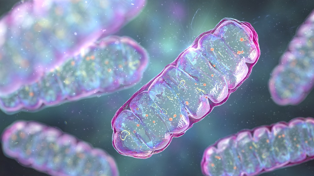
Mitochondrial diseases are a diverse group of disorders that arise when mitochondria malfunction. They are often caused by mutations in mitochondrial DNA (mtDNA) itself or in nuclear genes encoding mitochondria-related proteins.
Mitochondrial dysfunction is also considered one of the hallmarks of aging – no wonder, given that mitochondria are the main source of energy for most cellular processes. When mitochondria falter due to accumulating mutations or persistent damage, such as oxidative stress, no tissue or organ is safe. Parkinson’s disease is a prominent example of a neurodegenerative disease in which mitochondrial dysfunction plays a central role [2].
If only we could deliver healthy, functional mitochondria into diseased cells! However, researchers pursuing this enticing idea have encountered multiple hurdles. Physical approaches like optical tweezers or photothermal nanoblades can transfer mitochondria with precision, but only into a tiny number of cells, while simply injecting free-floating mitochondria into the bloodstream has produced only modest effects [3].
Success in a dish
In this new study published in Cell, a group of Chinese scientists attempted to solve this problem by encapsulating healthy mitochondria in cellular membranes taken from red blood cells (erythrocytes), hoping that this would protect mitochondria while in the bloodstream and facilitate their uptake by recipient cells. Conveniently, erythrocytes are just plasma membranes with no other organelles inside, which makes them an ideal and clinically safe source of membrane material.
Mitochondria were isolated from donor cells, mixed with erythrocyte plasma membranes from mice or humans, and allowed to self-assemble into capsule-like structures. Mitochondria inside the capsules showed two improved markers of mitochondrial function, higher membrane potential and higher ATP levels, than free mitochondria, suggesting the packaging actually preserves or enhances mitochondrial health.
Capsules containing fluorescently labeled donor mitochondria were then incubated with acceptor cells. Time-lapse videos showed donor mitochondria entering cells through membrane fusion. By 48 hours, donor mitochondria fused with the cell’s endogenous mitochondrial network, with about 80% of acceptor cells acquiring donor mitochondria.
Transplanted mitochondria maintained normal membrane potential, and donor mtDNA reached 71% of the total mtDNA pool. Critically, capsule-mediated delivery was dramatically more efficient than delivering free mitochondria.
In the next experiment, rho zero (ρ0) cells, which have been deliberately depleted of all their mtDNA, were treated. These cells can survive in supplemented culture, but they have severely impaired mitochondrial function.
Donor mitochondria entered ρ0 cells in large numbers, and mitochondrial morphology recovered from swollen (a sign of dysfunction) to normal tubular forms. MtDNA levels were restored to near-normal and persisted for at least 21 days. mtDNA-encoded transcripts and proteins were detected, confirming the DNA was being read and translated.
Next, the researchers treated GM04516 cells – human fibroblasts with a large fragment deletion in mtDNA – with capsules loaded with mitochondria from normal human fibroblasts. 86% of patient cells acquired donor mitochondria. The proportion of mtDNA carrying the deletion fell from 14.4% to 2.67%, while oxygen consumption, ATP production, and cell viability increased.
The team then treated human fibroblasts harboring m.3243A>G, the most common pathogenic mtDNA point mutation, which causes mitochondrial encephalomyopathy, lactic acidosis, and stroke-like episodes (MELAS) and a range of other syndromes. The mutation rate fell from 92.6% to 73.3%, while mitochondrial protein levels increased. The reduction was smaller than in the deletion model, but cells usually tolerate mitochondrial dysfunction until the fraction of mutant mtDNA exceeds a critical threshold (typically 60-90%, depending on the mutation and cell type).
Improvements in Leigh and Parkinson’s models
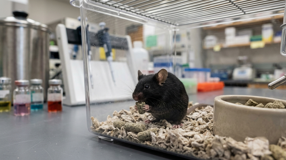
The researchers then moved to in vivo experiments, injecting mitochondrial capsules into mice via different routes: intramuscular injection, direct injection into the substantia nigra (a region of the brain important for movement control), and intravenous injection. They also performed intramuscular injection in two cynomolgus monkeys.
After intramuscular injection, transplanted mitochondria were detected in surrounding muscle tissue, after direct brain injection, they were found in both the substantia nigra and cortex, and after IV injection, donor mitochondria were distributed systemically. In cynomolgus monkeys, mitochondria were successfully delivered to muscle tissue.
Next came the turn of mice with a severe Leigh syndrome phenotype. In humans, it is a rare and fatal inherited mitochondrial disorder, usually appearing in infancy and resulting in death within few years. Median survival increased from 48.5 days (untreated) to 61 days (free mitochondria) to 74 days with mitochondrial capsules – impressive in a model with a severe, fully penetrant phenotype.
The big test was a mouse model of Parkinson’s disease. The animals received a toxin that caused mitochondrial dysfunction and cell death specifically in dopaminergic neurons, and then IV injections of mitochondrial capsules twice weekly for one month. The number of functioning dopaminergic neurons was significantly rescued by the treatment. Behavioral testing showed substantial reversal of bradykinesia, the slow movement that is characteristic of Parkinson’s.
The researchers confirmed improvement of mitochondrial function. The effects persisted for at least three months after treatment – the timeframe of the experiment. Free mitochondria failed to produce comparable effects at the same dose and schedule.
Finally, rather than systemic IV injection, the authors injected mitochondrial capsules directly into the substantia nigra. A single intracerebral injection produced a high local abundance of transplanted mitochondria in both the substantia nigra and cortex, significant neuron recovery, and improvements in motor behavior and mitochondrial function. This shows that targeted delivery can achieve therapeutic effects with a minimal number of doses, and that the IV results did not come from a systemic (such as anti-inflammatory) effect.
Literature
[1] Du, S., Long, Q., Zhou, Y., Fu, J., Wu, H., Yang, L., … & Liu, X. (2026). Transplantation of encapsulated mitochondria alleviates dysfunction in mitochondrial and Parkinson’s disease models. Cell.
[2] Schapira, A. H. V., Cooper, J. M., Dexter, D., Clark, J. B., Jenner, P., & Marsden, C. D. (1990). Mitochondrial complex I deficiency in Parkinson’s disease. Journal of neurochemistry, 54(3), 823-827.
[3] Nakai, R., Varnum, S., Field, R. L., Shi, H., Giwa, R., Jia, W., … & Brestoff, J. R. (2024). Mitochondria transfer-based therapies reduce the morbidity and mortality of Leigh syndrome. Nature metabolism, 6(10), 1886-1896.