An investigation into the aging immune system identified age-related changes, including sex-dependent differences, in immune cell subpopulations and gene expression. In general, females showed greater age-related changes than males, including greater changes in autoimmune gene expression [1].
Cell-by-cell analysis
Aging results in changes to the function and composition of immune cells, collectively referred to as immunosenescence. This decline in immune function manifests as increased susceptibility to infections, cancer, autoimmune diseases, and vascular diseases [2] as well as a persistent low-level inflammatory state known as inflammaging.

Many of these changes are sex-specific; unfortunately, sex-specific aspects of biology are often understudied. As Marta Melé, leader of the Transcriptomics and Functional Genomics group at Barcelona Supercomputing Center (BSC) and director of the study, said, “Many studies still do not take sex into account in their analyses, or directly only use data from men, so they leave key questions unanswered. Our research was born precisely from this need and combines a scientific outlook with a sex perspective, inclusive data, and great computational power.”
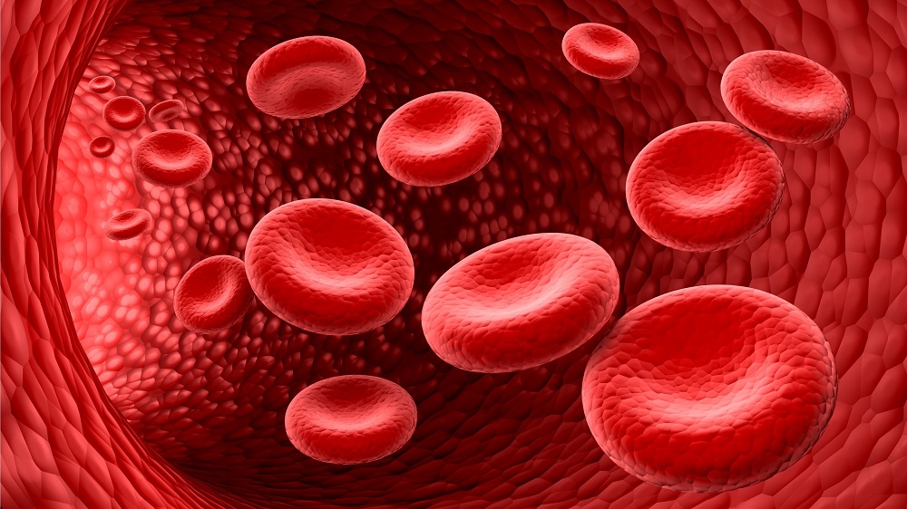
The authors of the study focused specifically on aging-related sex-specific changes in immune cell subpopulations. To address that, they analyzed gene expression levels in single cells, rather than the typical bulk analysis, in over 1 million peripheral blood mononuclear cells (PBMCs) from 416 male and 566 female donors aged 19 to 97 years.
“Until now, most studies analyzed the immune system based on the average of many cells at once, which makes it difficult to capture the progressive effects of aging. With cell-by-cell analysis and a much larger sample, we were able to detect (…) patterns and compare them robustly between biological sexes,” explained Maria Sopena-Rios, researcher at BSC and first co-author of the study.
Sex-dependent immune aging
Initial analysis of the immune cells identified populations that showed similar age-dependent changes in both sexes, but the researchers also observed sex-specific differences. Further analysis indicated that women exhibited more pronounced aging-related changes in immune cells, suggesting greater immune system remodeling, including both increases and decreases in various cell populations. Since the changes appear in immune cell subpopulations with different functions, this suggests differences in how male and female immune systems function as they age.
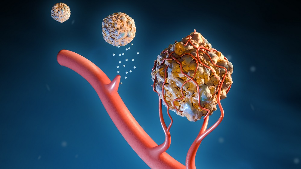
The researchers identified three sex-specific, age-enriched groups: CD8+ TEM and CD14+ monocytes in females, and naive B cells in males. CD8+ TEM was enriched for cytotoxic markers, cell-killing, and natural killer activation signatures. Those activities are important for destroying pathogen-infected and tumorous cells [3]. CD14+ monocytes showed an increase in inflammatory markers, potentially contributing to inflammaging, but they are also involved in viral defense.
The observation that naive B cells are enriched with age in men, specifically the accumulation of their subset CD5+ B cells (more pronounced in some donors), has clinical importance since it has been previously reported that “these expansions may represent the early stages of monoclonal B cell lymphocytosis, a precursor to chronic lymphocytic leukemia, which is more prevalent in older males.” [4]
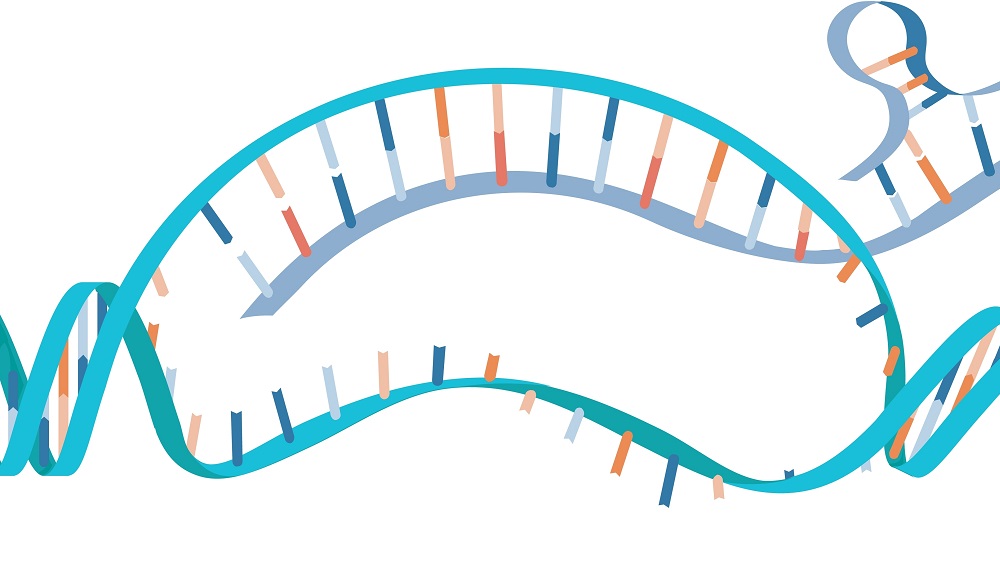
This analysis of cell types was accompanied by an analysis of gene expression, which indicated that 25% of age-associated changes were shared between sexes but a significant number were sex-specific. Women had more such unique age-associated differentially expressed genes across most cell types (2,306 in women and 1,122 in men), suggesting that women have stronger gene expression responses to this type of aging. Analysis of the various pathways in which those genes were involved indicated that around half were shared between the sexes, and the remainder were female-specific.
Grouping individuals into early (<50 years old), mid (40-60 years old), and late (≥50 years old) age groups revealed that most of the gene expression changes occurred in the late group, especially “around age 70 in female participants and slightly later in male participants.”

Differences in disease susceptibility
Immunosenescence does not affect both sexes in the same way. While the previous results suggest that males are more susceptible to leukemia, females face different immune system-related problems.
Women generally have stronger immune responses, resulting in greater resistance to infections [5,6]. However, they pay a high price for this effectiveness: a significantly increased prevalence of autoimmune diseases compared to men [7]. Multiple pieces of evidence from this study, including age-related changes in cell subpopulations and gene expression, support the previous findings and help explain some of the observations at the molecular level.
An analysis of age-related differentially expressed genes showed enrichment for autoimmune-related functions in an immune cell subtype associated with autoimmune disease pathogenesis. Further investigation showed that autoimmune-related gene expression increases significantly with age in both men and women; however, in women older than 50, these genes show significantly higher levels than in men. All of these observations suggest a potential enhancement of autoimmune susceptibility in women. They also agree with previous observations that certain autoimmune diseases, such as multiple sclerosis, inflammatory bowel disease, rheumatoid arthritis, and psoriasis, generally worsen with age [8-12].
Beyond the immune system
The authors of this study suggest that the identified changes can be used as biomarkers of immunosenescence and disease risk. However, the immune system is not an isolated system; it also affects other organs. As Aida Ripoll-Cladellas, researcher at BSC and first co-author of the study, puts it, “The immune system plays a fundamental role throughout the organism; therefore, the differences we observed have a very important generalized impact on the entire body. Better understanding the aging of the immune system can help us understand processes that go beyond the blood and affect multiple tissues.”
Literature
[1] Sopena-Rios, M., Ripoll-Cladellas, A., Omidi, F., Ballouz, S., Alquicira-Hernandez, J., Oelen, R., Hewitt, A. W., Franke, L., van der Wijst, M. G. P., Powell, J. E., & Melé, M. (2026). Single-cell analysis of the human immune system reveals sex-specific dynamics of immunosenescence. Nature aging, 10.1038/s43587-026-01099-x. Advance online publication.
[2] Aw, D., Silva, A. B., & Palmer, D. B. (2007). Immunosenescence: emerging challenges for an ageing population. Immunology, 120(4), 435–446.
[3] Vojdani, A., Koksoy, S., Vojdani, E., Engelman, M., Benzvi, C., & Lerner, A. (2024). Natural Killer Cells and Cytotoxic T Cells: Complementary Partners against Microorganisms and Cancer. Microorganisms, 12(1), 230.
[4] Molica S. (2006). Sex differences in incidence and outcome of chronic lymphocytic leukemia patients. Leukemia & lymphoma, 47(8), 1477–1480.
[5] Furman, D., Hejblum, B. P., Simon, N., Jojic, V., Dekker, C. L., Thiébaut, R., Tibshirani, R. J., & Davis, M. M. (2014). Systems analysis of sex differences reveals an immunosuppressive role for testosterone in the response to influenza vaccination. Proceedings of the National Academy of Sciences of the United States of America, 111(2), 869–874.
[6] Fischinger, S., Boudreau, C. M., Butler, A. L., Streeck, H., & Alter, G. (2019). Sex differences in vaccine-induced humoral immunity. Seminars in immunopathology, 41(2), 239–249.
[7] Jacobson, D. L., Gange, S. J., Rose, N. R., & Graham, N. M. (1997). Epidemiology and estimated population burden of selected autoimmune diseases in the United States. Clinical immunology and immunopathology, 84(3), 223–243.
[8] Park, E., & Ciofani, M. (2025). Th17 cell pathogenicity in autoimmune disease. Experimental & molecular medicine, 57(9), 1913–1927.
[9] Baecher-Allan, C., Kaskow, B. J., & Weiner, H. L. (2018). Multiple Sclerosis: Mechanisms and Immunotherapy. Neuron, 97(4), 742–768.
[10] Kebir, H., Ifergan, I., Alvarez, J. I., Bernard, M., Poirier, J., Arbour, N., Duquette, P., & Prat, A. (2009). Preferential recruitment of interferon-gamma-expressing TH17 cells in multiple sclerosis. Annals of neurology, 66(3), 390–402.
[11] Chen, L., Wu, B., Mo, L., Chen, H., Zhao, Y., Tan, T., Chen, L., Li, Y., Yao, P., & Tang, Y. (2024). Associations between biological ageing and the risk of, genetic susceptibility to, and life expectancy associated with rheumatoid arthritis: a secondary analysis of two observational studies. The lancet. Healthy longevity, 5(1), e45–e55.
[12] Puche-Larrubia, M. Á., Ladehesa-Pineda, L., López-Montilla, M. D., Barbarroja, N., Escudero-Contreras, A., Vazquez-Mellado, J., Collantes-Estévez, E., & López-Medina, C. (2023). Differences between early vs. late-onset of psoriatic arthritis: Data from the RESPONDIA and REGISPONSER registries. Joint bone spine, 90(4), 105563.