How Intestinal Aging Encourages Harmful Bacteria
- Immune dysfunction and dysbiosis are tightly linked.
In Aging Cell, researchers have elucidated the relationship between intestinal aging and age-related changes to the gut microbiome.
Two interdependent biologies
The human gut works through the interaction of two entirely different sets of cells. The first is the body’s actual cells, including the intestinal barrier between the gut and the rest of the body, various types of ordinary immune cells, and Peyer’s patches with follicle-associated epithelium (FAE) areas that contain microfold cells (M cells), which perform crucial immunoregulatory tasks [1]. The second is the gut microbiome, the various types of bacteria that help us digest food.
In a healthy system, these co-evolved biologies support one another. Short-chain fatty acids generated by beneficial microbiomes support immune function [2], perform Toll-like receptor (TLR) signaling, and summon T regulatory cells (Tregs) [3]. With aging, however, this relationship begins to decline. Beneficial Bifidobacterium and Faecalibacterium populations dwindle, replaced by harmful Enterobacteriaceae that produce different metabolites, which do not include the needed SCFAs [4].
A decline in immune function
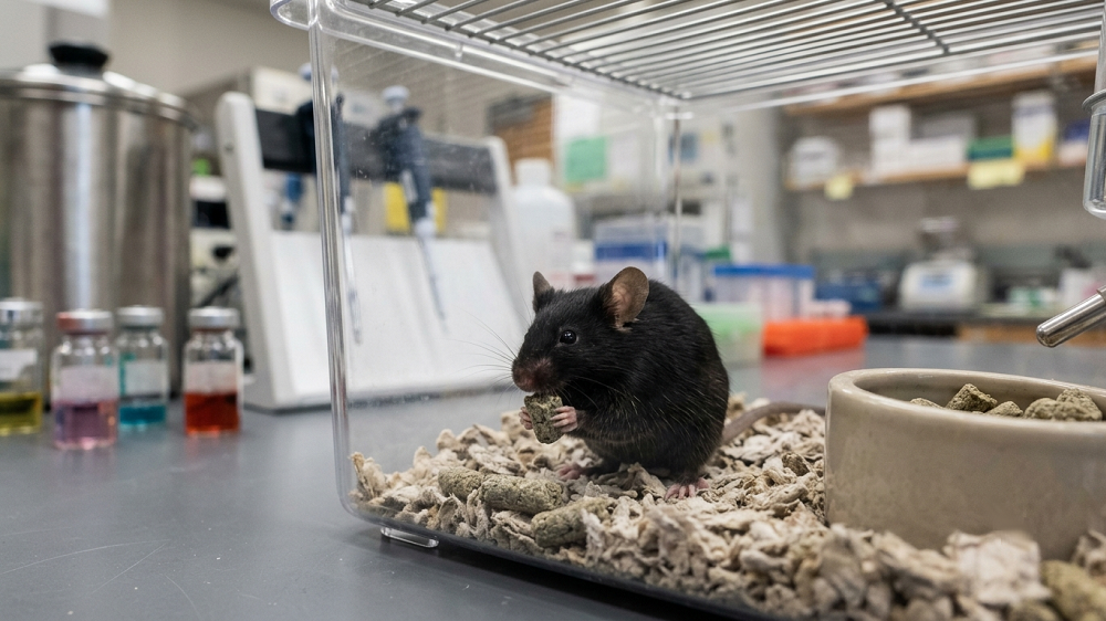
To investigate this relationship, the authors first examined the intestines of 3-month-old and 24-month-old wild-type Black 6 mice. Unsurprisingly, like in other parts of the body, the aged mice had more of the senescence-associated secretory phenotype (SASP), including inflammatory factors and the senescence biomarker p16, while markers relating to intestinal barrier function were decreased.
The overall population of T helper cells in the intestines declined with age, and the cells that remained were disproportionally of the Th1 and Th17 subtypes, signifying increased inflammation. Immunoglobin A (IgA) excretions were reduced in aged mice as well. Meanwhile, lipopolysaccharide (LPS) in excretions remained unchanged, despite an age-related increase in serum; this may signify a decline in intestinal barrier function, as bacterial components make their way from the intestines into the bloodstream.
The researchers also investigated FAE cells. Compared to youthful mice, these cells in aged mice had 446 upregulated genes and 132 downregulated genes. As expected, the affected genes were similar to the changes found in the excretions, relating to IgA, mucosal immunity, and various immunoregulatory processes.
Bacterial changes and intestinal aging are tightly intertwined
While Bacillota (formerly known as Firmicutes) species remained the overwhelming majority in both younger and older mice, the proportion of these bacteria were found to decrease with age, replaced with Bacteroidota; Lactobacillus; Desulfovibrio, which produces hydrogen sulfide [5]; Candidatus Saccharimonas, a pathogenic bacterium that promotes intestinal adenoma [6]; and various other species, some of which are poorly documented.
There was also an increase in Marvinbryantia, a bacterial type that may be beneficial instead of harmful with aging in some contexts; these researchers note another study that has found it to be protective against cirrhosis [7]. However, in this study, Marvinbryantia, along with Candidatus Saccharimonas and Desulfovibrio, was found to be associated with the failure of M cells to properly recognize antigens, as measured by changes in related genes.
When an even more pathogenic species, Clostridium difficile, was introduced into the intestines, the older mice had significantly more immune infiltration and more visibie inflammation. However, only the young mice demonstrated a higher level of short-term inflammation as measured by IL-17A. There was no statistically significant increase in the older mice, which had much higher levels of IL-17A even in the absence of this pathogen.
The researchers summarize the failures of the intestinal barrier and the increases in senescence and inflammation as a “complex, interdependent feedback loop” that leads to an “age-related disruption of host-microbiota coordination”, a deteriorating relationship that encourages the colonization of hostile bacteria that take advantage of the decreased protection, kicking off a downward spiral that causes further damage. The authors note that they were unable to prove whether bacterial changes or native organismal aging were the initial drivers of this spiral.
This was a mouse study, and laboratory mice live under far different conditions than human beings, who live near many more sources of bacteria and eat many different foods that impact bacterial populations. Additionally, some of the bacterial species that play key roles in murine gut health are not prevalent in people and do not have exact equivalents. For example, one group of “Segmented Filamentous Bacteria”, which was found to play a key role in T helper activation in mice, does not have a human counterpart. The researchers comment that organoids made from human gut cells may be necessary in properly understanding the relationship between the human gut and the bacteria it hosts.
Literature
[1] Nakamura, Y., Kimura, S., & Hase, K. (2018). M cell-dependent antigen uptake on follicle-associated epithelium for mucosal immune surveillance. Inflammation and regeneration, 38(1), 15.
[2] Xiao, Y., Feng, Y., Zhao, J., Chen, W., & Lu, W. (2025). Achieving healthy aging through gut microbiota-directed dietary intervention: Focusing on microbial biomarkers and host mechanisms. Journal of Advanced Research, 68, 179-200.
[3] Thevaranjan, N., Puchta, A., Schulz, C., Naidoo, A., Szamosi, J. C., Verschoor, C. P., … & Bowdish, D. M. (2017). Age-associated microbial dysbiosis promotes intestinal permeability, systemic inflammation, and macrophage dysfunction. Cell host & microbe, 21(4), 455-466.
[4] Fadzuli, N. I. A., Lim, S. M., Neoh, C. F., Majeed, A. B. A., Tan, M. P., Khor, H. M., … & Ramasamy, K. (2024). Faecal intestinal permeability and intestinal inflammatory markers in older adults with age-related disorders: a systematic review and meta-analysis. Ageing Research Reviews, 101, 102506.
[5] Singh, S. B., Carroll-Portillo, A., & Lin, H. C. (2023). Desulfovibrio in the gut: the enemy within?. Microorganisms, 11(7), 1772.
[6] Guo, C., Xu, Y., Han, X., Liu, X., Xie, R., Cheng, Z., & Fu, X. (2021). Transcriptomic and proteomic study on the high-fat diet combined with AOM/DSS-induced adenomatous polyps in mice. Frontiers in Oncology, 11, 736225.
[7] Yuan, M., Hu, X., Yao, L., Chen, P., Wang, Z., Liu, P., … & Li, L. (2023). Causal relationship between gut microbiota and liver cirrhosis: 16S rRNA sequencing and Mendelian randomization analyses. Journal of Clinical and Translational Hepatology, 12(2), 123.