More Autophagy Reduces Toxin-Induced Kidney Failure in Mice
- A well-studied protein is again a promising target.

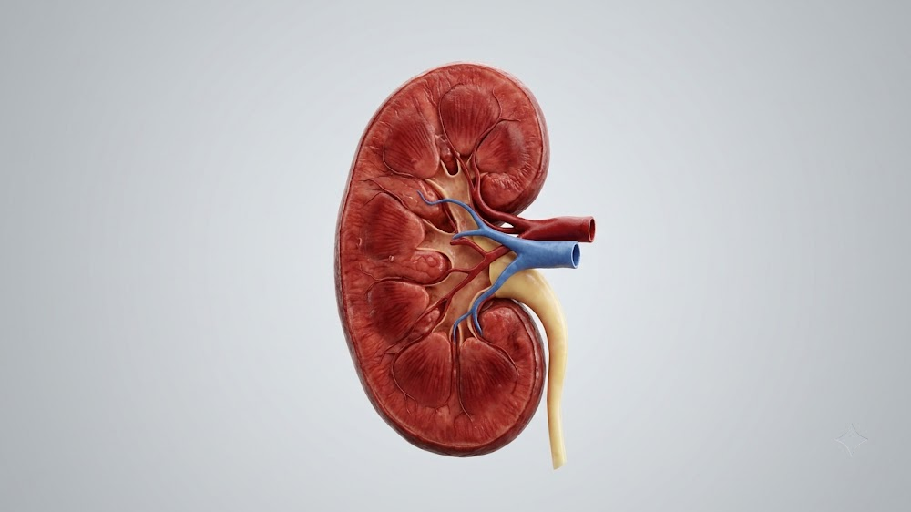
- In mice given a poison that induces kidney failure, younger mice had better outcomes than older mice.
- This was found to be related to autophagy, a maintenance process that decreases in kidney cells with senescence.
- Older mice given C1, which increases autophagy, had better outcomes than untreated older mice when poisoned in this way.
Autophagy, which increases in younger mice under toxic stress, does not increase in older mice and leaves them susceptible to acute kidney injury (AKI). Increasing autophagic flux by targeting a key molecule, transcription factor EB (TFEB), alleviates some of this damage.
An acute disease with lasting effects
AKI is a rapidly developing condition that causes patients to lose kidney function within days, sometimes within hours. In severe cases, kidney dialysis may become required to save the patient. Aging is a risk factor for AKI, for both incidence and severity [1], and in older people, it frequently leads to systemic inflammation and death [2].
In intensive care settings, blood poisoning (sepsis) caused by invading pathogens is a common cause of AKI [3]. However, while it is possible to artificially purify the blood and administer drugs to kill off pathogens, there are currently no methods of directly treating AKI.
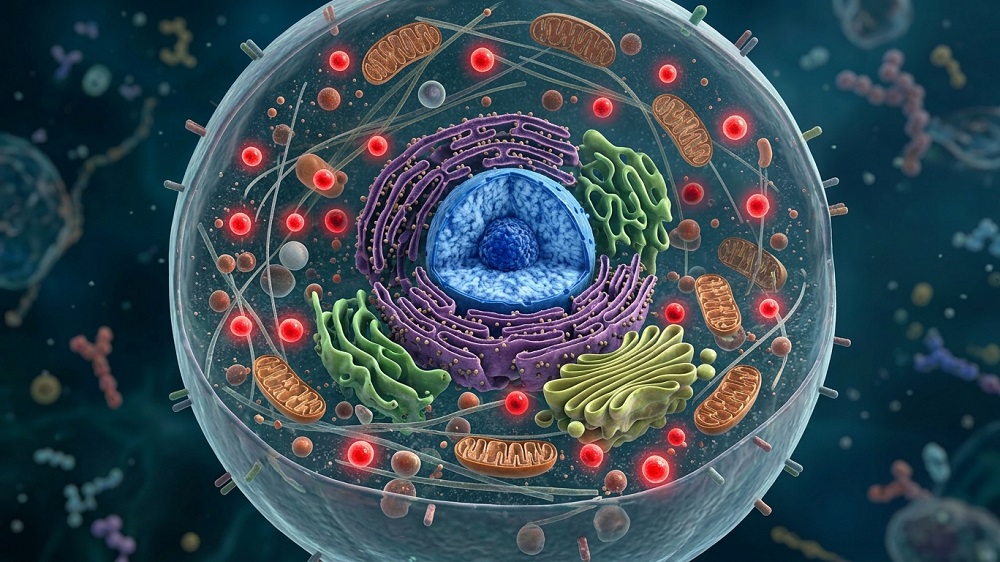
The researchers have hit upon TFEB as a potential avenue for such a treatment. We have previously reported that this protein improves the maintenance of other proteins and increases cellular survival in senescence due to its effects on autophagy, a cellular maintenance process in which cells consume their own organelles. TFEB is directly related to autophagy, directly activating several associated proteins [4], and substantial previous work has found that TFEB is downregulated in various models of AKI, including septic AKI [5].
Older mice are less poison resistant
This paper’s first experiment involved administering lipopolysaccharide (LPS), a poison that is known to induce AKI, to 2-month-old and 18-month-old mice. After LPS administration, creatinine, blood urea nitrogen (BUN), tubular damage, and many other biomarkers of kidney damage were higher in the older mice than the younger mice.
LC3, a marker of autophagy, was slightly higher in the older mice before LPS administration. However, after LPS administration, LC3 was notably upregulated in the younger mice but not significantly upregulated in the older mice, and LPS caused autophagic flux to be twice as high in the younger animals as the older animals.
A cellular experiment found this to be related to senescence; ordinary kidney cells upregulated LC3 in response to this toxin, while senescent cells were unresponsive in this respect, and cells derived from older animals were less responsive from cells derived from younger animals. LC3 was found within puncta within the senescent cells, but it was not flowing freely, reflecting a lack of autophagic flux rather than an increase.
Administering Tat-Beclin 1, a peptide that directly induces autophagy, to kidney cells diminished the damage caused by LPS. Chloroquine, which inhibits autophagy, increased this damage, leading to both increased markers of cellular senescence and increased cellular death by apoptosis. Therefore, the relationship appears to be bidirectional; senescent cells exhibit less autophagy, and a lack of autophagy under toxic stress conditions contributes to cellular senescence.
Using TFEB to fight back
These results were confirmed by a gene expression analysis, which found significant decreases in autophagy-related genes in older mice compared to younger ones. One of the most downregulated proteins was TFEB; normally, older and younger mice express similar levels of TFEB, but LPS exposure caused TFEB in younger mice to decrease while it decreased even more in older mice. This finding was confirmed in cells, with LPS causing senescent cells to express significantly less TFEB than similarly treated non-senescent cells.
Using RNA to force senescent cells to express more TFEB under LPS conditions partially restored autophagy to these cells. Other autophagy-related genes were increased by this expression, and inflammatory cytokines were decreased.
The researchers then tested a curcumin analog, C1, which increases the nuclear translocation of TFEB, in older kidney cells exposed to LPS and a population of older mice also exposed to the toxin. In cells, this treatment improved autophagy by encouraging the beneficial translocation of TFEB even though it did not increase the total amount. In mice, this treatment partially but significantly reduced multiple markers of kidney damage, including creatinine, BUN, and NGAL, an established biomarker of kidney injury.
This work was done in a group of toxically injured mice and may not reflect conditions for real kidney patients. However, the findings are promising within this context. It remains to be seen whether C1 or another autophagy enhancer can ameliorate the damage caused by sepsis-induced or other AKI.
Literature
[1] Hsu, R. K., McCulloch, C. E., Dudley, R. A., Lo, L. J., & Hsu, C. Y. (2013). Temporal changes in incidence of dialysis-requiring AKI. Journal of the American Society of Nephrology, 24(1), 37-42.
[2] Rex, N., Melk, A., & Schmitt, R. (2023). Cellular senescence and kidney aging. Clinical Science, 137(24), 1805-1821.
[3] Pais, T., Jorge, S., & Lopes, J. A. (2024). Acute kidney injury in sepsis. International journal of molecular sciences, 25(11), 5924.
[4] Di Malta, C., Cinque, L., & Settembre, C. (2019). Transcriptional regulation of autophagy: mechanisms and diseases. Frontiers in cell and developmental biology, 7, 114.
[5] Li, R., Zhao, X., Zhang, S., Dong, W., Zhang, L., Chen, Y., … & Liang, X. (2021). RIP3 impedes transcription factor EB to suppress autophagic degradation in septic acute kidney injury. Cell Death & Disease, 12(6), 593.
About the author
Josh Conway