
In Aging, researchers have described the differences between the secretions of cells driven senescent through a cancer gene and cells that stopped proliferating due to p16 expression.
A difference of kind

The SASP is a mix of many components that have various effects on the cells exposed to it. While inflammatory factors, such as cytokines and matrix metalloproteinases, are the most well-known of these, it also consists of ions, various metabolites, and exosomes: encapsulated cellular contents that cells use to signal one another. While exosomes have been researched for their therapeutic value [1], and we have reported on beneficial exosomes’ life-extending effects, exosomes secreted by senescent cells do the opposite [2].
This paper focuses on cells exhibiting the biomarker p16, which is encoded by the gene CDKN2A. This particular biomarker has been heavily linked to senescence, and purging p16-expressing cells from mice lengthens their lives [3]. However, Judith Campisi’s lab had noted that cells that were induced into senescence through p16 express different SASP components [4].
A SASP without inflammation
These researchers took a closer look at such differences, using IMR-90 fibroblasts to directly compare the secretions of ordinary senescent cells to those of p16-expressing cells and non-senescent cells. The first group was driven senescent through the oncogene H-RASG12V (RAS), while the second was made to express p16 through a specific viral vector.
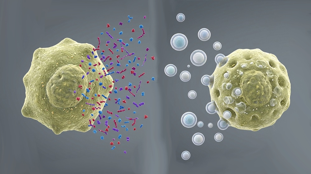
As expected, the secretions of all three groups was substantially different. The RAS group secreted the inflammatory SASP component IL-1β, while the other two did not secrete it at all. While mRNA analysis found that the p16 group was producing IL-6, it secreted even less of it into its environment than the control group did. However, while the control group did not secrete any small extracellular vesicles (sEVs), the RAS group secreted many, and the p16 group secreted slightly more.
Cells spread their own type of senescence
These sEVs caused recipient non-senescent cells to behave much like the cells that secreted them. sEVs from p16 cells induced p16 expression in their recipients, yet they did not encourage the recipients to secrete inflammatory factors such as IL-6 and IL-8. sEVs from RAS cells were inconsistent in encouraging p16 expression, but they did encourage the production of these inflammatory cytokines. sEVs from both types stimulated DNA damage as measured by the biomarker γH2AX.
The researchers confirmed their findings by exposing babies’ fibroblasts in vitro to sEVs derived from the fibroblasts of older people. Even without the rest of the SASP, these older EVs were fully capable of driving the young cells into senescence, as measured by the biomarker SA-β-gal.
This paper holds that the SASP is fundamentally divided into two different portions: a soluble fraction (the sSASP), which consists of proteins and other relatively simple components, and a fraction that consists of extracellular vesicles (the evSASP). While the p16-expressing cells used in this experiment did not secrete the sSASP, they secreted the evSASP, which was found to have negative effects even without classical inflammatory factors.
This is exploratory cellular research, and the specific contents of the sEVs were not discovered, nor was any RNA transcription done in the recipient cells. The researchers suggest that their cargo may be genotoxic in nature and activates stress responses in the recipient cells. Whether or not it is feasible to target sEVs or their originating machinery is not yet clear.

Literature
[1] Ripoll, L., Zickler, A. M., Vader, P., El Andaloussi, S., Verweij, F. J., & Van Niel, G. (2026). Biology and therapeutic potential of extracellular vesicle targeting and uptake. Nature Reviews Molecular Cell Biology, 27(5), 358-376.
[2] Takasugi, M., Okada, R., Takahashi, A., Virya Chen, D., Watanabe, S., & Hara, E. (2017). Small extracellular vesicles secreted from senescent cells promote cancer cell proliferation through EphA2. Nature communications, 8(1), 15729.
[3] Baker, D. J., Childs, B. G., Durik, M., Wijers, M. E., Sieben, C. J., Zhong, J., … & Van Deursen, J. M. (2016). Naturally occurring p16Ink4a-positive cells shorten healthy lifespan. Nature, 530(7589), 184-189.
[4] Coppé, J. P., Rodier, F., Patil, C. K., Freund, A., Desprez, P. Y., & Campisi, J. (2011). Tumor suppressor and aging biomarker p16INK4a induces cellular senescence without the associated inflammatory secretory phenotype. Journal of Biological Chemistry, 286(42), 36396-36403.