The Blog

Building a Future Free of Age-Related Disease

Human Heart

Fortilin Protects Against Cell Death in the Heart

Researchers have found that the protein fortilin, which is abundant in the heart, protects it from failure by blocking apoptotic activity. This discovery opens new possibilities in treating age-related heart diseases [1].

When your heart fails you

As menacing at it sounds, heart failure (HF) does not mean that your heart has completely failed, just that it doesn’t pump blood as well as it should anymore. However, HF is still a debilitating and often deadly condition caused by structural and functional impairments of heart muscle.

As with all cardiovascular diseases, HF prevalence has been on the rise. It currently affects 5.7 million Americans and tens of millions more people all over the world. HF is age-related as well [2]: it is the leading cause of hospitalization in people older than 65 and a major cause of mortality.

30-40% of HF patients die within 1 year after the diagnosis and 60-70% within 5 years. This is because HF keeps progressing through pervasive death of cardiomyocytes by apoptosis, even after its upstream causes, such as coronary artery disease and hypertension, have been treated.

Cardiomyocytes (CMs) are the muscle cells of the heart that are responsible for its contraction and relaxation. For decades, the dogma was that CMs stop proliferating soon after birth, similar to what was previously believed about brain cells. Both of those dogmas turned out to be not entirely true: CMs do proliferate in adults, albeit at a very slow rate that does not change the overall picture [3].

Scientists are not entirely sure why CMs stop dividing in humans and many other species, in contrast to some others, such as the zebrafish. If we can find ways to re-ignite proliferation in adult CMs chemically or genetically, this could lead to a breakthrough in treating age-related heart diseases [4]. Until such ways are found, though, we must protect our CMs as well as we can.

Fortilin and p53

Scientists have long been interested in the protective role of fortilin, a protein abundantly expressed in a healthy heart. In people diagnosed with HF, fortilin levels tend to drop sharply. It has been known that fortilin acts as an inhibitor of the p53 protein, and this new paper seeks to elucidate some important aspects of this connection.

P53 is an anti-tumor protein and is sometimes called the guardian of the genome. It acts by detecting specific alterations in the DNA that are characteristic of tumorigenesis and binding to those loci. Then, P53 triggers a cascade of signals that eventually cause the cell to die via apoptosis.

In longevity research, p53 is widely known as a marker of cellular senescence, which illustrates the dual role that senescence plays in the body. Many types of senescent cells strongly express p53 but do not undergo apoptosis (this is what senescence is all about – cells stopping short of apoptotic death). It seems that in patients with HF, p53’s role is more deleterious than beneficial.

Connecting the dots

First, the researchers created genetically engineered mice with fortilin knocked out specifically in the heart. While normal at birth and fertile, those mice started to die as early as 6 weeks since birth and were all dead by 9 weeks. Autopsy showed that their hearts were dilated and thinned, with signs of severe fibrosis and apoptosis in CMs. P53 levels were very high, and the pro-apoptotic genes regulated by p53 were also overexpressed.

Next, these fortilin-KO mice were crossed with p53-KO mice. Full deletion of both p53 and fortilin significantly improved these mice’s lifespan and healthspan and decreased apoptosis in CMs, although not to the level of wild-type mice. The researchers suggest that this is due to fortilin playing an additional protective role by mediating endoplasmic reticulum stress.

Further experiments showed that fortilin affects p53 at least on three levels: it downregulates p53 by suppressing its transcriptional activation; it binds to p53, preventing it from activating pro-apoptotic genes; and, finally, it promotes degradation of p53 by proteasomes, the cellular recyclers of proteins.

The researchers suggest that bumping up fortilin levels in the heart can be a promising avenue in anti-HF therapies. However, we should be careful when downregulating p53, because of its anti-cancer effect. Additionally, since some types of cellular senescence depend heavily on p53, it is possible that fortilin will be tested as a senolytic.

Conclusion

Although the discovered interplay between fortilin and p53 inspires hope, it also highlights the fact that age-related pathologies often spawn from mechanisms that protect us earlier in life. If further research shows that p53 can be downregulated in the heart without sacrificing its anti-tumor activity, this may lead to the development of new therapies against heart failure – at least until we learn to how to make CMs proliferate again.

We would like to ask you a small favor. We are a non-profit foundation, and unlike some other organizations, we have no shareholders and no products to sell you. All our news and educational content is free for everyone to read, but it does mean that we rely on the help of people like you. Every contribution, no matter if it’s big or small, supports independent journalism and sustains our future.

Literature

[1] Chunhacha, P., Pinkaew, D., Sinthujaroen, P., Bowles, D. E., & Fujise, K. (2021). Fortilin inhibits p53, halts cardiomyocyte apoptosis, and protects the heart against heart failure. Cell Death Discovery7(1), 1-10.

[2] Li, H., Hastings, M. H., Rhee, J., Trager, L. E., Roh, J. D., & Rosenzweig, A. (2020). Targeting age-related pathways in heart failure. Circulation research126(4), 533-551.

[3] Mollova, M., Bersell, K., Walsh, S., Savla, J., Das, L. T., Park, S. Y., … & Kühn, B. (2013). Cardiomyocyte proliferation contributes to heart growth in young humans. Proceedings of the National Academy of Sciences110(4), 1446-1451.

[4] Yutzey, K. E. (2017). Cardiomyocyte proliferation: teaching an old dogma new tricks. Circulation research120(4), 627-629.

Clinical Trial

A Review of Clinical Trials of Senolytics

A team of researchers, including Dr. James Kirkland of Mayo Clinic, has published a review of the ongoing efforts of researchers to clear senescent cells through senolytics.

A growing push for senolytics

We have recently published a discussion on the various roles of senescent cells within the body, some of which are helpful but many of which are harmful, especially in people who are significantly affected by aging. Today’s review highlights the potential therapeutic benefits of directly removing these cells through senolytics. While senomorphics (drugs that alter senescent cells to lose their senescent qualities) are mentioned, they are not currently being explored as thoroughly.

Acknowledging the landmark 2013 paper The Hallmarks of Aging, the researchers promote a Unitary Theory of Fundamental Aging Processes. With a great deal of supporting evidence, this theory holds that the Hallmarks of Aging are largely bi-directional; hallmarks that are thought of as “downstream” can significantly influence the “upstream” hallmarks. This review singles out cellular senescence as being able to affect all eight of the other hallmarks, and it explains the effects of the SASP.

The reviewers also promote the Translational Geroscience Network (TGN), which is comprised of 8 institutions, including Mayo Clinic. One of its stated aims is to conduct new assays for fundamental markers of aging and disease, including senescent cells. It intends to navigate the obstacles involved in creating clinical trials of interventions that target the basic mechanisms of aging.

Drug discovery and preclinical trials

This review discusses the history and current state of senolytic drug discovery. It includes the well-known combination of dasatinib and quercetin (D+Q), the flavonoid fisetin, and navitoclax; however, navitoclax has off-target effects that include platelet deficiency and neutropenia, a serious immune disorder. Efforts are being made to reduce these effects [1], and high-throughput screening is being used to identify more targeted and potent senolytics [2].

The reviewers also showcase the preclinical research involved in these drugs. The researchers cite multiple mouse studies showing the effectiveness of D+Q against metabolic disorders, heart problems, frailty, pulmonary fibrosis, and other age-related issues. Fisetin may be effective against neurodegenerative disorders [3], although more thorough research needs to be done in this regard.

Human trials

The reviewers list several ongoing human trials involved in senolytics. These include a recently completed Phase 1 trial of D+Q against Alzheimer’s disease with results that have yet to be published, a D+Q trial against kidney disease, fisetin trials against frailty and inflammation, fisetin trials against osteoarthritis, and a fisetin trial that specifically targets COVID-19. Additionally, both D+Q and fisetin are being tested for their effects on skeletal health in people.

Future trials will include fisetin against COVID-19, further trials of senolytics as therapies for Alzheimer’s disease, and a trial showing the effectiveness of senolytics in adult survivors of childhood cancer.

Conclusion

The reviewers conclude by saying that senomorphics, along with multiple aging therapeutics in combination therapies, require further exploration. They caution that while some combinations may have synergistic effects, others may have reduced efficacy instead, and they champion the need for collaborative efforts, such as the TGN, in discovering and developing effective combination approaches.

We would like to ask you a small favor. We are a non-profit foundation, and unlike some other organizations, we have no shareholders and no products to sell you. All our news and educational content is free for everyone to read, but it does mean that we rely on the help of people like you. Every contribution, no matter if it’s big or small, supports independent journalism and sustains our future.

Literature

[1] He, Y., Zhang, X., Chang, J., Kim, H. N., Zhang, P., Wang, Y., … & Zhou, D. (2020). Using proteolysis-targeting chimera technology to reduce navitoclax platelet toxicity and improve its senolytic activity. Nature communications, 11(1), 1-14.

[2] Munoz-Espin, D., Rovira, M., Galiana, I., Gimenez, C., Lozano-Torres, B., Paez-Ribes, M., … & Serrano, M. A versatile drug delivery system targeting senescent cells. EMBO Mol Med. 2018; 10 (9).

[3] Maher, P. (2020). Preventing and treating neurological disorders with the flavonol fisetin. Brain Plasticity, 6(2), 155-166.

Michael Geer Interview

Michael Geer: “Digital Markers Are the Future”

Michael Geer is a successful serial entrepreneur who came to the longevity field to get things going. He and his co-founder Pete Ward have recently launched the app Humanity, which offers its users ways to monitor their rates of aging and makes actionable suggestions as to how to slow those rates down. We talked to Michael about why apps like this are important and went into the details of how Humanity works.

The market for consumer-oriented aging biomarkers is exploding. What is their importance not just to Humanity with a capital H but to humanity and to the longevity field?

Basically, people want to be healthier for longer. They want to be fully functional, disease-free – or as disease-free as possible for as long as possible. So, they want to have feedback that would tell them whether they are heading in a better direction or in a reverse direction.

But is it working? The fitness industry sells a lot of flashy things that do have some relation to health, but they don’t necessarily tell you whether you’re going to be healthier in the future. This is why people are so interested in a holistic measure of future health, that’s why what we’re doing is so attractive.

Do you feel like this is new to most people, this mindset of thinking of aging as something that can be slowed or reversed?

The words may be different, but I guess what many people think about health is that they generally want to keep being the way they are now, keep functional, keep doing all those things they like. It’s a simple thought that means staying mobile, not having a disease. Now, it may be framed as aging, which sounds a bit new conceptually, but it’s mostly the label that changed.

On the other hand, the understanding that their body is slowly losing function, moving towards greater susceptibility to disease – that’s something new. The way that most people, including doctors, still talk about health is somewhat binary: you either have a disease or you don’t. This concept that you’re gradually moving towards a higher susceptibility to disease is new and quite powerful because this leads to the question, what do you do now? How do you decrease the speed of this sliding towards greater susceptibility to disease? This is a call for action.

You are one of the cohort of passionate entrepreneurs who have discovered the longevity field and are moving it forward. How did you become involved?

In a past life, I was on the founding team of Badoo, a dating site. Around the time I left Badoo, a couple of people close to me got a late-stage cancer diagnosis and both passed away fairly soon after those diagnoses. From that moment, my biggest question was, why aren’t we screening everybody for cancer? Why are people finding out at stage four? I asked a bunch of doctors, and tons of different answers were given, as you can imagine.

The main answer, which made sense, was there’s too many false positives. If you screened everybody for cancer, no matter their age or family history, you would end up with telling many people that they might have cancer when they don’t, and you might end up subjecting them to unnecessary, dangerous procedures.

This did seem like a logical answer. I reached out to a few people asking “How do we take the false positives out of these tests?” The answer at the time (which is actually coming to fruition with a few companies now) was to use genetic sequencing [cancer tests based on circulating DNA].

Being of the founder mentality, I just Googled top people in genetics. Two names come up when you do that, or at least back then, this was the case: Craig Venter and George Church. I reached out to George, and he was very kind to spend some time with me. He answered a lot of my questions, which were, I’m sure, very stupid at the time and hopefully are slightly less stupid now.

I started talking to George, and then I moved out to the Valley to help run a consumer VPN company.  When I was out there, I began talking to people at UCSF [University of California – San Francisco]. Then serendipitously, through some events, I also met people from Buck Institute.

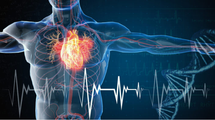
All those people – stem cell researchers, immunologists, geneticists – were spending a lot of time in their labs trying to measure the loss of function in the systems they were looking at. That really intrigued me because I had never had that concept before. This is how we started our conversation today – that concept of our systems losing function gradually, over a long period of time, and that it can be measured. In all those labs, scientists are trying to measure this because they need to test new compounds, they need a way to know if the compound actually decreases the loss of function in the system, because that’s the goal.

You mean, we need biomarkers of aging.

Yes. And then I was lucky enough to get connected with Kristen Fortney [CEO of BioAge Labs], and she turned me onto this idea that you could go to the biobanks, to these longitudinal data sets and see the biomarkers in the past, and also the future real health outcomes.

A few minutes after that conversation, I began thinking that we definitely need to bring this to consumers if we can, and it sounded like since there are digital markers in some of those biobanks; you could actually do that as opposed to having to test everybody’s blood.

Then I pulled my co-founder, Pete Ward, who has a similar background (he created one of the first popular social networks), and we started looking into it. We went to what we called science fantasy camps. We’d fly into Boston; we’d do our whole genome, but the real reason we were there was because we wanted to meet up with George again and other people in that space. So, that’s what we did. I was a believer after spending enough time just sitting in people’s labs, and Pete also was in enough meetings to become a believer too. The rest is history.

Can you give me your elevator pitch for Humanity?

Humanity is an app that can tell you how fast you’re aging and then, importantly, can guide you towards the actions that will slow down your aging. Our focus is on answering the two questions that everybody has about health. First, what should I be doing to be healthier? And the second question is, how do I know when it’s working? Humanity is set up to answer those two questions. Our laser focus is to help people to know what they can do to slow their aging, and then be able to see whether the thing that they’re trying is actually working.

What is your business model – subscription-based, selling data, or something else?

Definitely not selling data. Yes, it’s freemium subscription. The whole business model is based on subscription. Freemium is a great model because it means 95% of your users are getting a ton of value for free, and then the 5% that want the premium features upgrade, and that makes the whole business run. It’s a surprisingly good model because it lets you make the impact that you want. We want to be as radically inclusive and impactful as possible, and this can only be done if most of the service is free or semi-free.

When up to 95% of your users are using it for free, and you still have a great viable business, that’s what you aim for. There will be other things – upsells, different tests, et cetera, but the mainstay will always be those different subscription levels. There’s a digital subscription that we already have, and then there’ll be a higher subscription, which will allow you to do things like DNA methylation testing.

Hopefully, soon you will be sitting on a mountain of data, which will be your significant competitive advantage. On the other hand, you want to make this data widely available. How do you plan to strike the balance here? 

We’ve created and sold a couple of companies at this point. Our balance is far pushed to the impact side. We want to be, and we already are, very open and collaborative. We want our impact to not just be within the walls of the app. We want to help the entire space, to learn more and be able to do more. A part of that is getting the learnings from the data out to others.

There are a few different ways we can do that effectively at scale. One of the most interesting ways that I’m looking quite a bit at is federated learning. Basically, this means the data stays where it is. Wherever the user put the data, it stays there.

Then machine learning algorithms come down into these siloed data sets, do some kind of learning, but then they only take the gradient change of those learnings out. And you have a privacy layer that checks to make sure that there’s no way to reverse engineer back to the actual data.

That’s done at scale by companies like Google. They do it for their predictive texts on Android phones.  That’s one way to cooperate, and the beauty of that is that the user always has full control over their data.

When you actually anonymize data, the user loses a bit of control, because even though people would say that the data can’t be tracked back to the user, it’s still their data and it’s still out there. With federated learning, it’s a better system. If the user wants us to delete all their data, Humanity has this ability.

Let’s dive deeper into the app. It takes in an impressive variety of metrics: physical activity, stress levels, nutrition, sleep quality, et cetera, and it returns a single biological age score. How is it calculated? What’s the science behind it?

There are two main algorithms that the user data is feeding. On the first side, you have a health navigation system. It’s seeing all the actions that a certain type of person is taking and how it’s affecting their rate of aging. Many of those things are being collected so that we can then suggest to that user and to other users different combinations of actions because they seem to be the ones that lead to the path to better health.

On the second, the rate of aging side, two main things are going into that. You’ve got the movement patterns. Basically, it’s accelerometer data from the phone and/or wearable that shows your movement patterns throughout the day. If you have a wearable, there’s also the heart rate. We are then calibrating with other predictors that can be found in our data and the external longitudinal datasets, including chronological age, gender, wearable sensor types, etc.

A lot of this data comes from the UK Biobank right now, but we will expand out from there. In this external dataset, we have people who have been monitored for 10, 15, 20 years. In the UK Biobank dataset, in addition to biomarkers, they also put accelerometers on a ton of people and recorded continuous heart rates from a bunch of people in the past.

And then, in the future, in the next 10 years or so, scientists follow up and collect all the health data, all the health events that happen to those people. One person had a heart attack. Another one got diagnosed with this or that type of cancer. Any morbidities, and in some cases, unfortunately, mortalities, get recorded. With the longitudinal data set like that, you’re able to get a signature: what does a person who’s going to have a heart attack in 10 years, or who’s going to be diagnosed with cancer, look like.

Then we can go to our internal user base and gather those same markers on the digital side – the movement patterns, the heart rate, and we can compare those signatures. This is how we build the probabilistic health trajectory of every person.

In our case, we’re working with a great partner, Gero. It provides a significant portion of that digital side that comes into our model. And then we do more internal calibration. First, calibration within devices because Fitbits and Apple watches and everything else are not created equal. We also do some other calibrations. I won’t go into all the details, but that’s why we believe that the accuracy is as high as possible.

Still, non-invasive biomarkers of aging, like those based on movement, are unorthodox. 

I’ll get a little bit deeper into the logistics side of it. There’s a lot of variance in blood markers, and these markers are usually only measured once or twice a year. But with movement, you have continuous monitoring.

So, even if the blood markers measure the state of the body better, which I don’t think is actually the case, I would say it is still outweighed or at least counterbalanced by the frequency of the measurement of the digital markers. I think there’s a lot of obvious reasons to say that digital markers will be the future.

I’m acquainted with Gero’s concept, and I agree with you about its benefits. Still, it’s not exactly proven yet. Which means you’re basically building a biological age clock on the go, training your models on users’ data, is that correct?

We will definitely do that, but the most important thing you need in order to improve upon the model is to get real health outcomes. So, I think the bigger thing we’ll do in the next year is to expand the external datasets that we’re training our models on.

As you move forward one year, two years, three years, and you get more access (which is actually happening quite nicely, as Apple and others are making it easier for the electronic health records to be connected up), as the users opt in to give you access to their data, you can definitely start using your internal data to make the models even better.

Having access to the actual health outcomes of the users does seem crucial.

It’s crucial to have that in the external dataset. It’s not as crucial to have it on the internal users, but it will make things more accurate when we do it, sure.

Has your clock been tested against existing biological age clocks?

We have stacked it up against some other clocks. We haven’t released any numbers though. Importantly, the stance that we’ve taken is collaboration. I think that ‘the clock to win them all’ is not going to get anybody anywhere. We’d like to be very open and transparent about it. Let’s openly talk about clocks’ strengths, their weaknesses, and how can we make them better. We’re very interested in that conversation, and we’re already having it.

Through marketing pressures, sometimes people are pushed into saying that theirs is the best measure, but I think, if we’re all honest with each other, all the clocks have their strengths and weaknesses. So how can we help? How we can put them together so that we get the best coverage of predictiveness? This is the question.

Let’s go back to the app. After several decades, the longevity field still doesn’t have a magic pill. The best interventions that we have are the good old stuff like physical activity and a healthy diet. Do your recommendations to people consist mainly of that?

Yes, right now, the guidance is very much lifestyle actions. I think it will continue to be that way for some time. Supplements will also be part of the guidance. The beauty of this is that if and when new, better things emerge, such as better senolytics, we’ll be poised to allow people to try that as well, and, importantly, people will be monitoring themselves with the app to actually know if it’s working or not.

I think we’re talking about 20, 30 years of healthy life that people can add with lifestyle actions and maybe some supplementation, and that’s our goal. Then, hopefully, more things will continue to come through. We’re super excited about all the stuff that David Sinclair and other people are working on. We’re rooting for them, but there are things people can do right now to add decades to their lives. That’s our current focus.

You and I know how important these lifestyle interventions are for longevity, but from the consumer’s point of view, do you think it might be just a little bit underwhelming – “these guys say that they can slow my rate of aging, but what they’ve been telling me is actually what my Apple watch tells me every day”?

To be very honest with you, the mainstream consumer loves it. For a biohacker, there is probably a little bit of that thought. “I have all my charts and all my measures, and they’re just telling me all those usual things.” But as a company, because we’re a startup, we need to concentrate on helping the majority of people.

Biohackers might feel the need to supplement our app with other stuff they’re doing, but from our 25 thousand users, we already see that they actually are engaging and changing their behavior quite a bit. At least for the mainstream user, it’s already quite enough.

We built this thing so we could start slowing down people’s aging immediately. I think the longevity field needs more of that because there’s a lot of drug discovery going on, but the timelines there are long. Meanwhile, we need to be extending people’s healthspan and lifespan now.

To which camp do you belong in terms of longevity – meaning, are you a fan of extreme life extension, or do you think it’s unrealistic or even not the best thing to pursue?

I’m a very big believer in the idea that we create the world around us. I think it’s quite apparent that there are things we can do to greatly lengthen our healthy lives.

We launched Humanity to start proving to people that, first, they are aging at different rates, and second, that there are things they can do right now to change that rate. In the long term, physically or scientifically, I don’t see anything that says that we couldn’t greatly extend human lifespan.

I’m in camp “everybody should have a choice”. I want to have a choice of when to die, and I think many people would like to have that choice too. And, scientifically, we will probably have the ability  to allow that choice if someone is like: “Hey, I want another 50 years.”

We would like to ask you a small favor. We are a non-profit foundation, and unlike some other organizations, we have no shareholders and no products to sell you. All our news and educational content is free for everyone to read, but it does mean that we rely on the help of people like you. Every contribution, no matter if it’s big or small, supports independent journalism and sustains our future.
Injured Hand

Potential Mechanisms Behind Skin Aging Identified

The latest research out of the Journal of Cell Biology shines a light on why our skin loses its healing abilities as we age.

The skin’s ability to regenerate dramatically decreases with age. Injuries that would have healed in days without a trace in childhood can take weeks to heal, leave behind scars, or even become chronic in late adulthood. Aging is complex and likely impacts many parts of the skin regeneration process. However, there is some initial evidence to suggest that the skin’s decreased regenerative abilities with age disproportionately impair the healing phase known as re-epithelialization [1].

During this stage, skin cells (especially keratinocytes) multiply and migrate to the damaged area and separate the wound from the outside world by laying down new epithelial tissue [2]. As such, the proliferation and migration of these cells has been of great interest to researchers, but much is still unknown about how these activities are regulated during skin regeneration.

Researchers at the Tokyo Medical and Dental University have recently used human keratinocyte colonies as a model to identify some of the growth factors and signaling pathways that are essential for re-epithelialization [3].

Keratinocyte mobility is associated with expansive cell division

In a cellular culture, the keratinocytes formed both “expanding” and “stacking” colony structures. Cells in expanding colonies were highly proliferative and mobile, while the stacking colony contained fewer dividing cells that mostly remained in the same positions. Within both types of colonies, cells that were more proliferative also displayed more movement.

Cell tracking experiments showed that mobility was associated with expanding rather than stacking behaviors. Mobile cells were able to find additional space on the basal layer after dividing instead of layering on top of one another. A previously established computer simulation of keratinocyte cell culture further confirmed these observations, showing that increased cell rotational speed increased cell expansion.

Understanding the mechanisms behind keratinocyte mobility and proliferation

Next, the researchers compared cells from wounds in young and adult mice, focusing on receptor tyrosine kinases (RTKs). Among the RTKs, epidermal growth factor (EGF) receptor expression and phosphorylation emerged as the best candidate that was both associated with wound healing relative to normal skin and decreased in the wounds of older mice.

Returning to their cell culture model, the researchers found that stimulating keratinocytes with EGF increased their mobility, increased their formation of expanding colonies, and decreased their stacking behavior. Further investigation determined that EGF stimulation increased the COL17A1 protein, likely through activation of TIMP1 reducing COL17A1’s degradation. In additional experiments, blocking COL17A1 expression had effects that were the opposite of EGF stimulation, which suggests that the two are acting as a part of the same biochemical pathway. Lastly, the authors linked COL17A1’s effects on cell mobility to its control over actin and keratin filament networks.

Although further investigations are still required, COL17A1 stabilization by regulating its proteolysis is a candidate therapeutic approach for the improvement of age-associated impaired skin regeneration, including ulcers.

Conclusion

These results shed light on the decline of skin’s regenerative capabilities with age and possibly identify novel targets to reverse this trend. However, cells often behave very differently in culture than they do in the body. It is not clear how well the experimental model used in this study captures the wound healing process in vivo. Therefore, even though these experiments were carried out in human cells, animal studies will need to confirm these findings before there is enough evidence to move on to human trials.

We would like to ask you a small favor. We are a non-profit foundation, and unlike some other organizations, we have no shareholders and no products to sell you. All our news and educational content is free for everyone to read, but it does mean that we rely on the help of people like you. Every contribution, no matter if it’s big or small, supports independent journalism and sustains our future.

Literature

[1] Holt, D.R. et al. Effect of age on wound healing in healthy human beings. Surgery (1992). https://pubmed.ncbi.nlm.nih.gov/1641768/

[2] Rousselle, P., Braye, F., and Dayan, G. Re-epithelialization of adult skin wounds: Cellular mechanisms and therapeutic strategies. Adv Drug Deliv Rev (2019) https://doi.org/10.1016/j.addr.2018.06.019

[3] Nanba, D. et al. EGFR-mediated epidermal stem cell motility drives skin regeneration through COL17A1 proteolysis. Journal of Cell Biology (2021). https://doi.org/10.1083/jcb.202012073

Research fundamentals

Cellular Senescence: It’s Complicated, but There’s Hope

Two of the most prominent experts in the field have published a review of cellular senescence in the context of metabolism, and we bring you the highlights [1].

We rarely cover review papers, but when Cristopher D. Wiley and Judith Campisi, two of the most prominent experts on senescent cells, publish a review on senescence and metabolism, we are happy to make an exception.

Senescent cells: helpers or traitors?

Senescent cells are cells that stop dividing but do not undergo death and removal from the organism. Such cells acquire altered morphology and linger in the body, releasing a complex of mostly pro-inflammatory chemicals called the senescence-associated secretory phenotype (SASP). Senescence can be induced by various types of stress, including replicative (after numerous divisions), genomic (DNA damage), radiation, and chemical. Senescent cells accumulate in our tissues with age and have been linked to multiple age-related disorders. For instance, SASP is probably a major factor in age-related systemic inflammation (inflammaging) [2].

Cellular senescence is not 100% bad, although the related biochemistry is complicated. Senescence is a powerful mechanism that inhibits carcinogenesis. It also plays an important role in wound healing. Wiley and Campisi note that cellular senescence might be an example of antagonistic pleiotropy, in which a mechanism that is beneficial earlier in life becomes deleterious in advanced age. Thus, it escapes evolutionary pressure, as nature doesn’t care what happens to you after you stop reproducing. In young organisms, the anti-cancer and regenerative effects of cellular senescence probably far outweigh its drawbacks, but as the burden of senescent cells grows with age, their darker side starts to dominate.

Cellular senescence was discovered back in the 1960s, but it is so complex a phenomenon that even today, many questions remain unanswered. As the authors note, senescence phenotypes vary between tissues, and no pathway of senescence is like another. The SASP has many variations as well [3]. This new review published in Nature seeks to make some sense of the complex relationship between cellular senescence and various aspects of our metabolism.

Causes of senescence

First, the authors discuss various causes of senescence, starting with mitochondrial dysfunction. Senescence caused by it has its own unique profile and name – mitochondrial dysfunction-associated senescence (MiDAS). It also results in a distinct SASP.

The authors mention that phenotypes of cellular senescence also depend on the oxygen available in the cell. This is easy to overlook in research, particularly when working on cell cultures subjected to room air, where oxygen concentration is higher than in tissues. As a rule, higher oxygen levels promote senescence, although it has been shown that hyperbaric therapy lowers markers of senescence in peripheral blood mononuclear cells. Hence, it is important to account for oxygen levels when studying senescent cells.

NAD+ is a compound that gets a lot of attention in the longevity field. Its age-related loss appears to be a major driver of senescence. Moreover, the authors suggest that SASP that seeps into the intracellular environment can trigger additional loss of NAD+ by interacting with macrophages. This creates a vicious cycle that can explain why age-related NAD+ loss is so harmful.

Hyperglycemia has also been identified as a major driver of senescence, though scientists still do not know how this works, since there are many potential mechanistic explanations. It is possible that, just like NAD+ deficiency, hyperglycemia drives senescence via loss of sirtuins. The authors note that “the links between diabetes and senescence are growing and increases in senescent cells at sites of diabetic complications indicate that this is an important area for future research.”

The metabolism of senescence

Moving to the metabolism of senescent cells, the authors note that there is a growing body of evidence that senescence considerably alters lipid processing in cells. In general, metabolic breakdown of lipids is upregulated in senescent cells since it is needed to produce many SASP components. In some types of senescent cells, particularly in the brain, lipid droplets are known to accumulate.

Research shows that lowering lipid levels in cell culture media prevents both lipid droplet accumulation and the upregulation of many SASP factors. Per the authors, this suggests that “lipid droplets, or at least the presence of lipids, is required for some key aspects of cellular senescence and the SASP”.

Another important process associated with cellular senescence is autophagy, the breaking down and recycling of organelles and macromolecules inside a cell. Autophagy is disturbed in senescent cells, and its inhibition has been shown to induce senescence. On the other hand, in line with the dual nature of senescence, activation of autophagy is a possible driver of some types of senescence.

Lysosomes, the organelles where autophagy takes place, are also dysregulated in senescent cells. Lysosome dysregulation increases acidity inside the cell, but senescent cells have ways to counter this. The authors propose to explore this pathway as a possible senolytic – basically, to help lysosomes kill senescent cells via increased acidity.

Possible interventions

This brings us to the last part of the review, which is dedicated to potential metabolic interventions. First, the authors describe the role of senescent cells in diabetes: according to them, senescent cells are responsible for several complications of diabetes, which means that some senolytics should also have anti-diabetic effects.

Statins, the drugs of choice against hypertension, in addition to lowering blood pressure, also inhibit many pro-inflammatory aspects of the SASP in fibroblasts and prevent senescence in endothelial progenitor cells. This may partly explain statins’ efficacy against atherosclerosis, since the latter is associated with senescence. Hence, it should be possible to use statins as senolytics outside the context of atherosclerosis.

An honorary mention goes to metformin, an anti-diabetes drug that has been shown to increase healthspan and lifespan in model organisms (read our interview with Nir Barzilai about the upcoming metformin study in humans). Metformin is known to protect against senescence in some cell types in vitro, along with murine models of intervertebral disc degeneration and chronic kidney disease. This might explain a lot of metformin’s hailed anti-aging qualities.

Finally, the authors mention two kinds of interventions that most of us can start applying right now: dietary changes and exercise. Caloric restriction, widely regarded as having anti-aging effects, prevents senescence in the kidneys of aged animals, while a high-calorie diet increases senescence. According to the review, ketogenic diets have been found to reduce markers of senescence in vascular smooth muscle cells and endothelial cells.

As for exercise, it is known to prevent senescence in hearts, livers, and adipose tissue of aged mice, and it has been linked to reduced markers of endothelial and leukocyte senescence in humans.

Conclusion

Cellular senescence is a multifaceted phenomenon, and its interplay with various functions of the body is far from being fully elucidated, as evidenced by this major NIH effort to create an atlas of senescent cells. Senescence can be both beneficial and deleterious, and its phenotypes vary among tissues. Hence, attempts, such as this review, to systematize our vast but insufficiently deep knowledge of cellular senescence are very important.

We would like to ask you a small favor. We are a non-profit foundation, and unlike some other organizations, we have no shareholders and no products to sell you. All our news and educational content is free for everyone to read, but it does mean that we rely on the help of people like you. Every contribution, no matter if it’s big or small, supports independent journalism and sustains our future.

Literature

[1] Wiley, C. D., & Campisi, J. (2021). The metabolic roots of senescence: mechanisms and opportunities for intervention. Nature Metabolism, 1-12.

[2] Coppé, J. P., Desprez, P. Y., Krtolica, A., & Campisi, J. (2010). The senescence-associated secretory phenotype: the dark side of tumor suppression. Annual Review of Pathology: Mechanisms of Disease5, 99-118.

[3] Wu, S. K., Ariffin, J., & Picone, R. (2020). The Senescence-Associated-Secretory-Phenotype induced by centrosome amplification constitutes a pathway that activates Hypoxia-Inducible-Factor-a. bioRxiv.

Spinal Injury

Resveratrol Aids Spinal Cord Regeneration in Rats

A team of researchers publishing in Aging have shown that resveratrol reduces inflammation and partially restores function in a rat model of spinal injury.

In line with previous research

This is far from the first study that aimed to use approaches associated with aging research in order to spur regeneration. For example, we have previously reported that removing senescent cells aids in spinal cord regeneration in a rodent model, at least partially because of the associated reduction in inflammation.

The researchers cite multiple, related other studies relating to resveratrol and the spinal cord. It reduces inflammation in lung tissues after spinal injury [1], aids in spinal cord regeneration through the mTOR pathway and autophagy [2], protects against spinal cord hypoxic injury through Nrf-2 [3], and aids in spinal regeneration by reducing inflammation through the Notch signaling pathway [4].

However, their study shows a different pathway for the effects of resveratrol: the Wnt/ß-catenin signaling pathway, which plays a significant role in basic cellular maintenance in multiple species.

Motor function restored

Rats that withstand the specific injury used in this experiment normally regain some motor function in their lower bodies according to the widely used Basso, Beattie, Bresnahan (BBB) locomotor scale. However, rats given resveratrol after this injury had BBB scores that were nearly those of uninjured rats after 28 days.

The inclined plane test, on which rats balance on a tilting surface, was also used. Spinally injured rats normally regain only a small amount of balance. This, too, was improved with the use of resveratrol, although not to the level of uninjured rats.

Rats regain very little of their sensory function after this type of injury. Resveratrol was shown to slightly improve this, although not to the level of statistical significance. Taken together, these results appear to show that resveratrol increases the normal regeneration of rats after spinal injury.

Inflammation decreased, regenerative factors increased

Resveratrol significantly decreased the inflammatory factors IL-6, IL-1ß, and TNF-α to approximately half that of untreated injured rats. IL-6 in particular was reduced to the level of uninjured rats.

Spinal injury substantially reduces the levels of GAP43 and NF421, two critical factors involved in nerve regeneration, while increasing GFAP, an astrocyte marker. Resveratrol restored all three of these factors nearly to the levels seen in uninjured rats.

Apoptosis (cellular death) was also measured, both in vitro and in vivo. In both this rat model and a cellular model that used hydrogen peroxide to stimulate apoptosis, resveratrol substantially decreased the rate of apoptosis.

Finally, the researchers examined the expression of multiple Wnt proteins and ß-catenin, whose expression decreases with spinal injury. They found that in rats given resveratrol, the levels of ß-catenin and these Wnt proteins were greatly increased, sometimes well above the levels seen in uninjured rats. GSK-3ß, which increases with spinal injury, was suppressed with resveratrol.

Finally, the researchers set out to prove the role of the Wnt/ß-catenin signaling pathway. They did this by administering XAV939, an inhibitor of this pathway, to a different group of rats that were given resveratrol after injury. As expected, XAV939 partially reversed the effects of resveratrol in both locomotor and biochemical measurements.

Conclusion

This study, along with the studies before it, provides strong evidence that resveratrol helps to restore spinal function through multiple methods. While resveratrol is by no means a cure for spinal injury, its anti-inflammatory and signaling effects make it a clear candidate to be examined as a treatment for spinal injury in humans, possibly alongside stem cell treatments or other therapies that use other approaches to restoring spinal function.

We would like to ask you a small favor. We are a non-profit foundation, and unlike some other organizations, we have no shareholders and no products to sell you. All our news and educational content is free for everyone to read, but it does mean that we rely on the help of people like you. Every contribution, no matter if it’s big or small, supports independent journalism and sustains our future.

Literature

[1] Liu, J., Yi, L., Xiang, Z., Zhong, J., Zhang, H., & Sun, T. (2015). Resveratrol attenuates spinal cord injury-induced inflammatory damage in rat lungs. International journal of clinical and experimental pathology, 8(2), 1237.

[2] Meng, H. Y., Shao, D. C., Li, H., Huang, X. D., Yang, G., Xu, B., & Niu, H. Y. (2018). Resveratrol improves neurological outcome and neuroinflammation following spinal cord injury through enhancing autophagy involving the AMPK/mTOR pathway. Molecular medicine reports, 18(2), 2237-2244.

[3] Kesherwani, V., Atif, F., Yousuf, S., & Agrawal, S. K. (2013). Resveratrol protects spinal cord dorsal column from hypoxic injury by activating Nrf-2. Neuroscience, 241, 80-88.

[4] Zhang, S., Botchway, B. O., Zhang, Y., & Liu, X. (2019). Resveratrol can inhibit Notch signaling pathway to improve spinal cord injury. Annals of Anatomy-Anatomischer Anzeiger, 223, 100-107.

Neuron Connections

New Mechanism of Neurodegeneration Identified

Researchers have identified an important mechanism of neurodegeneration that may be useful in treating age-related diseases such as Alzheimer’s and dementia [1].

These neurons must die

Age-related neurodegenerative diseases, including Parkinson’s and Alzheimer’s, have become more prevalent mostly because of the successes of modern medicine. Today, more people become old enough to fall prey to neurodegeneration, and without solving neurodegeneration, we cannot hope to achieve meaningful lifespan and healthspan extension. Some longevity experts say half-jokingly that even if we will be able to replace every organ in the human body, we cannot replace the brain, so we must learn how to fix it. Yet, in recent decades, this field has seen little progress.

Neurodegeneration is, basically, the death of neurons. It happens as a result of various types of stress, including injuries. It has been known for some time that astrocytes, cells that perform various housekeeping functions in the central nervous system, are activated by disease, injury, and systemic inflammation in a way that leads them to kill neurons. This activation, at least when related to systemic inflammation, is induced by several proteins, including interleukin-1a (IL-1a) and tumor necrosis factor (TNF) [2].

Looking for the murder weapon

Until now, scientists did not know how exactly activated astrocytes kill neurons. The group of researchers behind this new paper was one of many to study this question. For some time, they thought that the instrument of murder must be a protein or a group of proteins, but attempts to pinpoint such killer proteins have been unsuccessful.

This time, after analyzing the full proteome in activated and quiescent astrocytes, the researchers found changes in expression of only a small fraction of proteins, and none of those proteins stood up to scrutiny as a possible culprit. However, among the proteins that were most notably upregulated in activated astrocytes were APOE and APOJ.

The main function of APOE and APOJ is to transport lipids (fats) by packing them into particles called lipoproteins [3]. Two types of those particles are widely known as LDL and HDL (low- and high-density lipoproteins: “bad” and “good” cholesterol, respectively). Neurons need cholesterol to function, and by forming lipoproteins, APOE and APOJ participate in the cholesterol supply chain. Interestingly, one of the alleles of the APOE gene is strongly associated with Alzheimer’s.

Further investigation showed that astrocytes’ cytotoxicity is indeed regulated by lipoprotein particles. However, since those particles consist of both lipids and proteins, it had to be determined which of them are responsible. Removing the protein fraction by knocking out APOE and APOJ genes in mice did not diminish the cytotoxicity, while removing the lipids did.

Then, the researchers reconstituted lipoparticles by taking the lipids from the activated cells and the proteins from the quiescent cells. These human-made lipoparticles were just as toxic, proving again that lipids and not proteins were to blame. Moreover, even when not packed into lipoparticles, lipids from activated astrocytes retained their toxicity, while lipids from quiescent astrocytes and HEKs (human embryonic kidney cells used as controls) were non-toxic.

The last step was to determine which lipids were so toxic. By analyzing the entire lipidome (lipid profile) of activated vs. quiescent astrocytes, the researchers found that two classes of lipids were significantly upregulated in reactive astrocytes: long-chain saturated free fatty acids (FFAs) and much less pronounceable phosphatidylcholines with very-long-chain fatty acid acyl chains (VLCPCs).

Less neuronal death in vivo

In order to prove their theory in vivo, the researchers had to find a way to stop the production of these two specific types of lipids in live mice while disrupting as few other cellular processes as possible. This was done by knocking out ELOVL1, the enzyme that is specifically responsible for the synthesis of longer-chain, fully saturated lipids. In wild-type mice with induced optical nerve injury, massive neuronal loss and no signs of vision restoration were observed. However, in mice with ELOVL1 knocked out, there was substantially less neuronal death.

It is worth noting that this same group of researchers had previously achieved even better results (close to zero neuronal death) by knocking out the aforementioned proteins that actually activate astrocytes. While targeting those factors may seem a better strategy, it is probably not, since they perform many other functions in the body. On the contrary, while having a less pronounced effect, targeting those two types of lipids is a gentler intervention that can be performed after the astrocytes become activated (for instance, as a result of injury or age-related inflammation).

Conclusion

After years of painfully slow progress in this field, this groundbreaking discovery showcases a mechanism of neuronal death that is at least partly responsible for neurodegeneration, including in age-related diseases such as Parkinson’s and Alzheimer’s, which have been notoriously hard to crack. It also offers a specific target for a prospective intervention.

We would like to ask you a small favor. We are a non-profit foundation, and unlike some other organizations, we have no shareholders and no products to sell you. All our news and educational content is free for everyone to read, but it does mean that we rely on the help of people like you. Every contribution, no matter if it’s big or small, supports independent journalism and sustains our future.

Literature

[1] Guttenplan, K. A., Weigel, M. K., Prakash, P., Wijewardhane, P. R., Hasel, P., Rufen-Blanchette, U., … & Barres, B. A. (2021). Neurotoxic reactive astrocytes induce cell death via saturated lipids. Nature, 1-6.

[2] Liddelow, S. A., Guttenplan, K. A., Clarke, L. E., Bennett, F. C., Bohlen, C. J., Schirmer, L., … & Barres, B. A. (2017). Neurotoxic reactive astrocytes are induced by activated microglia. Nature541(7638), 481-487.

[3] Huang, Y., & Mahley, R. W. (2014). Apolipoprotein E: structure and function in lipid metabolism, neurobiology, and Alzheimer’s diseases. Neurobiology of disease72, 3-12.

Journal Club

Targeted clearance of senescent cells

The next Journal Club will be on Tuesday 2nd November and Dr. Oliver Medvedik will be taking a look at a new paper called “Targeted clearance of senescent cells using an antibody-drug conjugate against a specific membrane marker“. Join us live on Facebook at 1 PM Eastern US time on Tuesday November 2nd.

Abstract

A wide range of diseases have been shown to be influenced by the accumulation of senescent cells, from fibrosis to diabetes, cancer, Alzheimer’s and other age-related pathologies. Consistent with this, clearance of senescent cells can prolong healthspan and lifespan in in vivo models. This provided a rationale for developing a new class of drugs, called senolytics, designed to selectively eliminate senescent cells in human tissues. The senolytics tested so far lack specificity and have significant off-target effects, suggesting that a targeted approach could be more clinically relevant. Here, we propose to use an extracellular epitope of B2M, a recently identified membrane marker of senescence, as a target for the specific delivery of toxic drugs into senescent cells. We show that an antibody–drug conjugate (ADC) against B2M clears senescent cells by releasing duocarmycin into them, while an isotype control ADC was not toxic for these cells. This effect was dependent on p53 expression and therefore more evident in stress-induced senescence. Non-senescent cells were not affected by either antibody, confirming the specificity of the treatment. Our results provide a proof-of-principle assessment of a novel approach for the specific elimination of senescent cells using a second generation targeted senolytic against proteins of their surfaceome, which could have clinical applications in pathological ageing and associated diseases.

Journal Club

Journal Club October 2021 – Targeted clearance of senescent cells

The next Journal Club will be on Tuesday 2nd November and Dr. Medvedik will be taking a look at a new paper called “Targeted clearance of senescent cells using an antibody-drug conjugate against a specific membrane marker“. Join us live on the call at 1 PM Eastern US time on Tuesday November 2nd.

Abstract

A wide range of diseases have been shown to be influenced by the accumulation of senescent cells, from fibrosis to diabetes, cancer, Alzheimer’s and other age-related pathologies. Consistent with this, clearance of senescent cells can prolong healthspan and lifespan in in vivo models. This provided a rationale for developing a new class of drugs, called senolytics, designed to selectively eliminate senescent cells in human tissues. The senolytics tested so far lack specificity and have significant off-target effects, suggesting that a targeted approach could be more clinically relevant. Here, we propose to use an extracellular epitope of B2M, a recently identified membrane marker of senescence, as a target for the specific delivery of toxic drugs into senescent cells. We show that an antibody–drug conjugate (ADC) against B2M clears senescent cells by releasing duocarmycin into them, while an isotype control ADC was not toxic for these cells. This effect was dependent on p53 expression and therefore more evident in stress-induced senescence. Non-senescent cells were not affected by either antibody, confirming the specificity of the treatment. Our results provide a proof-of-principle assessment of a novel approach for the specific elimination of senescent cells using a second generation targeted senolytic against proteins of their surfaceome, which could have clinical applications in pathological ageing and associated diseases.

How to join the Zoom call https://lifespan-io.zoom.us/j/82570486171?pwd=RXhxZElhS1FBcmZYdGE0eHN2dE1VUT09 Meeting ID: 825 7048 6171 Passcode: 516322 One tap mobile +16699006833,,82570486171#,,,,*516322# US (San Jose) +12532158782,,82570486171#,,,,*516322# US (Tacoma) Dial by your location +1 669 900 6833 US (San Jose) +1 253 215 8782 US (Tacoma) +1 301 715 8592 US (Washington DC) +1 312 626 6799 US (Chicago) +1 346 248 7799 US (Houston) +1 646 876 9923 US (New York) Meeting ID: 825 7048 6171 Passcode: 516322 Find your local number: https://lifespan-io.zoom.us/u/kyB1us5Sk
Links

Energy Metabolism Decline Linked To Alzheimer’s

Researchers publishing in Aging have discovered that an energy metabolism compound is associated with a protein that suppresses amyloid beta in a mouse model of Alzheimer’s.

The compounds involved

The researchers describe nicotinamide mononucleotide adenylyltransferase 2 (Nmnat2) as a key co-factor for energy metabolism. This compound is involved in creating NAD+, which is essential in basic metabolic processes, from NMN, a well-known compound that is commonly taken as a supplement.

A disintegrin and metalloprotease domain 10 (ADAM10) is responsible for cleaving amyloid precursor protein (APP), which is a source of amyloid beta, the aggregates of which are the key hallmark of Alzheimer’s. If ß- and ?-secretases are used to cleave APP, amyloid beta is the result [1]. However, ADAM10 is responsible for a-secretases, which do not cleave APP into amyloid beta [2] and instead form secreted APP, which has been shown to be neuroprotective [3].

AMP-activated kinase (AMPK) is a regulator of metabolism and is upregulated when nutrients are in short supply, such as during caloric restriction. Increased expression of AMPK is associated with longevity [4].

The experiments

The researchers discovered several relationships between these compounds. First, they examined Tg2576 mice, a model of Alzheimer’s disease, and compared them to their wild-type counterparts. They found that the brain cortices of these Alzheimer’s-prone mice were severely deficient in both ADAM10 and Nmnat2.

They then tested the effect of Nmnat2 in a cellular culture. After they transfected cells with a plasmid responsible for producing Nmnat2, they found that ADAM10 and the associated a-secretases were increased by approximately one and a half times and that amyloid beta was decreased by approximately half.

The researchers also examined the relationship between Nmnat2 and AMPK. They compared the cortical cells of Tg2576 and wild-type mice, finding that both of these compounds were halved in the Alzheimer’s-prone mice. They also found that Nmnat2 in cellular culture led to a substantial increase in AMPK.

The NAD/NADH ratio, a marker of metabolic activity, was found to be upregulated in Nmnat2-transfected cells and downregulated in mice prone to Alzheimer’s disease.

Finally, the researchers performed another test to try to understand the cause-and-effect relationship involved. The researchers examined the AMPK promoter AICAR and the AMPK inhibitor Compound C. Cells that were given AICAR had more ADAM10 and less amyloid beta; cells that were given both Nmnat2 and Compound C did not enjoy the positive effects associated with Nmnat2. The researchers, therefore, conclude that AMPK is critical in the increased production of ADAM10.

Conclusion

This is a mouse and cellular study that relies on models and chemical analysis to determine effects. The effects of increasing Nmnat2 were not studied in live animals, and this study certainly does not provide any evidence that increasing NAD+ through any supplement can help to alleviate Alzheimer’s in human beings.

However, it illustrates a very interesting and perhaps targetable relationship, and more investigation is needed in order to determine if Alzheimer’s disease can be affected on a metabolic level.

We would like to ask you a small favor. We are a non-profit foundation, and unlike some other organizations, we have no shareholders and no products to sell you. All our news and educational content is free for everyone to read, but it does mean that we rely on the help of people like you. Every contribution, no matter if it’s big or small, supports independent journalism and sustains our future.
Literature

[1] Vassar, R., & Citron, M. (2000). Aß-generating enzymes: recent advances in ß-and ?-secretase research. Neuron, 27(3), 419-422.

[2] Postina, R., Schroeder, A., Dewachter, I., Bohl, J., Schmitt, U., Kojro, E., … & Fahrenholz, F. (2004). A disintegrin-metalloproteinase prevents amyloid plaque formation and hippocampal defects in an Alzheimer disease mouse model. The Journal of clinical investigation, 113(10), 1456-1464.

[3] Mattson, M. P. (1997). Cellular actions of beta-amyloid precursor protein and its soluble and fibrillogenic derivatives. Physiological reviews, 77(4), 1081-1132.

[4] Salminen, A., & Kaarniranta, K. (2012). AMP-activated protein kinase (AMPK) controls the aging process via an integrated signaling network. Ageing research reviews, 11(2), 230-241.

Three Fates

Lack of Klotho Associated With All-Cause Mortality

A recent study in The Journals of Gerontology, Series A has associated low klotho levels with mortality in Americans over the age of 40.

Klotho and aging

Prediction of mortality is a major interest of the aging field and the broader medical field. Epigenetic clocks, senescent cell accumulation, and telomere length have all received recent attention for their abilities to predict mortality beyond chronological age. The klotho gene has been associated with aging and mortality in various observational and interventional studies, with lower levels resulting in shorter lifespans and vice versa [1]. Therefore, it may also serve as a measure of biological aging and a risk factor for mortality.

Initial studies have found this to be true in patients with chronic kidney disease [2] and older adults [3]. A recent collaboration between the University of North Carolina at Chapel Hill and the National Institute of Environmental Health Sciences within the NIH has investigated this in a more general population [4].

Are serum klotho levels predictive of mortality?

Data from the American National Health and Nutrition Examination Survey (NHANES) was used in this study. 10,069 participants qualified for inclusion in the study, ranging from 40 to 79 years old with an average age of 56. The participant population was slightly healthier compared to the general American population of the same age based on BMI, physical activity levels, smoking history, and alcohol consumption. Of these participants, 616 (4.3%) had deceased after a mean follow-up of 5 years.

Serum klotho levels were measured for each of these participants. Age, BMI, income, and alcohol consumption were weakly and negatively associated with klotho levels. Females, non-Hispanic blacks, college graduates, and people who had never smoked had higher klotho levels than their counterparts.

Grouping by quartile based on klotho concentration, the lowest quartile had a 31% higher rate of death than the highest quartile. Further adjustments for kidney function, BMI, income, ethnicity, smoking status, and education very slightly decreased this association, but alcohol consumption had no effect. Looking into specific causes of death, cause-specific associations were each similar to all-cause mortality.

Impact of physical activity

One novel finding from this study was the relationship between klotho, physical activity, and mortality. No difference in serum klotho levels was found based on activity level. However, when relating klotho levels to mortality, adjusting for physical activity did slightly reduce the correlation between klotho and mortality.

Additionally, those who participated in little to no exercise had an even stronger klotho-to-mortality association than the dataset as a whole. Meanwhile, klotho levels were not associated with mortality in people who achieved the recommended amount of weekly exercise. A similar effect was not found for any of the other surveyed participant characteristics.

Abstract

a-Klotho (klotho) is a protein involved in suppressing oxidative stress and inflammation. In animal models, it is reported to underlie numerous aging phenotypes and longevity. Among a nationally representative sample of adults aged 40 to 79 in the United States, we investigated whether circulating concentrations of klotho is a marker of mortality risk. Serum klotho was measured by ELISA on 10,069 individuals enrolled in the National Health and Nutrition Examination Survey between 2007-2014. Mortality follow-up data based on the National Death Index were available through December 31, 2015. After a mean follow-up of 58 months (range: 1-108), 616 incident deaths occurred. Using survey-weighted Cox regression models adjusted for age, sex and survey cycle, low serum klotho concentration (< 666 pg/mL) was associated with a 31% higher risk of death (compared to klotho concentration > 985 pg/mL, HR: 1.31, 95% CI: 1.00, 1.71, P= 0.05). Associations were consistent for mortality caused by heart disease or cancer. Associations of klotho with all-cause mortality did not appear to differ by most participant characteristics. However, we observed effect modification by physical activity, such that low levels of serum klotho were more strongly associated with mortality among individuals who did not meet recommendation-based physical activity guidelines. Our findings suggest that, among the general population of American adults, circulating levels of klotho may serve as a marker of mortality risk.

Conclusions

This was the first survey done comparing klotho serum levels to mortality in participants who were younger than 65 and/or without chronic kidney disease. This study did not include young adults, and therefore these findings cannot be applied to that population. However, mortality risk is extremely low below age 40 and is mostly caused by accidents, physical injury, and self-harm – factors that klotho levels would be unlikely to predict.

One unanswered question surrounding klotho is whether low levels are simply detrimental or if high levels can also be protective. For mortality in this dataset, the second, third, and highest quartile all had similar survival curves. Meanwhile, survival of participants in the lowest quartile for klotho concentration was significantly decreased. This could suggest that higher levels of klotho are not associated with a survival benefit, at least within physiological levels. If klotho is a driver rather than a result of the aging process, it is possible that klotho-targeting treatments could still provide benefits by boosting levels beyond these ranges or by only treating patients with low klotho levels.

Perhaps the most interesting component of this study is the impact of exercise on the klotho-mortality relationship. Notably, the survey only collected data about current levels of activity, not a history of exercise, which may have a larger impact on overall health. There could be a number of explanations for why klotho was only predictive of mortality in low-exercise participants, which warrants further investigation.

Lastly, it is important to note that this was an association study that did not look at cause and effect. These results are still positive news for klotho-targeting interventions, at least for non-Hispanic white Americans over the age of 40 with low exercise levels and in the bottom quartile of serum klotho concentrations. Nevertheless, it cannot be said from this experimental design whether or not klotho contributed to the increase in mortality in these participants.

We would like to ask you a small favor. We are a non-profit foundation, and unlike some other organizations, we have no shareholders and no products to sell you. All our news and educational content is free for everyone to read, but it does mean that we rely on the help of people like you. Every contribution, no matter if it’s big or small, supports independent journalism and sustains our future.

Literature

[1] Kuro-O.M. The Klotho proteins in health and disease. Nature Reviews Nephrology (2019). https://doi.org/10.1038/s41581-018-0078-3

[2] Charoenngam, N., Ponvilawan, B., and Ungprasert, P. Lower circulating soluble Klotho level is associated with increased risk of all-cause mortality in chronic kidney disease patients: a systematic review and meta-analysis. Int Urol Nephrol (2020). https://doi.org/10.1007/s11255-020-02510-1

[3] Semba, R.D. et al. Plasma klotho and mortality risk in older community-dwelling adults. J Gerontol A Biol Sci Med Sci (2011). https://doi.org/10.1093/gerona/glr058

[4] Kresovich, J.K. and Bulka, C.M. Low serum klotho associated with all-cause mortality among a nationally representative sample of American adults. J Gerontol A Biol Sci Med Sci (2021). https://doi.org/10.1093/gerona/glab308

Erase bad cells

Scientists Propose a New Targeted Senolytic

A group of researchers has developed a delivery system that identifies senescent cells and injects them with a deadly compound. This offers a potential solution to the problem of targeted delivery of senolytics [1].

Cellular senescence has been implicated in numerous aging-related disorders, including fibrosis, diabetes, cancer, and Alzheimer’s. Today, senolytics, drugs that remove senescent cells from tissues, are one of the most promising avenues in longevity research.

However, their development has not always been smooth. One of the problems that researchers run into is that current senolytics may not be targeted enough. Killing cells is a risky business; you don’t want to miss too often.

Right on target

The targeting issue is neither new nor restricted to senolytics. Scientists have been developing methods of targeted drug delivery to certain cell types, such as cancer cells, for decades. One of these methods is ADCs – antibody-drug conjugates. An ADC consists of a drug molecule connected to an antibody that recognizes a particular protein displayed on the surface of a target cell. When the antibody part of the molecule attaches itself to the protein, the whole ADC gets internalized by the cell and cleaved in a lysosome. Then, the antibody part is recycled, while the drug part does what it was designed to do. ADCs are already used in several approved anti-cancer medications.

By analyzing the “surfaceome” of senescent cells – i.e., the profile of proteins displayed on their surface – the researchers behind this new study had previously identified the target. It is a transmembrane protein  called B2M, and it is highly expressed in senescent cells with moderate to no expression in normal cells.

Yet, senescent cells themselves are not particularly well defined, which is why the NIH has initiated the creation of an atlas of senescent cells. To begin with, there is more than one way for a cell to become senescent. One such process is replicative senescence, when after several passages (divisions), the cell gets exhausted and stops dividing. There is also stress-induced senescence caused by various types of stress – radiation, chemical, etc.

Scientists have identified several markers of cellular senescence, such as the proteins ß-galactosidase, p16, p21, and p53, but not all senescent cells express all of these markers. For instance, p53 is a hallmark of cells that underwent stress-induced senescence, while replicative senescence is better indicated by the presence of p21. B2M is expressed almost exclusively in cells that became senescent via the p53 pathway.

(Don’t) kill ’em all

The B2M-targeting ADC that the researchers developed binds to the extracellular domain of B2M via an antibody and uses the drug duocarmycin, which kills cells by inflicting heavy DNA damage. The study showed that B2M-ADCs successfully targeted cells that expressed B2M on their surface. Yet, the result was far from the total elimination of senescent cells. Rather, B2M-ADCs cleared on average 32% of p53-expressing senescent cells but, as expected, were ineffective against other senescence pathways. They also killed 6% of non-senescent cells – what the researchers thought was a negligible amount.

These results might seem underwhelming, but the fact that B2M-ADCs do not clear all or even most senescent cells might not be a bad one. Cellular senescence is a multi-faceted mechanism that participates in many important functions, such as wound healing and cancer prevention [2]. The problems associated with aging-related cellular senescence probably emerge when too many senescent cells accumulate. The authors offer their thoughts on that too: they suggest that this might be an example of antagonistic pleiotropy – a trait or a mechanism that is beneficial earlier in life but becomes deleterious in older age. Hence, controlling the senescent cell population might be preferable to wiping it out completely, although this demands further research.

More to come

Scientists continue to perfect methods of targeted delivery and develop new ones. It is worth noting that this same group of researchers had previously proposed another method of targeted delivery of B2M to senescent cells [3]. This method is based on molecularly imprinted polymer nanoparticles (nanoMIPs), which are essentially synthetic antibodies.

Conclusion

This study combines the newly identified senescence-associated surface protein B2M with a trusted method of targeted delivery – antibody-drug conjugates. Despite what might seem like less than ideal results, the idea of using senescence-associated surface proteins as targets holds a lot of promise, and this particular research constitutes an important proof of concept. As scientists learn more about senescent cells and try various combinations of drugs and delivery systems, we expect to hear good news on that front more often.

We would like to ask you a small favor. We are a non-profit foundation, and unlike some other organizations, we have no shareholders and no products to sell you. All our news and educational content is free for everyone to read, but it does mean that we rely on the help of people like you. Every contribution, no matter if it’s big or small, supports independent journalism and sustains our future.

Literature

[1] Poblocka, M., Bassey, A. L., Smith, V. M., Falcicchio, M., Manso, A. S., Althubiti, M., … & Macip, S. (2021). Targeted clearance of senescent cells using an antibody-drug conjugate against a specific membrane marker. Scientific Reports11(1), 1-10.

[2] Campisi, J. (2013). Aging, cellular senescence, and cancer. Annual review of physiology75, 685-705.

[3] Ekpenyong-Akiba, A. E., Canfarotta, F., Abd H, B., Poblocka, M., Casulleras, M., Castilla-Vallmanya, L., … & Macip, S. (2019). Detecting and targeting senescent cells using molecularly imprinted nanoparticles. Nanoscale Horizons4(3), 757-768.

White Blood Cells

NR Temporarily Improves Immune Function in Mice

A study published in the Nature journal Aging and Mechanisms of Disease has shown that supplementation with nicotinamide riboside (NR) substantially improves the immune systems of older mice due to its effects on stem cell fate. However, the benefits do not persist after NR is removed.

The destiny of blood cells

As the researchers explain, hematopoietic stem cells (HSCs) are driven towards one of three fates: erythroid, myeloid, or lymphoid. Erythroid cells are the well-known, basic red blood cells that carry oxygen to tissues. Myeloid cells are responsible for innate immunity, including macrophages, megakaryocytes (which produce platelets), and neutrophils. Lymphoid cells become the cells of the adaptive immune system: B cells and T cells.

In old mice, the balance of these cells shifts: lymphoid cells are produced in greatly reduced quantities, and myeloid cells increase. After initial experiments with a mouse model of accelerated aging, the researchers investigated NR as a treatment for this condition in aged, wild-type mice.

Substantial, significant results

The experiment on old wild-type mice was fundamentally a success: lymphoid cell fate was increased in old mice with NR supplementation, and myeloid cell fate was decreased, thus restoring the balance more towards youth.

The levels of 11 out of 13 inflammatory cytokines were decreased in aged mice after NR supplementation, many of them dramatically, despite an increase in inflammatory monocytes. Neutrophils, which normally increase with age, were somewhat decreased in treated old mice.

NR had little discernable effect on young wild-type mice in most of these categories. In these mice, lymphoid cell counts were practically identical, and neutrophil count was not decreased to a level of statistical significance. Giving NR to lymphoid cells, even young lymphoid cells, in vitro dramatically decreased their propensity to become B cells, although this was not shown in living animals.

This led the researchers to hypothesize that other signals also have strong influences on cell fate in vivo. They examined what genetic transcriptional changes were occurring in the cells of living animals given NR treatment. They found that myeloid-related genes in cells given NR were downregulated, while lymphoid-related genes were upregulated, showing the broad effects of NAD+ supplementation in living mice.

Withdrawal is a problem

Unfortunately, nearly all of the gains that were experienced by these aged mice were reverted once the supplement was removed from their diet, often leaving them worse than when they started. Neutrophils and inflammation, which were reduced during NR supplementation, were worse after its removal than in untreated wild-type mice. Therefore, the researchers hold that if NR is to be used to combat immune aging, it needs to be taken over the long term.

The researchers also reported not being able to reproduce positive results shown in other studies, such as the change in regulation of genes associated with sirtuins and CD38.

Conclusion

This research provides edifying, if somewhat unwelcome, evidence for the effects of NR and NAD+ repletion on immune cells. While the cell fate and anti-inflammatory effects are difficult to deny, if the immune systems of older people can truly become ‘addicted’ to NR as this mouse study shows, then it is unlikely to be a popular choice for human use. However, this research may lead us towards ways to restore cell fate, and thus youthful immune function, in a more permanent way.

We would like to ask you a small favor. We are a non-profit foundation, and unlike some other organizations, we have no shareholders and no products to sell you. All our news and educational content is free for everyone to read, but it does mean that we rely on the help of people like you. Every contribution, no matter if it’s big or small, supports independent journalism and sustains our future.
Rejuvenome Logo

$70 Million Rejuvenome Initiative Launches

Recently, the Astera Institute has announced that it will be collaborating with the Buck Institute on its Rejuvenome project.

The project is a focused research organization (FRO), a new model of organization that is laser-focused on a specific scientific challenge that cannot be solved by existing academic, industrial, or governmental organizations.

Nicholas Schaum, former Stanford postdoctoral scholar, is leading the project and is supported by an advisory board that includes Morgan Levine of the Yale Center for Research on Aging, Joao Pedro de Magalhaes of Liverpool’s Integrative Genomics of Ageing Group, and Tony Wyss-Coray, distinguished professor of neurology and neurological sciences at Stanford University.

The goal of Rejuvenome is to coordinate with the wider aging scientific research community in order to create an open dataset to detail how aging biomarkers are impacted by interventions during the lifespans of mice.

This data will provide a foundation to help guide future aging research by showing the impact of interventions on aging. This information will then inform the direction and development of future therapies that target the aging processes themselves.

Many studies in the past have focused on interventions that only target a single aging process, but in biology, nothing works in isolation. Rejuvenome seeks to address this shortfall. This project aims to test various combinations of interventions that could potentially address multiple aging processes at once. Given that there are nine proposed reasons we age, this is a good thing.

Press Release

The Rejuvenome Project is a first of its kind study seeking to fully characterize aging and validate optimal longevity intervention combinations for extending lifespan

The Buck Institute and the Astera Institute today announced a comprehensive, multi-omics study of the biological effects of longevity interventions. Through a series of large-scale lifespan studies in genetically diverse mice, researchers will test interventions, both alone and in combination, known or suspected to impede or reverse aging and extend longevity.

The work aims to create an open and comprehensive dataset to better understand the biology of aging and the underlying mechanisms of how to potentially impede the aging process. This dataset, which will be freely accessible to the research and drug discovery communities, will provide the most complete picture of the impact of aging interventions in mice across multiple biomarkers and clinically relevant phenotypes. Leading scientists and thought leaders across the field will participate in the selection and design of the interventions.

Research on aging is at a critical inflection point, with breakthroughs in basic science and multiple compounds being tested in clinical trials. While the field is starting to have tools and treatments that target the biology of aging and improve health, a deep and fundamental understanding of how they work, and the models used to validate such findings, is still lacking. Further, because of vision, funding constraints, infrastructure limitations and other impediments, smaller projects are conducted independently of each other and there is little to no research into combination therapies, even though this will likely be the only avenue to achieving meaningful results.

“The Rejuvenome Project was launched to target these bottlenecks,” says Nicholas Schaum, PhD, Scientific Director of Rejuvenome. “We hope to do that by characterizing treatments and regimens, both established and newly invented, for which we have reason to believe improve health and longevity.”

“The breadth and depth of this project centered around an unprecedentedly extensive and deep whole-body functional and multi-omic assay panel has the potential to redefine scientific understanding of how to best intervene in the aging process,” says Eric Verdin, MD, President and CEO of the Buck Institute. “We are delighted to partner with Astera on this very significant work.”

The Rejuvenome Project is expected to take approximately seven years to complete. All wet lab operations will be centered at the Buck while the dry lab computational aspects of the project will reside at the Astera Institute in Berkeley. “The Rejuvenome is the quintessential moonshot project in longevity,” says Astera’s founder Jed McCaleb. “If we are successful it will provide the most complete picture ever of how best to intervene in aging and will produce powerful new avenues for drug development.”

Source – Full press release

Conclusion

Could combining senolytics to reduce systemic inflammation along with stem cell therapy create a powerful synergy? Could multiple senolytics used together create a more efficient way to remove senescent cells? Might partial cellular reprogramming, in concert with filtering harmful cytokines from aged blood, lead to more impressive increases of healthy lifespan?

Ultimately, these questions and others like them will need to be answered. Aging is a combination of processes all working together, so the interventions against those processes must be the same. Rejuvenome will seek to plug this important gap in our knowledge, which, to date, the research community has largely failed to address.

We would like to ask you a small favor. We are a non-profit foundation, and unlike some other organizations, we have no shareholders and no products to sell you. All our news and educational content is free for everyone to read, but it does mean that we rely on the help of people like you. Every contribution, no matter if it’s big or small, supports independent journalism and sustains our future.

Balanced stones

Allele Associated With Alzheimer’s Improves Cognition

Scientists have demonstrated that “the Alzheimer’s allele” APOE e4 helps cognition earlier in life prior to the onset of the disease [1]. This might point at a possible reason for some aspects of aging.

What makes us stronger kills us later

The risk of developing Alzheimer’s, one of the deadliest age-related diseases, is considerably elevated in people carrying one of the four alleles of the APOE gene: APOE e4 [2]. People heterozygous for this allele have a three to four-fold higher risk of developing Alzheimer’s, and homozygous people have a 12 to 16-fold higher risk. APOE e4 is also the ancestral allele, the oldest one among the alleles of this gene.

If APOE e4 is so harmful, why wasn’t it weeded out by evolution long ago? The answer might be antagonistic pleiotropy. This theory of aging stipulates that some genetic variants might be beneficial earlier in life and become deleterious only in old age, which is why they keep being passed down from generation to generation. If something helps us while we are young, from the standpoint of evolution, it does not matter if it kills us later. As cruel as it sounds, nature does not seem to care what happens to us past our reproductive age.

Scientists have already found some evidence in support of this theory. The peculiar case of APOE e4 might be another piece of it: a group of researchers has just shown that people with APOE e4 enjoy an advantage in some cognitive tasks over people with other alleles – at least prior to the development of clinical Alzheimer’s.

What was where?

The researchers recruited around 400 people from the longitudinal British study Insight 46, so called because it involves people born in one week back in 1946. These people have been periodically assessed by scientists since they were children, which makes it one of the longest-running longitudinal studies in the world. This enabled the authors of this new paper to control for various factors, such as childhood cognitive ability and lifestyle.

The age of the participants provided an additional benefit: at this age, the rates of dementia are still low, but a substantial proportion of people are already showing signs of preclinical Alzheimer’s disease. This enabled the researchers to catch the moment when both the beneficial and the deleterious effects of APOE e4 are visible. The idea of documenting the interplay between those effects also dictated exclusion from the study of people who had already developed severe cognitive symptoms. Of the 398 subjects who made it to the study, 120 – around 30% – carried the APOE e4 allele.

Alzheimer’s is strongly associated with the accumulation of the protein amyloid ß in the brain. The beginning of this accumulation precedes the clinical stage of the disease by several years. Not surprisingly and consistent with previous research, subjects carrying APOE e4 showed abnormal amyloid ß accumulation much more often: in 37.5% of the cases vs. 9.7% among people with other alleles.

What was surprising, though, is that APOE e4 carriers actually fared better in a cognitive test that involved seeing objects on the screen for a short time and then correctly recalling their shape and location (the so-called What Was Where test). In this test, people with abnormal amyloid ß levels on average fared slightly worse than the others, but this effect was not pronounced enough to offset the seeming benefits of APOE e4. Yet, the results suggest that later in life, the balance probably shifts, with the “dark side” of the allele taking over.

Not the only advantage

This was not the first study that showed APOE e4 having some benefits for its carriers. Several advantages have been previously reported, including increased fertility, resistance to infections, decreased perinatal and infant mortality, and also some slight cognitive advantages [3].

Certainly, this is not an ironclad proof of the antagonistic pleiotropy theory of aging. Cognitive ability has many faces, and one test can only get us so far. We also cannot know how significant the cognitive advantage that APOE e4 seems to provide is in terms of survival. The study itself had some limitations – for instance, it only included people who were willing and able to visit a research facility, and this created a selection bias. The researchers also wish they had more subjects who are homozygous for APOE e4, but such people are a rarity.

Conclusion

This study provides evidence that antagonistic pleiotropy is a factor in aging. How much of a factor it truly is remains to be seen, but at least it offers an interesting explanation as to why nature seems to be focused on killing us after we stop reproducing.

We would like to ask you a small favor. We are a non-profit foundation, and unlike some other organizations, we have no shareholders and no products to sell you. All our news and educational content is free for everyone to read, but it does mean that we rely on the help of people like you. Every contribution, no matter if it’s big or small, supports independent journalism and sustains our future.

Literature

[1] Lu, K., Nicholas, J. M., Pertzov, Y., Grogan, J., Husain, M., Pavisic, I. M., … & Crutch, S. J. (2021). Dissociable effects of APOE e4 and ß-amyloid pathology on visual working memory. Nature Aging, 1-8.

[2] Frisoni, G. B., Manfredi, M., Geroldi, C., Binetti, G., Zanetti, O., Bianchetti, A., & Trabucchi, M. (1998). The prevalence of apoE-e4 in Alzheimer’s disease is age dependent. Journal of Neurology, Neurosurgery & Psychiatry65(1), 103-106.

[3] Rusted, J. M., Evans, S. L., King, S. L., Dowell, N., Tabet, N., & Tofts, P. S. (2013). APOE e4 polymorphism in young adults is associated with improved attention and indexed by distinct neural signatures. Neuroimage65, 364-373.