The Blog

Building a Future Free of Age-Related Disease

THC logo

Transhuman Coin Debuts on International Markets

A novel cryptocurrency, Transhuman Coin, has recently announced its debut. When a transaction occurs, this coin is automatically distributed back into funding its own growth and the development of research into cures for various disabilities, including age-related disorders.

We at lifespan.io support Transhuman Coin donations on our donations page, and THC has donated 10 million of its coins to lifespan.io to support our work.

Press Release

AUSTRALIA, September 23, 2021 — Transhuman Coin, a research and charity-oriented cryptocurrency token, has been announced by Transhuman co-founders Dr. Charles Awuzie and Peter Xing. Founded behind an ideology of enhancing human life through science and technology, the token is one of the first on the market to support such causes domestically and internationally.

As a research and charity-oriented token, a portion of every transaction is allocated to fund science and technology initiatives. After the buy, sale, or transfer of a Transhuman Coin, two percent of the transacted value is sent directly to the Liquidity Pool to strengthen the value of Transhuman Coin, while another two percent is redistributed to holders, including the Transhuman Fund Wallet, and another two percent is sent to the Marketing Wallet. Allocated money will be used to support cutting-edge research and development for solutions to diseases, disabilities, and death through science, technology, and human enhancement while rewarding Transhuman Coin token holders.

“We are proud to be the first research and charity-oriented cryptocurrency token on the market,” remarked Dr. Charles Awuzie, a Co-Founder of Transhuman Coin. “We are using the power of cryptocurrencies to fund human enhancement technologies that can positively impact us all. Research into age reversal and longevity is proving increasingly promising, and it is exciting to be supporting initiatives on this front,” he added.

A major goal of the project is to build a Research Center to facilitate cross-disciplinary work between scholars, students, and experts in these areas of science and technology. The center will enhance research opportunities in related fields of research, promote academic excellence, knowledge creation, and solve real-world problems. Organizers plan to launch the center by 2023.

“The Transhuman Fund Wallet is dedicated to this project,” remarked Peter Xing, a Co-Founder of Transhuman Coin. “We embraced a unique vision and ideology of our project. Transhumanism is about transcending the limitations of our human biology through science and technology. The Transhuman Fund Wallet will donate to projects which increase our healthy human lifespans, enhance our intelligence, and improve our wellbeing” he added.

Transhuman Coin is currently listed on Pancakeswap and Coinsbit. To learn more, please read the Transhuman Coin whitepaper.

Website: www.transhumancoin.finance

Alyse SUE (Dev Team) Transhuman Coin admin@transhumancoin.finance

We would like to ask you a small favor. We are a non-profit foundation, and unlike some other organizations, we have no shareholders and no products to sell you. All our news and educational content is free for everyone to read, but it does mean that we rely on the help of people like you. Every contribution, no matter if it’s big or small, supports independent journalism and sustains our future.
Longevity Science Foundation

The Longevity Science Foundation Launches

The Longevity Science Foundation has launched today. This new Swiss foundation has confirmed that it will be committing over $1 billion in the next ten years to research, institutions, and projects advancing healthy human longevity and extending the healthy human lifespan to more than 120 years.

We had the opportunity to speak to Garri Zmudze, the Executive Coordinator at the Longevity Science Foundation, about the launch.

Is the foundation starting with a pool of capital to disburse, or are you currently in the process of raising funds?

The foundation starts with a certain pool of funds, which has been pre-committed by our early adopters but enters a fundraising mode in parallel.

Can you explain more about how donations and voting works?

We will be releasing a separate announcement and a paper explaining our governance/voting mechanism. In short, we want to empower our donators to participate in the final funding decisions, once the candidates have been pre-vetted by our DD team and the Visionary Board.

How does IP work in this new foundation?

The Foundation does not claim any IP in the projects funded, and it does not claim any equity.

It sounds like you are going to focus on translational research. What are the current barriers that our field faces to getting therapies against aging into the clinic, and how will the foundation be helping overcome those barriers?

We believe that a key ingredient to provide the longevity space with a qualitative push is to be able to fund early-stage research, which can be then developed to the stage of being differentiable as a potential therapy/product and, thus, be eligible for venture funding. By increasing the amount of such cases, you are ultimately stimulating the risk capital inflow in the industry as well as multiplying the amount of early-stage ventures en route to their clinical validation.

We, therefore, believe that the key barrier is lack of transparent, equity-free funding for early-stage longevity tech, which is not yet eligible for venture funding yet needs capital to define a distinguishable therapy/product to work on.

You state that you are “seeking to fund projects that can make a significant difference in people’s lives as soon as possible.” So, therapy-wise, what do you consider the low-hanging fruit that can deliver in this short timeframe?

One can name several, starting from senescent cell research or immune-oncology, as well as epigenetic reprogramming and other longevity-specific tech domains. We really, however, do not differentiate therapies in terms of more or less promising, as a lot of fields in longevity can be considered potentially equally impactful, provided that these are supported by early funding. The Foundation’s Visionary Board shall clearly identify the areas that we will prioritize in our first project call.

What about more ambitious approaches, such as partial cellular reprogramming, which are likely decades away but may lead to big gains in healthy lifespan?

As noted, they are of equal importance for us to support. The eligibility for funding fundamentally depends on the clear strategy presented by the research team, a strong background and IP position, as well as a well-defined vision. We do, therefore, urge the groups working on “decades away” approaches to consider us to support them.

Press Release

September 30, 2021 – Zug, Switzerland – A consortium of biotech founders, clinicians, and leading longevity research institutions announced today the launch of the Longevity Science Foundation. The new Swiss foundation has committed to distributing more than $1 billion over the next ten years to research, institutions and projects advancing healthy human longevity and extending the healthy human lifespan to more than 120 years.

The Foundation is advised by a Visionary Board of leading longevity researchers, led by Evelyne Bischof and joined by Andrea B. Maier, Eric Verdin, Matt Kaeberlein, and Alex Zhavoronkov.

The Longevity Science Foundation will provide funding to promising longevity research institutions and groups around the world. The focus of the Foundation will be to select support projects in four major areas of healthy longevity medicine and tech – therapeutics, personalised medicine, AI, and predictive diagnostics. The Foundation is seeking to fund projects that can make a significant difference in people’s lives as soon as possible – even within five years.

One of the main focuses of the Foundation is in driving longevity medicine from theoretical concepts to real-world applications. The Foundation’s donations will support the transformation of scientific findings and deep technological advances into treatments and solutions that can be used in the clinic today. By identifying and funding the most promising and cutting-edge advances, the Foundation seeks to address one of the most pressing issues in the science and applicability of longevity medicine – radical inequality in accessing and understanding longevity-focused treatment. Significant funding gaps remain an obstacle to bringing longevity medicine out of the laboratory and into the real world.

“The symbiotic efforts of the Foundation are a splendid chance for the field of Longevity Medicine to argue its position as a novel, most progressive and crucial medical specialty, as well as to accelerate the bridging of the gap between the gerosciences and the clinic. This includes advancing a patient-centered and personalised approach. After all, we are all patients suffering from aging, and as such we should all be engaged in finding a solution,” said Evelyne Yehudit Bischof, chairman of the Visionary Board for the Longevity Science Foundation and renowned longevity physician at Human Longevity Inc.

“The Longevity Science Foundation enables the rapid acceleration of basic science of human longevity and its translation towards human trials and clinical practice. In 5 years, healthy longevity will not only exist as a lab-proven concept, but will become part of everyone’s life,” said Andrea B. Maier, member of the Visionary Board and co-director of the Centre for Healthy Longevity at the National University of Singapore.

“Age is the greatest risk factor for nearly every major cause of death and disability in developed nations. Therapeutically targeting biological aging is key to fulfilling the promise of 21-st century medicine, and the Foundation is poised to play a central role in making this a reality,” said Matt Kaeberlein, CEO of the American Aging Association and Professor at the University of Washington, where he leads several major initiatives including the NIH Nathan Shock Center of Excellence in the Basic Biology of Aging and the Healthy Aging and Longevity (HALO) Research Institute.

Alex Zhavoronkov, Visionary Board member and an expert in AI-powered drug discovery noted that “the Foundation has created a unique and transparent mechanism for boosting early longevity research worldwide and ensuring mass public participation in decision making. This approach finally allows us to speak about getting closer to the idea of mass adoption of longevity ideas and treatments.”

“This new Foundation comes at a particularly important time to support the rapidly growing field of aging research. We have more possible targets than we can follow. The Foundation will allow our field to initiate new lines of investigation in aging research and to pursue them all the way to the clinic with the ultimate goal of increasing healthspan and lifespan in humans,” said Eric Verdin, Visionary Board Member and CEO and President of the Buck Institute.

The Foundation will also empower people from all over the world to directly support the development of longevity research. All contributors to the foundation will receive voting rights as part of their donation and will be able to help decide which projects receive funding. All projects will be first pre-selected by the Foundation’s board and evaluated for their technical soundness and potential impact on human longevity. The Foundation will also research essential topics in longevity research and share findings with the public to enhance awareness of longevity care and available treatments.

Press Contact:

press@longevity.foundation 

About the Longevity Science Foundation

The Longevity Science Foundation is a Swiss non-profit organisation advancing human longevity by funding research and development of medical technologies to extend the healthy human lifespan to more than 120 years. The long-term mission of the Longevity Science Foundation is to help make longevity-focused care accessible to everyone, no matter their background, by bringing cutting-edge science on aging out of the laboratory and into the mainstream. To learn more, visit www.longevity.foundation.

We would like to ask you a small favor. We are a non-profit foundation, and unlike some other organizations, we have no shareholders and no products to sell you. All our news and educational content is free for everyone to read, but it does mean that we rely on the help of people like you. Every contribution, no matter if it’s big or small, supports independent journalism and sustains our future.
Small meal

The Effects of Caloric Restriction on Stem Cells

A review published in The Malaysian Journal of Medical Sciences has outlined what effects caloric restriction (CR) has on the development and differentiation of stem cells.

Stem cells and their exhaustion

The reviewers began by discussing stem cell exhaustion, a hallmark of aging and a common concern of aging research that we have covered extensively. They highlight its place in the hallmarks and the diseases that can arise from it, such as aplastic anemia and failure of the bone marrow.

The researchers highlighted four key areas in which caloric restriction has been shown to have benefits for stem cell potency: muscle tissue, the intestinal lining, the blood, and the skin and hair.

Caloric restriction and muscle tissue

The researchers explain that cells within muscle tissue are replenished from pools of satellite cells, which contain stem cells. Upon injury, these cells become activated in order to repair damaged tissue [1].

The reviewers cite a 2012 study in which both young and old mice were fed a calorically restricted diet and compared to controls [2]. Both young and old mice on CR had stem cells thrice the size of controls, and there were more satellite cells in each gram of muscle. According to that study, this is because the mitochondria receive energy from oxidative processes rather than glycolysis, which promotes energy generation and aids stem cells in their development. On the other hand, other studies suggested the importance of glycolysis in activating these cells, such as during high-intensity exercise [3].

CR also has beneficial effects on signaling, which affects stem cell activity [2]. CR caused more satellite cells to express Sirt1 and Foxo3, two well-known metabolic regulators. These benefits were preserved when the cells were taken outside the mice, and this study found that, if both the recipient and the donor mouse are undergoing caloric restriction, muscle engraftment is four times as effective due to the anti-inflammatory properties of CR and the increase in satellite cells.

Caloric restriction and the intestinal lining

The intestinal lining is consistently maintained through a population of columnar base cells (CBCs) and intestinal stem cells (ISCs) [4]. While CBCs are more active, ISCs are responsible for restoring the intestinal walls after DNA damage, such as from radiation [5].

The primary positive effect of CR on the intestines is its benefit in preserving the population of ISCs [6]. One study found that CR for 4 to 28 weeks increased the intestinal stem cell populations of mice by 35%, compared to a control group, and increased these stem cells’ potency [7].

However, CR also had a negative effect here. The cells were able to better self-renew and maintain their populations under CR, but they were less likely to differentiate. This led to a decrease in the amount of intestinal villi and smaller intestines overall [7].

The reviewers also cite multiple studies outlining the complicated relationship between CR and the well-known metabolic factor mTORC1.

Caloric restriction and hematopoietic stem cells (HSCs)

The term “hematopoietic” refers to the ability of stem cells to create blood cells of all types, and the reviewers point out that stem cell exhaustion in this area leads to blood cell defects and a decreased ability to fight infection [7]. Here, the reviewers hold, CR directly combats the effects of aging.

One strain of mouse, when young, had little effect on its HSCs from CR, but that strain was substantially affected by CR as it aged. The researchers of that study show that CR improves the ability of HSCs to renew themselves and differentiate, thus improving bone marrow function [8]. It also decreases the risk of cancer and reduces p16INK4a, a known marker of senescence.

Caloric restriction and skin stem cells

One study found that by increasing the pool of stem cells, caloric restriction had a substantially beneficial effect on the fur coats of mice; the fur was thicker and less thermally conductive. On the other hand, it also impeded vasoconstriction (which decreases heat loss), and it naturally decreased fat reserves; for small animals such as mice, the ability to burn fat for energy in order to maintain body temperature is important [9]. Here, the benefits seem applicable to human beings, but the downsides are likely to be considerably less so.

Conclusion

While caloric restriction is far from a panacea for any problem, including stem cell exhaustion, its benefits have been shown to be substantial in both human beings and animal models. While not all of the data is positive, the wide variety of studies outlined in this review show how the metabolic effects of CR are largely beneficial towards stem cell proliferation.

By researching this line of inquiry further, it may one day be possible to discover ways to affect the mechanisms of action directly and so develop drugs and therapies that go beyond the effects of simple caloric restriction, directly stimulating stem cells to proliferate and replace losses.

We would like to ask you a small favor. We are a non-profit foundation, and unlike some other organizations, we have no shareholders and no products to sell you. All our news and educational content is free for everyone to read, but it does mean that we rely on the help of people like you. Every contribution, no matter if it’s big or small, supports independent journalism and sustains our future.

Literature

[1] Wagers, A. J., & Conboy, I. M. (2005). Cellular and molecular signatures of muscle regeneration: current concepts and controversies in adult myogenesis. Cell, 122(5), 659-667.

[2] Cerletti, M., Jang, Y. C., Finley, L. W., Haigis, M. C., & Wagers, A. J. (2012). Short-term calorie restriction enhances skeletal muscle stem cell function. Cell stem cell, 10(5), 515-519.

[3] Okabe, K., Mukai, K., Ohmura, H., Takahashi, T., & Miyata, H. (2016). Effect of acute high-intensity exercise in normobaric hypoxia on Thoroughbred skeletal muscle. The Journal of sports medicine and physical fitness, 57(5), 711-719.

[4] Barker, N., Van Es, J. H., Kuipers, J., Kujala, P., Van Den Born, M., Cozijnsen, M., … & Clevers, H. (2007). Identification of stem cells in small intestine and colon by marker gene Lgr5. Nature, 449(7165), 1003-1007.

[5] Montgomery, R. K., Carlone, D. L., Richmond, C. A., Farilla, L., Kranendonk, M. E., Henderson, D. E., … & Breault, D. T. (2011). Mouse telomerase reverse transcriptase (mTert) expression marks slowly cycling intestinal stem cells. Proceedings of the National Academy of Sciences, 108(1), 179-184.

[6] Yousefi, M., Nakauka-Ddamba, A., Berry, C. T., Li, N., Schoenberger, J., Simeonov, K. P., … & Lengner, C. J. (2018). Calorie restriction governs intestinal epithelial regeneration through cell-autonomous regulation of mTORC1 in reserve stem cells. Stem cell reports, 10(3), 703-711.

[7] Liang, Y., Van Zant, G., & Szilvassy, S. J. (2005). Effects of aging on the homing and engraftment of murine hematopoietic stem and progenitor cells. Blood, 106(4), 1479-1487.

[8] Ertl, R. P., Chen, J., Astle, C. M., Duffy, T. M., & Harrison, D. E. (2008). Effects of dietary restriction on hematopoietic stem-cell aging are genetically regulated. Blood, The Journal of the American Society of Hematology, 111(3), 1709-1716.

[9] Forni, M. F., Peloggia, J., Braga, T. T., Chinchilla, J. E. O., Shinohara, J., Navas, C. A., … & Kowaltowski, A. J. (2017). Caloric restriction promotes structural and metabolic changes in the skin. Cell reports, 20(11), 2678-2692.

Exercise

Activity and Diet Shown to Slow Aging (Again)

Scientists have used DNA methylation markers to measure the anti-aging effects of healthy diet and physical activity, reaching interesting, if at times contradictory, results [1].

Move, eat healthy, check your biological age

Physical activity (PA) and healthy diet are two powerful interventions that have time and again shown their potential to slow aging. Their importance is bolstered by the current lack of approved age-reversing drugs and therapies.

We have discussed methods of measuring the potential anti-aging effects of interventions through epigenetic clocks that derive our biological age from methylation patterns in our DNA. Although epigenetic clocks are not without their imperfections, as we discuss in this recent interview with Dr. Morgan Levine, the second-generation clock GrimAge is widely considered one of the best [2].

Epigenetic clocks give us the amazing ability to check any intervention for its anti-aging potential by simply adding a methylation blood test to the trial protocol. We should probably expect a deluge of such data in the near future, which would increase our understanding of the aging processes.

Introducing EML

In this new study, the researchers applied GrimAge to the dataset that originated in DAMA – a two-year randomized interventional trial of physical activity and healthy diet in 219 postmenopausal women [3]. DAMA required from its participants some changes in physical activity and in eating habits. The dietary changes were mostly along the lines of the Mediterranean diet, such as consuming more fruits, vegetables, legumes, fish, and whole grains along with less meat, refined grains, desserts, dairy products, and alcohol. The study also mandated the exclusive use of olive oil as dressing and cooking fat.

On the physical activity (PA) front, the subjects were required to up their PA levels to at least one hour of moderate activity per day at three times the resting metabolic rate (MET), thus providing 3 MET-hours’ worth of activity. This was in addition to more strenuous activity that added 6 to 10 MET-hours weekly. The subjects were divided into four groups: the PA group, the diet group, the PA + diet group, and the control group.

Interestingly, in addition to GrimAge, the researchers used a second metric called epigenetic mutation load (EML). Contrary to epigenetic clocks that use a specific subset of methylation sites that are thought to be associated with aging, EML is defined as the sum total of extreme methylation values per sample. EML is rarely used as a biomarker of aging, but it is known to increase with age. Although much of this increase can probably be explained by epigenetic drift (stochastic mutations that accumulate with time), EML has also shown some correlation with factors that are known to accelerate aging, such as smoking and alcohol intake, and with epigenetic clocks [4]. The researchers suggest that EML and epigenetic clocks might be complementary in that they describe different aspects of aging.

Results: Some expected, some surprising

Using GrimAge, the researchers measured aging acceleration (AA), which they defined as the difference between predicted biological age and chronological age. Both GrimAgeAA and EML measurements were taken twice, at the beginning (baseline) and at the end of the study. Measuring at baseline proved informative in itself, since the values could be linked to various aspects of the subjects’ current lifestyle. For instance, GrimAgeAA was strongly affected by smoking and body mass index, with obesity being several times more detrimental than simply being overweight.

Among dietary habits, GrimAgeAA was heavily correlated with the intake of fruits and vegetables (you can guess the nature of the correlation: that’s right, eat more fruits and veggies if you want to live longer), but not with meat consumption. EML was correlated with processed meat consumption, but this was the only statistically significant correlation that EML showed at baseline.

The post-interventional results were a bit of a mixed bag as well. GrimAgeAA showed strong association with dietary interventions, and EML with the increase in physical activity, but not vice versa. Since GrimAge is a much more established biomarker of aging, it is tempting to conclude that physical activity might not be so good at slowing aging after all, but this is just one study. There are others that confirm the positive effect of physical activity on lifespan and healthspan [5]. The discrepancies can also stem from the fact that while a 200+ sample size is considered respectable for an interventional study, it might be too small for accurate statistical analysis, especially when divided into four groups.

Conclusion

Importantly, this is an interventional anti-aging study in humans, and we still have few of those. It poses some interesting questions and pioneers the use of EML as a possible biomarker of aging, though its potential as such is still unclear. Despite its limitations, the study largely confirms what we know about the anti-aging potential of healthy diet and physical activity. As the researchers justly note, DAMA required from its participants only moderate and easily achievable changes in their lifestyle. The study seems to confirm that such changes, even if applied later in life, can have a meaningful anti-aging effect.

We would like to ask you a small favor. We are a non-profit foundation, and unlike some other organizations, we have no shareholders and no products to sell you. All our news and educational content is free for everyone to read, but it does mean that we rely on the help of people like you. Every contribution, no matter if it’s big or small, supports independent journalism and sustains our future.

Literature

[1] Fiorito, G., Caini, S., Palli, D., Bendinelli, B., Saieva, C., Ermini, I., … & Masala, G. (2021). DNA methylation-based biomarkers of aging were slowed down in a two-year diet and physical activity intervention trial: the DAMA study. Aging Cell, e13439.

[2] Lu, A. T., Quach, A., Wilson, J. G., Reiner, A. P., Aviv, A., Raj, K., … & Horvath, S. (2019). DNA methylation GrimAge strongly predicts lifespan and healthspan. Aging (Albany NY), 11(2), 303.

[3] Masala, G., Assedi, M., Caini, S., Ermini, I., Occhini, D., Castaldo, M., … & Palli, D. (2014). The DAMA trial: a diet and physical activity intervention trial to reduce mammographic breast density in postmenopausal women in Tuscany, Italy. Study protocol and baseline characteristics. Tumori Journal, 100(4), 377-385.

[4] Bergsma, T., & Rogaeva, E. (2020). DNA methylation clocks and their predictive capacity for aging phenotypes and healthspan. Neuroscience insights, 15, 2633105520942221.

[5] Reimers, C. D., Knapp, G., & Reimers, A. K. (2012). Does physical activity increase life expectancy? A review of the literature. Journal of aging research, 2012.

NR molecule

What is Nicotinamide Riboside? A Summary of NR

Nicotinamide riboside (NR) is part of the B3 vitamin family. Like other forms of vitamin B3, nicotinamide riboside gets converted into nicotinamide adenine dinucleotide (NAD+), a coenzyme essential for life. For this reason, it is often called a NAD+ precursor because it is part of the series of chemical steps that are required to create NAD+.

What does nicotinamide riboside do?

An important function of NR is to act as an intermediary, convert itself into NAD+, and provide fuel to our cells so that they can keep working. NR is one of four ways in which our body can create NAD+.

This chart shows the various pathways through which NAD+ is created, including the steps that NR goes through from being consumed in food or taken as a supplement.

NAD and NMN

NR is commonly marketed as an NAD+ booster or anti-aging supplement. Indeed, there is some evidence that NR can increase NAD+ levels in humans and not just mice [1]. Tru Niagen and Elysium Basis are two of the most common suppliers.

What is NAD+?

NAD+ is a critical coenzyme found in all living cells and is essential to life. It is a dinucleotide, which means that it consists of two nucleotides joined through their phosphate groups. One nucleotide contains an adenine base, and the other contains nicotinamide. NAD+ is one of the most versatile molecules in the body, and is an area of intense focus for aging research.

NAD+ acts as fuel for many key biological processes:

  • Converting nutrients into energy
  • Repairing DNA damage
  • Fortifying cells’ defense systems
  • Regulating the circadian rhythm

Unfortunately, as we age, the levels of NAD+ decline leading to increasingly less energy for cellular functions. This can contribute to the development of various age-related diseases such as diabetes,

You can learn more about NAD+ by clicking on this topic box:

Nicotinamide riboside in food

NR is present in beer, yeast, and cows’ milk, but before you rush out to grab a pint or two of your favorite beer, do bear in mind it only contains a trace amount of NR [2]. It should go without saying that we do not recommend excessive beer consumption as a good approach to getting more NR into your diet or for longevity.

Nicotinamide riboside benefits

The majority of research on NR has been conducted on animals rather than via human clinical studies. Because of this, there are no firm conclusions regarding its effectiveness in humans, especially in the context of aging.

However, there may be some potential benefits of NR, though more human studies really should be done, especially comparisons between it and other NAD+ precursors like NMN and Niacin. In general, the human studies that have been conducted offer evidence that NR provides benefits to various aspects of health.

Nicotinamide riboside may support heart health

The leading cause of death in the world is heart disease, and aging is a leading risk factor for it. Aging causes our blood vessels to become stiffer and less flexible. This causes our blood pressure to rise and forces the heart to work harder to pump the blood around our body. Over time, high blood pressure raises the risk of heart disease and could have fatal consequences.

In a human study, the administration of nicotinamide riboside reduced aortic stiffness and blood pressure in middle-aged and older adults [3].

Another human study showed that NR reduced systolic blood pressure and aortic stiffness, and it increased NAD+ associated metabolites, including adenosine and adenosine triphosphate [4]. This supports the idea that NR does increase systemic NAD+ levels and other molecules associated with the regulation of energy production and metabolism.

While there is significant interest in the potential of NR for treating cardiovascular disease, more research is needed.

Nicotinamide riboside may help with weight control

One study found that nicotinamide riboside supplementation altered body composition and skeletal muscle acetylcarnitine concentrations in otherwise healthy obese humans [5]. The study saw healthy overweight or obese men and women take one gram a day for a 6-week period.

It was found that markers of NAD+ synthesis were increased in skeletal muscle when compared to the control group. Acetylcarnitine metabolism was also shown to be increased by the presence of NR in skeletal muscle tissue. Acetylcarnitine metabolism has been linked with metabolic flexibility and improved metabolic health.

However, the reason why NR influences metabolism in this way is still unclear. NR also appeared to induce minor changes in body composition and sleeping metabolic rate in the participants.

This study showed absolutely no effect on insulin sensitivity, mitochondrial function, hepatic and intramyocellular lipid accumulation, cardiac energy status, cardiac ejection fraction, ambulatory blood pressure, plasma markers of inflammation, or energy metabolism.

These mixed results are once again another indication that much more research is needed around NR before it is properly understood.

More mixed results for nicotinamide riboside

A small 2019 human study found no clinical benefit to supplementation with the drug [6]. During the study, the impact of the drug on a few markers of disease and frailty was analyzed. Grip strength and muscle blood flow and metabolism saw no improvement following NR.

However, meNAM, a protein thought to be increased in type 2 diabetes and insulin resistance, did see a significant increase. However, the study found no impact on type 2 diabetes or insulin resistance, so it is unclear what these changes mean.

Finally, the study did show that levels of a protein called NAAD, a biomarker associated with NAD+ boosting supplements, was significantly increased in muscle. The study also reported an increase of NAM excretion products – potentially the result of the target muscle tissues already having enough NAD+ before the study began.

Nicotinamide riboside side effects

No serious adverse effects have been reported in human studies, though most of the studies so far have been short in duration and low in participant numbers. The need for larger scale and more robust human studies is critical if NR is to be properly evaluated.

To date, some people have reported mild to moderate side effects, including nausea, fatigue, headaches, diarrhea, stomach upset and indigestion. While that seems to suggest NR is likely safe, the lack of large scale long-term studies means that this cannot be confirmed.

As always, if you do decide to take a NR supplement and experience any adverse effects, you should cease taking it immediately and consult your doctor.

Disclaimer

This article is only a very brief summary. It is not intended as an exhaustive guide and is based on the interpretation of research data, which is speculative by nature. This article is not a substitute for consulting your physician about which supplements may or may not be right for you. We do not endorse supplement use or any product or supplement vendor, and all discussion here is for scientific interest.

We would like to ask you a small favor. We are a non-profit foundation, and unlike some other organizations, we have no shareholders and no products to sell you. All our news and educational content is free for everyone to read, but it does mean that we rely on the help of people like you. Every contribution, no matter if it’s big or small, supports independent journalism and sustains our future.

Literature

[1] Trammell, S. A., Schmidt, M. S., Weidemann, B. J., Redpath, P., Jaksch, F., Dellinger, R. W., … & Brenner, C. (2016). Nicotinamide riboside is uniquely and orally bioavailable in mice and humans. Nature communications, 7(1), 1-14.

[2] Lee, H. J., Hong, Y. S., Jun, W., & Yang, S. J. (2015). Nicotinamide riboside ameliorates hepatic metaflammation by modulating NLRP3 inflammasome in a rodent model of type 2 diabetes. Journal of medicinal food, 18(11), 1207-1213.

[3] Martens, C., Denman, B., Mazzo, M., Armstrong, M., Reisdorph, N., McQueen, M., … & Seals, D. (2017). NAA1 NICOTINAMIDE RIBOSIDE SUPPLEMENTATION REDUCES AORTIC STIFFNESS AND BLOOD PRESSURE IN MIDDLE-AGED AND OLDER ADULTS. Artery Research, 20(C), 49-49.

[4] Martens, C. R., Denman, B. A., Mazzo, M. R., Armstrong, M. L., Reisdorph, N., McQueen, M. B., … & Seals, D. R. (2018). Chronic nicotinamide riboside supplementation is well-tolerated and elevates NAD+ in healthy middle-aged and older adults. Nature communications, 9(1), 1-11.

[5] Remie, C. M., Roumans, K. H., Moonen, M. P., Connell, N. J., Havekes, B., Mevenkamp, J., … & Schrauwen, P. (2020). Nicotinamide riboside supplementation alters body composition and skeletal muscle acetylcarnitine concentrations in healthy obese humans. The American journal of clinical nutrition, 112(2), 413-426.

[6] Elhassan, Y. S., Kluckova, K., Fletcher, R. S., Schmidt, M. S., Garten, A., Doig, C. L., Cartwright, D. M., Oakey, L., Burley, C. V., Jenkinson, N., Wilson, M., Lucas, S., Akerman, I., Seabright, A., Lai, Y. C., Tennant, D. A., Nightingale, P., Wallis, G. A., Manolopoulos, K. N., Brenner, C., … Lavery, G. G. (2019). Nicotinamide Riboside Augments the Aged Human Skeletal Muscle NAD+ Metabolome and Induces Transcriptomic and Anti-inflammatory Signatures. Cell reports, 28(7), 1717–1728.e6. https://doi.org/10.1016/j.celrep.2019.07.043

[7] Trammell, S. A., Schmidt, M. S., Weidemann, B. J., Redpath, P., Jaksch, F., Dellinger, R. W., … & Brenner, C. (2016). Nicotinamide riboside is uniquely and orally bioavailable in mice and humans. Nature communications, 7, 12948.

Brain anatomy

Our Brains’ Metabolism Changes as We Age

A study conducted in Sweden and published in Scientific Reports has outlined the relationship between aging and the metabolites present in cerebrospinal fluid (CSF), which cushions and supports the brain and spine.

A detailed, metabolic analysis

The researchers extracted CSF from 41 people aged 20 to 74 who don’t have neurological disorders. They investigated somewhat fewer than 2,000 metabolites that they could correlate across at least three-quarters of the samples. Through computational analysis, they determined which metabolites were associated with gender and which were associated with aging.

Comprehensive, if not fully clear, results

The researchers found that while men and women express different metabolites in CSF, these differences are largely orthogonal to aging. Once this was confirmed, the researchers ceased to investigate sex-based differences and focused solely on age-related metabolites. Most of these metabolites increased with aging; the researchers found only five that decreased instead.

While it is largely arcane with chemical identifiers, the full list of the strongest aging-linked metabolites is interesting in its detail. Only a few of them are directly connected with known neurodegenerative disorders, such as dopaquinone, which is labeled as being linked to Parkinson’s disease.

Many of these metabolites were listed as being of microbial origin. While this does not provide conclusive proof on its own, this provides additional and useful evidence showing the relationship between the gut and the brain. We have published multiple articles on how the gut microbiome and brain chemistry interact.

A similar number of age-related metabolic changes were related to the cytochrome P450 system, which affects the blood-brain barrier, and the researchers cite prior research showing how gene expression harms this system [1]. Inflammation, immunity, and basic, fundamental parts of cellular metabolism were affected as well, such as the NAD+ cycle and purine metabolism. The researchers hypothesize that many of these changes are related to increased expression of compounds that mediate stress and repair damaged DNA.

Probably the most fascinating metabolites, however, are simply listed as “Unknown” on the table. The researchers did not identify them nor any purpose they might have had; these compounds were simply found to be higher or lower in aged individuals than younger ones. An investigation into what these metabolites are and what they do is clearly a promising subject for future research.

Conclusion

This study is illuminating but preliminary, and it consisted of only 41 participants among a wide range of ages. The researchers were unable to control for things like caffeine intake and prescription drug use. A broader and more in-depth study could reveal the impact and extent of such compounds.

Additionally, because many of the metabolites were found to be of microbial origin, such a future study could potentially benefit from a side-by-side examination of the gut microbiome. Using other biological markers of aging, such as epigenetic clocks, could also shed more light on this relationship.

We would like to ask you a small favor. We are a non-profit foundation, and unlike some other organizations, we have no shareholders and no products to sell you. All our news and educational content is free for everyone to read, but it does mean that we rely on the help of people like you. Every contribution, no matter if it’s big or small, supports independent journalism and sustains our future.

Literature

[1] Wauthier, V., Verbeeck, R. K., & Buc Calderon, P. (2007). The effect of ageing on cytochrome p450 enzymes: consequences for drug biotransformation in the elderly. Current medicinal chemistry, 14(7), 745-757.

Underdog Pharma

Underdog Pharmaceuticals Concludes $10M Financing Round

The investment company Kizoo has announced that one of the companies in its portfolio, Underdog Pharmaceuticals, has received $10 million in funding from a Series Seed II round. Mike Kope, who is well-known for his work in SENS Research Foundation, is the co-CEO of Underdog.

As we have previously reported, this company is focused on removing atherosclerotic plaque through UDP-003, which removes oxidized cholesterol from the bloodstream, and it recently received an Innovation Passport under the United Kingdom’s Innovative Licensing and Access Pathway.

Mountain View, CA, U.S., September 23, 2021 – Underdog Pharmaceuticals, a key investment of Michael Greve’s rejuvenation biotech VC Kizoo has closed a $10 million round of capital. Kizoo led the company’s Series Seed II round, which saw participation from both existing and new investors.

Accounting for up to 40% of deaths in Europe and the United States, cardiovascular disease, heart attacks, and strokes are by far the world’s most life-threatening conditions. Their primary cause is atherosclerosis – the buildup of plaque in the arterial walls. Underdog’s engineered synthetic carbohydrate compound, UDP-003, is designed to target and remove toxic oxidized cholesterol, a key driver of arterial plaque accumulation.

Underdog was recently awarded the Innovation Passport under the United Kingdom’s Innovative Licensing and Access Pathway (ILAP). This will enable faster patient access to its groundbreaking treatment for the prevention of heart attacks and stroke. The company expects to begin clinical trials in 2023.

Mike Kope, Underdog Co-CEO, said “The ILAP award together with the new funds will enable us to further advance to deliver a simple and affordable preventive therapy for the world”.

“We are really into this. This is the first deployment from our $360M commitment to fund the most promising rejuvenation startups through follow-up rounds to advance the therapies from clinical development to public availability”, said Frank Schueler, Managing Director of Kizoo Technology Capital.

About Underdog

Underdog Pharmaceuticals, Inc., is pursuing a mission to treat the underlying causes of age-related disease. The company develops simple and direct interventions targeting oxidized cholesterol using rationally designed molecules, to provide the first true disease-modifying treatments for common age-related conditions such as atherosclerosis, heart attack, and stroke. Its products are based on novel derivatives of a well-known, safe compound and a new way of looking at cardiovascular disease. For more information, please visit www.underdogpharma.com.

About KIZOO

Kizoo provides seed and follow-on financing with a focus on rejuvenation biotech. Having been entrepreneurs, VC, and mentors in both high-growth tech and biotech companies ourselves for many years with multiple exits and massive value created for the founders, Kizoo now brings this experience to the emerging field of rejuvenation biotech. We see it as a young industry that will eventually outgrow today’s largest technology markets.

As part of Michael Greve’s Forever Healthy Group, Kizoo directly supports the creation of startups turning research on the root causes of aging into therapies and services for human application. Investments include Cellvie, Underdog, Revel Pharmaceuticals, Elastrin Therapeutics, and others.

Forever Healthy’s other initiatives include the evaluation of new rejuvenation therapies, evidenced-based curation of the world’s cutting-edge medical knowledge, funding research projects on the root causes of aging, and hosting the annual Undoing Aging Conference.

For more information, please visit: www.kizoo.com and www.forever-healthy.org

We would like to ask you a small favor. We are a non-profit foundation, and unlike some other organizations, we have no shareholders and no products to sell you. All our news and educational content is free for everyone to read, but it does mean that we rely on the help of people like you. Every contribution, no matter if it’s big or small, supports independent journalism and sustains our future.
Protein

Getting Proteins Right to Live Longer

Changing a single amino acid in a single protein boosts the fidelity of protein synthesis, and that is enough to increase lifespan in a variety of organisms, according to new research [1].

Proofreading proteins

The loss of proteostasis is one of the hallmarks of aging. However, much of the research on the topic has focused on areas such as degradation, with less attention having been given to how differences in protein synthesis could affect aging. This is particularly striking because DNA mutations have gotten a lot of attention despite being several orders of magnitude less common than errors in proteins.

The translation of DNA into proteins (via mRNA) is a complex, highly orchestrated process involving a panoply of molecular players. Mutations in ribosomal proteins involved in the process have been shown to affect the fidelity of translation, and there’s even some evidence that they affect lifespan, though most of the work has been done in single-celled organisms. Prompted by this, an international team of researchers decided to investigate the consequences of a similar mutation in multicellular creatures.

The team focused on the ribosomal protein RPS23 because of its role in proofreading during protein synthesis. Examining the RSP23 sequence in a wide range of organisms revealed that the 60th amino acid in the protein was nearly entirely conserved throughout evolution, in organisms from archaea to eukaryotes. The only exceptions were in archaea that live in extremely hot environments, where the amino acid had switched from a lysine to an arginine. Given that such extreme environments can lead to errors in protein synthesis and folding, it seemed reasonable to hypothesize that the lysine-to-arginine switch might improve the accuracy of the process.

Correction at a cost

The team tested this idea by inducing the same mutation in model animals, such as fruit flies, yeast, and nematodes. They found that the mutation increased protein translation accuracy and, intriguingly, that it prevented the increase in translation errors normally seen with age. It also made the test animals more resistant to heat stress and increased their lifespan.

These findings prompted an obvious question: if this change is so beneficial, why is it confined to one group of organisms instead of being spread throughout the tree of life? The researchers hypothesized that the benefits were offset by negative trade-offs. Consistent with this, they found that the mutant yeast colonies were smaller than their wild-type counterparts. Likewise, the mutant nematodes and fruit flies developed more slowly than wild types, and behavioral measurements in nematodes showed that the mutation decreased size-related behaviors but not other behaviors. In other words, the increase in accuracy comes at the cost of decreased or delayed growth and reproduction. In extreme conditions, the boost in accuracy is worth it, but in other contexts, the slower, more accurate mutants would lose out to their faster-breeding lysine-carrying kin.

Finally, the team tested whether longevity drugs such as rapamycin might act by increasing translation fidelity. They found that pharmacological interventions like rapamycin do reduce translation errors, but rapamycin nevertheless increased the lifespan of the mutant organisms. This suggests that increased translation fidelity is only part of the anti-aging activity of rapamycin, and it also demonstrates that drug treatments that improve translation accuracy can increase lifespan, holding out hope that these findings could be used as the basis for developing novel longevity therapeutics.

Loss of proteostasis is a fundamental process driving aging. Proteostasis is affected by the accuracy of translation, yet the physiological consequence of having fewer protein synthesis errors during multi-cellular organismal aging is poorly understood. Our phylogenetic analysis of RPS23, a key protein in the ribosomal decoding center, uncovered a lysine residue almost universally conserved across all domains of life, which is replaced by an arginine in a small number of hyperthermophilic archaea. When introduced into eukaryotic RPS23 homologs, this mutation leads to accurate translation, as well as heat shock resistance and longer life, in yeast, worms, and flies. Furthermore, we show that anti-aging drugs such as rapamycin, Torin1, and trametinib reduce translation errors, and that rapamycin extends further organismal longevity in RPS23 hyperaccuracy mutants. This implies a unified mode of action for diverse pharmacological anti-aging therapies. These findings pave the way for identifying novel translation accuracy interventions to improve aging..

Conclusion

This is an elegant and intriguing study. It’s particularly interesting to see that tweaking protein translation can significantly affect lifespan in multicellular organisms given the recent claim that the evolution of the hallmarks suggests that efforts should focus on the “metacellular hallmarks”. This work demonstrates that addressing the simpler mechanisms can help, and it highlights the value of focusing on ways to improve protein translation accuracy and fidelity.

We would like to ask you a small favor. We are a non-profit foundation, and unlike some other organizations, we have no shareholders and no products to sell you. All our news and educational content is free for everyone to read, but it does mean that we rely on the help of people like you. Every contribution, no matter if it’s big or small, supports independent journalism and sustains our future.

Literature

[1] Martinez-Miguel, VE, et al. Increased fidelity of protein synthesis extends lifespan. Cell Metabolism (2021), doi: 10.1016/j.cmet.2021.08.017
Levine Interview

Dr. Morgan Levine on Building a Better Epigenetic Clock

In her lab at Yale, Dr. Morgan Levine tackles some of the most exciting and difficult problems in geroscience. She specializes in bioinformatics and is working on creating and finessing clocks that measure biological age. We talked with Dr. Levine about her work, the impact that biological age clocks have had on the longevity field, and how due diligence is needed when applying the powerful methods of machine learning to biology research.

This year, we mark 10 years since the creation of the first epigenetic (methylation) clock [1]. How has this invention advanced the longevity field, where do we stand today, and what are the next steps?

Epigenetic clocks hold a lot of promise for the field. One of the problems in aging research is that biological aging is a latent variable – you can’t actually observe it. Therefore, people had to find proxies for testing whether specific interventions affect aging. Lifespan has been used in most studies, but now more research is being done in humans. This requires more immediate outputs that can determine an effect on biological aging.

The epigenetic clocks are one way to do this, but I still don’t feel we have a biomarker of aging that is perfect and has no caveats. There’s a lot of room for improvement in the epigenetic clocks field, and I’m excited that we’re going in that direction, whether it’s understanding mechanisms or what the clocks are actually capturing.

It is important to understand which kinds of methylation changes are critical for lifespan and healthspan, because you can change the clock, but it doesn’t mean you changed the important components that will actually translate into a longer healthier life. Disentangling that part will be critical. And then getting them to be really robust, like N-of-1, on an individual level – that would be awesome.

Can we say that the clocks have revolutionized our field, or would that be an overstatement?

I think people are now seeing the benefits of having biomarkers of aging in real research (and I’m not saying epigenetic clocks are the best ones). Epigenetic clocks were the first to be taken seriously as something that can be used to proxy aging. While many of their critics have valid points, it’s an amazing first step to show that we can do this. Maybe not perfectly, but we’re getting there.

You have made a breakthrough with the new clock that, as of July, didn’t even have a name. Does it now, and could you walk our readers through that?

So, this clock is technically not published yet, and we’re still improving it to make it even better, but right now, it works pretty well. At least internally, we’re calling it SystemsAge clock, but it still doesn’t have an agreed-upon name. Once we actually publish it, we’ll have to come up with a final name. I’m not as creative with naming as Steve [Horvath] was with his GrimAge clock.

What we tried to do was somewhat similar to the PhenoAge or GrimAge clocks – to use clinical chemistry or proteomic measures as something to train on. But in this case, we used a priori evidence of which organ systems different biomarkers reflect.

We grouped the biomarkers into a bunch of different systems: inflammation and immune functioning, kidney functioning, liver, etc. And then, like with PhenoAge, we actually made aging measures of that and trained methylation predictors on each of the systems.

Then we combined them into a full systems clock, which captures the combination of all these things. But even more exciting is that we can cluster people based on their profiles across systems. Two people can have the same epigenetic age and even the same chronological age, but they could have gotten there through different routes. One might have chronic inflammation, and another person might have impaired liver functioning. We want to understand those individual profiles, what’s associated with them, and what those profiles mean for different outcomes. That’s where we are right now.

In another paper, your group was able to drastically lower the sample sizes needed to detect effects of interventions [2].

Yes, that was us redoing existing clocks. We found that almost all the existing epigenetic clocks in the literature are very noisy. Say, I take a blood sample from someone, split it in two and run it twice. As a result, I can get differences of up to nine years in the predicted age – and that’s in the same batch, run on the same array. So, it’s not like a weird batch effect, it’s just a noise issue.

We realized that to be used in a clinical trial, or even at an N-of-1 for people who are interested in biohacking or quantified self, we have to be very reliable at least in the numbers that people get. So, we employed a new kind of statistical method that we developed for making these clocks where instead of training clocks based on DNA methylation values from CpGs [methylation sites in the genome where cytosine is followed by guanine], we first ran a principal component analysis.

We used essentially all the principal components as input to the elastic net, the same way that CpGs used to be input. The nice thing about that is that random technical variation will not be captured in the principal components. They won’t pick that up. This way, you almost completely remove this kind of random or stochastic signal from your measures and you just get whatever the non-stochastic signals or functions are in your data.

We found that now, we can get most of the technical replicates within a year of biological age of each other. Then we made new versions of the most used clocks like the original Horvath clock, the skin-blood Horvath clock, GrimAge, PhenoAge, Hannum – basically most of those that are widely applied.

And we showed that this can increase the power of clinical trials. If you’re expecting your clinical trial to produce a difference of, say, two years of change in epigenetic age, because we’ve taken out the noise component, you actually don’t need as many samples to find that.

This sounds huge. Do you think it will be used by other people in their clocks?

Yes, we’re already having people contact us, and we’re putting up a GitHub, so people can actually download the code. We want people to use it. And before that’s up, if people email us, we’re also trying to make sure we can distribute the code so that they can just calculate these clocks in their own data.

Going back for a second to your new clock. So, it is superior, at least in some parameters, to the existing clocks? 

We are trying to test a number of different outcomes. In terms of just overall mortality prediction, it’s doing better than not just GrimAge, but also this new cleaned-up version of it that we’ve made. It also is much better in measuring functional aging outcomes, like cognitive functioning, physical functioning, etc. We have found that GrimAge is very strongly driven by cardiovascular diseases, and it’s not as good in predicting things that are a little separated from that. So, our new clock does better in cardiovascular mortality and way better in these other aging domains as well.

Methylation clocks have always been sort of a black box. We know that their readings correlate with aging (even if we may not agree on the definition of aging), but we don’t know how they do that, what the biological underpinnings are. How can we solve that?

That’s something we’re most interested in – figuring out what’s under the hood, what is actually driving these measures. And our hypothesis is that there’s not a singular phenomenon, there are different types of methylation changes throughout the genome that represent different biological phenomena or pathways. They map to different things, perhaps different hallmarks. We are trying to deconstruct epigenetic clocks to really understand which age-related methylation changes track together across tissues and samples.

And from there, we’ll be able to do a better job at connecting them to some functional or mechanistic underpinnings. Whereas if you look at the clock as a whole, it’s kind of a grab bag of many things that are contributing to it. We’re also doing a lot of in vitro work, both in human and in mouse cells, where we can induce things and see the changes.

Is it possible that the abundance of epigenetic clocks leads to some researchers cherry-picking them to find those that will yield the best result?

I think that probably happens. My guess is that people will test a few clocks and then they put out the one that worked. And I’m not saying I know of anyone specifically doing that, but I do think it’s definitely not just possible, but it’s probable that people are going to do that because there are so many options now.

What we like to do, and what I like to tell others who I collaborate with: test multiple clocks, but – and I think it’s important – put them all out because knowing how things map to different clocks is informative, especially with the second-generation clocks, which were trained not on chronological age, but on biological aging correlates. That gets us back to this whole idea of deconstructing the clocks, understanding why certain clocks work in certain contexts and others don’t.

The unwillingness to publish results that are less than flattering is indeed a major problem.

Yes. If only people just published their data. That’s what I would like to see: just publish the data and anyone should be able to test different clocks on it — including new clocks that come along.

You have found that reprogramming drastically reduces cellular biological age. This is a hot topic, with Altos Labs now joining Calico and others. What are your thoughts on cellular reprogramming in the context of aging?

Reprogramming is something we’re extremely interested in. We are not approaching it necessarily with the idea that it’s translational at this point. We’re interested in it as a fundamental biological question. And it seems to be very much dictated by the epigenome. We want to know what epigenetic patterns dictate cellular state. How do cells traverse this landscape and end up in a different cellular state, and what’s dictating that? Do cells directly go back up the same path they came down? What would it mean if you age that cell again in vitro? We’re very curious about all these questions.

What does your scientific intuition tell you? Could this be the Holy Grail, the thing that will end aging?

I don’t know if it will end aging. I like to think of aging the same way you would think of an ecosystem. The living systems are important for the ecosystem, but there also are non-living systems – in our case, all these byproducts in the microenvironment.

Such as the intracellular matrix?

Yes, the dynamics between both the living entities and their environment are critical. I think reprogramming is probably not going to solve aging on its own, but it is a very exciting scientific question. I think that potentially we can figure out feasibility and how to do it safely. We can start from a small scale. If you just intervene in one cell type, does that get you any benefit – and that’s without trying to completely move the entire organism back to some stage.

Are there any actionable strategies that are known to decrease epigenetic age, such as caloric restriction?

In mouse studies, caloric restriction repeatedly seems to reduce epigenetic age across different clocks in various labs. I would say, that and reprogramming are the two things I’m very confident about – in mice – both at least make it look like the epigenetic clock has reversed.

In humans, it’s less clear. Definitely, cellular reprogramming still shows this. With dietary restriction studies, we’re not sure yet, but I hope that they would, and I think they might. More research needs to be done on fasting and on different dietary restriction regimens other than just straightforward caloric restriction.

Yes, that would be very interesting. And is there something that you personally do to, so to speak, keep aging at bay? 

I eat almost exclusively plant-based food and I do time restriction eating like a lot of people do. I also try to get a lot of exercise. My sleep is one area I’m not sure I do well in.

I agree, that’s hard. Well, let us look at the broader picture. How important are bioinformatics to medicine as a whole and to the longevity field in particular?

They can be critically important, but we shouldn’t assume that all applications of them are perfect and somehow the magic answer to everything. From people outside of bioinformatics and computational biology, there’s too much leeway or optimism about what certain models actually can provide. It’s like “garbage in – garbage out”. If they’re applied in a thoughtful, creative way, they can be very powerful, especially with aging, which is such a complex process with so many different variables that are changing. The only way to truly capture that is through approaches based on computational biology or informatics. But again, not every model is useful.

Yes, we are seeing a lot of machine learning in research lately, and there’s a feeling that these tools sometimes are used without proper training or understanding. Do you think this might be spreading a bit too fast?

Yes. The fact is that it’s become a lot easier for people who don’t really understand what the tools are doing or how to creatively use them together, or what assumptions you’re making in your data. Anyone can just apply it. I can give undergrads or brand-new graduate students in my lab some R or Python library, give them some script, and they can put new data in and run it. But that doesn’t mean it’s a good model.

The other thing I find that often is lacking is creativity in terms of how we’re doing this. We need to think more about what question we’re trying to answer and what assumptions we’re making, and then figure out creatively how to do that. It’s not enough to throw something into a neural net and say, oh, see, I can get some predictions, so it must be useful.

Maybe people like you can do something to educate the scientific community on how to use these tools properly?

I hope so. It’s kind of what you alluded to – that people can apply these tools really quickly, but it takes some level of intuition about the data to be able to actually use them in a smart way. That only comes from doing a lot of data analysis for a long time and knowing what data looks like and what things to look out for. So, yes, I think we can talk about it openly, so that the general community doesn’t just accept that every single thing that comes out of bioinformatics is gold.

A couple of words on your collaboration with Elysium. You have developed an at-home epigenetic age test for them. Could you please tell our readers more about that?

I actually have a book – like everyone else – coming out. It will be published next May, and this is basically the topic of the book: how can we measure aging, track aging, quantify it? How can that be used by people and what can you eventually do about it?

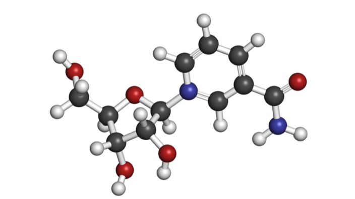
Elysium approached me right after I started my position at Yale or maybe a year in. My understanding is that they were selling NAD precursors that they suggested would slow aging, but they had no way to verify that in humans. So, they were looking for biomarkers of aging. It was shortly after the PhenoAge paper had come out. We developed this collaboration where at first, they were licensing a different version of PhenoAge, but then we discovered this issue with reproducibility where you can measure the same sample multiple times and get a different answer. For a consumer-facing product this is a huge problem. So, I got kind of obsessed with solving this problem and eventually I figured out how to do this. I developed another clock, which is what they eventually licensed and now provide for this consumer-based tests. I’m serving more as an advisor but not doing a lot of hands-on work with them besides providing ideas, answering questions about how things can be applied.

Just to get the idea: how do results look for the consumer?

You get a kit in the mail with a saliva test, you send it back, and then you get an email that tells you your epigenetic age, your rate of aging, which is your epigenetic age divided by your chronological age. A difference of two years is, obviously, a bigger deal when you’re 20 versus when you’re 80.

And then they just give you a straightforward lifestyle recommendation sheet based on observational studies. We actually don’t know anything causal, it’s just epidemiological data, and it’s nothing people probably don’t already know – like don’t smoke, eat plant-based, exercise.

So, the added value is that it can alert people of their condition and motivate them to change something in their lifestyles. And they also offer a subscription because the longitudinal aspect is important, I guess?

Yes. I actually don’t know a lot because I’m not really involved in any of the business stuff. I believe they offer that. As a scientist I would say that the longitudinal aspect is important, assuming you can afford it, and the question is how often someone should be measuring. It is important because I don’t necessarily care what my epigenetic age is today, I just care that over the next 10 years, I’ve only increased it by less than 10 years. You want to keep that rate slower.

One last question: are you familiar with any research into the methylation patterns of centenarians and supercentenarians? Do you think it’s an important thing to study?

The hard thing with doing research in centenarians and supercentenarians is there’s no control group to compare them against. All the individuals who had died – you don’t know what they would look like if somehow you kept them alive. What has been done, including in some of the studies I’ve been on, is comparing offspring of centenarians to control. And in these age-matched controls, we do find lower epigenetic age using mostly the second-generation clocks. But this is the issue with looking at centenarians: let’s say we find that, on average, they’re five years or even 10 years younger than their chronological age. The problem is that epigenetic clocks don’t increase very linearly with chronological age. And we do see a slowing or at least a decrease, even starting at age 70 or 80. So we don’t know if they are along the trajectory that would have continued, or if they are truly younger.

We would like to ask you a small favor. We are a non-profit foundation, and unlike some other organizations, we have no shareholders and no products to sell you. All our news and educational content is free for everyone to read, but it does mean that we rely on the help of people like you. Every contribution, no matter if it’s big or small, supports independent journalism and sustains our future.

Literature

[1] Bocklandt, S., Lin, W., Sehl, M. E., Sánchez, F. J., Sinsheimer, J. S., Horvath, S., & Vilain, E. (2011). Epigenetic predictor of age. PloS one, 6(6), e14821.

[2] Higgins-Chen, A. T., Thrush, K. L., Wang, Y., Kuo, P. L., Wang, M., Minteer, C. J., … & Levine, M. E. (2021). A computational solution for bolstering reliability of epigenetic clocks: Implications for clinical trials and longitudinal tracking. bioRxiv.

Spinal Disc Degeneration

Senolytics Alleviate Spinal Disc Degeneration in Mice

A group of researchers has found that early long-term treatment of mice with the popular senolytic duo dasatinib and quercetin alleviates symptoms of intervertebral disc degeneration, a major age-related cause of disability [1].

Age-related disease with early onset

Conventional wisdom says that our spines just were not built to last for as long as humans live today, especially considering how many of us lead sedentary lives. In most people, the constant strain causes the development of spinal abnormalities quite early in life. Intervertebral discs, which give the spine its flexibility and the ability to absorb shocks, are especially vulnerable. They degenerate faster than other musculoskeletal tissues, with the first signs usually appearing in our early teens [2].

One study found that the prevalence of intervertebral disc degeneration over the entire spine was 71% in men and 77% in women aged less than 50 years and more than 90% in both men and women aged more than 50 years [3]. Intervertebral disc degeneration is the leading cause of low back pain, which, in turn, is a major cause of age-related disability.

Intervertebral discs and senescence

Intervertebral discs consist of dense fluid, rich in the protein aggrecan, packed into an even denser flexible envelope made mainly of collagen fibers. Disc degeneration is caused by the degradation of its extracellular matrix (ECM) along with increased inflammation and cellular death. These factors alter the discs’ mechanistic properties and hamper their ability to withstand loads.

Several studies have linked intervertebral disc degeneration to cellular senescence, so it was only natural for scientists to turn their attention to senolytics. Previous research showed promising results, but this time, the scientists decided to investigate the effects of long-term treatment.

They chose the increasingly popular senolytic cocktail of dasatinib and quercetin, commonly known as D + Q. The researchers used healthy mice, dividing them into three groups by the ages when the rodents began receiving regular doses of D + Q: 6, 14, and 18 months. The experiment stopped at 23 months, a respectable old age for mice.

Mice are also prone to intervertebral disc degeneration, so the animals naturally developed such symptoms with age. The study showed that the D + Q cocktail decreased cellular senescence and age-related inflammation, helped preserve healthy ECM, limited aggrecan degradation, reduced fibrosis, and preserved cellular phenotype in the nucleus pulpous (disc fluid).

Not too early, not too late

The results differed across the groups. The most pronounced effects were observed in mice that began receiving their D + Q at the age of 14 months, more or less in parallel with the onset of the symptoms. The group that began receiving the treatment earlier in life, at the age of 6 months, benefited from it somewhat less, while in the third group, treated since the age of 18 months, the positive effects of the drug cocktail were practically indiscernible.

The treatment was well-tolerated by all mice, but it did not result in any meaningful increase in the animals’ life expectancy. It did, however, lead to some increase in grip strength compared to control groups, which might indicate a positive effect on healthspan (less frailty).

No effect on the knees

In addition to intravertebral disc degeneration, the researchers analyzed the effects of the treatment on another, closely related, disorder: osteoarthritis in knee joints. Unfortunately, no difference between the knee joints of treated and untreated mice was found, including in levels of senescence markers. It seems that D + Q, at least in this regimen, fails to alleviate age-related osteoarthritis. Additionally, no positive change was observed between the study groups and the controls in several other parameters, such as bone density.

The scientists conclude that long-term D + Q treatment “shows differential effects on progression of aging pathologies depending on the skeletal tissue type (i.e., disc, bone, or cartilage) and starting point of treatment”. The most important takeaway from the study, according to its authors, is that there seems to be a therapeutic window for senolytic intervention in age-related intervertebral disc degeneration, and that, if administered during this window, the D + Q cocktail might be able to produce meaningful improvements.

Conclusion

Following previous research that showed the link between cellular senescence and intervertebral disc degeneration, this new study demonstrates that senolytic drugs have the potential to treat this disorder if the treatment begins soon after the early signs of disc degeneration appear. It does not mean, however, that hope is lost for humans with more advanced symptoms (including yours truly). This is a limited-scale study that paves the way for more serious research that might yield better results.

We would like to ask you a small favor. We are a non-profit foundation, and unlike some other organizations, we have no shareholders and no products to sell you. All our news and educational content is free for everyone to read, but it does mean that we rely on the help of people like you. Every contribution, no matter if it’s big or small, supports independent journalism and sustains our future.

Literature

[1] Novais, E. J., Tran, V. A., Johnston, S. N., Darris, K. R., Roupas, A. J., Sessions, G. A., … & Risbud, M. V. (2021). Long-term treatment with senolytic drugs Dasatinib and Quercetin ameliorates age-dependent intervertebral disc degeneration in mice. Nature Communications, 12(1), 1-17.

[2] Urban, J. P., & Roberts, S. (2003). Degeneration of the intervertebral disc. Arthritis Res Ther, 5(3), 1-11.

[3] Teraguchi, M., Yoshimura, N., Hashizume, H., Muraki, S., Yamada, H., Minamide, A., … & Yoshida, M. (2014). Prevalence and distribution of intervertebral disc degeneration over the entire spine in a population-based cohort: the Wakayama Spine Study. Osteoarthritis and cartilage, 22(1), 104-110.

Bone structure fails as we age but can be rejuvenated

Epigenomic Rejuvenation for Restoring Bone Stem Cells

Researchers have shown that age-related loss of bone maintenance is caused by changes in gene expression [1]. Perhaps most importantly, those changes can also be reversed and the bone marrow stem cells rejuvenated.

Bones become weaker as we age as stem cell function declines

With advancing age, it is typical for our bones to become thinner and structurally weaker. The risk of suffering fractures and developing bone diseases such as osteoporosis also rises as we get older. With this, the danger of falls rises, which can have deadly consequences.

One of the reasons this happens is because the bone marrow stem cells stop functioning properly and the upkeep and supply of fresh replacement bone cells declines. This results in increasingly poorer bone integrity and structure.

A team of researchers from the Max Planck Institute for Biology of Ageing and CECAD Cluster of Excellence for Ageing Research at the University of Cologne have demonstrated that this loss of function is caused by epigenetic alterations.

Epigenetic alterations change what genes are expressed, and those changes follow a predictable pattern as we age. The researchers showed that these alterations occur in bone marrow stem cells too.

They compared the epigenomes of stem cells taken from young and old mice. They found that there were significant differences between the two. In particular, genes that play a key role in bone structure were less active in old mice.

But, how do these epigenetic alterations happen? There are four main ways these changes occur:

  • Changes to DNA methylation patterns modify which genes are expressed or silenced, which can cause harmful alterations to cell function and behavior.
  • Histone modification changes how tightly DNA is packed into the nucleus and helps condense it into chromatin. More histone expression supports longevity.
  • Transcriptional alterations represent a variance in gene expression, leading to increasingly harmful influence over cellular function.
  • Remodeling of chromatin is an alteration to the DNA support structure that helps or hinders gene expression.

You can learn more about epigenetic alterations and why they are an important cause of aging here:

Rejuvenating old stem cells

Perhaps more exciting was that the researchers were also able to reverse these epigenetic changes in the bone marrow stem cells by adding acetate. This paves the way for rejuvenation of the bone-marrow stem cells and combating diseases such as osteoporosis.

They exposed bone marrow stem cells from mice to a nutrient solution that included sodium acetate. The stem cells use the acetate to allow enzymes to attach to the histones, which, in turn, increases access to genes and can increase their expression. The result of this increased gene activity was to rejuvenate the epigenome; in other words, it made the stem cells functionally younger and able to produce more bone cells.

Next, the researchers investigated if these epigenetic alterations also increased the risk of bone fractures or osteoporosis in people. To do this, they examined mesenchymal stem cells taken from patients following hip surgery. They found that the cells from older people who also had osteoporosis had the same epigenetic alterations as seen in mice.

Abstract

Aging is accompanied by a general decline in the function of many cellular pathways. However, whether these are causally or functionally interconnected remains elusive. Here, we study the effect of mitochondrial–nuclear communication on stem cell aging. We show that aged mesenchymal stem cells exhibit reduced chromatin accessibility and lower histone acetylation, particularly on promoters and enhancers of osteogenic genes. The reduced histone acetylation is due to impaired export of mitochondrial acetyl-CoA, owing to the lower levels of citrate carrier (CiC). We demonstrate that aged cells showed enhanced lysosomal degradation of CiC, which is mediated via mitochondrial-derived vesicles. Strikingly, restoring cytosolic acetyl-CoA levels either by exogenous CiC expression or via acetate supplementation, remodels the chromatin landscape and rescues the osteogenesis defects of aged mesenchymal stem cells. Collectively, our results establish a tight, age-dependent connection between mitochondrial quality control, chromatin and stem cell fate, which are linked together by CiC.

Conclusion

This study opens the door for potential ways to combat osteoporosis and other, similar conditions by rejuvenating the bone marrow stem cells. The challenge will be to deliver acetate only to the target cells, as the use of acetate is very specific to them. If such a therapy can be made to work, it will be further confirmation that targeting the aging processes directly is the way to go.

Of course, partial cellular reprogramming also has the potential to address this by rewinding the aging clock on these and other cells. You can read more about how cells can be reprogrammed to become younger in our article Yamanaka Factors and Making Old Cells Young.

We would like to ask you a small favor. We are a non-profit foundation, and unlike some other organizations, we have no shareholders and no products to sell you. All our news and educational content is free for everyone to read, but it does mean that we rely on the help of people like you. Every contribution, no matter if it’s big or small, supports independent journalism and sustains our future.

Literature

[1] Pouikli, A., Parekh, S., Maleszewska, M., Nikopoulou, C., Baghdadi, M., Tripodi, I., … & Tessarz, P. (2021). Chromatin remodeling due to degradation of citrate carrier impairs osteogenesis of aged mesenchymal stem cells. Nature Aging, 1-16.

Evolution

Understanding How We Evolved to Age

In a paper in Frontiers in Genetics, Maël Lemoine of France’s CNRS offers an evolutionary perspective on the hallmarks of aging [1]. This review examines the appearance of the hallmarks in different groups across the tree of life, with the aim of providing a comprehensive picture of how the mechanisms of aging have evolved. With this in hand, longevity researchers would have a better framework for contextualizing their findings, and an evolutionary perspective could also be valuable in guiding which hallmarks would best reward research efforts with effective interventions.

General theories

The evolutionary biologist Theodosius Dobzhansky once wrote an essay entitled “Nothing in Biology Makes Sense Except in the Light of Evolution,” and that sentiment has become something of a tenet among evolutionary biologists and perhaps even among biologists more broadly. Despite the undisputed centrality of evolutionary processes to our understanding of biology, evolutionary biology has remained peripheral to longevity research.

There have been broad theories about the evolution of aging. For example, Medawar’s hypothesis proposes that mutations that affect organisms later in life are progressively ‘invisible’ to selection because the organism will already have reproduced by the time the mutation’s deleterious effects come into play. Another theory, antagonistic pleiotropy, proposes that some traits could be beneficial in early life and deleterious in later life. Finally, the disposable soma theory suggests that a trade-off between reproduction and repair underlies aging. While these ideas offer a broad framework for the evolutionary ‘why’ behind aging, they don’t address the evolutionary appearance of aging – that is, when aging or aspects of it first appear in the evolutionary record.

Lemoine set out to do just that. He uses the hallmarks of aging published in 2013 as his framework, but instead of keeping the nine hallmarks from the paper, he breaks them down into 20 more specific hallmarks. He then tracks the appearance of these hallmarks across the tree of life, using a phylogenetic tree published in a 2010 study of cancer genes. His goal is to find the earliest groups that exhibit each of the hallmarks, which allows him to infer the order in which the hallmarks evolved.

A more precise progression

According to Lemoine, the earliest hallmark to appear is loss of proteostasis, which he reports is present in prokaryotes. This is based on the fact that aggregated proteins seem to increase the chance of a bacterium dying. The aggregates are unevenly allocated during cell division, partitioning the damage between the daughter and mother cells.

The same approach is used to pin down the order of the other hallmarks that Lemoine is studying. Genomic instability seems to be the next to appear, with the common ancestor of archaea and eukaryotes apparently having histones and chromatin to help structure their DNA – though Lemoine notes that there isn’t currently any evidence that either is involved in archaeal aging.

This highlights an inevitable challenge of such an ambitious project. In many cases, components of a mechanism of aging are present, but there isn’t any evidence that these components are involved in aging, often because there hasn’t been any research on that question. Lemoine notes this clearly and nevertheless continues through the tree of life, adding hallmarks as he makes his way towards bilaterians, mammals, and humans. It’s interesting to see that hallmarks such as the accumulation of mutations and the shortening of telomeres are the most recent to have evolved, appearing only in bilaterians.

Though all of the hallmarks have appeared in bilaterians, Lemoine points out that more specific hallmarks exists in narrower groups. For example, he points out that human aging is characterized by a reduction in the repertoire of T cells, a very specific hallmark that applies only to a narrow evolutionary group.

The end result of this research isn’t just a list of hallmarks appearing in a particular order. Lemoine discovered that the hallmarks could be divided into two groups based on when they appeared, which he calls unicellular and metacellular. The unicellular hallmarks are involved in the aging of individual cells, whereas the metacellular hallmarks underlie the aging of multicellular organisms. A key finding is that the earliest multicellular organisms to appear are not senescent. They solved the problems of unicellular aging by renewing tissues and are simple enough in structure that the metacellular hallmarks don’t pose a significant problem.

Abstract

The evolutionary theory of aging has set the foundations for a comprehensive understanding of aging. The biology of aging has listed and described the “hallmarks of aging,” i.e., cellular and molecular mechanisms involved in human aging. The present paper is the first to infer the order of appearance of the hallmarks of bilaterian and thereby human aging throughout evolution from their presence in progressively narrower clades. Its first result is that all organisms, even non-senescent, have to deal with at least one mechanism of aging – the progressive accumulation of misfolded or unstable proteins. Due to their cumulation, these mechanisms are called “layers of aging.” A difference should be made between the first four layers of unicellular aging, present in some unicellular organisms and in all multicellular opisthokonts, that stem and strike “from the inside” of individual cells and span from increasingly abnormal protein folding to deregulated nutrient sensing, and the last four layers of metacellular aging, progressively appearing in metazoans, that strike the cells of a multicellular organism “from the outside,” i.e., because of other cells, and span from transcriptional alterations to the disruption of intercellular communication. The evolution of metazoans and eumetazoans probably solved the problem of aging along with the problem of unicellular aging. However, metacellular aging originates in the mechanisms by which the effects of unicellular aging are kept under control – e.g., the exhaustion of stem cells that contribute to replace damaged somatic cells. In bilaterians, additional functions have taken a toll on generally useless potentially limited lifespan to increase the fitness of organisms at the price of a progressively less efficient containment of the damage of unicellular aging. In the end, this picture suggests that geroscience should be more efficient in targeting conditions of metacellular aging rather than unicellular aging itself.

Conclusion

This is both an ambitious effort and a valuable contribution. While there can doubtless be quibbles about specific details, the overall idea is worth pursuing, and the resulting framework enhances our understanding of aging biology. A key takeaway that Lemoine highlights is the division between unicellular and metacellular hallmarks. According to this framework, because the metacellular hallmarks are the main drivers of aging in many multicellular organisms, research that targets the processes behind the aging of individual cells is less likely to increase our longevity than research that targets the processes behind the metacellular hallmarks.

We would like to ask you a small favor. We are a non-profit foundation, and unlike some other organizations, we have no shareholders and no products to sell you. All our news and educational content is free for everyone to read, but it does mean that we rely on the help of people like you. Every contribution, no matter if it’s big or small, supports independent journalism and sustains our future.

Literature

[1] Lemoine, M. The evolution of the hallmarks of aging. Frontiers in Genetics (2021), doi: 10.3389/fgene.2021.693071
Elderly exercising

MoveAge: A New Movement-Based Biological Aging Clock

Researchers have developed a biological aging clock based on movement data from wearable devices. Using their clock and the same dataset, they identified a few possibly life-prolonging nutrients and drugs [1].

I move, therefore I am

Developing reliable biomarkers of aging is crucial for geroscience, since deducing the geroprotective effect of interventions from mortality alone, thankfully, takes a long time. Numerous biological aging clocks have been developed, with methylation-based clocks being among the most precise [2]. Yet, methylation clocks, like many others, use blood-derived data. Drawing blood is an intrusive and rather unpleasant process that can only be performed occasionally in a medical facility, and getting the results involves a lot of “wet lab” work.

This raises the need for alternative biomarkers of aging. Several groups have proposed physical activity and movement as a measure of biological age. Wearable devices, which have become ubiquitous in recent years, emerged as a natural source of data for these markers. These devices continuously monitor various parameters in individual users and can potentially be a goldmine of personal health data. We covered one such biological aging clock in this interview with Dr. Fedichev, whose company, Gero, has launched a downloadable app that derives the user’s biological age from physical activity.

Since a vast majority of wearable devices are made by for-profit corporations and users are often unwilling to share their data, such data is not readily available. This new study gets around that by making use of the National Health and Nutrition Examination Survey (NHANES) database. This major “program designed to assess the health and nutritional status of adults and children in the United States” has been going on continuously since the 1960s. Long before the current era of wrist-worn devices, in the early 2000s, thousands of NHANES participants were asked to wear movement-measuring devices on their ankles to register their physical activity levels.

The workings of MoveAge

The researchers trained their model, which they called MoveAge, on data from 2,634 people aged 18+ using a random forest machine learning method, and then they tested it on a different part of the dataset. The model proved to be fairly predictive of chronological age and general mortality in people aged 40-70. The researchers suggest that younger people’s mortality is too low, and older people’s mortality is too high, to give a strong signal among those age groups; in the young, death is an exception, while in the elderly, it’s the norm.

Since the NHANES database also contains information on dietary habits and drug intake, the researchers tried to find correlations between these variables and biological age. A strong negative correlation was observed between biological age and the consumption of fiber, magnesium, and vitamin E, in line with previous research. Correlation was weaker with drugs, but one candidate did emerge: doxazosin, which is used to treat benign prostate hyperplasia (BHP) and hypertension.

Finally, the researchers tested the effect of doxazosin on C. elegans worms, an animal model widely used in geroscience. The scientists were able to achieve a modest lifespan extension in worms treated with the drug. It is worth noting that results from studies in C. elegans tend to not translate well to humans, and numerous other interventions have yielded much greater increases in the worms’ lifespan [3].

A glimpse into the future

The model described in this study is far from perfect. To begin with, a “movement clock” can be contaminated by many factors, such as a temporary drop in physical activity due to increased workload or a spike due to a vacation (or vice versa, depending on the nature of one’s job and personal preferences). The strength of such clocks should be in their ability to take many measurements over a long period of time, i.e., longitudinally. Unfortunately, MoveAge was trained on data from just one week of physical activity. Still, its success hints at the vast potential of such clocks.

Wearable devices are quickly becoming a staple of modern life, and the variety of data they gather constantly increases. Today, it includes heart rate, blood pressure, stress levels, and sleep quality, and the best is probably yet to come. The rather primitive MoveAge offers us a glimpse into our nearest future, in which multiple types of data will be routinely gathered and cross-referenced by billions of devices worldwide. This will deliver a clearer picture of our health and help advance medicine (including personalized medicine) using the power of machine learning.

The accessibility of such data is crucial, and it depends both on corporations and on end users. Some concerns about personal data use are understandable, and medical data is highly sensitive by nature, but these concerns are often overblown. Allowing the use of your personal health data might benefit everyone, including you.

Conclusion

Whether the ability to move really is “the most downstream measure of healthy aging”, as the authors of this study suggest, remains to be seen, but what counts is the amount of correlation between a measure and biological age. This research is another proof of concept for biological age clocks that use data from wearable devices. We might expect that such non-intrusive clocks based on longitudinal measurements will become more widespread and might soon threaten the hegemony of blood-based biomarkers of aging.

We would like to ask you a small favor. We are a non-profit foundation, and unlike some other organizations, we have no shareholders and no products to sell you. All our news and educational content is free for everyone to read, but it does mean that we rely on the help of people like you. Every contribution, no matter if it’s big or small, supports independent journalism and sustains our future.

Literature

[1] McIntyre, R. L., Rahman, M., Vanapalli, S. A., Houtkooper, R. H., & Janssens, G. E. (2021). Biological age prediction from wearable device movement data identifies nutritional and pharmacological interventions for healthy aging. Frontiers in Aging, 26.

[2] Bergsma, T., & Rogaeva, E. (2020). DNA methylation clocks and their predictive capacity for aging phenotypes and healthspan. Neuroscience insights, 15, 2633105520942221.

[3] Lan, J., Rollins, J. A., Zang, X., Wu, D., Zou, L., Wang, Z., … & Chen, D. (2019). Translational regulation of non-autonomous mitochondrial stress response promotes longevity. Cell reports, 28(4), 1050-1062.

The human thymus

Key Source of Thymic Aging Discovered

Publishing in Nature Communications’ Cell Death Discovery, a team of Chinese researchers has discovered a reason for thymic involution, the age-related decline of the thymus.

The thymus and its involution

The thymus is a boot camp for T cells. Originating from bone marrow, T (for thymus) cells are chemically trained in the thymus, where they learn to attack antigens and not to attack the body’s tissues. However, over time, thymic tissue is gradually replaced by fat tissue, and its function decreases accordingly. This process is known as involution, and combating thymic involution has long been a goal of aging research.

In their introduction, the researchers of this study explain that thymic function is governed by thymic epithelial cells (TECs). When TECs are dysregulated and the function of the thymus falters, this can lead to both infection susceptibility [1] and autoimmune disorders [2].

Dendritic cells, Jagged, and Notch

Dendritic cells are part of the adaptive immune system. As the researchers explain, dendritic cells are tissue-resident cells that migrate into lymph nodes and provide multiple necessary functions, including transforming naive T cells into effector T cells. However, these cells can and do enter the thymus itself, where their activity can be negative.

The researchers showed that through direct cell-to-cell communication, dendritic cells express a ligand called Jagged, which binds to the Notch protein. This protein is critical to the proper functioning of the thymus, and this study provides evidence that Notch3 is expressed by TECs. Through this intercellular communication, the dendritic cells activated the Notch signaling pathway in a way that induced cellular death (apoptosis) of the TECs and inhibited their proliferation.

The researchers conducted further experiments to verify their results. Injecting dendritic cells led to thymic atrophy, as did injection of the Jagged ligand.

Abstract

Multiple proinflammatory conditions, including chemotherapy, radiotherapy, transplant rejection, and microbial infections, have been identified to induce involution of the thymus. However, the underlying cellular and molecular mechanisms of these inflammatory conditions inducing apoptosis of thymic epithelial cells (TECs), the main components of the thymus, remain largely unknown. In the circulation, mature dendritic cells (mDCs), the predominant initiator of innate and adaptive immune response, can migrate into the thymus. Herein, we demonstrated that mDCs were able to directly inhibit TECs proliferation and induce their apoptosis by activating the Jagged1/Notch3 signaling pathway. Intrathymic injection of either mDCs or recombinant mouse Jagged1-human Fc fusion protein (rmJagged1-hFc) into mice resulted in acute atrophy of the thymus. Furthermore, DAPT, a ?-secretase inhibitor, reversed the effects induced by mDC or rmJagged1-hFc. These findings suggest that acute or aging-related thymus degeneration can be induced either by mass migration of circulating mDCs in a short period of time or by a few but constantly homing mDCs.

Conclusion

Although this is a basic research study that has not examined any potential treatments, it paves the way for the possible development of signaling-based interventions that use the relationship between Jagged and Notch to slow and/or reverse thymic involution. However, while the researchers have clearly shown the role of Notch signaling as a cause of thymic aging in human beings, they also note that it is not the only cause. These signals are part of an intricate network, so directly inhibiting Notch signaling may or may not be a fruitful endeavor.

Similarly, this research should be looked at in light of treatments that have been reported to restore thymic function, such as the HGH and DHEA treatment championed by Intervene Immune.

We would like to ask you a small favor. We are a non-profit foundation, and unlike some other organizations, we have no shareholders and no products to sell you. All our news and educational content is free for everyone to read, but it does mean that we rely on the help of people like you. Every contribution, no matter if it’s big or small, supports independent journalism and sustains our future.

Literature

[1] Kellogg, C., & Equils, O. (2021). The role of the thymus in COVID-19 disease severity: implications for antibody treatment and immunization. Human Vaccines & Immunotherapeutics, 17(3), 638-643.

[2] Abramson, J., & Anderson, G. (2017). Thymic epithelial cells. Annual review of immunology, 35, 85-118.

Cholesterol

Underdog Pharmaceuticals Receives Accelerated Designation

Underdog Pharmaceuticals (Now called Cyclarity Therapeautics) has just issued a press release announcing that UDP-003, a compound being studied for its effectiveness against oxidized cholesterol, has been given an Innovation Passport under the UK’s Innovative Licensing and Access Pathway program. This is similar to the Fast Track designation given by the FDA in the United States.

Underdog is a spin-out company from SENS Research Foundation, and Matthew O’Connor, who is quoted in this article, is well-known for his previous activities at SENS Research Foundation.

Mountain View, CA, U.S., September 8, 2021 –

Underdog Pharmaceuticals, Inc. (Underdog), a pre-clinical stage pharmaceutical company focusing on the treatment and reversal of age-related diseases, has been awarded an Innovation Passport under the United Kingdom’s Innovative Licensing and Access Pathway (ILAP), to pursue fast patient access to its groundbreaking treatment for cardiovascular disease.

Accounting for more than 40% of deaths in Europe and the United States, and with annual treatment costs more than £29 billion in the UK alone, cardiovascular disease is by far the world’s most life-threatening condition, and its primary cause is atherosclerosis. Underdog’s engineered synthetic carbohydrate compound, UDP-003, is designed to target and remove toxic oxidized cholesterol, a key driver of arterial plaque accumulation. Underdog is one of the first companies, and UDP-003 one of the very few pre-clinical therapies, to receive an ILAP designation.

ILAP, introduced by the Medicines and Healthcare products Regulatory Agency (MHRA) in January, is designed to accelerate development of and access to promising medicines of significant potential public health benefit. The program provides enhanced early input and interactions with MHRA, the National Institute for Health and Care Excellence (NICE), the Scottish Medicines Consortium (SMC), and the National Health Service (NHS). The ILAP program also provides the potential for significant downstream regulatory advantages, including rolling clinical reviews, accelerated assessment, and supervised early reimbursed use under adaptive authorization.

“This is an honor for us and a wonderful step forward for our program,” said Matthew S. O’Connor, Underdog CEO of Scientific Affairs. “There is an enormous public need for drugs which can significantly reverse established atherosclerosis, rather than manage or delay its onset.”

“I believe the MHRA is sending a powerful message by reviewing and admitting preclinical stage programs like UDP-003 into ILAP,” noted Mike Kope, Underdog CEO of Corporate Affairs. “We’re pursuing a new target, with a new compound and seeking evidence of genuine disease modification; and though the potential is fascinating, the clinical challenges are real. The advice and consultation we’ll receive will be invaluable to the success of the program.”

About Underdog

Underdog Pharmaceuticals, Inc., is pursuing a mission to treat the underlying causes of age-related disease. The company develops simple and direct interventions targeting oxidized cholesterol using rationally designed molecules, to provide the first true disease-modifying treatments for common age-related conditions such as atherosclerosis, heart attack and stroke. Its products are based on novel derivatives of a well-known, safe compound and a new way of looking at cardiovascular disease. For more information, please visit www.underdogpharma.com or send an email to press@underdogpharma.com

Forward Looking Statements

The information and statements contained herein contain forward-looking statements, which are subject to risks and uncertainties that could cause actual results to differ materially from such forward-looking statements and should not be considered as an indication or guarantee of future performance. Forward-looking statements involve known and unknown risks and uncertainties which could cause the Company’s prospects or actual results to differ materially from those in the forward-looking statements. The Company does not undertake any obligation to update the information or forward-looking statements contained herein whether as a result of new information or future events or otherwise.

Contact:

Mike Kope

Co-CEO

Underdog Pharmaceuticals Inc

press@underdogpharma.com

We would like to ask you a small favor. We are a non-profit foundation, and unlike some other organizations, we have no shareholders and no products to sell you. All our news and educational content is free for everyone to read, but it does mean that we rely on the help of people like you. Every contribution, no matter if it’s big or small, supports independent journalism and sustains our future.