The Blog

Building a Future Free of Age-Related Disease

Oranges and Grapes

Resveratrol, Vitamin C Drop Oxidative Stress After Menopause

In a randomized, controlled trial published in Nutrients, researchers tested supplementation with resveratrol, vitamin C, and a combination of both. They learned that all of the treatments had a similar positive impact on oxidative stress in postmenopausal women [1].

Menopausal transition

Around the world, a significant proportion of women have reached the post-menopausal stage. In Mexico, where the study was conducted, 15% of women have reaced this threshold. As life expectancy increases, this fraction will also increase in the coming years.

Menopause is followed by decreased antioxidant capacity, resulting in an imbalance between oxidant molecule generation and antioxidant capacity. Such imbalance creates oxidative stress (OS). During oxidative stress, oxidizing agents can attack and break down molecules that are essential to cells and tissues, such as lipids and proteins.

This group of researchers previously reported “that postmenopausal women present higher concentrations of OS markers than women of reproductive age” [2]. Therefore, using antioxidants seems like a reasonable strategy to reduce the detrimental effect of oxidative damage on lipids and proteins.

Those researchers chose resveratrol and vitamin C because previous research had described them as having “cardioprotective, anti-sclerotic, anti-inflammatory, and antioxidant properties“ [3].

Resveratrol is a natural phytoestrogen and a polyphenolic flavonoid that is well tolerated and not toxic. Resveratrol was previously described to have a role in increasing both the expression and activity of antioxidant enzymes while reducing oxidative load [4, 5].

Clinical trial of postmenopausal females

The researchers conducted a pilot randomized, double-blind clinical trial. They recruited women between 50 and 60 years old who were in the early postmenopause stage and had insulin resistance.

Among the exclusion criteria were the use of hormone replacement therapy or drugs such as anticoagulants, metformin, bezafibrate, statins, or any antioxidant in the three months before the beginning of the study. Active smokers and women with some health conditions were also excluded.

The study participants were divided into one of three groups: 13 participants in the resveratrol group plus a vitamin C placebo (group A), 15 participants in the resveratrol and vitamin C (group B), and 14 participants in the vitamin C plus a resveratrol placebo (group C). Depending on the group assignment, the participants received 500 mg resveratrol capsules, vitamin C/ascorbic acid tablets, or placebo tablets for three months.

Reduced oxidative stress

First, the researchers analyzed baseline clinical and biochemical blood test results. In the intra-group analysis, baseline measurements were compared to after-treatment measurements, and no significant differences were found in measurements such as weight, BMI, glucose, insulin, lipid profile, and uric acid.

When groups were compared with each other after a three-month intervention, the researchers observed significantly lower total cholesterol levels and significantly higher triglyceride levels in the vitamin C group compared to the resveratrol group. The researchers also observed significantly lower triglyceride concentrations in the combined group compared to the vitamin C-only group.

Comparing the baseline and after-treatment levels of lipohydroperoxides (LPH), a measure of oxidative deterioration of lipids, showed a significant decrease of 33% in the combined group. The resveratrol group and the vitamin C group had 25% and 15% decreases in LPH levels, respectively. However, those differences were not statistically significant. The researchers hypothesize that it was due to the sample size being too small, and future research with a bigger sample size might lead to significant results in those groups as well.

The researchers also analyzed the levels of MDA, the end product of lipoperoxidation. All three groups showed statistically significant differences between initial measurement before the treatment and following the 3-month treatmnet. MDA levels were reduced by 26% in the resveratrol group, 32% in the combined group, and 38% in the vitamin C group.

Previous experiments conducted on rats that received resveratrol achieved similar results [6]. Similarly, a study of people under 18 taking vitamin C also showed MDA reduction [7].

Resveratrol Vit C 1

After measuring lipid damage, the researchers analyzed protein oxidative damage. A statistically significant reduction was also observed in all groups. The differences between before and after treatment measurements show 39% reduction in the combined group and 29% in both the resveratrol and vitamin C groups.

Resveratrol Vit C 2

There were also differences in antioxidant capacity. However, this time, there was an increase of 30% for the combined group and 28% for the vitamin C group following the treatment compared to baseline.

Resveratrol Vit C 3

No improvements in insulin resistance

Despite previous research linking oxidative state and insulin resistance, none of this study’s groups demonstrated statistically significant differences in insulin resistance.

The researchers discuss previous studies on humans that, except for one, all reported that resveratrol does not affect insulin resistance. Those studies looked at different populations of participants with different health conditions and variable durations and doses of treatment.

Similarity of all groups

The authors caution when interpreting the results, as the research has some limitations. For example, the generalizability of the results from a homogenous group of participants, including only females from the Valley of Mexico metropolitan area, was limited.

Additionally, the researchers did not include a control, untreated group. However, as the authors explain, their question addressed how the combined treatments compare with a single treatment and whether there is possible synergy between them. Their experimental setup allowed for those comparisons and comparisons of the changes that the women experienced compared to baseline.

The researchers conclude that despite the differences in the effects of different measurements of oxidative stress, “none of the three interventions were superior to the others.”

We would like to ask you a small favor. We are a non-profit foundation, and unlike some other organizations, we have no shareholders and no products to sell you. All our news and educational content is free for everyone to read, but it does mean that we rely on the help of people like you. Every contribution, no matter if it’s big or small, supports independent journalism and sustains our future.

Literature

[1] Montoya-Estrada, A., García-Cortés, A. Y., Romo-Yañez, J., Ortiz-Luna, G. F., Arellano-Eguiluz, A., Belmont-Gómez, A., Lopéz-Ugalde, V., León-Reyes, G., Flores-Pliego, A., Espejel-Nuñez, A., Solis-Paredes, J. M., & Reyes-Muñoz, E. (2024). The Administration of Resveratrol and Vitamin C Reduces Oxidative Stress in Postmenopausal Women-A Pilot Randomized Clinical Trial. Nutrients, 16(21), 3775.

[2] Montoya-Estrada, A., Velázquez-Yescas, K. G., Veruete-Bedolla, D. B., Ruiz-Herrera, J. D., Villarreal-Barranca, A., Romo-Yañez, J., Ortiz-Luna, G. F., Arellano-Eguiluz, A., Solis-Paredes, M., Flores-Pliego, A., Espejel-Nuñez, A., Estrada-Gutierrez, G., & Reyes-Muñoz, E. (2020). Parameters of Oxidative Stress in Reproductive and Postmenopausal Mexican Women. International journal of environmental research and public health, 17(5), 1492.

[3] Breuss, J. M., Atanasov, A. G., & Uhrin, P. (2019). Resveratrol and Its Effects on the Vascular System. International journal of molecular sciences, 20(7), 1523.

[4] Xia, N., Daiber, A., Förstermann, U., & Li, H. (2017). Antioxidant effects of resveratrol in the cardiovascular system. British journal of pharmacology, 174(12), 1633–1646.

[5] Livraghi, V., Mazza, L., Chiappori, F., Cardano, M., Cazzalini, O., Puglisi, R., Capoferri, R., Pozzi, A., Stivala, L. A., Zannini, L., & Savio, M. (2024). A proteasome-dependent inhibition of SIRT-1 by the resveratrol analogue 4,4′-dihydroxy-trans-stilbene. Journal of traditional and complementary medicine, 14(5), 534–543.

[6] Kong, D., Yan, Y., He, X. Y., Yang, H., Liang, B., Wang, J., He, Y., Ding, Y., & Yu, H. (2019). Effects of Resveratrol on the Mechanisms of Antioxidants and Estrogen in Alzheimer’s Disease. BioMed research international, 2019, 8983752.

[7] Ismy, J., Soebadi, A., Mangunatmadja, I., Monica, M., Sari, T. T., & Yuliarti, K. (2024). Role of antioxidants in reducing oxidative stress and seizure frequency in drug-resistant epileptic patients. Narra J, 4(2), e790.

Turn.bio logo

Turn Biotechnologies Announces ERA™ Bone Marrow Study

Turn Biotechnologies, a cell rejuvenation and restoration company developing novel mRNA medicines for untreatable, age-related conditions, announced its latest study to assess the efficacy of using epigenetic reprogramming to rejuvenate bone marrow stem cells.

The study, which is being funded by Methuselah Foundation, is the first to evaluate use of Turn Bio’s unique RNA-based ERA™ therapy to rejuvenate bone marrow function to improve the quality of donor cells used in stem cell transplantation. Healthy marrow releases blood cells into the bloodstream. As adults age, their bone marrow produces fewer disease-fighting B and T cells and other products that help reduce inflammation throughout the body. Aged stem cells also collect changes in their DNA known as clonal hematopoiesis that can serve as a prelude to leukemia development.

“Multiple studies of clinical bone marrow transplant outcomes have identified that patients who receive donor stem cells from young donors have superior outcomes, owing to more durable correction of underlying blood and immune defects, with lower risks of graft dysfunction and donor clonal hematopoiesis,” said Timothy Olson, MD, PhD, principal investigator of this study, and Medical Director of the Blood and Marrow Transplant and Co-Chief of the Cellular Therapy & Transplant Section at Children’s Hospital of Philadelphia (CHOP). “We hope the study shows that epigenetic reprogramming can help to make bone marrow transplants both more effective and more accessible to patients.”

Rejuvenating older bone marrow cells could improve the body’s ability to fight disease, speed wound healing, improve the blood cells’ capacity to carry oxygen, and enhance the success of donor-to-patient transplantations. This could allow the use of older donors for transplants.

Transplants replace a patient’s diseased blood-forming cells with healthy cells. The procedures are used to treat certain types of cancer, blood disorders, and autoimmune diseases. Nearly half of the world’s annual 50,000 bone marrow transplants are performed in the United States.

Methuselah Foundation is interested in increasing survival rates for marrow transplant and gene therapy patients where current survival rates for patients with nonmalignant diseases is over 70% with a matched sibling donor and over 36% with unrelated donors.

By rejuvenating donor stem cells, researchers aim to increase the safety and efficacy of these intensive treatments. Additionally, with the growing use of gene therapy and gene editing, the ability to rejuvenate stem cells during these processes can potentially make the one-time treatments safer and more effective.

“We believe this study will confirm that epigenetic reprogramming can effectively rejuvenate bone marrow cells and restore their youthful performance —which will potentially extend the healthy human lifespan,” said David Gobel, co-founder and CEO of Methuselah Foundation, the world’s first biomedical non-profit focused on human longevity.

The year-long study will measure the effectiveness of Turn’s ERA solution by treating mouse blood progenitor cells and transplanting those cells into irradiated mice of the same genetic background.

“This study represents a significant milestone for Turn Bio as it demonstrates how we are taking our science beyond individual therapeutics to create solutions for a variety of diseases. We are optimistic it will validate the dramatic impact of epigenetic reprogramming in improving the human healthspan,” said Anja Krammer, Turn Bio CEO. “We have repeatedly demonstrated that ERA treatments can safely rejuvenate human cells. This is an opportunity to bring theory to life.”

The study will be conducted by Timothy S. Olson, MD, PhD, Associate Section Chief, Cell Therapy & Transplant Program at Children’s Hospital of Philadelphia (CHOP), with the support of Turn Bio Scientific Advisors: Joseph Hai Oved, MD, Leader of the Primary Immunodeficiency and Immune Dysregulation Program at Memorial Sloan Kettering Cancer Center  in New York, and Kevin J. Curran, MD, Director of the Immune Effector Cell Program, Memorial Sloan Kettering Cancer Center.

About Turn Biotechnologies

Turn Bio is a pre-clinical-stage company focused on repairing tissue at the cellular level and developing transformative drug delivery systems. The company’s proprietary mRNA-based ERA™ technology restores optimal gene expression by combatting the effects of aging in the epigenome. This restores cells’ ability to prevent or treat disease and heal or regenerate tissue. It will help to fight incurable chronic diseases. Its eTurna™ Delivery Platform uses unique formulations to precisely deliver cargo to specific organs, tissues, and cell types.

The company is completing pre-clinical research on tailored therapies targeting indications in dermatology and immunology, and developing therapies for ophthalmology, osteo-arthritis, and the muscular system. For more information, see www.turn.bio.

We would like to ask you a small favor. We are a non-profit foundation, and unlike some other organizations, we have no shareholders and no products to sell you. All our news and educational content is free for everyone to read, but it does mean that we rely on the help of people like you. Every contribution, no matter if it’s big or small, supports independent journalism and sustains our future.
Mehmood Khan Interview

Mehmood Khan on Aging Policy and Collaboration

I first met Dr. Mehmood Khan in 2022 at the inaugural Longevity Summit Dublin, where organizers Aubrey de Grey and Martin O’Dea made a bold decision to include policy and advocacy discussions alongside the traditional focus on longevity research. Like many others in the audience, I was captivated by the forceful, engaging, and compelling one-man show that Khan delivered with apparent ease. “This is how public persuasion is done,” I remember thinking. “We need more people like him on our side.”

Khan had already enjoyed an illustrious career before joining the longevity movement, holding high-profile roles such as Vice Chairman and Chief Scientific Officer of Global Research and Development at PepsiCo and President of Global R&D at Takeda Pharmaceuticals. However, Khan is not just a high-profile bureaucrat. He has a solid background in science and healthcare, having served as Chief of the Endocrine Division at Hennepin County Medical Center in Minnesota and later as a faculty member in endocrinology at the Mayo Clinic and Medical School.

It’s this rare combination of qualifications and interests that led him to his current role as CEO of Hevolution, the world’s best-funded healthspan-focused non-profit, created by, no less, a royal Saudi order. Despite being a relatively new player in the field, Hevolution has swiftly established itself as a dominant force, leveraging its overflowing war chest both boldly and strategically.

Hevolution is the largest sponsor of the $100-million-plus XPRIZE Healthspan and supports dozens of researchers in the fundamental biology of aging across the globe. It has also begun investing in longevity startups. With so much happening, an interview with Mehmood Khan was long overdue. We sat down to discuss Hevolution’s vision, its progress, and his thoughts on the future of the field.

Let’s start with your personal journey to the longevity field. I know it’s an unusual one.

Unlike most experts, I haven’t been in the field long directly. Indirectly, it’s been my whole career. I was an endocrinologist, practiced at the Mayo Clinic and the University of Minnesota, saw a lot of patients with diabetes, metabolic disease, obesity. Much of my early research, if you go back 25 years or so, was around metabolic disease and obesity, and this was before aging was itself considered a discipline.

My career has progressed from being focused in a specific subject area to being fascinated by how you can impact health at a large scale, which is what bridged my interest from medicine to food. I had a faculty appointment in food science as well as in medicine because I trained in food science at the College of Food Science and Agriculture. This was 35 years ago, and that progressed to thinking about global challenges, whether it’s food supply, water, or the carbon footprint of large industries like PepsiCo.

The chance to converge all of this together crystallized with the whole field of healthspan. When I started looking at this field, everyone was still primarily talking about longevity. And it was clear to me that longevity is not the target here – healthspan is. What we’ve spent the last years doing, and it’s been my privilege to lead the team, is to focus Hevolution much more on healthspan than longevity.

This distinction between healthspan and longevity seems important to you. Yes, we have seen increases in longevity but not in healthspan in recent decades, which isn’t good, but do we really have to uncouple these terms to such an extent?

I think sometimes you have to uncouple it because it’s the vocabulary that you use that gets the traction with policymakers. I’ve spoken to a lot of health ministers, finance ministers, and several heads of state over the last few years. If we make this purely about getting people to live longer, it becomes interpreted as more dependency – more people to take care of, bigger pension bills, bigger health bills for Medicare, National Health Service in the UK, Japan, China, wherever you go.

If you talk about longevity, you end up with not a lot of interest at the very senior policymaker level. Health ministers, yes, but they’re not the ones holding the purse strings. You’ve got to convince the finance ministers, the labor ministers, the economy ministers that this is of national interest. It isn’t about people living longer for the sake of living longer, but about productivity.

If you talk to individuals (and we’ve surveyed them), and you ask, “Do you want to live longer?” – that won’t be their number one priority. But if you ask, “Do you want to live healthy as long as possible?” – yes, almost unanimously. So, as scientists, we may not uncouple it, but the average person on the street understands those two things differently.

More importantly, policymakers view these as two different things. I think it’s important for us as leaders in the field to say that our primary goal is to keep populations healthy as long as possible. The secondary benefit of that might be that people live longer. But if we start with the argument that we want people to live longer, that’s not going very far. It hasn’t gone for decades.

If you focus on life extension, you inevitably get sucked into debates such as “Is this against God (if you’re religious) or nature?” You get into all other kinds of ethical debates. But the minute you say, “I want people to live healthy for as long as possible,” there’s no disagreement about that.

Yes, that makes a lot of sense. Let’s now talk about Hevolution, which emerged in the field just a couple of years ago and has since become a major player.

Hevolution was created by a royal order from the King. It is chaired by His Royal Highness, Prince Muhammad, the Crown Prince and Prime Minister. It was the vision to take on a global challenge that would benefit as many people on the planet as possible. Healthy aging touches every human being. It’s a global challenge that affects every country, including the kingdom, so it’s very relevant locally across the Gulf, but globally as well.

It was set up as a nonprofit because the primary driver was to fund the science to move the field forward, accelerate the science into the marketplace with patient capital willing to take risks, and put it under a nonprofit umbrella. So, the incentives are aligned – we want our venture capital arm to succeed, attract other investors, and grow the size of the pie.

One part of that mission statement was particularly important to us: to extend healthy lifespan for the benefit of all. That’s something that is uniquely possible under the umbrella of a nonprofit. We want to democratize everything we do. We put that lens on everything we evaluate. Whatever we do, we look at it through the scalability lens. We’re not interested in technologies that might touch a few privileged people in the world, even though that may be where it starts. We look at it and ask ourselves, “How does this scale? Is there a path?”

Naturally, we care about another important issue: the bioethics of healthspan extension. It’s not just asking the question “Can it be done?” but also “Should it be done?”, and that shouldn’t be left just to the scientists. I am a scientist, and I should not be the one leading that conversation. I’m a strong believer that this should be something that experts in ethics are engaging with from day one.

Before we funded a single science grant, we put together a global ethics team, and that, to my knowledge, is the first time global bioethicists have been convened around the field of aging and asked to guide us and hold us accountable. Arthur Caplan, who is at the helm, is a world expert on ethics. He’s the chair of bioethics at NYU, previously Johns Hopkins, New Orleans. Julian Savulescu, founding chair of bioethics at Oxford, is another example.

This is a very interesting aspect of Hevolution I admit I’ve never heard of. I think many aging researchers don’t really want bioethicists around – they see them mostly as a nuisance. Can you name some of the insights this team has come up with?

There are a couple of things that they’ve started to push us to think about. One is, “How do you make this inclusive?” Not only the output, but the research. Initially, like everybody else, we were looking at the world and saying, let’s fund North America, the UK, but usually absent at the research table are people from the African subcontinent, South America, Latin America. Our bioethicists asked us why.

If you’re really going to address global aging, you have to understand aging in the context of the world. The continent that’s going to have the most elderly people in the world in the next 50 years is Africa, and yet, you almost never see people from Africa at aging meetings. It was a good push. We went to the WHO to explore possible collaborations: they’ve got a much better footprint there than we do. How do we collaborate with them? I don’t have an answer yet, but we’re working on it.

Another example is how we think about aging as a process in terms of when we would intervene versus not intervene. When is the earliest you should intervene? If you ask biologists, they’ll tell you that aging as a process starts around 14 days after conception. Should you intervene in an embryo? Should you intervene in a child?

I had the privilege of giving a couple of lectures at Yale Law School three or four years ago on the distinction between law and ethics in business. Something you learn when you look at this as a scientist (the lawyers already know that) is that the law is only a memorialization of a society’s ethics. Then, isn’t it our job as scientists to help convene that ethics discussion and move it forward alongside the scientific field rather than have it follow?

What’s your philosophy in choosing projects?

First, we want to fund the underlying science in the biology of aging. That’s our primary target. We don’t want to just fund another research study on Alzheimer’s, diabetes, dementia, osteoporosis. There’s lots of research going on and lots that’s been done. Most of it has come from working backwards from the end case. They say, now you’ve got osteoporosis, what can I do to increase your bone mineral density? If you have dementia, what can I do to slow it down? Not what can I do to prevent it.

So, understanding the biology is the key. The second thing is understanding how and when to intervene. When you start asking those questions, one thing that will surface is that we still don’t really know how to measure aging as a biological process. Yes, there’s DNA methylation, there’s this and that. Every six months you’ll hear of a new biomarker, then another, but most of them come from relatively small studies correlated with one or two variables. We have A-glycohemoglobin (HbA1c) for diabetes, but not its equivalent for aging. We know what cardiovascular risk profiling looks like based on biomarkers – LDL, HDL – but we don’t have the same for aging.

So, one bucket is how do you measure aging? This has to be predictive and correlate with treatment. Ideally, a good biomarker will reverse with treatment. This is why we funded the global meeting on biomarkers of aging at Cold Spring Harbor earlier this year. We brought together the world’s experts, both clinicians and biomarker experts and regulators, to start a series of discussions.

I strongly believe that we should fund this research and facilitate this debate. For instance, we should think about how we make biomarkers of aging open source. You know, if LDL cholesterol had been patented, we probably wouldn’t have statins.

Right now, we’ve got about 150 labs and 200 principal investigators around the world that are funded by us. We’re also funding partnerships where we’re a partner in the scientific grants. Basically, just like the NIH, we say, send us your best idea. We then send it to independent review; we don’t like to review the grants ourselves. I don’t need to take phone calls saying, “Hey, Mehmood, I just submitted this grant. What do you think?” I wouldn’t even know what you’re talking about because it goes to an independent panel.

I’ll give you an example. We’re interested in proteostasis at the cellular and tissue level. How do we bring the best minds together? In this case, we went to Richard Morimoto at Northwestern, one of the world’s academic leaders in proteostasis, and said, if you would put your dream team together and develop a series of research plans around that, we’ll fund you. And he did. It’s almost 30 million dollars. That’s an example of a partnership where we targeted the area, found the world expert, and asked that PI to put together a multi-center team.

The value of that is he’s going to collaborate with people who might have been competing with him for grants in the past. Now he can say, “Listen, we can get funding together if we come together.” That’s the catalyst role that we play as Hevolution. We’re catalyzing the field by catalyzing collaboration.

Another way you’ve been catalyzing the field is through XPRIZE Healthspan, where Hevolution is the lead and single-largest funder. Can you tell me more about this collaboration? In particular, since you talked about biomarkers, what do you think about their idea of reversal of age-related loss of function as the endpoint?

It’s very exciting to see how they’re incentivizing the world. They’ve done this successfully in other fields, putting the best minds, ideas, and resources to answer the question. This is the moonshot, this is the challenge: show us 10 years of reversal (of loss of function). Let’s go!

Evidence shows that in prizing mechanisms such as XPRIZE, the multiplier is about 8 to 10. For every dollar of the prize money, the collective investment brought in by the competitors is eight to ten times that. For a chance to win 80 million I’m going to put in two million, three million from grants or investors. This scenario repeats itself a hundred times over, everybody’s competing, and finally, the 80-million prize draws 800 million into that field.

That’s the beauty of this model. The reason we support it is that, in the back of my mind, a hundred-million-dollar prize means a billion dollars will potentially be mobilized. That’s the leverage. Every team that’s competing is going to either find investors, or government funding, or donors.

Regarding the specific biomarkers, when we went to the table, I said that nobody from Hevolution or any sponsor should be involved in defining the endpoints or sit on the judging committee. We helped find independent judges, we helped put together the endpoint committee to come up with the definition, but that was the limit of our involvement.

I was delighted when Professor Patrick Maxwell accepted. He’s the Dean of the Medical School at the University of Cambridge – a world-class researcher. Ironically, some of the people we approached turned us down for a very interesting reason. When they learned about the details of the prize, they said they couldn’t do it because they now wanted to compete. We still counted that as a win.

Tell me about the upcoming second Hevolution Summit. What is special about this particular longevity conference?

Actually, we did a small regional one early in 2023 in partnership with the National Academy of Medicine, and that one was focused on the Gulf area. So, it’s our third event of this type but the second Healthspan Summit.

At the first Healthspan Summit, we brought a lot of people together – it was bigger than anybody expected. We had people with subject matter expertise and people who were there because they were curious. The delightful part is we had people travel from all over the world, and we didn’t pay anybody to speak – no honorarium. We said, if you want to come and speak, it’s a privilege, and we’ve held that bar.

People loved the fact that we convened experts from different disciplines. We had regulators talking with investors, talking with scientists. People also appreciated the depth of the discussion on all the topics.

For the second summit, we’re taking the best of what we did and making it better. Now we have a chance to tell the world what we’ve accomplished since the first one. We can showcase research programs we’ve funded, partnerships – not ourselves, but we get the experts, the funded scientists to stand up and talk about their field.

We’ve now announced three companies we’ve invested in. We can showcase not only what those investments are but why we’ve invested in them. By then we may have announced more as well. We look to bring investors and these startups even closer together for matchmaking.

We’ll have updates from the XPRIZE – by then, we’ll be able to show the world how many teams there are, where they’re located, what types of fields they’re involved in, what science they’re interested in. The second summit is about the proof points.

The challenge I have right now is focus. Think about what I just shared with you: 2000 grants reviewed, 200 PIs funded, three companies announced, potentially five partnerships. I can’t put everybody on stage. It’s a good problem to have, but I think the world wants to hear from these people. They don’t want to hear from me.

The last piece, which I think will be the most exciting in the future, is matchmaking between our ecosystem of scientists. One thing we can do better than any government funding agency is cross borders. We can fund teams across borders much more easily than the NIH or the MRC because their priorities lie first and foremost within their own borders. We’re trying to cross these borders.

I have heard you on several occasions talking about the need to work across the entire ecosystem, including with public opinion and decision makers. However, I understand that Hevolution hasn’t funded any projects in that particular field. Why is that?

We’re very careful. We see ourselves as advocating for the field, convening different experts, but we’re not a lobby. We can’t legally lobby, nor do we want to.

However, we’ve had several policymakers, ministers, former ministers, attending our meetings – in the UK, in the US, in the Middle East. We’ve had very senior people from industry: Jon Symonds, Chairman of GlaxoSmithKline, was there as an example. Numerous CEOs from big pharma, small pharma, all engaging.

This means we have a chance to bring these parties together and hope that will shape the discourse around policy. We’re seeing early signs of that traction. Recently, I had a meeting right here in Riyadh with a former health minister. He was on a formal visit, but he asked his staff to arrange a private lunch meeting with me. That tells us that people like him are looking to Hevolution as a credible source, they are interested.

I was delighted to hear that when ARPA-H was announced by President Biden, one of the pillars this year was going to be aging. That’s a huge win for the field. Nobody’s going to be able to put in the sort of resources that the U.S. federal government can: the NIH’s budget is 44-plus billion dollars. Put 10 percent of that toward aging, and you’ve made a massive impact on the field’s progress.

How optimistic are you about the near future of our field?

I’m quite optimistic for several reasons. One, it’s a public health imperative. Two, it’s an economic imperative. Three, the science is now optimistically telling us that something can be done about it. You can have the first two, but if nothing can be done about it, so what?

The fourth reason is your field – the media. Today, I see three bodies in the media. One is mostly about the throw-away terms, the sound bites, those are people who don’t do their homework. Whatever the latest vitamin cure is, they’ll push it. That hurts our field. The second is the outlets that not only understand the science but find a way to communicate it to the readers who might not have a scientific background. The third is core science – the fact that Nature now has Nature Aging is huge. One of the top two journals in the world having a whole series on aging tells you something.

What then frustrates you? What bottlenecks do you see that you’d love to change in terms of public opinion, regulation, the scientific environment?

I see two major things. One is that sometimes, people in this field get over-enthusiastic and ahead of their own data. While I love passion and energy, the challenge is the risk to credibility. If you suddenly start making statements like, “In my lifetime, you’re going to live to be 150,” it sets an expectation and that hurts the field. Let’s not get ahead of ourselves. That frustrates me. Let’s stick with the evidence. Let the data push this field forward.

The old African proverb we often quote is “If you want to go fast, go alone. If you want to go far, take everybody with you.” Sometimes people in our field forget this wisdom. This is going to take a village. It’s a team sport. It’s going to take multiple players in different scientific and non-scientific disciplines. We’ve got to bring them together.

Just trying to create likes on social media in the short term gets a lot of publicity, sometimes gets you stardom, but that’s usually short-lived. Sometimes in that process, one forgets that it’s about the field, not oneself. I’ve been a CEO for a long time. I’ve been running large organizations and small organizations. One of the things you learn as a senior executive, whether at organizations as big as PepsiCo or as small as a biotech company, is that it’s always about the field, the business, the enterprise, not oneself. And if you remember that, you can move mountains.

We would like to ask you a small favor. We are a non-profit foundation, and unlike some other organizations, we have no shareholders and no products to sell you. All our news and educational content is free for everyone to read, but it does mean that we rely on the help of people like you. Every contribution, no matter if it’s big or small, supports independent journalism and sustains our future.
Three supplements

A Clinical Trial of a Three-Part Treatment for Inflammaging

Reseachers publishing in Antioxidants have combined three antioxidant and anti-inflammatory compounds and tested their effects in human beings.

Three complementing choices

The first component the researchers included was AM3, a compound that is core to the Inmunoferon supplement and is an immunomodulator that has been found in trials to aid against infections [1]. Some prior work has found that AM3 also assists against immunosenescence [2]. However, as these researchers note, little work has been done in that area, and that work did not establish whether or not it could do anything to curtail age-related immune dysfunction.

The second component was spermidine, a polyamine that has been reported to improve the cellular maintenance process known as autophagy, thereby also ameliorating immunosenescence [3]. Spermidine has also been reported to assist in gut function by returning macrophage polarization to a less inflammatory state [4].

The third component was hesperidin, a flavonoid that recent prior work had found to have potential effects against multiple diseases, including hepatitis [5] and several metabolism-related disorders, such as diabetes [6], obesity [7], and non-alcoholic fatty liver disease [8]. The researchers hold that these effects most likely originate from its effects against inflammation, such as its suppression of the senescence-related protein MMP-9 [9], and on immune response [10].

All three of these ingredients are sold in various parts of the world as supplements and are generally considered nontoxic. No side effects were noted in this study.

Effects on inflammation, oxidation, and immune function

A total of 35 healthy people aged between 30 and 60 years old completed this study, which lasted for two months. The doses of these three compounds are distinctly different: 150 milligrams of an AM3-containing compound and 50 milligrams of hesperidin were included alongside only .6 of a milligram of spermidine.

As their primary target, the researchers utilized ImmunolAge, an immune system-based metric that calculates such factors as neutrophil activity and natural killer activity [11], as their measurement of biological age. They noted that the participants in both the placebo and treatment groups had, on average, an ImmunolAge of 20 years over chronological age, which the researchers ascribed to the stress and anxiety that the participants were reporting at baseline.

The placebo effect was not statistically significant, while ImmunolAge was significantly decreased in the supplement group by approximately 10 years. While this finding is strongly positive, the researchers also note that this was still higher by a decade than the participants’ chronological ages.

This difference in ImmunolAge was due to stronger responsiveness of both neutrophils and lymphocytes along with an increase in phagocytosis, the ability of immune cells to engulf and consume pathogens. In general, the cells were more responsive to perceived threats and more willing to attack them. Despite these benefits to other immune cell types, natural killer cells were unaffected.

Immune Cell Responsiveness

This increase in immune responsiveness was accompanied by significant decreases in circulating inflammation. The well-known inflammatory factors TNF-α and IL-1β were significantly decreased, while the anti-inflammatory factor IL-10 was increased. However, the inflammatory factor IL-6 was also increased.

Oxidative stress was also significantly affected by this supplement combination. The natural antioxidant glutathione was found to be more active, while the amount of used, oxidized glutathione in the blood was decreased.

Moreover, the researchers hold that this supplement combination has significant effects on oxi-inflammaging, a combination of oxidative stress and inflammaging that has been suggested to have significant effects on lifespan [12].

More research needed

While this was a randomized, controlled trial with significant positive results, it was a pilot trial of only 35 people, not a Phase 2 or larger Phase 3 trial. This trial solely used an immune system-based calculation as a proxy for biological age; no epigenetic clock was used, and other lifespan-related biomarkers were not obtained. Cellular senescence, which the researchers had mentioned and was likely to be affected by the circulating biomarkers studied here, was also not directly analyzed. This study was conducted on a middle-aged group; it is unclear whether or not older people would have responded in the same way.

The researchers also noted that such factors as diet were not altered, with participants encouraged to continue their usual eating habits, and it was unclear how many natural antioxidants the participants were already consuming. Additionally, it is infeasible to determine if such a supplement combination can actually extend lifespan in healthy people through direct analysis, and the researchers recommend animal studies in further work.

We would like to ask you a small favor. We are a non-profit foundation, and unlike some other organizations, we have no shareholders and no products to sell you. All our news and educational content is free for everyone to read, but it does mean that we rely on the help of people like you. Every contribution, no matter if it’s big or small, supports independent journalism and sustains our future.

Literature

[1] JA, G. M., & Schamann, F. (1992). Immunologic clinical evaluation of a biological response modifier, AM3, in the treatment of childhood infectious respiratory pathology. Allergologia et Immunopathologia, 20(1), 35-39.

[2] Villarrubia, V. G., Koch, M., & MC, C. C., González, S. and Alvarez-Mon, M.(1997) The immunosenescent phenotype in mice and humans can be defined by alterations in the natural immunity reversal by immunomodulation with oral AM3. Immunopharmacology and Immunotoxicology, 19, 53-74.

[3] Zhang, H., & Simon, A. K. (2020). Polyamines reverse immune senescence via the translational control of autophagy. Autophagy, 16(1), 181-182.

[4] Niechcial, A., Schwarzfischer, M., Wawrzyniak, M., Atrott, K., Laimbacher, A., Morsy, Y., … & Spalinger, M. R. (2023). Spermidine ameliorates colitis via induction of anti-inflammatory macrophages and prevention of intestinal dysbiosis. Journal of Crohn’s and Colitis, 17(9), 1489-1503.

[5] Li, S., Hao, L., Hu, X., & Li, L. (2023). A systematic study on the treatment of hepatitis B-related hepatocellular carcinoma with drugs based on bioinformatics and key target reverse network pharmacology and experimental verification. Infectious Agents and Cancer, 18(1), 41.

[6] Mirzaei, A., Mirzaei, A., Khalilabad, S. N., Askari, V. R., & Rahimi, V. B. (2023). Promising influences of hesperidin and hesperetin against diabetes and its complications: a systematic review of molecular, cellular, and metabolic effects. EXCLI journal, 22, 1235.

[7] Xiong, H., Wang, J., Ran, Q., Lou, G., Peng, C., Gan, Q., … & Huang, Q. (2019). Hesperidin: A therapeutic agent for obesity. Drug design, development and therapy, 3855-3866.

[8] Morshedzadeh, N., Ramezani Ahmadi, A., Behrouz, V., & Mir, E. (2023). A narrative review on the role of hesperidin on metabolic parameters, liver enzymes, and inflammatory markers in nonalcoholic fatty liver disease. Food Science & Nutrition, 11(12), 7523-7533.

[9] Lee, H. J., Im, A. R., Kim, S. M., Kang, H. S., Lee, J. D., & Chae, S. (2018). The flavonoid hesperidin exerts anti-photoaging effect by downregulating matrix metalloproteinase (MMP)-9 expression via mitogen activated protein kinase (MAPK)-dependent signaling pathways. BMC complementary and alternative medicine, 18, 1-9.

[10] Camps-Bossacoma, M., Franch, À., Pérez-Cano, F. J., & Castell, M. (2017). Influence of hesperidin on the systemic and intestinal rat immune response. Nutrients, 9(6), 580.

[11] Martínez de Toda, I., Vida, C., Díaz-Del Cerro, E., & De la Fuente, M. (2021). The immunity clock. The Journals of Gerontology: Series A, 76(11), 1939-1945.

[12] Miquel, J. (2009). An update of the oxidation-inflammation theory of aging: the involvement of the immune system in oxi-inflamm-aging. Current pharmaceutical design, 15(26), 3003-3026.

Rejuvenation Roundup November 2024

Rejuvenation Roundup November 2024

While the United States has just celebrated a holiday that opposes caloric restriction, researchers there and around the world continue to discover the effects of it and other interventions against aging. Let’s see what’s been done in November.

Interviews

Rozalyn Anderson InterviewRozalyn Anderson Explains Caloric Restriction: At the University of Wisconsin-Madison, Dr. Rozalyn Anderson is studying one of the oldest-known and yet most powerful anti-aging interventions: caloric restriction (CR).

Advocacy and Analysis

AI in Longevity: The Reality Today: Back in 2006, a website called “The Death Clock” appeared on the internet, with a promise to answer one of life’s greatest questions: “When will I die?” Just like the fascination with death dates drove internet traffic to that particular site in the 90s, so too does the interest in a healthier, longer lifespan drive longevity investment in 2024.

GSA 2024The Best Talks of GSA 2024: We bring you a selection of presentations from the annual conference organized by the Gerontological Society of America.

Research Roundup

Towards Fine-Tuned Control of Gene Expression: In a groundbreaking Nature paper, researchers have developed synthetic regulatory sequences that could prevent targeted gene therapies from having effects in unwanted cell types.

Ketogenic DietKetogenic Diet Effective Against Multiple Sclerosis in Mice: Scientists have found that both a ketogenic diet and oral supplementation with ketone bodies alleviate symptoms of multiple sclerosis, a serious autoimmune disorder, in a mouse model.

Mesenchymal Stem Cells Rejuvenate Aged Mice: In a new study, the researchers administered human umbilical cord-derived mesenchymal stem cells to aged mice and observed reduced degeneration in multiple organs, changes to microbial composition, metabolic alterations, improvements in behavior and ability, and reduced fearfulness.

Vesicles for jointsExtracellular Vesicles for Treating Arthritis: A study in Pharmacological Research has found that small extracellular vesicles derived from embryonic stem cells alleviate osteoarthritis in cells and mice.

Rejuvenating the Hippocampus With Metabolites: Researchers have reported in Aging Cell that injecting specific one-carbon metabolites into the hippocampus can rejuvenate its cells.

OvariesA Senolytic Accelerates Reproductive Aging in Aged Mice: In a new study, researchers tested the impact of the senolytic drug ABT-263 on the reproductive systems of old female mice. ABT-263 treatment did not rescue age-related changes in hormonal levels, further depleted ovarian reserves, and didn’t improve most of the tested signs of reproductive aging.

Preventing Alzheimer’s Proteins From Accumulating: Boosting a key autophagy-related protein discourages a core component of Alzheimer’s from taking hold, according to a study published in Aging Cell.

Cell illustrationStudy of Direct Reprogramming Challenges Consensus: Scientists have shown that in a direct cellular reprogramming scenario, neurons are produced almost exclusively by a rare subtype of multipotent cells. Their findings, they claim, change our understanding of reprogramming, but not everyone agrees.

Engineering T Cells to Fight Brain Cancer: Researchers publishing in Nature have reported a new advance in developing chimeric antigen receptor (CAR) T cells to fight solid tumors in the brain.

Growing neuronIntranasal Spray Alleviates Early Alzheimer’s in Mice: A novel therapy based on induced neuronal stem cells shows promise in a mouse model of Alzheimer’s disease – and it can be administered intranasally.

Dietary Diversity Is Associated With Delayed Aging: An analysis of data from over twenty thousand people has indicated that greater dietary diversity is associated with slower biological aging.

Older man exercisingHow Exercise Preserves Function in Motor Nerves: In Aging Cell, researchers have described the specific cell types that give exercise protective effects against motor nerve degeneration.

Finding Cells That Send Signals Against Arthritis: In Aging, researchers have reported that deriving extracellular vesicles from mesenchymal stromal cells (MSCs) in fat tissue has beneficial effects in models of osteoarthritis.

MenopauseBuilding an Atlas of Human Ovarian Aging: The researchers of a Nature Aging paper used multi-omics approaches to investigate cellular, molecular, and genetic drivers of human ovarian aging.

Time-restricted eating reveals a “younger” immune system and reshapes the intestinal microbiome in human: Overall, TRE showed multiple anti-aging effects, which may help humans maintain a healthy lifestyle to stay “young”.

Association of dietary anthocyanidins intake with all-cause mortality and cardiovascular diseases mortality in USA adults: This research indicates that an appropriate dietary intake of anthocyanins is associated with a reduction in overall mortality rates

Acute exercise boosts NAD+ metabolism of human peripheral blood mononuclear cells: These results demonstrate that acute exercise triggers NAD+ biosynthesis of human PBMCs with potential implications for immunometabolism, immune effector function, and immunological exercise adaptions.

High-intensity interval exercise is more efficient than medium intensity exercise at inducing neurogenesis: These findings suggest that HIIT enhances neurogenesis more robustly than MIIT in both niches, with HCA1 playing a crucial role in V-SVZ neurogenesis.

Pterostilbene Targets Hallmarks of Aging in the Gene Expression Landscape in Blood of Healthy Rats: The findings provide a rationale for pre-clinical and clinical longevity studies and encourage investigations on PTS in maintaining cellular homeostasis, decelerating the process of aging, and improving conditions with chronic inflammation.

Effect of nicotinamide riboside on airway inflammation in COPD: a randomized, placebo-controlled trial: In exploratory analyses, treatment with NR showed indications of upregulated gene pathways related to genomic integrity in the airways and reduced epigenetic aging, possibly through a reduction in cellular senescence.

Investigating the efficacy of ergothioneine to delay cognitive decline in mild cognitively impaired subjects: These results add to existing data that ergothioneine is safe for extended consumption and may hold the potential to delay cognitive decline in elderly adults.

Sex Differences in Response to Diet Enriched with Glutathione Precursors in the Aging Heart: This work supports the concept that aged male and female hearts are phenotypically different. These basic differences may affect the response to pharmacological and diet interventions, including antioxidants.

The Administration of Resveratrol and Vitamin C Reduces Oxidative Stress in Postmenopausal Women: Vitamin C increased the total antioxidant capacity of postmenopausal women with insulin resistance by up to 33%. However, using resveratrol and/or vitamin C alone or in combination did not present significant differences in insulin resistance.

Human Supplementation with AM3, Spermidine, and Hesperidin Enhances Immune Function, Decreases Biological Age, and Improves Oxidative-Inflammatory State: The researchers propose that this supplement may be a strategy to rejuvenate biological age and achieve healthy aging.

Metabolite signatures of chronological age, aging, survival, and longevity: These results characterize many metabolites involved in aging and point to nutrition as a source of intervention for healthy aging therapeutics.

A small-molecule screen identifies novel aging modulators by targeting 5-HT/DA signaling pathway: This study offers a way for the discovery of drugs that promote healthy aging, and provides potential interventions for preventing behavioral deterioration in the elderly.

Mendelian randomization analyses support causal relationships between gut microbiome and longevity: These microbial taxa and pathways may potentially play a protective role in promoting longevity or have a suppressive effect on lifespan.

Gut microbiota and epigenetic age acceleration: a bi-directional Mendelian randomization study: This study implicates the potential causal effects of specific microbiota on EAA, potentially providing novel insights into the prevention aging through specific gut microbiota.

Slowed epigenetic aging in Olympic champions compared to non-champions: The data suggest that rigorous and long-term exercise from adolescence to adulthood has beneficial effects on epigenetic aging.

Attitudes towards geroprotection: measuring willingness, from lifestyle changes to drug use: Future research may want to delve deeper into the role of facilitating trust relations between medical institutions and the public in promoting the use of geroprotective drugs.

Negative effects of lifespan extending intervention on resilience in mice: None of the three age-sensitive assays responded to the health-extending interventions in the way the researchers expected, and for some assays, including anesthesia response, interventions actually worsened outcomes.

Stem cell transplantation extends the reproductive life span of naturally aging cynomolgus monkeys: These findings demonstrate the beneficial effects of M-cell transplantation on aging ovaries and expand the understanding of the molecular mechanisms underlying ovarian aging and stem cell-based alleviation of this process.

Restoration of hair follicle inductive properties by depletion of senescent cells:  These data revealed that senolytic treatment of cultured human dermal papilla cells markedly increased their inductive potency in hair follicle regeneration.

News Nuggets

Found the needleNew Crowdfunding Project Looks for a “Better Rapamycin”: Ora Biomedical and the Rapamycin Longevity Lab have launched a project to screen more than 600 mTOR inhibitors in the hope of finding some that are superior to geroscience’s poster child, rapamycin.

Coming Up

The Longevity Summit Announces Fourth Annual Event: The Longevity Summit, a leading conference bringing together the complete ecosystem of longevity science and biotechnology, will hold its fourth annual event on December 3-4, 2024, at the Buck Institute for Research on Aging in Novato, California.

We would like to ask you a small favor. We are a non-profit foundation, and unlike some other organizations, we have no shareholders and no products to sell you. All our news and educational content is free for everyone to read, but it does mean that we rely on the help of people like you. Every contribution, no matter if it’s big or small, supports independent journalism and sustains our future.

Menopause

Building an Atlas of Human Ovarian Aging

The researchers of a Nature Aging paper used multi-omics approaches to investigate cellular, molecular, and genetic drivers of human ovarian aging [1].

An early decline

A decline in ovarian function occurs before a similar decline in other organs. Already in their mid-to-late thirties, women can experience symptoms such as fertility problems or increased frequency of offspring disabilities [2]. Fertility-related issues are not the only symptoms: this decline in ovarian function also impacts the proper functioning of other bodily systems, leading to various health problems and impacting lifespan [3, 4].

Sequencing ovarian cells

In this new study, the researchers used human ovarian tissues from four young (23-29 years) and four reproductively aged (49-54 years) healthy donors and performed two types of sequencing: one that provides a gene expression profile and another that provides information about chromatin accessibility across the genome.

Sequencing data analysis identified eight clusters representing all major somatic cell types in the ovary. The proportion of each cell type differed between young and aged ovaries, suggesting an impact of aging on ovarian architecture.

In aged ovaries compared to young ovaries, the researchers observed significantly decreased numbers of components of ovarian follicles, granulosa, and theca cells, as well as blood vessel and lymphatic endothelial cells. Since the number of follicles decreases with age, it is unsurprising that the number of cellular components that build them also would decrease. Only epithelial cell numbers were higher in aged ovaries, and the authors suggest that it is linked to “lifetime ovulation-induced rupture and repair.”

Comparing the changes in gene expression in ovarian tissues and eight age-matched human tissues indicated “high coordination in aging-related transcriptomic changes” among ovarian cells but not in cells from other tissues. Therefore, the researchers believe that the nature of ovarian aging is distinct from that of other tissues.

Ovary-specific mTOR

Identifying differentially expressed genes (DEGs) indicated that most changes among aging-associated DEGs were similar and shared among cell types; however, some were cell type-specific.

Analysis of pathways and ovarian aging-specific genes suggested mTOR signaling as an ovary-specific pathway with a central role in ovarian aging. This is particularly interesting given that previous research has shown that mTOR can delay mouse ovarian aging [5] and that mTOR has a sex-specific effect on lifespan extension, being more potent in females [6].

Aging-related changes

Aging results in many changes on the molecular level. The researchers in this article looked into cellular senescence and cellular communication. Aged ovarian tissues showed an increase in one of the senescence markers and presented with a subset of upregulated senescence-associated secretory phenotype (SASP) genes.

On the other hand, the researchers generally observed reduced intracellular communication with age. However, there were some differences between different cell types, with epithelial cells experiencing more and more robust interactions with all cell types.

Researchers described pathways, such as ones essential for follicle development and growth, that were higher in young granulosa cells and oocytes but decreased in aged cells. However, there were also pathways related to cell adhesion and fibroinflammation that showed higher communication probability in most cell types in aged ovaries.

Losing identity with age

The researchers investigated the master transcription factor networks that define cell identity since cell identity loss is an age-related change that cells undergo. First, they identified transcription factor motifs related to each ovarian cell type and compared their activity in young and aged cells. They noted significantly decreased motif activity of identity-associated transcription factors in most cells during ovarian aging. The exception was epithelial cells that had increased identity-associated transcription factor activity.

Additional proof of age-related cell identity loss came from analysis of the expression of the top 100 cell-type-specific genes. For young ovarian cells, the expression of those genes was high except for smooth muscle and epithelial cells. In aged granulosa, immune, and theca cells, the level of cell-type-specific genes was low, suggesting cell identity loss.

Building regulatory networks

The researchers used the data they obtained to build cell-type-specific transcription factor regulatory networks for human ovarian aging in different ovarian cell types. This network highlighted the important role of the CEBPD protein in human ovarian aging. “CEBPD target genes were enriched in key aging processes, including mTOR signaling, MAPK signaling and cellular senescence, across multiple cell types.”

Age at menopause

Age at natural menopause (ANM) impacts female fertility and other health aspects. Multiple genome-wide association studies (GWASs) underscore the importance of genetics in determining ANM, with almost all of the locations in the genome (genomic loci) associated with ANM located in the non-coding regions and related to cell type-specific gene regulation.

The researchers aimed to better understand the relationship between ANM-associated gene variants and ovarian aging. They used data from previous studies to identify ANM-associated variants and compared them to the putative enhancers and promoters they identified in this study. Ths search for regulatory elements again pointed to the involvement of mTOR signaling and DNA damage response.

The researchers focused more deeply on one of the genetic variants related to the latter, located in the HELB gene’s putative promoter, which was associated with delayed ANM. HELB encodes DNA helicase B, a negative regulator of homologous recombination. The researchers identified one genetic variant that could decrease the activity of transcription factors to bind to the HELB promoter and potentially affect its expression. This could potentially affect genome maintenance and delay ANM.

In-depth functional analysis into HELB expression regulation, using human stem cells differentiated into ovarian cell types, confirmed that a late ANM-associated HELB regulatory genetic variant reduced HELB gene expression.

Further, the post-GWAS analysis allowed the authors to identify candidate gene regulatory variants and their putative target genes associated with ANM and narrow the list to ovarian tissue-specific hits. This list can inform future studies of non-coding variants that impact human ovarian aging.

From genetic insights to therapies

The authors of this study created resources that can be used to further the understanding of female ovarian aging. Their analysis already pointed out a few critical observations, such as “upstream factors (for example, CEBPD) and downstream effectors (for example, mTOR signaling) contributing to ovarian aging.”

The importance of mTOR and its role in ovarian aging can be a potential target for developing future therapies to slow it down. However, to achieve that, a deeper mechanistic understanding of molecular processes is necessary.

Additionally, while this study brought valuable new tools, future studies should include larger sample sizes with women of different ages and reproductive life stages. The stages of the menstrual cycle should also be considered as a variable.

We would like to ask you a small favor. We are a non-profit foundation, and unlike some other organizations, we have no shareholders and no products to sell you. All our news and educational content is free for everyone to read, but it does mean that we rely on the help of people like you. Every contribution, no matter if it’s big or small, supports independent journalism and sustains our future.

Literature

[1] Jin, C., Wang, X., Yang, J., Kim, S., Hudgins, A. D., Gamliel, A., Pei, M., Contreras, D., Devos, M., Guo, Q., Vijg, J., Conti, M., Hoeijmakers, J., Campisi, J., Lobo, R., Williams, Z., Rosenfeld, M. G., & Suh, Y. (2024). Molecular and genetic insights into human ovarian aging from single-nuclei multi-omics analyses. Nature aging, 10.1038/s43587-024-00762-5. Advance online publication.

[2] Nagaoka, S. I., Hassold, T. J., & Hunt, P. A. (2012). Human aneuploidy: mechanisms and new insights into an age-old problem. Nature reviews. Genetics, 13(7), 493–504.

[3] Muka, T., Oliver-Williams, C., Kunutsor, S., Laven, J. S., Fauser, B. C., Chowdhury, R., Kavousi, M., & Franco, O. H. (2016). Association of Age at Onset of Menopause and Time Since Onset of Menopause With Cardiovascular Outcomes, Intermediate Vascular Traits, and All-Cause Mortality: A Systematic Review and Meta-analysis. JAMA cardiology, 1(7), 767–776.

[4] Cargill, S. L., Carey, J. R., Müller, H. G., & Anderson, G. (2003). Age of ovary determines remaining life expectancy in old ovariectomized mice. Aging cell, 2(3), 185–190.

[5] Heng, D., Sheng, X., Tian, C., Li, J., Liu, L., Gou, M., & Liu, L. (2021). Mtor inhibition by INK128 extends functions of the ovary reconstituted from germline stem cells in aging and premature aging mice. Aging cell, 20(2), e13304.

[6] Harrison, D. E., Strong, R., Sharp, Z. D., Nelson, J. F., Astle, C. M., Flurkey, K., Nadon, N. L., Wilkinson, J. E., Frenkel, K., Carter, C. S., Pahor, M., Javors, M. A., Fernandez, E., & Miller, R. A. (2009). Rapamycin fed late in life extends lifespan in genetically heterogeneous mice. Nature, 460(7253), 392–395.

GSA 2024

The Best Talks of GSA 2024

Today, we bring you a selection of presentations from the annual conference organized by the Gerontological Society of America.

Most of our savvy readers, if asked to name the biggest gerontology conference, would probably go with the venerable ARDD in Copenhagen or the new favorite, Hevolution Foundation’s Healthspan Summit in Riyadh. Yet, both pale in comparison with the enormous conference (still not the biggest in the world though) organized annually by the Gerontological Society of America. GSA 2024 was held in my hometown of Seattle earlier this month, and I was there to witness all the grandeur.

And big it was! There were 4,000 participants over four days (five if you count the pre-conference workshops), and it occupied the entire four floors of the Seattle Convention Center. However, the lion’s share of the conference was focused not on the biology of aging or longevity biotech companies but rather on caring for the elderly and the societal aspects of aging. We in the longevity field tend to forget about this immense industry of helping people during their last decades of life and the legions of dedicated people who provide this help.

In sharp contrast, the biology of aging track at the conference was tiny, with a couple of dozen people struggling to fill the modestly sized auditorium. Nevertheless, it featured high-quality talks and some of the biggest names in the longevity field, such as Harvard Professor David Sinclair. Sinclair’s talk was a part of the pre-conference workshop on the hallmarks of aging, which makes it a great starting point for this selection of talks.

Cellular reprogramming is moving closer to the clinic

Fame and controversy notwithstanding, Sinclair remains one of the leading and most productive geroscientists, conducting cutting-edge research out of his Harvard lab and overseeing a few companies. In his talk, he provided an overview of his information theory of aging and the recent research by his team, which we have previously featured on our site.

Information, Sinclair said, is becoming ever more relevant to understanding aging. All the hallmarks of aging “talk to each other.” However, with time, this biological information becomes corrupted by “noise,” which is introduced in several ways, including via DNA mutations and unwanted changes in gene expression (epimutations).

Mutations and epimutations are inextricably linked by the fact that certain proteins (namely, some of the members of the sirtuin family) participate both in regulating gene expression by repressing chromatin and in repairing DNA breaks. When those proteins are summoned to a DNA break, they abandon their posts as chromatin guardians and do not always successfully return.

Sinclair’s lab has created a mouse model involving induced changes to the epigenome (ICE) into which the researchers can induce a modest amount of DNA breaks, which are faithfully repaired, thus not leading to mutations – however, the repair process leads to epimutations. This allowed the team to uncouple genetic and epigenetic changes and look at how the latter affect aging. According to Sinclair, experiments with ICE mice have confirmed that epigenetic changes induce aging across multiple hallmarks, even in the absence of DNA mutations.

Sinclair’s idea is that the information required to restore the epigenome to its original or at least younger state, which happens during cellular reprogramming, must be stored somewhere in the cell; therefore, every epigenetic change is “recorded” by yet unknown molecular mechanisms. Sinclair believes his group might have taken the first steps in identifying these mechanisms: they showed that the enzymes SIRT1 and TET2, which are involved in epigenetic alterations, bind to the same sites in certain genes as the Yamanaka reprogramming factors.

Another direction that Sinclair pursues is cellular reprogramming’s clinical applications. His team has developed a protocol that uses three of the four classic Yamanaka factors (OCT4, SOX2, and KLF4, or “OSK”), deliberately omitting c-Myc, which is an oncogene. In addition to improving safety, this allows for continuous expression of the reprogramming factors without throwing the cells back into pluripotency. For more details, read our recent interview with Sharon Rosenzweig-Lipson, CSO of Life Biosciences, one of Sinclair’s companies.

Life builds on Sinclair’s work in restoring vision via cellular reprogramming. In his talk, Sinclair showed a video of a formerly blind aged mouse responding to visual stimuli after just three weeks of OSK treatment. Experiments have also been performed on rats and non-human primates. Life Biosciences might be very close to bringing cellular reprogramming to the clinic, as Sinclair announced human trials scheduled for August 2025.

While gene therapy with these factors would be expensive, the Sinclair lab is developing more affordable alternatives. The researchers have identified cocktails of small molecules that can induce partial cellular reprogramming.

The research has progressed from cells to miniature tissue models (organoids). The researchers’ work with cellular senescence has delivered particularly promising results, with senescent cells showing dramatic improvements after just ten days of treatment.

While the initial focus is on eye diseases such as glaucoma and AMD (age-related macular degeneration, a very hard disease to tackle), Sinclair hopes that their findings “might be relevant to a whole variety of diseases, not just the eye.”

From toast to aging: the hidden impact of glycation

For most of his career, Prof. Pankaj Kapahi of the Buck Institute on Aging has been studying glycation, a non-enzymatic chemical reaction where sugar molecules, such as glucose, bond to proteins, lipids, or DNA. Glycation is also the chemical process that makes toast brown and flavorful (“We’re slowly toasting away,” Kapahi joked).

Glycation impairs molecules’ normal function and is known to contribute to aging and diseases like diabetes and Alzheimer’s. Advanced glycation end-products are abbreviated as AGEs.

Kapahi’s recent research focuses on methylglyoxal (MGO), a highly reactive molecule produced whenever cells use glucose and a precursor to AGEs. “It’s about 1,000 times more reactive than glucose,” Kapahi explained. “It has both aldehyde and ketone groups that can bind DNA, proteins, and lipids through covalent bonds, and there’s no escape from its production.”

This reactivity affects multiple hallmarks of aging, including mitochondrial function and epigenetic state. The researchers discovered that it also drives cellular senescence. Things are complicated by the fact that MGO might not be just a harmful byproduct but also have regulatory roles, including appetite regulation and glycolysis control.

“Our current research shows glycation accelerates aging in multiple organs: heart, eye, fat cells, brain (by affecting myelination), and pancreas,” Kapahi said. “It also increases senescence markers in fat and affects glucose homeostasis.”

The team screened about 600 compounds to find AGE-lowering agents. Bictinamide emerged as a promising candidate, and the team has developed a five-compound cocktail. It includes lipoic acid, which effectively reduces the burden of methylglyoxal and AGE across tissues.

This cocktail, called Gly-Low, improves aortic stiffening, glucose tolerance even in normal mice, and neuromuscular balance. Impressively, it extends lifespan in mice when administered late in life (around 24 months). In high-fat diet models, it reduces blood glucose and improves glucose tolerance while significantly reducing inflammatory cytokine loads.

The researchers are now investigating the mechanisms behind these effects. “We’re using click chemistry to identify methylglyoxal-modified proteins,” Kapahi explained, “hypothesizing that these modifications might make proteins appear foreign to the immune system, triggering inflammatory responses. This work should help us understand how glycation drives aging and age-related diseases.”

When life gives you fibroblasts, make neurons!

Larissa Traxler from Jerome Mertens’s Lab at UC San Diego gave a talk titled “From Old Skin to Old Neurons: Direct Conversion to Explore the Interface between Cellular Aging and Disease.”

One of the reasons we lack cures for diseases like Alzheimer’s, she said, is because aging, a major risk factor, is so heterogeneous. No unifying etiology for Alzheimer’s exists, and almost all cases are sporadic. This inspired Traxler’s team to develop individual-specific analysis approaches that can capture this heterogeneity, which they achieve by using direct cellular reprogramming of patient-derived fibroblasts into neurons.

Starting from patient skin biopsies, they then use lentiviral factors to induce direct conversion with 40-50% efficiency. After cell sorting, more than 95% purity is achieved. The resulting induced neurons (iNs) are a combination of excitatory and inhibitory types and are mostly similar to frontal cortex neurons. Using various combinations of transcription factors such as Ngn2 and Ascl1, the team can generate different neuronal subtypes.

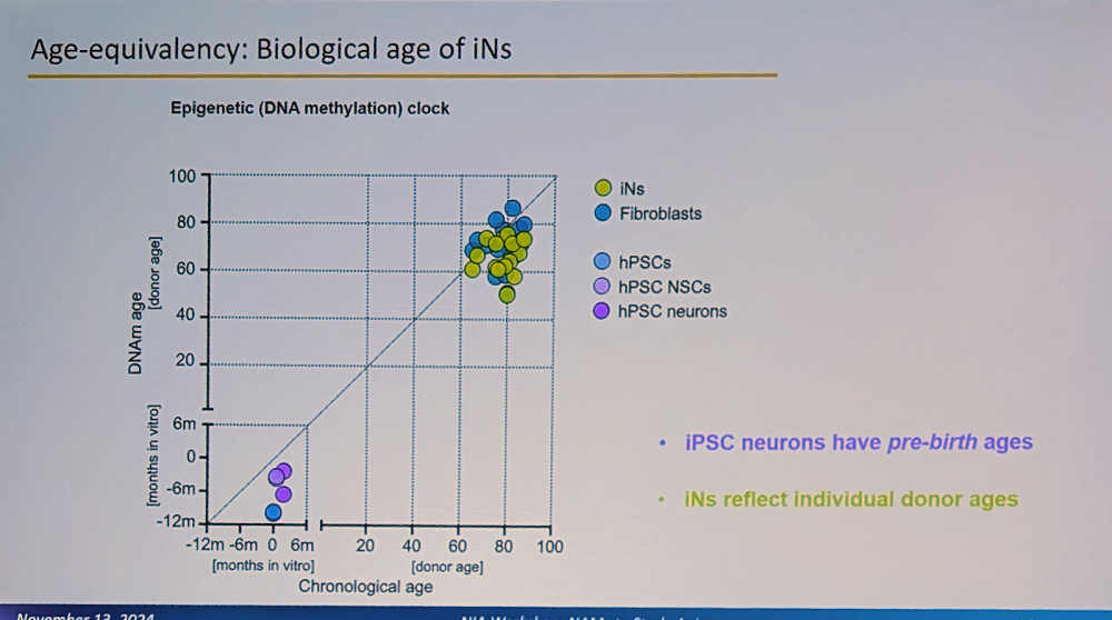
A crucial aspect of the system is that these iNs maintain the biological age of the donor and their specific aging signature. “This is fundamentally different from iPSC-derived neurons, which reset to pre-birth ages according to methylation clocks,” Traxler said. “Our iNs reflect individual donor ages and display adult-stage characteristics, while iPSC neurons resemble fetal stages.”

GSA2024 1

This age retention is particularly visible in adult neuronal splicing, such as in MAPT (Tau) genes, where the researchers observe adult-specific combinations of isoforms and their phosphorylation. Interestingly, humans have much more alternative splicing than mice, a feature that cannot be properly modeled in mouse systems (mouse models of Alzheimer’s have indeed been unreliable, and their relevance to human Alzheimer’s is questioned).

The group went even further, having developed multicellular constructs that combine iNs with glial cells on polymer scaffolds. These form dense three-dimensional structures with synaptic connections. In these 3D constructs, the researchers have observed amyloid deposition with clear differences between familial and sporadic AD cases.

Interestingly, we recently reported on a study claiming that low efficiency of direct fibroblast-to-neuron reprogramming happens because only a small subset of stemlike cells (neuron crest progenitor cells) embedded within differentiated skin cells can produce neurons. However, the much higher efficiency cited by Traxler seems to contradict this claim. It is possible that her team uses a stronger reprogramming protocol that can cause a wider variety of skin cells to transition into neurons.

Can this diabetes drug extend lifespan?

Carolina Solis-Herrera from the University of Texas spoke at a session focused on repurposing existing drugs for slowing aging. Sodium-glucose cotransporter-2 (SGLT2) inhibitors, originally developed as glucose-lowering medications for type 2 diabetes, have attracted considerable attention in the longevity field after evidence appeared that this class of drugs positively affects both lifespan and healthspan. In particular, canagliflozin was among the handful of drugs that produced significant life extensions in mice in the rigorous Interventions Testing Program (ITP) trials.

Solis-Herrera’s group is trying to unravel the mechanisms of action behind those benefits. Recent evidence from trials shows that patients on SGLT2 inhibitors, both with and without diabetes, experienced fewer cardiovascular events, reduced hospitalization for heart failure, and improved kidney function. Cardiovascular and renal problems are, of course, two major causes of death in both diabetic and non-diabetic people.

“The cardiovascular protection we see with SGLT2 inhibitors emerges remarkably quickly – between six to eight weeks, far too fast to be explained by traditional risk factor improvements,” Solis-Herrera said. “This suggests there must be other mechanisms at work.”

Results show that SGLT2 inhibitors work along several pathways. They enhance the clearance of senescent cells; induce calorie loss by promoting urinary glucose excretion, which resembles caloric restriction; modulate key nutrient-sensing pathways involved in aging, such as mTOR and AMPK; and reduce age-related low-grade inflammation (inflammaging) and oxidative stress.

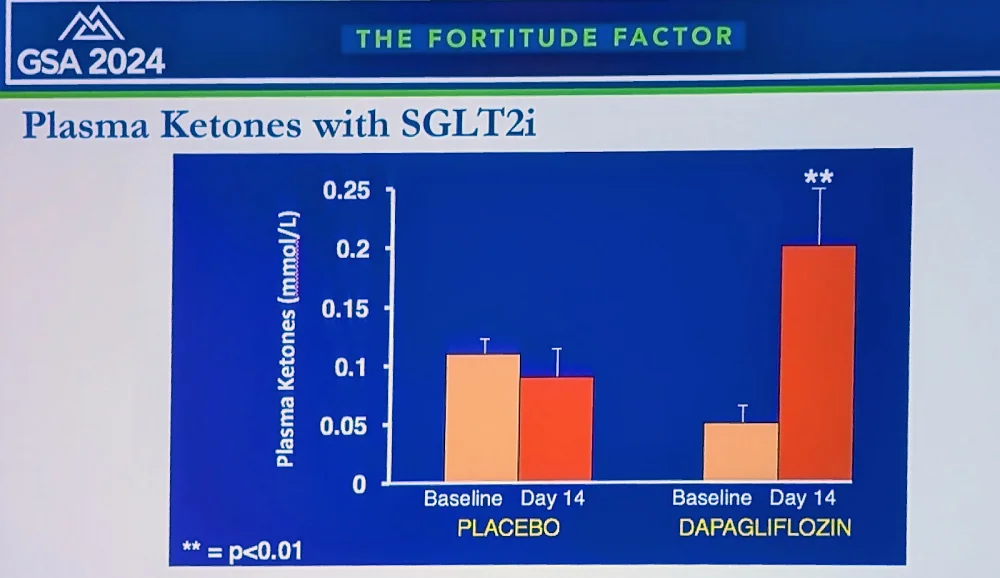
The researchers currently focus on the “ketone hypothesis”: that SGLT2 inhibitors increase the production of ketone bodies, such as beta-hydroxybutyrate (BHB). “We found that ketones reduce oxygen consumption and increase efficiency in various organs, including heart and kidneys,” Solis-Herrera said. “We’re now investigating applications in Alzheimer’s, diabetic retinopathy, dementia, and post-transplant patients for reducing rejection – that’s why we call SGLT2 inhibitors “the gift that keeps giving.”

GSA2024 2

Solis-Herrera reported on a new study in humans, where patients on SGLT2 inhibitors showed decreased inflammatory biomarkers, including TNF-alpha; a decrease in oxidation markers and senescent cell burden; and a significant decrease in visceral fat across multiple organs. “In summary,” she concluded, “SGLT2 inhibitors, originally created for diabetes, have emerged as potentially powerful anti-aging compounds. Their mechanisms likely involve a metabolic shift from glucose excretion to ketogenesis, which appears to be beneficial rather than maladaptive. They modulate key molecular pathways like mTOR and AMPK, reducing inflammation and oxidation.”

Looking beyond weight loss

John Newman, another representative of Buck Institute on Aging at the conference, talked about an even more hyped type of drug: glucagon-like peptide (GLP-1) receptor agonists, such as semaglutide, the principal ingredient of Ozempic and Wegovy. These drugs have revolutionized the treatment of diabetes and obesity, but many researchers believe they provide anti-aging benefits outside this context. Newman said he was very enthusiastic about GLP-1 agonists, but “there are critical gaps that need filling before we let that enthusiasm run away.”

Newman explained that GLP-1, a small peptide hormone secreted by L cells in the gut epithelium and circulating throughout the body, reduces motility in the gut and slows glucose absorption. In the pancreas, it enhances glucose-stimulated insulin secretion to reduce hyperglycemia. However, endogenous GLP-1 is rapidly degraded, with a half-life in plasma “in the order of minutes.”

The GLP-1 receptor also interacts intracellularly with various aging-related pathways, such as mTOR and FOXO, enhancing mitochondrial function and dampening inflammation. “All these pathways,” Newman said, “are very familiar to geroscientists, and this integration of GLP-1 receptor signaling with mechanisms of aging is part of why the idea of GLP-1 receptor agonists as gerotherapeutics is so tempting.”

GSA2024 3

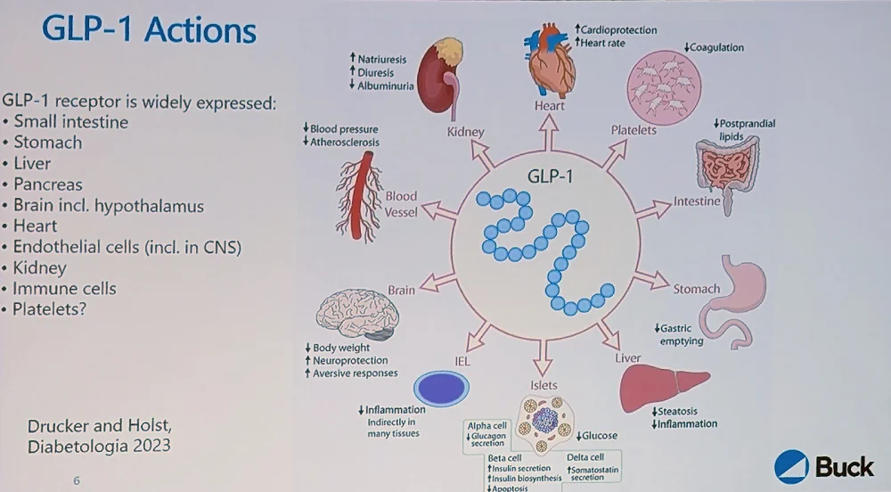
The GLP-1 receptor is widely distributed in the body, including in hypothalamic neurons, which is how it regulates satiety and appetite. It is also present in the heart, which might explain the cardiovascular benefits.

Large-scale trials have shown a significant decrease in cardiovascular mortality in obese and/or diabetic patients. However, gastrointestinal side effects are a major problem: virtually all patients on GLP-1 receptor agonists will eventually experience them, and they are severe enough to cause a 5%-8% dropout in studies.

“The results are striking for diabetes treatment and, within either diabetes or obesity, for atherosclerotic disease, kidney disease, and heart failure with preserved ejection fraction,” Newman said. “But does this make them gerotherapeutics? Not necessarily – it makes them highly effective diabetes and obesity treatments.”

For us to decide that these drugs are indeed geroprotectors, he explained, we have to see efficacy in diseases outside this metabolic cluster, such as neurodegenerative disease, cancer, and osteoporosis. Small trials in Parkinson’s and Alzheimer’s diseases showed some promise, but the results were not dramatic. A large Alzheimer’s trial (EVOKE) is ongoing. Importantly, this is one of the first large trials in non-obese people.

“The big picture,” Newman summarized, “is that while these are very effective agents in obesity and diabetes, we don’t know if benefits extend beyond these conditions or if cardiovascular and kidney protection is independent of weight loss – crucial questions for their potential as gerotherapeutics. Questions remain about the effects on sarcopenic obesity and diseases of aging not caused by obesity or diabetes. Much work remains to be done.”

Cellular reprogramming for organ transplantation

Pradeep Reddy of Juan Carlos Izpisua Belmonte’s lab gave a fascinating talk on cellular reprogramming. Belmonte is one of the pioneers of partial reprogramming and the first to demonstrate significant life extension in progeroid mice. Like several other first-tier geroscientists, Belmonte, previously at Salk Institute, was recruited by Altos Labs. Reddy’s talk presented a rare opportunity to gauge how things are going at the best-funded longevity startup in the world.

Reddy started by bringing up the lab’s work from several years ago on Hutchinson-Gilford progeria syndrome (HGPS). When the researchers reprogrammed fibroblasts from HGPS patients to induced pluripotent stem cells (iPSCs), “one striking observation was that all the aging hallmarks their cells initially showed were totally reversed, even though the mutation was still present,” Reddy said.

When these iPSCs were re-differentiated back to somatic lineages, they started to manifest the disease phenotype again. However, the team realized the importance of the first part: “that it’s possible to take a pre-diseased cell and reset or reverse those disease markers.”

Previously, partial reprogramming was mostly discussed in the context of aging. However, their results led the team to look for opportunities to apply reprogramming to contexts other than aging, “changing the trajectory of cells from diseased to healthy.”

“We conducted several studies in aged animals across different tissues,” Reddy said. “It’s not specific to only certain cell types – it can be a broad, agnostic approach. One thing that happens during loss of chromatin stability in disease or aging is loss of cell identity, which leads to decreased functional fidelity.”

One epigenetic alteration that is ubiquitous and important in aging is the epithelial-to-mesenchymal transition (EMT), in which epithelial cells lose cellular adhesion and become more motile. EMT can play a beneficial role in wound healing, but it also harms the original function of epithelial cells and is one of the central mechanisms of invasion and metastasizing in cancer.

The researchers observed increased EMT signatures in models of liver disease. “Similarly, we see the same phenotype in different cardiomyopathies, lung, and kidney disease,” Reddy said. “It’s a common phenotype, not specific to one tissue.” Partial reprogramming led to the erasure of these mesenchymal signatures in a matter of two to four days, which can explain early benefits.

One area where the researchers attempted to apply partial reprogramming is cellular senescence since senescence cells undergo drastic epigenetic changes. They saw decreased levels of SASP (senescence-associated secretory phenotype) elements such as p16, increased resilience, less hair graying, and improved wound healing in treated mice.

Delivering reprogramming factors via viral vectors remains a challenge since, with systemic delivery, most particles end up in the liver. Reddy’s team sought to rejuvenate kidneys, but the delivery problem seemed insurmountable until they decided to take a page from clinical practices, where donor organs are often connected to perfusion machines ex vivo for up to several hours to keep them viable. The idea was to add reprogramming factors to the perfusion solution in order to increase the organ’s fitness.

In collaboration with a clinic in Barcelona, the researchers worked on kidney transplantation in rats. As expected, organs from old donors were less viable, but reprogramming during perfusion showed promising results.

GSA2024 4

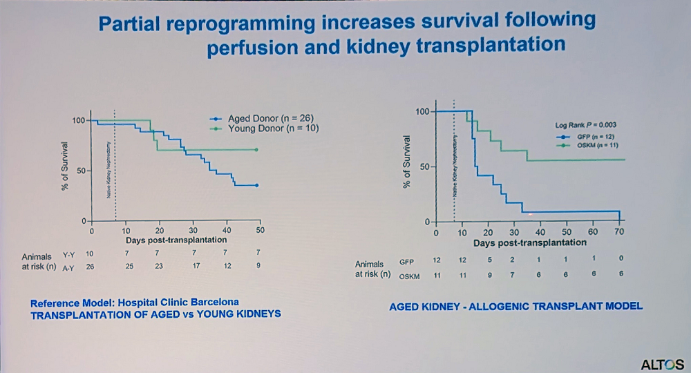
“This platform could help expand the donor pool by allowing us to improve suboptimal organs that would otherwise be discarded,” Reddy said. “We’ve built a modified perfusion system that doesn’t require complete transplantation – organs can remain connected to the body while undergoing perfusion, making it more applicable for age-related disease cases.”

No limit for New Limit

Not as hyped as Altos Labs, New Limit is nevertheless another exciting company working in the field of cellular reprogramming. Jacob Kimmel, who co-founded the company after stints at UCSF and Alphabet’s anti-aging company Calico, gave a captivating overview of its research.

New Limit is built on the premise that the classic Yamanaka reprogramming factors (OSKM) are not special, and many other rejuvenating factors and combinations can be found, tailored to specific cell types and contexts. “We plan to formulate these combinations into medicines using mRNA technology, similar to what many of us experienced with COVID vaccines,” Kimmel said. “Finally, we want to deploy these clinically to treat pathologies that will eventually affect all of us.”

While New Limit is working on several cell types and indications, in this talk, Kimmel focused on their T cell program, which targets infectious diseases by improving resilience. “There’s an enormous number of possible combinations,” he said. “We can’t experiment our way through that, so we need to be both efficient and smart about which experiments we choose.”

To solve this problem, New Limit has developed a proprietary high-throughput discovery process that begins with predictive computer models. The work then moves into primary human cells from multiple young and old donors.

When the researchers introduce pools of transcription factors, “due to the stochasticity of delivery, each cell picks up a different subset of factors,” Kimmel explained. “The result is a dish where all possible subcombinations up to a certain number are represented. We’ve attached DNA barcodes to these factors, allowing us to use single-cell genomics downstream to measure what happened – which genes the cell is expressing and which transcription factors achieved that outcome.”

To detect if any combination resulted in rejuvenation, the researchers use machine learning models to predict cell age from gene expression profiles. Using this system, they have screened around 9,000 combinations of transcription factors for their effect on cell age – “about 500 times more than the roughly 19 combinations tested in academic literature,” Kimmel said.

Interestingly, the researchers have found that many different transcription factors can reverse T cell aging, often to the same degree as the Yamanaka factors. That said, those factors were not just variants of Yamanaka’s but are “broadly distributed across different transcription factor families, suggesting multiple paths to reprogram cell age.” Kimmel reported seeing a lot of synergy as if transcription factors tend to work better in combinations.

GSA2024 5

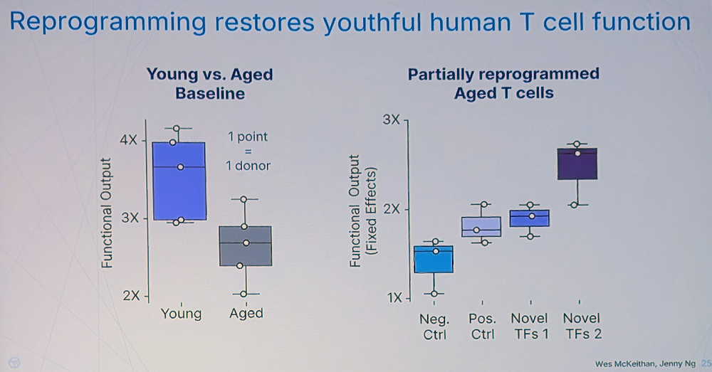
Rejuvenation apparently leads to improved T-cell fitness. “We found canonical cytotoxicity functions of human T cells are significantly impaired with age – something not clearly established in the literature before our work,” Kimmel said. However, the team has found many novel combinations that restore T cells’ fitness even stronger than the Yamanaka factors.

The question of durability, according to Kimmel, is crucial – effects that only last while mRNAs are expressed would be difficult to translate into treatments. “But we found that some combinations produce durable changes, measurable many days after turning off the factors,” he said.

Y is this happening?

Nick Chavkin, Assistant Professor at the Department of Pediatrics at Seattle Children’s Research Institute (hence, a local), gave a talk about a particularly interesting mutation affecting blood cells: the loss of the Y chromosome. Due to hematopoietic clonal expansion (when hematopoietic stem cells with certain mutations become more successful in reproduction and, as a result, dominate the cell pool), this mutation is quite prevalent in aged people, but for obvious reasons, only in men. According to the UK Biobank, by age 70, about 45% of men show appreciable Y chromosome loss, which, according to Chavkin, makes it the most common known human post-zygotic mutation.

Large biobank datasets also revealed the link between this long-known condition and all-cause mortality. Men with Y chromosome loss are about twice as likely to die at any given age compared to men without Y loss.

“The first associations were with cancer mortality and Alzheimer’s disease diagnosis – men with Y loss show higher rates of both,” Chavkin said. “We also demonstrated an increased rate of cardiovascular disease. This led us to investigate the mechanistic aspects. While the correlations are interesting, we wanted to know: could Y loss actually promote these disease states, or is it just an age-related phenomenon associated with genomic instability?”

The researchers created a mouse model with 80-90% Y-loss in hematopoietic stem cells. Compared to controls, those mice showed diminished survival, age-related cardiomyopathy, pulmonary and renal fibrosis, and cognitive decline: all the known hallmarks of Y loss.

Y-loss mice also showed exacerbated heart failure conditions. Looking for mechanistic explanations, the researchers discovered that Y-loss macrophages have a preference for fibrotic polarization, unlike some other known clonal mutations that promote inflammation.

Chavkin’s team then looked for the specific Y chromosome genes that drive these effects. “This was relatively straightforward because the Y chromosome is often considered a genetic “wasteland” post-puberty,” Chavkin said. “In mouse macrophages, only four Y chromosome genes are appreciably expressed, all within about a million base pairs: KDM5D, EIF2S3Y, UTY, and DDX3Y.”

Three knockouts had no effect, but UTY knockout recapitulated the full Y-loss phenotype. UTY is an epigenetic modifier that probably has broad regulatory effects.

Further experiments suggested that UTY inhibits pro-fibrotic macrophage polarization by regulating genomic DNA accessibility. UTY knockout probably allows certain transcription factors to bind and promote this polarization, leading to fibrosis.

The team’s current hypothesis is that Y chromosome loss leads to UTY insufficiency in monocytes. This increases chromatin accessibility for pro-fibrotic genes, allowing fibrotic transcription factor activity and ultimately leading to pro-fibrotic polarization and myocardial fibrosis.

“In summary, Y chromosome loss appears to be an age-related somatic mutation contributing to male mortality,” Chavkin said. “Our work suggests UTY plays a key role in this process. This mutation affects multiple hallmarks of aging – these X0 cells show intrinsic genomic instability, epigenetic alterations, effects on chronic inflammation, and altered intercellular communication.”

We would like to ask you a small favor. We are a non-profit foundation, and unlike some other organizations, we have no shareholders and no products to sell you. All our news and educational content is free for everyone to read, but it does mean that we rely on the help of people like you. Every contribution, no matter if it’s big or small, supports independent journalism and sustains our future.
MSCs

Finding Cells That Send Signals Against Arthritis

In Aging, researchers have reported that deriving extracellular vesicles from mesenchymal stromal cells (MSCs) in fat tissue has beneficial effects in models of osteoarthritis.

Which source to use?

Earlier this month, we wrote about how small extracellular vesicles derived from embryonic cells (ESC-sEVs) alleviate arthritis in rodent models. While these researchers acknowledge the potential of ESC-based therapies, this study focuses on EVs from a different source: MSCs, specifically those derived from fat (adipose) tissue (ASCs). ASCs from ruminants’ antler tissues, just like EVs derived from other tissues, have been found to alleviate osteoarthritis in rat models [1] for the same fundamental reason: the alleviation of cellular senescence.

Similar results were found in a study of ASC-EVs in human cells derived from patients with advanced osteoarthritis [2], and a conditioned medium derived from ASCs was found to do similar things [3]. However, these researchers also realize that most studies, such as the one we covered earlier this month, are more focused on cells driven senescent by inflammation rather than DNA damage [4], and cells driven senescent by different origins can have different effects.

Positive effects against different senescence origins

This paper, therefore, conducts experiments on cartilage-generating chondrocytes that were driven senescent by DNA damage, which was inflicted through the administration of the toxin etoposide. As expected, these cells started exhibiting the senescence marker SA-β-gal along with the DNA damage marker γH2AX, with a trend towards an increase in the SASP.

Exposing these cells to ASC-EVs along with etoposide blunted the effects of the toxin. SASP markers, SA-β-gal expression, and even DNA damage as measured by γH2AX were all reduced. These changes were found to be at least partially due to a restoration of the balance between the metabolic buildup process of anabolism and the breakdown process of catabolism.

The researchers then turned to the more conventional method of inducing senescence through the inflammatory factor IL-1β. Compared to the etoposide-induced group, cells exposed to this factor did not exhibit DNA damage, although they had still had enlarged nuclear surfaces just as the etoposide group did. The SASP factors induced by IL-1β, however, were markedly increased compared to the etoposide group.

Fortunately, most of these factors were significantly downregulated when ASC-EVs had been previously introduced. The interleukins IL-6 and IL-8, two major SASP components, were affected, as were matrix metalloproteinases (MMPs). The researchers describe the effect as “senoprotective”, as it had prevented the cells from going senescent.

There was a very interesting difference between this study and the study from two weeks ago. In that study, the researchers reported that FOXO1 was upregulated, and FOXO3 was not; this study, on the other hand, reported the exact opposite. This suggests a distinction between the two EV sources and a possibility of combination treatments that use EVs derived from both sources.

Benefits in a mouse model

The researchers replicated their findings in a mouse model of induced osteoarthritis through collagen destruction with collagenase. Most notably, 24 days after the introduction of ASC-EVs, the treated group’s osteoarthritis score was nearly identical to that of the arthritis-free control group, and even after 42 days, the treatment still appeared to be effective in most mice. While not all of the many tested biomarkers went in the desired direction at 9 days or 14 days, an analysis of the expression of various genes led these researchers to conlude that ASC-EVs have a “therapeutic effect” in these mice.

This paper, like others before it, spends a considerable amount of time characterizing and diagnosing the target cells to which the treatment is targeted. With the various sources of EVs being shown to have effects in cells, this may be enough to bring these sorts of treatments into clinical trials, possibly if EVs from these sources are combined. However, it might also be of value to closely examine just what is in these tiny packages being sent from the donor cells and if it is possible to include or exclude any of their contents.

We would like to ask you a small favor. We are a non-profit foundation, and unlike some other organizations, we have no shareholders and no products to sell you. All our news and educational content is free for everyone to read, but it does mean that we rely on the help of people like you. Every contribution, no matter if it’s big or small, supports independent journalism and sustains our future.

Literature

[1] Lei, J., Jiang, X., Li, W., Ren, J., Wang, D., Ji, Z., … & Wang, S. (2022). Exosomes from antler stem cells alleviate mesenchymal stem cell senescence and osteoarthritis. Protein & cell, 13(3), 220-226.

[2] Tofiño-Vian, M., Guillén, M. I., Pérez del Caz, M. D., Castejón, M. A., & Alcaraz, M. J. (2017). Extracellular vesicles from adipose‐derived mesenchymal stem cells downregulate senescence features in osteoarthritic osteoblasts. Oxidative Medicine and Cellular Longevity, 2017(1), 7197598.

[3] Platas, J., Guillén, M. I., Del Caz, M. D. P., Gomar, F., Castejón, M. A., Mirabet, V., & Alcaraz, M. J. (2016). Paracrine effects of human adipose-derived mesenchymal stem cells in inflammatory stress-induced senescence features of osteoarthritic chondrocytes. Aging (Albany NY), 8(8), 1703.

[4] Philipot, D., Guérit, D., Platano, D., Chuchana, P., Olivotto, E., Espinoza, F., … & Brondello, J. M. (2014). p16 INK4a and its regulator miR-24 link senescence and chondrocyte terminal differentiation-associated matrix remodeling in osteoarthritis. Arthritis research & therapy, 16, 1-12.

Older man exercising

How Exercise Preserves Function in Motor Nerves

In Aging Cell, researchers have described the specific cell types that give exercise protective effects against motor nerve degeneration.

A different level of back problems

With aging, the prevalence of nerves connecting to muscle tissue dwindles, a phenomenon known as denervation [1]. This has been linked to a depopulation of the alpha motor neurons in the spinal cord [2]. Unless interventions are undertaken, the muscles themselves dwindle as a result [3], being gradually replaced by non-muscle fibrosis instead [4].

This fibrotic replacement is driven by meschenchymal fibroblasts [5] instead of the muscle fibroblasts that would replenish the normal tissue [6]. Fibroblasts have also been reported to secrete factors that encourage nerve regrowth [7], as have muscle stem cells [8].

While it is disputed, some work even suggests that, in middle age, human beings’ dwindling nerves are replaced by these natural processes [9] and that this results in more complex nerve centers than younger people have, particularly in people who exercise regularly [10]. While the accelerated decline caused by runaway aging processes leads to degradation without regeneration of the nervous tissue [11], intensive exercise has been found to efficiently protect against this [12].

The precise molecular mechanisms and cells involved, however, had not been fully described. This research focuses on muscle fibroblasts and stem cells in an effort to discover precisely what makes exercise such a powerful treatment for motor nerve degradation.

Cell type matters

In the first experiment, primary motor neurons were cultured from rat embryos, while muscle fibroblasts and stem cells were taken from human muscle biopsies. Despite the species difference, these cells were found to interact in a compatible way. Four of the human volunteers were young, four were old and sedentary, and six were old people who had exercised throughout their lives.

Muscle stem cells and fibroblasts were found to have starkly different gene expression profiles and stimulate the rat neurons in starkly different ways: a total of 11% of the neurons’ genes were expressed differently between the two groups, with roughly equal amounts of upregulation and downregulation. Considering the functions of these genes, culturing with fibroblasts seemed to encourage neural growth in a way that culturing with stem cells did not, including two genes that code for synapse transmission and a gene for the formation of new neurons (neurogenesis).

Culturing the rat neurons with conditioned media from these cell types, instead of directly, yielded somewhat similar results. Here, the researchers found that motor neurons are affected in complementary ways by each of these cell types, with fibroblasts still appearing to be more important for growth and development.

Lifelong exercisers have multiple advantages

The researchers then started comparing the cells of the different groups. Interestingly, they found that, while the young people were still better at exerting force than the older people and had slightly more lean mass, the older exercisers’ muscles appeared to be more efficient than younger people and older sedentary people. CAF, a key biomarker of neurological impairment, was not elevated in the older exercisers compred to the younger people, while older sedenary people had significantly elevated amounts.

Culturing the motor neurons with cells grown from older sedentary people had substantially negative effects: a full 53% more of the neurons survived after being cultured with cells derived from older exercisers versus their sedentary counterparts. The older exercisers’ cells even appeared to promote neuronal health more than those taken from younger people, although there was no statistically significant difference found with this small group.

Neuronal survival with exercised cells

This was a relatively small study, and it did not delve into the precise molecular mechanisms involved in this communication between muscle fibroblasts, muscle stem cells, and motor neurons. However, it has clearly identified the cell types involved. An analysis of such factors as extracellular vesicles and other intercellular communication will be necessary to translate these findings into a useful treatment.

Despite its limitations, however, this study makes it absolutely clear that exercise is necessary for long-term muscle health and defense against motor neuron loss with age. It is unknown when life-changing biological interventions will enter the clinic; right now, according to this and many other studies, exercise remains one of the best treatments that is broadly available.

We would like to ask you a small favor. We are a non-profit foundation, and unlike some other organizations, we have no shareholders and no products to sell you. All our news and educational content is free for everyone to read, but it does mean that we rely on the help of people like you. Every contribution, no matter if it’s big or small, supports independent journalism and sustains our future.

Literature

[1] Soendenbroe, C., Andersen, J. L., & Mackey, A. L. (2021). Muscle-nerve communication and the molecular assessment of human skeletal muscle denervation with aging. American Journal of Physiology-Cell Physiology, 321(2), C317-C329.

[2] McNeil, C. J., Doherty, T. J., Stashuk, D. W., & Rice, C. L. (2005). Motor unit number estimates in the tibialis anterior muscle of young, old, and very old men. Muscle & Nerve: Official Journal of the American Association of Electrodiagnostic Medicine, 31(4), 461-467.

[3] McPhee, J. S., Cameron, J., Maden-Wilkinson, T., Piasecki, M., Yap, M. H., Jones, D. A., & Degens, H. (2018). The contributions of fiber atrophy, fiber loss, in situ specific force, and voluntary activation to weakness in sarcopenia. The Journals of Gerontology: Series A, 73(10), 1287-1294.

[4] Madaro, L., Passafaro, M., Sala, D., Etxaniz, U., Lugarini, F., Proietti, D., … & Puri, P. L. (2018). Denervation-activated STAT3–IL-6 signalling in fibro-adipogenic progenitors promotes myofibres atrophy and fibrosis. Nature cell biology, 20(8), 917-927.

[5] Rebolledo, D. L., González, D., Faundez-Contreras, J., Contreras, O., Vio, C. P., Murphy-Ullrich, J. E., … & Brandan, E. (2019). Denervation-induced skeletal muscle fibrosis is mediated by CTGF/CCN2 independently of TGF-β. Matrix Biology, 82, 20-37.

[6] Wosczyna, M. N., & Rando, T. A. (2018). A muscle stem cell support group: coordinated cellular responses in muscle regeneration. Developmental cell, 46(2), 135-143.

[7] Theret, M., Rossi, F. M., & Contreras, O. (2021). Evolving roles of muscle-resident fibro-adipogenic progenitors in health, regeneration, neuromuscular disorders, and aging. Frontiers in Physiology, 12, 673404.

[8] Liu, W., Klose, A., Forman, S., Paris, N. D., Wei-LaPierre, L., Cortes-Lopez, M., … & Chakkalakal, J. V. (2017). Loss of adult skeletal muscle stem cells drives age-related neuromuscular junction degeneration. Elife, 6, e26464.

[9] Deschenes, M. R. (2011). Motor unit and neuromuscular junction remodeling with aging. Curr Aging Sci 4 (3): 209–220.

[10] Jones, E. J., Piasecki, J., Ireland, A., Stashuk, D. W., Atherton, P. J., Phillips, B. E., … & Piasecki, M. (2021). Lifelong exercise is associated with more homogeneous motor unit potential features across deep and superficial areas of vastus lateralis. GeroScience, 1-11.

[11] Snow, L. M., Mcloon, L. K., & Thompson, L. V. (2005). Adult and developmental myosin heavy chain isoforms in soleus muscle of aging Fischer Brown Norway rat. The Anatomical Record Part A: Discoveries in Molecular, Cellular, and Evolutionary Biology: An Official Publication of the American Association of Anatomists, 286(1), 866-873.

[12] Soendenbroe, C., Heisterberg, M. F., Schjerling, P., Kjaer, M., Andersen, J. L., & Mackey, A. L. (2022). Human skeletal muscle acetylcholine receptor gene expression in elderly males performing heavy resistance exercise. American Journal of Physiology-Cell Physiology, 323(1), C159-C169.

Diverse diet

Dietary Diversity Is Associated With Delayed Aging

An analysis of data from over twenty thousand people has indicated that greater dietary diversity is associated with slower biological aging [1].

Your health is what you eat

Good dietary habits are linked to many health benefits, and different diets were previously reported to impact the speed of aging and senescence. For example, adherence to the Mediterranean diet is positively associated with increased lifespan and healthspan.

We have also previously reported on some health benefits linked to different dietary patterns, such as associations linking an anti-inflammatory diet and the Mediterranean diet with a reduced risk of dementia, the positive impact of a ketogenic diet on symptoms of multiple sclerosis, the impact of Mediterranean, keto, and plant-based diets on cancer risk and progression, and the metabolic benefits of a ketogenic diet and the Mediterranean diet in pre-diabetes and Type 2 diabetes patients.

The authors of this study did not focus on any specific diet; instead, they focused on the diversity of food consumed by the study participants. They discuss the impact of a diverse diet, which is rich in macronutrients, micronutrients, antioxidants, and bioactive compounds, on the speed of aging.

Biological age is not just a number

Compared to chronological age alone, the relationship between biological age and chronological age is a better estimate of health and the risk of developing age-related diseases. A higher biological age suggests a higher possibility of developing age-related diseases and a higher chance of dying.

The researchers analyzed data from 22,600 participants (49.3% male) with an average age of 48 years from the National Health and Nutrition Examination Survey (NHANES), a cross-sectional survey conducted in the United States. People under 20 years of age, pregnant, and those with no available food intake or biological age data were excluded from the analysis.

The researchers in this study used phenotypic age and Klemera–Doubal method (KDM) biological age to represent the biological age of study participants. Those measures are based on the composite clinical biomarkers.

They used systolic blood pressure, blood creatinine, urea nitrogen, albumin, total cholesterol, glycosylated hemoglobin A1c, percentage of lymphocytes, mean erythrocyte volume, leukocyte count, and alkaline phosphatase as biomarkers for their assessment.

The more diverse, the better

The researchers assessed the dietary diversity score (DDS), which was described as simple, effective, and validated in clinical trials. It measures the number of food groups in one’s diet, based on five major food groups and 18 subgroups. “A higher DDS is generally indicative of a more varied diet and is associated with a broader intake of essential nutrients.” Previous research had reported an association between a higher DDS and a lower risk of chronic diseases such as diabetes mellitus [2] and cardiovascular diseases (CVD) [3].

In the analyzed group, the researchers measured DDS based on the average score from two self-reported 24-hour dietary recalls.

Higher diversity, lower biological age

The researchers used a few models to analyze the data. In the first model, they didn’t include any confounding variables. The second and third models were adjusted for different factors. Model two included demographic factors. The third model also included health metrics, such as cancer, smoking, alcohol consumption, and metabolic data. The researchers performed multiple modeling analyses using different variables (continuous and categorical) and corrected for multiple confounders.

Their results suggested an association between higher DDS and slower biological aging. They note that this relationship is both highly significant (overall p of under 0.001) and linear.

DDS 1

Analysis of the participants’ subgroups divided by different health or demographic factors suggested an inverse relationship between DDS and phenotypic age acceleration across subgroups; however, these results were mainly not statistically significant.

The researchers also performed a sensitivity analysis that ensured the robustness of their observations. They did this analysis using multiple adjustments and concluded that the consistency of all three models suggests “a higher dietary diversity is significantly associated with lower phenotypic age acceleration, regardless of the adjustment methods employed.”

The researchers also explored the idea of oxidative stress being the factor mediating the relationship between dietary diversity and aging. They observed that “the oxidative stress indicator GGT had a significant mediating effect on the association of DDS and phenotypic age acceleration.”

Glutamyltransferase (GGT) was one of the proteins that was significantly lower in people with higher DDS. White blood cell count and neutrophil-lymphocyte ratio, two indicators of inflammation, were also significantly reduced. In contrast, levels of albumin, a potential indicator of anti-inflammatory capacity, and serum klotho, a protein with anti-aging properties, were higher.

Robust results, but without mechanistic understanding

Since this study is based on observational data, it cannot determine the mechanism behind the observed association. Still, the researchers proposed some hypotheses. They believe that oxidative stress and inflammation could be key processes mediating the effect of diet on aging, as a more diverse diet includes more antioxidants and anti-inflammatory compounds that protect cells from aging-related processes. They also consider the possible role of gut microbiota since a diverse diet can help maintain microbial diversity, an important factor for gut health. However, that particular aspect was not explored in this study.

The researchers claim that their results are robust and can be extrapolated to multi-ethnic and otherwise varied populations due to this data’s consistency and analysis.

While this analysis suggested that some associations are mediators, it cannot establish causality, and potential confounding factors (beyond what was tested) cannot be ruled out. The reporting of food intake should also be optimized for future studies.

Overall, this study’s results align with previous research, which linked reduced food diversity to an increased risk of age-related chronic diseases and mortality [2-5]. As the authors note, “promoting dietary diversity may facilitate healthy aging, which has significant implications for public health.”

DDS 2
We would like to ask you a small favor. We are a non-profit foundation, and unlike some other organizations, we have no shareholders and no products to sell you. All our news and educational content is free for everyone to read, but it does mean that we rely on the help of people like you. Every contribution, no matter if it’s big or small, supports independent journalism and sustains our future.

Literature

[1] Liao, W., & Li, M. Y. (2024). Dietary diversity contributes to delay biological aging. Frontiers in medicine, 11, 1463569.

[2] Zheng, G., Cai, M., Liu, H., Li, R., Qian, Z., Howard, S. W., Keith, A. E., Zhang, S., Wang, X., Zhang, J., Lin, H., & Hua, J. (2023). Dietary Diversity and Inflammatory Diet Associated with All-Cause Mortality and Incidence and Mortality of Type 2 Diabetes: Two Prospective Cohort Studies. Nutrients, 15(9), 2120.

[3] Chalermsri, C., Ziaei, S., Ekström, E. C., Muangpaisan, W., Aekplakorn, W., Satheannopakao, W., & Rahman, S. M. (2022). Dietary diversity associated with risk of cardiovascular diseases among community-dwelling older people: A national health examination survey from Thailand. Frontiers in nutrition, 9, 1002066.

[4] Zheng, G., Xia, H., Lai, Z., Shi, H., Zhang, J., Wang, C., Tian, F., & Lin, H. (2024). Dietary Inflammatory Index and Dietary Diversity Score Associated with Sarcopenia and Its Components: Findings from a Nationwide Cross-Sectional Study. Nutrients, 16(7), 1038.

[5] Chalermsri, C., Rahman, S. M., Ekström, E. C., Ziaei, S., Aekplakorn, W., Satheannopakao, W., & Muangpaisan, W. (2023). Dietary diversity predicts the mortality among older people: Data from the fifth Thai national health examination survey. Archives of gerontology and geriatrics, 110, 104986.

Growing neuron

Intranasal Spray Alleviates Early Alzheimer’s in Mice

A novel therapy based on induced neuronal stem cells shows promise in a mouse model of Alzheimer’s disease – and it can be administered intranasally [1].

Who needs cells?

Stem cell therapies have made great strides in recent decades. They have been successfully employed against numerous diseases, from cancer to osteoarthritis. However, storing, transporting, and administering cells is not easy. Moreover, in some cases, cells, with their extraordinarily complex metabolisms, can produce unwanted side effects. For instance, neural stem/progenitor cells (NSCs) have been shown to improve symptoms of certain brain diseases [2] but can also trigger pathological changes in the brain [3].

However, the whole cell might not always be necessary. Cells have been known to communicate with each other by excreting extracellular vesicles (EVs), tiny membrane-bound bubbles that can carry various cargoes, such as RNA molecules and proteins. Scientists have learned to harvest vesicles produced by cells and administer them locally or systemically, often recapitulating much of the effect of cell administration.

Unlike stem cells, EVs can be frozen and thawed without compromising their therapeutic efficacy. Furthermore, cells of different types can now be easily produced from induced pluripotent stem cells (iPSCs). Those are somatic cells reverted into pluripotency by applying certain molecules, such as the original reprogramming cocktail of Yamanaka factors (OSKM).

Less inflammation

In a new study published in the Journal of Extracellular Vesicles, researchers from Texas A&M University took human iPSCs and re-differentiated them into NSCs. They then harvested the extracellular vesicles produced by the cells, purified them, and administered them intranasally to a mouse model of familial Alzheimer’s disease (5xFAD mice). Despite decades of research and tens of billions of dollars spent, a cure for Alzheimer’s remains elusive.

While mouse models of Alzheimer’s have their limitations, as mice naturally don’t develop this disease, 5xFAD mice have been widely used. These animals start displaying Alzheimer’s-like pathologies, such as amyloid beta (Aβ) plaques and increased neuroinflammation at the age of three months, which is when the treatment was administered. About two months later, the mice underwent cognitive and neuropathological assessments.

The researchers confirmed that the EVs were indeed taken up by the brain’s resident macrophages (microglia). In Alzheimer’s, these cells surround Aβ plaques, presumably in an attempt to remove them. They display increased activation and inflammation, which has been linked to disease progression.

“Prolonged activation causes microglia to lose their normal function and begin to harm neurons, leading to progressive neuron loss,” explains Ashok K. Shetty, Ph.D., a University Distinguished Professor and associate director at the Institute for Regenerative Medicine in the Department of Cell Biology and Genetics, and the corresponding author on the study.

RNA sequencing revealed that the treatment downregulated multiple inflammation-related pathways that were significantly upregulated in 5xFAD mice compared to healthy controls. Notably, this occurred without compromising the microglia’s phagocytosis function: their ability to engulf and destroy pathogens.

The treatment also led to a significant reduction in the burden of Aβ plaques and phosphorylated tau protein, two major hallmarks of Alzheimer’s. While both sexes showed improvements, males demonstrated a more robust response to the treatment.

EVs Alzheimer's Mice

By the age of five months, 5xFAD mice typically demonstrate significant cognitive decline, which was also observed in this study. The EV treatment, however, appeared to effectively block this decline. Tests included the object location test, in which cognitively healthy animals are expected to spend more time exploring an object in a novel place than in a familiar place, and the pattern recognition test, which measures the ability to discern novel objects from familiar ones.

Interestingly, the researchers also assessed the mice’s mood. Mood changes are increasingly gaining recognition as a clinically important aspect of Alzheimer’s disease. Just like many human Alzheimer’s patients, untreated 5xFAD mice exhibited anhedonia, the inability to enjoy things – in this case, sweetened water. The EV treatment restored the rodents’ joie de vivre – or at least their preference for sugar.

Similar effects in human cells

While the researchers did not test their treatment in human patients, they pursued the next best alternative: applying NSC-derived EVs to human microglia in vitro. When challenged with Aβ-42, an isoform of Aβ known for its high aggregation propensity and central role in the pathology of Alzheimer’s disease, microglia exhibited overactivation and an inflammatory phenotype. These effects, however, were reversed by the EV treatment.

According to the paper, EVs are superior to NSCs in that they do not replicate and readily cross the blood-brain barrier. Intranasal administration is easy, non-invasive, and characterized by rapid action. While the study was conducted on animals at early stages of the disease, constant advances in diagnostics make this less of a limitation.

“This approach is effective because the cargo carried by these extracellular vesicles could reduce the neuropathological changes in the brain,” says Shetty, who has filed a patent on the intranasal application of neural stem cell-derived extracellular vesicles for treating Alzheimer’s and other neurological disorders. “Our journey to advance the application of this therapy for Alzheimer’s disease is just beginning.”

We would like to ask you a small favor. We are a non-profit foundation, and unlike some other organizations, we have no shareholders and no products to sell you. All our news and educational content is free for everyone to read, but it does mean that we rely on the help of people like you. Every contribution, no matter if it’s big or small, supports independent journalism and sustains our future.

Literature

[1] Madhu, L. N., Kodali, M., Upadhya, R., Rao, S., Somayaji, Y., Attaluri, S., … & Shetty, A. K. (2024). Extracellular vesicles from human‐induced pluripotent stem cell‐derived neural stem cells alleviate proinflammatory cascades within disease‐associated microglia in Alzheimer’s disease. Journal of Extracellular Vesicles, 13(11), e12519.

[2] Temple, S. (2023). Advancing cell therapy for neurodegenerative diseases. Cell stem cell, 30(5), 512-529.

[3] Abdi, S., Javanmehr, N., Ghasemi-Kasman, M., Bali, H. Y., & Pirzadeh, M. (2022). Stem cell-based therapeutic and diagnostic approaches in Alzheimer’s disease. Current Neuropharmacology, 20(6), 1093-1115.

CAR T Cells Attack

Engineering T Cells to Fight Brain Cancer

Researchers publishing in Nature have reported a new advance in developing chimeric antigen receptor (CAR) T cells to fight solid tumors in the brain.

A difficult endeavor

CAR T cell-based therapies are normally discussed in the context of blood cancers, as getting them to effectively attack solid tumors has proven very difficult, despite intensive research on the subject [1]. They have been used to attack glioblastomas, the most aggressive brain tumors in adults [2], even though targeting of antigens associated with this cancer can have toxic, off-target effects [3].

One safer method involves targeting of EGVRvIII, a molecule that appears on roughly 40% of these tumors [4]. However, a clinical trial utilizing this approach failed: the tumors were able to mutate and protect themselves against the treatment [5]. New approaches are being developed, and tested in trials, to better combat these tumors [6].

The researchers note the challenges involved in this sort of work. Glioblastoma tumors are very unfriendly to the immune system, suppressing its functions with increasing severity as the tumor grows [7]. For example, these tumors will secrete CD47, a natural immunosuppressant, in order to prevent macrophages from consuming them [8]. However, targeting CD47 has been found to be ineffective, as the therapy fails to penetrate the tumor, and dangerous to other tissues [9].

These researchers, therefore, have developed a fourth-generation therapy to target these tumors. These anti-EGFRvIII CAR T cells also release SGRP, a protein that binds to CD47, thereby directly fighting the immunosuppressive environment; however, cells also need CD47 to function properly, and the researchers were pleased to note that this alteration did not interfere with the cells’ own function.

Effective in mice

In experiments against cultured glioblastoma cells, which do not have such a protective environment, these new cells performed just as well as other CAR T cells. These cells were also found to be target-specific: cells that do not produce EGVRvIII were not harmed by these engineered cells.

In these sorts of experiments, it is relatively easy to graft human cancer cells into a mouse model and then have the CAR T cells defeat them there; however, this does not sufficiently mimic the actual tumor microenvironment, so preclinical successes can lead to clinical failures. Therefore, the researchers chose a model that avoids this problem.

The previous anti-EGFRvIII therapy was found to be effective in this scenario, extending the tumor-grafted mice’s lives and offering a one-in-five survival rate after 90 days, versus the zero they had with ineffective treatments. However, the new one performed incredibly well in comparison: after 90 days, almost none of the mice had died at all, and about two-thirds of them were completely free of tumors. The systemic toxicity associated with some forms of CAR treatment was not found in the animals treated with the new approach.

CAR T SGRP Survival

Letting other cells do their jobs

The researchers believe that some of the benefits are due to immune cell invasion of the tumors, not just of these particular T cells but of endogenous immune cells of all types. The engineered cells’ expression of SGRP within these tumors appeared to be effective. These findings were further confirmed by an analysis of cellular consumption (phagocytosis). The CAR T SGRP treatment encouraged local cells to do their jobs and consume cancer cells at a higher rate.

Spurred by their findings, the researchers also tested their SGRP approach against a mouse model of lymphoma. While they were not able to obtain the same impressive results as their glioblastoma experiment, they were able to obtain a 20% survival rate after 80 days; none of the animals treated with SGRP-less CAR T cells survived that long. While this approach did not stop lymphoma growth, it greatly slowed it down, even with just one initial treatment.

Despite sharing some of the same qualities, the researchers believe that their approach is superior to previous anti-CD47 approaches because it is expressed consistently and directly into the tumor to which the CAR T cells are attracted, a task that even locally injected antibodies have been found unable to properly do. While this is still just a mouse experiment, it may yield better clinical trial results than previous approaches.

We would like to ask you a small favor. We are a non-profit foundation, and unlike some other organizations, we have no shareholders and no products to sell you. All our news and educational content is free for everyone to read, but it does mean that we rely on the help of people like you. Every contribution, no matter if it’s big or small, supports independent journalism and sustains our future.

Literature

[1] Hou, A. J., Chen, L. C., & Chen, Y. Y. (2021). Navigating CAR-T cells through the solid-tumour microenvironment. Nature reviews Drug discovery, 20(7), 531-550.

[2] Ostrom, Q. T., Cioffi, G., Gittleman, H., Patil, N., Waite, K., Kruchko, C., & Barnholtz-Sloan, J. S. (2019). CBTRUS statistical report: primary brain and other central nervous system tumors diagnosed in the United States in 2012–2016. Neuro-oncology, 21(Supplement_5), v1-v100.

[3] Luksik, A. S., Yazigi, E., Shah, P., & Jackson, C. M. (2023). CAR T cell therapy in glioblastoma: overcoming challenges related to antigen expression. Cancers, 15(5), 1414.

[4] Felsberg, J., Hentschel, B., Kaulich, K., Gramatzki, D., Zacher, A., Malzkorn, B., … & Weller, M. (2017). Epidermal growth factor receptor variant III (EGFRvIII) positivity in EGFR-amplified glioblastomas: prognostic role and comparison between primary and recurrent tumors. Clinical Cancer Research, 23(22), 6846-6855.

[5] O’Rourke, D. M., Nasrallah, M. P., Desai, A., Melenhorst, J. J., Mansfield, K., Morrissette, J. J., … & Maus, M. V. (2017). A single dose of peripherally infused EGFRvIII-directed CAR T cells mediates antigen loss and induces adaptive resistance in patients with recurrent glioblastoma. Science translational medicine, 9(399), eaaa0984.

[6] Choi, B. D., Gerstner, E. R., Frigault, M. J., Leick, M. B., Mount, C. W., Balaj, L., … & Maus, M. V. (2024). Intraventricular CARv3-TEAM-E T cells in recurrent glioblastoma. New England Journal of Medicine, 390(14), 1290-1298.

[7] Yeo, A. T., Rawal, S., Delcuze, B., Christofides, A., Atayde, A., Strauss, L., … & Charest, A. (2022). Single-cell RNA sequencing reveals evolution of immune landscape during glioblastoma progression. Nature immunology, 23(6), 971-984.

[8] Willingham, S. B., Volkmer, J. P., Gentles, A. J., Sahoo, D., Dalerba, P., Mitra, S. S., … & Weissman, I. L. (2012). The CD47-signal regulatory protein alpha (SIRPa) interaction is a therapeutic target for human solid tumors. Proceedings of the National Academy of Sciences, 109(17), 6662-6667.

[9] Sikic, B. I., Lakhani, N., Patnaik, A., Shah, S. A., Chandana, S. R., Rasco, D., … & Padda, S. K. (2019). First-in-human, first-in-class phase I trial of the anti-CD47 antibody Hu5F9-G4 in patients with advanced cancers. Journal of Clinical Oncology, 37(12), 946-953.

Cell illustration

Study of Direct Reprogramming Challenges Consensus

Scientists have shown that in a direct cellular reprogramming scenario, neurons are produced almost exclusively by a rare subtype of multipotent cells. Their findings, they claim, change our understanding of reprogramming, but not everyone agrees [1].

Direct action

In the world of cellular reprogramming, the two most well-known realms are pluripotent reprogramming, in which somatic cells are thrown back all the way into pluripotency, and partial reprogramming, in which these cells are rejuvenated without making them lose their cellular identity.

However, there is a third route called direct reprogramming, in which somatic cells are directly transformed into differentiated cells of another type [2]. A similar process happens in the body naturally in some cases, but scientists have learned to facilitate it by expressing certain factors. Transitions have been reported even between relatively distant cell types.

Both direct and pluripotent reprogramming are characterized by low efficiency, with only a fraction of the cells making the transition. There are two types of theories as to why this happens. According to one, due to multiple stochastic changes accumulated throughout cells’ lives, some of them lose the ability to transition. The other posits that from the start, only a small subset of cells has the properties necessary for transitioning.

It takes a special type of cell

In a new study published in Stem Cell Reports, researchers from the university of Toronto set out to test those theories in neonatal murine fibroblasts, which are thought to be able to transition into neurons.

The scientists obtained the cells for reprogramming from the head-and-neck region of mouse embryos. They then applied a standard protocol for direct reprogramming by virally delivering and overexpressing three neuron fate-specifying transcription factors: BRN2, ASCL1, and MYT1L, collectively known as BAM.

It is generally assumed that a large majority of cells in such a culture are murine embryonic fibroblasts (MEF) and that treating them with BAM should produce neurons, albeit with low effectiveness. What the researchers found, however, is that almost all induced neurons could be traced back to a subset of stem-like “neural crest cells”, specialized cells that originate in the developing embryo along the edges of the neural tube, the precursor to the central nervous system.

These cells can turn into a variety of cell types, including smooth muscle cells, osteoblasts, melanocytes, and fibroblasts. However, as the researchers found, they don’t disappear past the early development stage. Instead, a small number of NC cells lingers in the tissue. According to the paper, induced neurons originate almost exclusively from those NC cells.

The researchers performed several experiments to validate their findings, including the depletion of NC cells in culture, in which case, there was almost no transition into neurons.

Reprogramming paths

Overturning the consensus?

This study’s results question the idea that a differentiated cell can be induced to directly transition into a distantly related cell type.

“We believed that most cases of cell reprogramming could be attributed to a rare, multi-potential stem cell that is found throughout the body and lays dormant within populations of mature cells,” said Justin Belair-Hickey, first author on the study and graduate student of U of T’s Donnelly Centre for Cellular and Biomolecular Research. “It was not fully understood why reprogramming tends to be an inefficient process. Our data explain this inefficiency by demonstrating that the neural crest stem cell is one of the few stem cells that can produce the desired reprogrammed cell type.”

“I think claims about direct reprogramming are either overstated or based on inaccurate interpretations of the data. We set out to demonstrate that the identity of a cell is much more defined and stable than the field of cellular reprogramming has proposed. At first glance, it appears that we’ve found skin cells that can be reprogrammed into neurons, but what we’ve actually found are stem cells in the skin that are derived from the brain.”

“Neural crest stem cells may have gone unnoticed by others studying cell reprogramming because, while they are widespread throughout the body, they are also rare,” said Derek van der Kooy, principal investigator on the study and professor of molecular genetics at the Donnelly Centre and U of T’s Temerty Faculty of Medicine. “As such, they may have been mistaken for mature cells of various types of tissue that could be reprogrammed into other cell types. I think what we’ve found is a unique group of stem cells that can be studied to understand the true potential of cell reprogramming.”

Relevance to the field

While their new paper focuses on direct reprogramming, the researchers mention their earlier 2019 study into pluripotent reprogramming [3], in which “a subset of MEFs exhibited an a priori propensity for reprogramming and dominance.”

However, some other researchers caution against generalizing the results. Vittorio Sebastiano, associate professor at Stanford, who is also a co-founder and SAB chair at the reprogramming-related startup Turn Biotechnologies, said to lifespan.io: “The work is certainly interesting, but the conclusions cannot be generalized. iPSCs (induced pluripotent stem cells) can be made from many different cell types. While skin fibroblasts are poorly and loosely characterized (which may support the authors’ claims), in other reprogramming experiments, more defined and characterized cell types, such as blood cells, are used.”

“While it is important to understand the process of full reprogramming, at the end of the day, making sure you have iPSCs is what matters, and also that their pluripotency and differentiation potential are established. In summary, while understanding how the full reprogramming to pluripotency is important, these results do not really ‘cripple’ any of the findings that have been made so far.”

According to another cellular reprogramming entrepreneur, Yuri Deigin, co-founder and CEO of YouthBio Therapeutics, “these findings are specific to direct lineage reprogramming and do not impact our understanding of full pluripotent reprogramming using the Yamanaka factors.”

“Contrary to the overarching idea from the authors’ 2019 paper — that reprogramming efficiency is driven by rare ‘elite’ clones — recent work by Konrad Hochedlinger and colleagues demonstrated that transient inhibition of H3K36 methylation enables nearly 100% of somatic cells to reprogram into iPSCs,” he said. “This indicates that all cells have the potential for reprogramming when certain epigenetic barriers are overcome. While it is true that under standard conditions, only a small fraction of cells typically reach full pluripotency, this limitation is not inherent.”

We would like to ask you a small favor. We are a non-profit foundation, and unlike some other organizations, we have no shareholders and no products to sell you. All our news and educational content is free for everyone to read, but it does mean that we rely on the help of people like you. Every contribution, no matter if it’s big or small, supports independent journalism and sustains our future.

Literature

[1] Belair-Hickey, J. J., Fahmy, A., Zhang, W., Sajid, R. S., Coles, B. L., Salter, M. W., & van der Kooy, D. (2024). Neural crest precursors from the skin are the primary source of directly reprogrammed neurons. Stem Cell Reports.

[2] Wang, H., Yang, Y., Liu, J., & Qian, L. (2021). Direct cell reprogramming: approaches, mechanisms and progress. Nature Reviews Molecular Cell Biology, 22(6), 410-424.

[3] Shakiba, N., Fahmy, A., Jayakumaran, G., McGibbon, S., David, L., Trcka, D., … & Zandstra, P. W. (2019). Cell competition during reprogramming gives rise to dominant clones. Science, 364(6438), eaan0925.

Protein aggregates on neurons

Preventing Alzheimer’s Proteins From Accumulating

Boosting a key autophagy-related protein discourages a core component of Alzheimer’s from taking hold, according to a study published in Aging Cell.

Taking out the trash

Autophagy is the maintenance process of the cell, in which autophagosomes engulf unwanted organelles and other material and fuse together with lysosomes to be digested. As these unwanted components include such things as misfolded proteins, this is far from the first study to link autophagic deficiencies to Alzheimer’s [1, 2].

Along with the well-known amyloid beta, misfolded and modified tau is the key biomarker of Alzheimer’s disease. Tau is a necessary protein for brain function, as it provides key functions for structure and signaling [3]; however, it can also be modified in a very large number of ways, many of which lead to the death of neurons and thus cognitive decline [4]. The most well-known, and possibly most dangerous, is phosphorylation, and phosphorylated tau has been known to be core to Alzheimer’s for decades [5]. Even worse, an excess of misfolded tau can cause failures in autophagy, leading to a rapid increase in the related problems [6].

To fight back against this process and restore autophagy to distressed neurons, this research focuses on tectonin beta-propeller repeat-containing protein 1 (TECPR1), which encourages autophagosomes and lysosomes to fuse [7], accelerates the consumption of protein aggregates, including in stem cells [8], and repairs damaged lysosomes [9]. However, TECPR1 had never been previously investigated in the context of Alzheimer’s.

Tau tangles lead to impaired clearance

This study began by causing a harmful, mutated form of tau, P301S-tau, to form in HEK293 human kidney cells. They found two harmful effects: first, that P301S-tau was discouraging autophagosomes from forming in the first place and then that this form of tau was preventing autophagosomes and lysosomes from combining.

This finding was replicated in mice. Transgenic mice that expressed P301S-tau actually had more autophagosomes than wild-type mice; they were just unable to complete their jobs, being left free-floating within the cell. As the researchers expected, there was far less TECPR1 in the cells of the transgenic mice, including in hippocampal neurons, which are responsible for learning and memory; this held true whether the mice were born transgenic or transfected with a retrovirus at a young age. The levels of other autophagy-related proteins were also heavily dysregulated.

Transfecting HEK293 cells with TECPR1 appeared to do the opposite of P301S-tau. More autophagosomes were created in the TECPR1-transfected cells, and autosomal and lysosomal fusion was increased as well.

TECPR1 fights tau tangles in mice

With these positive results in hand, the researchers then turned to their mouse population. 8-month-old wild-type and P301S mice were transfected with a retrovirus that causes the overexpression of TECPR1, then studied a month later. In wild-type mice, this did nothing in terms of brain capability; there were no changes in learning ability nor behavior.

However, in the P301S group, there were a few marked changes. In the Morris water maze test, P301S mice were much slower to explore, and their memory was much worse. Transfection with TECPR1 brought these metrics much closer to those of wild-type mice. The transfection also caused benefits in object recognition; TECPR1-treated P301S mice were much better at distinguishing between new and old objects than their untreated, tau-tangled counterparts, and they had a greater ability to retain fear memories as well.

TECPR1 Mouse Results

These findings were confirmed when the mice’s brains were analyzed. While TECPR1 did nothing beneficial for wild-type mice, using it to combat the mutant tau caused the neurons to stay alive and to make more connections with other neurons. Fundamental proteins that were reduced with P301S were restored with TECPR1. Overall, the researchers concluded that TECPR1 restores neuroplasticity to tau-impaired mice.

Further work found that the mechanism of action was indeed as the researchers had believed: both total tau and phosphorylated tau were reduced in the hippocampi of the P301S mice. An examination of gene expression found that TECPR1 did not affect the production of tau, only its consumption, and further work found that administering other autophagy-inhibiting compounds will prevent TECPR1 from having any positive effect.

With these results, the researchers believe that TECPR1 is a good target for treating Alzheimer’s disease. However, there are no known methods of getting more TECPR1 into the neurons of living people. To begin a clinical trial, either a gene therapy, ideally one that only targets the affected neurons, must be developed or a small molecule or nanoparticle must be found to efficiently administer TECPR1 into the affected cells or to cause them to upregulate it themselves.

We would like to ask you a small favor. We are a non-profit foundation, and unlike some other organizations, we have no shareholders and no products to sell you. All our news and educational content is free for everyone to read, but it does mean that we rely on the help of people like you. Every contribution, no matter if it’s big or small, supports independent journalism and sustains our future.

Literature

[1] Zhang, Z., Yang, X., Song, Y. Q., & Tu, J. (2021). Autophagy in Alzheimer’s disease pathogenesis: Therapeutic potential and future perspectives. Ageing research reviews, 72, 101464.

[2] Zhang, W., Xu, C., Sun, J., Shen, H. M., Wang, J., & Yang, C. (2022). Impairment of the autophagy–lysosomal pathway in Alzheimer’s diseases: pathogenic mechanisms and therapeutic potential. Acta Pharmaceutica Sinica B, 12(3), 1019-1040.

[3] Wang, J. Z., & Liu, F. (2008). Microtubule-associated protein tau in development, degeneration and protection of neurons. Progress in neurobiology, 85(2), 148-175.

[4] Li, C., & Götz, J. (2017). Tau-based therapies in neurodegeneration: opportunities and challenges. Nature Reviews Drug Discovery, 16(12), 863-883.

[5] Braak, H., & Braak, E. (1991). Neuropathological stageing of Alzheimer-related changes. Acta neuropathologica, 82(4), 239-259.

[6] Feng, Q., Luo, Y., Zhang, X. N., Yang, X. F., Hong, X. Y., Sun, D. S., … & Wang, J. Z. (2020). MAPT/Tau accumulation represses autophagy flux by disrupting IST1-regulated ESCRT-III complex formation: a vicious cycle in Alzheimer neurodegeneration. Autophagy, 16(4), 641-658.

[7] Kim, J. H., Hong, S. B., Lee, J. K., Han, S., Roh, K. H., Lee, K. E., … & Song, H. K. (2015). Insights into autophagosome maturation revealed by the structures of ATG5 with its interacting partners. Autophagy, 11(1), 75-87.

[8] Wetzel, L., Blanchard, S., Rama, S., Beier, V., Kaufmann, A., & Wollert, T. (2020). TECPR1 promotes aggrephagy by direct recruitment of LC3C autophagosomes to lysosomes. Nature communications, 11(1), 2993.

[9] Corkery, D. P., Castro‐Gonzalez, S., Knyazeva, A., Herzog, L. K., & Wu, Y. W. (2023). An ATG12‐ATG5‐TECPR1 E3‐like complex regulates unconventional LC3 lipidation at damaged lysosomes. EMBO reports, 24(9), e56841.

Ovaries

A Senolytic Accelerates Reproductive Aging in Aged Mice

In a new study, researchers tested the impact of the senolytic drug ABT-263 on the reproductive systems of old female mice. ABT-263 treatment did not rescue age-related changes in hormonal levels, further depleted ovarian reserves, and didn’t improve most of the tested signs of reproductive aging [1].

Not created equal

Aging doesn’t seem to affect all organs equally. The female reproductive system appears to be an outlier in aging, as its functionality declines much earlier in life than the rest of the body. Not only does ovarian aging lead to infertility, it is associated with an increased risk of age-related diseases, including cardiovascular diseases, osteoporosis, and mortality [2].

The authors note that one of the issues with aged ovaries is the accumulation of senescent cells. Accumulation of senescent cells can be reversed by using senolytic drugs, such as ABT-263, a drug that has been shown to be able to induce apoptosis, “eliminate senescent cells in various disease models,” and attenuate mice’s ovarian aging when administered early [3]. However, fertility interventions are usually needed at later ages, for which ABT-263 has not been previously tested.

Lack of improvement

The researchers treated seven 16-month-old mice with ABT-263 and used another seven 16-month-old mice and six 2-month-old mice as controls. Sixteen months of age is the rough equivalent of a human who is 45 years old, which is around the age of menopause for human females. Mice were treated with ABT-263 for 7 consecutive days, had a 7-day break, and then another 7 days of treatment.

Following the treatment, the researchers examined the mice’s estrus cycles for six consecutive days. The estrus cycle of mice consists of hormonal, cyclical changes that allow for oocytes to mature and be ready for fertilization. It is akin to human females’ monthly cycles; however, the cycles in mice last just 4-5 days.

The researchers observed a difference between the young control group and the two groups with older animals. Older mice, ABT-263-treated and untreated, had irregular estrus cycles, and half of the ABT-263-treated older animals were stuck in the diestrus phase of the cycle. The diestrus phase is the last phase in the cycle, when ovulation has already occurred but the new cycle hasn’t started yet.

Those observations and measurements of levels of specific hormones indicated no significant differences between ABT-263-treated older mice and untreated aged mice, suggesting that ABT-263 treatment didn’t improve estrus cycles in aged mice.

An analysis of ovaries also highlighted a lack of improvements in aged mice despite the ABT-263 treatment. Compared to young mice, aged ABT-263-treated and untreated mice had smaller ovaries with bleeding outside of blood vessels (hematomas). The ovaries of ABT-263-treated aged mice were, as this image shows, even smaller than those of untreated aged mice.

Ovarian aging

More harm than good

The researchers analyzed the presence of different stages of ovarian follicles. The ovarian follicle is a cellular structure that releases hormones that impact menstrual cycles and releases an egg that can be fertilized. Each female has a set number of follicles at birth. As time passes, those reserves are depleted.

However, it is not only about the quantity of the follicles being reduced with age; the quality also suffers. When the process of activation, development, selection of dominant follicle, and follicle maturation (folliculogenesis) becomes dysregulated due to aging, it leads to poor oocyte quality.

The authors used α-SMA staining to localize follicles, as α-SMA is highly expressed around follicles and the corpus luteum, the remains of the follicle. As expected, young mice had plenty of follicles, and the number was lower in aged mice. The researchers expected the ABT-263 treatment to increase or at least maintain the number of follicles in aged ovaries, but this was not the case. ABT-263 treatment not only didn’t rescue this decrease in follicles in aged mice, it made it worse, suggesting that ABT-263 treatment reduced the ovarian reserves of these mice.

This is in line with measurements of serum AMH, another marker of ovarian reserve. AMH levels were lower in ABT-263-treated mice compared to aged untreated controls. However, the difference was not statistically significant. In light of the other results, this should be tested with a more significant cohort of animals to get a solid answer.

Hallmarks of ovarian aging

A reduced number of follicles is not the only aspect of ovarian aging. There is also the formation of multinucleated giant cells (MGCs), ovarian fibrosis, and a higher expression of γ-H2AX, a marker of DNA double-strand damage response in granulosa cells and ovarian stromal cells, which are different cell types that reside in the ovary. The number of ovarian stromal cells that expressed γ-H2AX wasn’t significantly different between the groups of mice.

DNA double-strand damage response is a process that drives apoptosis and senescence. However, the researchers noted a difference in apoptosis, with aged groups (ABT-263 treated and untreated) showing more apoptosis than the young mice, suggesting that cell apoptosis is independent of DNA damage in ovarian stromal cells. In this study, the researchers observed the presence of MGCs in the ovaries of aged mice, and ABT-263 treatment didn’t prevent MGCs formation in aged mice.

Despite the lack of positive results on multiple aspects of ovarian aging, there were also some positive effects of ABT-263 treatment; for example, age-related fibrosis in ovarian tissue was mitigated by ABT-263. However, this reduced fibrosis cannot prevent ABT-263’s negative impact on follicles and acceleration of ovarian aging in aged mice.

ABT-263 treatment also positively impacted the expression of genes impacting the production of steroid hormones (steroidogenic genes). However, almost all those changes were statistically insignificant. Increasing the number of animals in future experiments might show more conclusive results.

Context dependence

While this study was performed on a small number of animals, the results suggest that some interventions might be context-specific. In this case, a senolytic previously shown to be successful in attenuating mice’s ovarian aging when administered early doesn’t have the same effect on older mice. The mechanism behind these differences requires further investigation. Still, these results highlight the importance of testing the same drug or intervention in different contexts, such as different ages, and the importance of early intervention in preserving female fertility.

While this research was done in rodents and still needs to be tested in humans, the authors suggest that “ABT-263 treatment accelerated the depletion of ovarian follicles in aged mice, suggesting that the administration of senolytic drugs in aged women may expedite the process of reproductive aging.”

The researchers tested many phenotypes associated with reproductive aging and the impact of ABT-263 treatment on those phenotypes. However, the ultimate test for reproduction is whether an animal can produce healthy offspring. This, however, was not tested in this study, and future research should address this question.

We would like to ask you a small favor. We are a non-profit foundation, and unlike some other organizations, we have no shareholders and no products to sell you. All our news and educational content is free for everyone to read, but it does mean that we rely on the help of people like you. Every contribution, no matter if it’s big or small, supports independent journalism and sustains our future.

Literature

[1] Xia, X., Yang, Y., Liu, P., Chen, L., Dai, X., Xue, P., & Wang, Y. (2024). The senolytic drug ABT-263 accelerates ovarian aging in older female mice. Scientific reports, 14(1), 23178.

[2] Christensen, M. W., Kesmodel, U. S., Christensen, K., Kirkegaard, K., & Ingerslev, H. J. (2020). Early ovarian ageing: is a low number of oocytes harvested in young women associated with an earlier and increased risk of age-related diseases?. Human reproduction (Oxford, England), 35(10), 2375–2390.

[3] Yan, H., Miranda, E. A. D., Jin, S., Wilson, F., An, K., Godbee, B., Zheng, X., Brau-Rodríguez, A. R., & Lei, L. (2024). Primary oocytes with cellular senescence features are involved in ovarian aging in mice. Scientific reports, 14(1), 13606.