The Blog

Building a Future Free of Age-Related Disease

Rozalyn Anderson Interview

Rozalyn Anderson Explains Caloric Restriction

At the University of Wisconsin-Madison, Dr. Rozalyn Anderson is studying one of the oldest-known and yet most powerful anti-aging interventions: caloric restriction (CR). Back in 1935, CR helped launch the entire longevity field when a pioneering study by Clive McCay and his colleagues at Cornell University demonstrated that CR without malnutrition significantly extends the lifespan of laboratory rats.

Almost 90 years later, scientists still study CR, and Rozalyn is at the forefront of this research. The golden standard of animal models is non-human primates, but lifespan studies in them last decades. Rozalyn is leading one of the very few studies of CR in monkeys, which began in 1989. Finally, we have lifespan data and a trove of other information that Rozalyn has presented at several recent conferences. We talked to Rozalyn about her fascination with CR and aging and the profound insights her study had produced.

How did you end up working on the biology of aging?

I started my first foray into aging research in David Sinclair‘s lab at Harvard. I came over from Ireland after completing my PhD in yeast to work in David’s lab, which at the time was mostly using yeast models. He had been working on genome instability in yeast but at that time, he became interested in aging and caloric restriction (CR) in particular.

Because my training was as a biochemist, I was immediately interested in the metabolic component. It fascinated me that you could study aging in yeast. We used to talk about  “the awesome power of yeast genetics.” Using these tools, we uncovered a role for metabolism in delaying aging by CR.

This is how I got started in CR and the metabolism of aging. Toward the end of my post-doc, I discovered, quite by happenstance, that there were individuals studying mice on CR. That sparked my interest, so I moved to Wisconsin to work with Rick Weindruch. That was terrific, but then I discovered there were monkeys on CR, which led me to take an appointment with the Wisconsin National Primate Research Center, again with Rick Weindruch as my mentor.

As a scientific discipline, aging is fascinating. Aging animals aren’t broken – they’re just different. I try to stay away from the oversimplified idea that if something’s up with aging, we should bring it down, or if it’s down, we should bring it up. What we see with aging are often small changes from middle age to later age to advanced age. They’re not overt changes, just subtle alterations. I think this idea that things are altered rather than simply inhibited or activated is closer to the truth.

Another fascinating aspect is the heterogeneity of aging. Take inbred mice, for example. They’re genetically identical clones, yet in a lifespan study, you see a survival curve – some die sooner, some later. Even siblings or littermates in the same study can age very differently.

Instead of treating this heterogeneity as a problem to control for, we see it as a trait of aging. We’re doing quite a bit of work trying to understand this process, finding correlates among age-related changes and trying to trace back to individual events that might trigger the variation we see at advanced age. Each tissue has its job, each cell has its function, each zone has its control nodes, and they’re all massively integrated.

Let’s dive deeper into your area of research. Caloric restriction is among the most powerful anti-aging interventions we know.

It’s obviously something that delays aging when implemented correctly – we know different strains respond differently, sex is a factor, and diet composition matters, but you can extend longevity, not just survival, in animals on CR.

So, how does that work? What does it tell us about aging? You delay disease onset, reduce comorbidities, and extend survival time. What’s happening there that makes the pace of aging different in CR animals?

For me, it always comes back to metabolism. I believe all of life is about energy – metabolism and organization of energy, communication of energetic status, and decisions about where that energy is invested – it’s the ordering of energy that gives us life.

That resonates a lot with the perhaps simplified idea that it all comes down to the choice between growth and maintenance and that CR switches the organism toward maintenance. To what extent is this correct?

I think that’s absolutely it. We see metabolism and growth working as a tag team – not opposing each other but balanced with each other. One distinction I’d make is that when people talk about growth, they often think of things getting bigger. In fact, most of the time, it’s about growth signaling.

Growth signaling is a response to the availability of nutrients and energy so you can build and create. In adults, most cells are post-mitotic – they’re not dividing, which is good: if left unchecked, we’d be the size of a metro city bus by age 15. The mechanisms that regulate growth are also involved in how cells respond to things independent of increasing size, whether it’s an insult, a stimulus, or an inhibitory cue. Importantly, they work hand in hand with metabolism because executing any task requires available energy.

I think that’s the biggest missing piece in what we teach students now – the cost of being alive, the metabolic cost, the energetic cost. I remind students, and sometimes, it hits them like a revelation: we are precisely what we eat. Our entire body is synthesized based on what we’ve ingested. It’s amazing when you think about it.

If you’re a new parent nursing a child, you see how they grow based on what you provide before they transition to ingesting food. After that, for all of us, literally all the cells we replace, all the processes we maintain, all the experiences and tasks we execute – we do it using food as building blocks and as fuel.

A person’s body is most interesting for what it does, not what it looks like. When you think about what the body can do – growth and development through adulthood, producing offspring, responding to different environmental cues, healing itself, adjusting – it’s amazing.

What exactly does CR do to the organism? What are your main takeaways from these decades-long studies?

I think what happens with CR is that reduced nutrient availability stimulates a program involving increased efficiency and curtailed recruitment of growth signaling pathways. Growth signaling is dampened, and I believe the reduced immune decline and lower inflammatory tone we see are direct results of that growth signaling component.

People think of insulin as the diabetes hormone, but insulin is fundamentally a growth signal. When you have nutrients and energy, it’s time to make and store things. We’re so focused on insulin’s role in diabetes that its actual physiological role can be overlooked. When you have dysregulated growth signaling, you’ll have problems with any kind of adaptive response that requires coordination.

There’s an effect, a response, and a resolution of that response – all requiring tight coordination of energetic and signaling components. We think of this as homeostasis and resilience: the ability to respond and come back. These processes get leaky with age, and I think CR’s finer balance of improved efficiency and dampened growth helps prevent this dysregulation.

What is the role of fasting in CR?

It’s one of CR’s main features. What happens during fasting is a fuel switch – this is Biochemistry 101. When the body has no nutrients coming in, you get what’s called glucose sparing. The liver’s job is to use fat as a fuel source and deplete glycogen to give glucose to everyone else. Any tissue able to switch to lipid use will do so, preserving available stored glucose for cells and tissues that preferentially use glucose. As fasting extends, you get more gluconeogenesis, which is deriving glucose from other carbon sources.

Glycogen is like a big warehouse of glucose units that can be liberated. It’s periodic – if you’re fasting, you liberate it; if not, you won’t. When fed, you store it. When those glycogen levels get low, you ramp up gluconeogenesis, using amino acids or the glycerol component of triglycerides as the carbon source. These feed into biochemistry pathways to produce glucose.

During fasting, the idea is to share and spare glucose. If you’re the liver, you share it; everyone else agrees not to use glucose unless absolutely necessary – like being chased by a bear – and instead uses lipids. Lipids are a denser fuel and are entirely dependent on mitochondrial function because to use them as fuel, you need respiration.

I think this lipid turnover component is one of CR’s most important features. In obesity, you see lipids accumulating not just in cells but outside them, expansion of adipocytes in unwanted places, and elevated circulating lipids. Lipids are quite toxic – they’re hydrophobic and sticky, you don’t want them floating around.

CR clears everything out efficiently. The fat (adipose) tissue in CR animals is different – generally lower mass, smaller adipocyte size. We can see it by MRI in our monkeys. We also see fewer signatures of inflammation in CR adipose tissue compared to age-matched controls.

Adipose tissue is an endocrine organ, sending signals to regulate metabolism in other tissues. We’re particularly interested in a protein called adiponectin. It comes from adipose tissue and tells other tissues to use fat as a fuel source. Taking that clue from CR, where adiponectin levels are elevated, we started treating animals with a drug that mimics adiponectin to change their metabolism. In males, we can preserve physical function – they can run longer, faster, stronger when given this drug because it’s similar to CR.

Interestingly, adiponectin uses some of the same mechanisms as exercise – the kinase AMPK and the transcriptional coactivator PGC1 alpha. There’s an area of overlap in fuel use between exercise and CR. They’re not identical, but they share features. This explains why both exercise and CR produce health benefits, but with exercise, your growth signaling stays at full tilt, while with CR, it’s not. That’s why exercise improves health but not longevity, while CR does both.

Let’s go back for a moment. Can you give me some background on CR research on non-human primates? There were two big studies, the NIA study, and the UW study, and you’re continuing with the original UW study, correct?

That’s right. Our first paper, which showed that CR was effective in improving survival, came out in 2009. We published a follow-up later because some animals were still alive at the time of that first publication. We had MRI imaging showing differences in gray matter volume preservation in CR animals, and that was too significant to hold back until the lifespan component of the study was completed,

There had been earlier publications on things like sarcopenia and metabolic health, but we needed enough animals to pass on and reach sufficient age to discuss survival. In 2009, we had significant differences in age-related survival but not all-cause mortality because our numbers were small. By 2014, we had significant differences in both age-related and all-cause survival, plus differences in age-related conditions between controls and CR animals.

The NIH team, currently led by my friends Julie Mattison and Rafael de Cabo, published their study in 2012. Here at UW Madison, it’s currently Ricki Colman and me. Before, all of us were Don Ingram and George Roth at NIH, Rick Weinrauch, and Joe Kemnitz here at UW. The UW Madison study started in ’89, and while all our animals are gone now, I think they may still have a couple at NIA.

NIA didn’t seem to see the same effects we did, which created some controversy. Our groups got together to write a manuscript using data from both studies – something quite unusual. Julie and I even did a comedy sketch, giving talks together and poking fun at each other’s studies.

Looking at all the data together, one thing is absolutely clear: animals that eat less and weigh less live longer. The NIH had different cohorts, including very early onset CR, which in retrospect, might not be ideal for primates. In monkeys, very early onset probably has benefits outweighed by detrimental effects, but when CR begins in adulthood, things look good. Their old-onset cohort, starting at 20 years, included some of the oldest animals ever recorded in captivity, and those animals looked similar to our CR animals.

There’s a widespread perception in the longevity field that the UW group wasn’t receiving a very good diet, and CR simply minimized the deleterious effects of that diet, which led to the observed difference in lifespan.

That story was such a red herring! Let me tell you why. First, even though our control animals were pudgy – not slim and trim – they were only slightly overweight compared to the Primate Aging Database. The crucial point is that our CR animals got the exact same diet – they just ate less of it, were healthier, and lived longer. So, the diet itself doesn’t explain the differences we see.

The control animals were fed what we’d call an approximation of ad libitum, but their food was removed in the late afternoon. Both groups were effectively on time-restricted feeding with no food overnight. Our control animals completely align with the lifespan curve for monkeys in the US, sitting exactly on the line. Our median survival matched what everyone else in the monkey research world sees. They weren’t anomalous.

The diet issue came up because the formulations were different – purified diet at UW, naturally sourced at NIA. UW chose a purified diet to know exactly what was in it and avoid seasonal variation. Neither approach is better – they’re just different.

Rafa (de Cabo) invited me to join him in a study where he took both monkey diets – Wisconsin and NIA – and fed them to mice. His team gave ad libitum monkey diet to mice, and because of slight differences in density, they did a swap to pair-feed so animals got exactly the same calories. A separate group was put on CR using one or the other diet.

The results? Animals fed ad libitum lived exactly the same whether on a Wisconsin or NIA diet. The pair-fed animals lived the same as each other again, independent of diet, and the CR animals lived the longest of all, with no difference between Wisconsin and NIA diets. That was definitive proof that diet wasn’t the primary driver of differences between studies.

An interesting finding emerged – the pair-fed animals got their food in one meal, ate it within hours, and had no food until the next day. They were effectively time-restricted eating, unintentionally. They lived longer than ad libitum mice despite having the same amount of food. Studies since then have shown the fasting component contributes to lifespan extension with CR: the maximum benefit comes from fasting plus calorie reduction, but you can get some benefits just from fasting.

There’s not a lot of clarity on the effect size of time-restricted feeding, but you’re saying it does help?

Time-restricted feeding is a reasonable paradigm. When you look at all the evidence from caloric restriction, fasting, time-restricted feeding, and alternate-day eating across species – from yeast to flies to worms to mice to monkeys to people – it’s compelling. The fasting component is hugely beneficial.

One challenge – not that I’d tell anyone what to do – is the food people eat without realizing it. Snacks, especially in the evening, when you decide to have something nice, and before you know it, half the pack is gone.

There is some anecdotal evidence that time-restricted feeding might interfere with exercise. Interestingly, your CR monkeys at later ages remained fitter and had more muscle mass.

That’s right, we saw that effect. The idea of CR in opposition to exercise is interesting. It’s not difficult to see that exercise increases energy demand, and so it comes down to energy balance – you have to fuel the body to do what it needs to do. Look at master athletes or Olympians: they eat vast quantities of food to support enormous energetic output. The energy requirement for something like the pommel horse is mind-blowing – the strength, the training. That energetic demand must be met.

If you’re demanding energy through exercise while imposing energetic restriction, the system is at odds – you’re not delivering what the body needs for what you’re asking it to do. There are interesting data from the first phase of CALERIE, the NIH human caloric restriction study. The Pennington team compared a group of individuals on 25% calorie reduction with another group where a 12% reduction in calories was combined with a 12% increase in energetic demand. There were overlaps and similarities, but these two interventions were not the same. For a typical person with a normal life, the key might be some exercise and moderate eating rather than CR per se.

In terms of general health, it seems clear that metabolic fitness is crucial, and I’d separate that from body composition. Body composition usually correlates with metabolic fitness in population studies, but metabolic fitness is what’s important. Take diabetes: many people with diabetes are overweight or obese, but far from all overweight people have diabetes. The key difference is metabolic fitness. When you’re metabolically compromised, you’re open to chronic diseases.

Personally, I think the CR paradigm as a lifestyle is unappealing. From a scientific point of view, I think CR’s best value is in the insights it delivers about the biology of aging and sustaining health into older age.

Another interesting finding concerned the differences between visceral and subcutaneous fat. It’s a platitude that visceral fat is the “unhealthy” one, but you have shown how exactly it exerts its bad effects.

Adipose tissue is crucial, and lipids are important beyond just being fuel. All organelles are lipid membrane-bound; the cell has a lipid membrane, and receptors sit in lipid membranes. In many neurodegenerative diseases, we see imbalances in lipids or issues with how they’re used – as stores, structural components, or signaling molecules. We see subtle differences at the molecular level between subcutaneous and visceral adipose depots; metabolism pathways are enriched in the subcutaneous, and immune and inflammatory pathways are enriched in the visceral.

These tissues also respond differently to CR, which is likely to be highly relevant to humans. Adipose tissues are the major reservoir for lipids; they are released to be used as fuel and play a role structurally and as signaling molecules. When we look at the molecular level, the parallels between humans and monkeys are striking – how adipose tissue looks, subcutaneous versus visceral, matches what we see in humans, as does the response to CR.

When you run lipidomics in monkey plasma or other tissues, you see thousands of lipids, and we’ve only annotated and defined a small fraction. Exciting developments are emerging with spatial lipidomics. I saw a wonderful talk by Kristin O’Connell, who is based in the Jackson labs, showing spatial lipidomics in the brain – they can identify brain regions simply by lipid profiles. The brain is second only to adipose tissue in fat content. Differences in lipids across regions of the brain would have huge implications for neural growth, synaptic activity, and remodeling of neural networks.

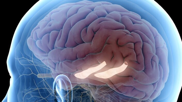
We have compelling data from our monkeys through MRI, looking at gray matter volume and white matter integrity. We can see both age and CR effects. In specific regions of the brain, gray matter volume tracks with insulin sensitivity, and white matter integrity shows interesting connections to circulating cholesterol and adiponectin. Our PET imaging examining glucose uptake also shows connections to insulin sensitivity. In all these measures, there’s a link to peripheral metabolic status. What happens from the neck down completely influences what happens in the brain. I bet that the lipid story is going to be fascinating and extremely important for brain aging and cognition.

We touched briefly on CR mimetics, like adiponectin. Do you believe CR-mimicking drugs are feasible and can change how people age?

All exploration is valuable. The beauty of drug interventions is that you can examine changes in old animals over different time frames, immediate to long term, and then remove the drug to look for lasting effects. While there are plenty of molecules targeting specific pathways, right now, the tools we have align more with the hallmarks of the aging idea than the CR mimetic concept.

Take rapamycin: I’m involved in the trial of rapamycin in the Dog Aging Project. Evidence suggests that low-dose intermittent rapamycin is beneficial for healthy aging, but it’s not a CR mimetic – it’s more about growth suppression.

That said, you can recapitulate some CR aspects through growth suppression, similar to what we see in genetic mutant long-lived dwarf mice. Reduced growth signaling correlates with a more youthful immune system and less inflammation. Other approaches target senescence directly through things like SASP inhibitors, focusing on cytokine release or senescent cell pathways that are important in chronic inflammation.

Metformin is closer to a CR mimetic, but it’s mainly effective in metabolically compromised animals, not healthy ones. The ketogenic diet is another interesting approach, acting as a fasting mimetic by recreating what happens during fasting.

What’s next for you after this extensive study?

NIH/NIA have been terrific in their support of this long-term big data monkey study. We’re still generating and analyzing the data. We’re collaborating with computer scientists, mathematicians, and bioinformaticians to build programs for data integration. There’s no established pipeline for this sort of thing – we’re blazing new trails in data analysis. It’s incredibly exciting.

Next year, we hope to publish findings on aging trajectories and inflection points specific to metabolites and lipids, developing biomarkers, and adding complexity. We have several studies on adipose tissue and brain in progress, with future plans for liver and skeletal muscle.

Thanks to funding from the Simons Foundation, we’ve maintained our brain research using the tissues from our Monkey Aging and CR study. One of the next things I’d love to examine is lipid tracers in the brain; it would be very cool to explore white matter integrity and neural network activity. We’re also engaged in studies of the effect of CR on neurovascular coupling and neural networks with live imaging capabilities, but those studies are only in mice. I am hoping to investigate how the brain takes out the garbage through a specialized lymph system.

One of my current interests is cells as communities. When you grow primary cells in isolation – neurons, astrocytes, hepatocytes – they do perform functions but not exactly as they would in a community. I’m fascinated by how cells communicate and support each other through information and metabolite delivery.

This represents the real frontier in aging biology – expanding from reductive research, which remains necessary, to understanding how everything works together. New technologies allow us to examine things spatially, study organoids in three dimensions, and observe cells growing together. The interactions between cells touching each other are different from cells merely sharing media and different still from isolated cells.

We would like to ask you a small favor. We are a non-profit foundation, and unlike some other organizations, we have no shareholders and no products to sell you. All our news and educational content is free for everyone to read, but it does mean that we rely on the help of people like you. Every contribution, no matter if it’s big or small, supports independent journalism and sustains our future.
Hippocampus back

Rejuvenating the Hippocampus With Metabolites

Researchers have reported in Aging Cell that injecting specific one-carbon metabolites into the hippocampus can rejuvenate its cells.

A small molecule approach

Epigenetic alterations have been found to impact the neuroplasticity of the brain, leading to problems with learning and memory [1]. This research team has previously found that reversing these alterations with Yamanaka factors has positive effects on memory retention, restoring it to a more youthful state [2]. However, these researchers contend that approaches using these factors are still out of reach for clinical use.

Instead, they focus on metabolites derived from one-carbon (1C) pathways, which are involved in the basics of epigenetic methylation. Previous work has found that exposing cells to these metabolites in a 1C-MIM cocktail results in epigenetic rejuvenation, restoring function to brain astrocytes and muscle strength to older mice [3]. However, two components of that cocktail were found to be harmful to neurons.

Effects on epigenetics and gene expression

These researchers decided to omit those two compounds, creating 1C-4-MIM. Because memory loss is prominent in aging, the researchers injected this reduced cocktail directly into the dentate gyrus, the part of the hippocampus most responsible for memory formation, in 12-month-old mice. They found that H4K20me3, an epigenetic marker that increases with age, was reduced in these mice; however, H3K9me3, which decreases with age, was unaffected.

Testing further on 4-month-old and 16-month-old mice, the researchers did not find evidence that a single dose of these metabolites reduces epigenetic age acceleration. However, 248 separate epigenetic loci were found to be significantly affected by the treatment. The researchers found that the two that were most affected are already known to have effects on the brain: Cacna2d2 controls calcium channels related to plasticity, while Zic4 affects a transcriptor associated with both memory and spatial learning [4].

Several other genes related to memory and learning were also affected, and, unsurprisingly, many more genes were significantly affected in older mice than younger mice. These results were replicated in RNA sequencing, which found that genes related to cellular proliferation were also affected; indeed, both proliferation in cells and the formation of new neurons (neurogenesis) in the mice were improved, as was the neuroplasticity marker GluN2B. Some of these effects may have been related to changes in oxidative stress, although the researchers could not confirm this finding.

Works on mice, but does it work on other animals?

Most importantly, these brain changes were found to have concrete effects on the mice’s behavior. Mice that had been injected with 1C-4-MM performed better in both a maze-based memory test and an object recognition test.

While it is unclear if this cocktail is safe or effective for human use, and this cocktail was not tested on truly old (22- to 24–month) mice, these are promising results that warrant further experimentation, possibly with a careful analysis of oxidative stress and with different species.

We would like to ask you a small favor. We are a non-profit foundation, and unlike some other organizations, we have no shareholders and no products to sell you. All our news and educational content is free for everyone to read, but it does mean that we rely on the help of people like you. Every contribution, no matter if it’s big or small, supports independent journalism and sustains our future.

Literature

[1] Creighton, S. D., Stefanelli, G., Reda, A., & Zovkic, I. B. (2020). Epigenetic mechanisms of learning and memory: implications for aging. International Journal of Molecular Sciences, 21(18), 6918.

[2] Rodríguez-Matellán, A., Alcazar, N., Hernández, F., Serrano, M., & Ávila, J. (2020). In vivo reprogramming ameliorates aging features in dentate gyrus cells and improves memory in mice. Stem Cell Reports, 15(5), 1056-1066.

[3] Hernandez-Benitez, R., Wang, C., Shi, L., Ouchi, Y., Zhong, C., Hishida, T., … & Belmonte, J. C. I. (2024). Intervention with metabolites emulating endogenous cell transitions accelerates muscle regeneration in young and aged mice. Cell Reports Medicine, 5(3).

[4] Chiavellini, P., Lehmann, M., Canatelli Mallat, M., Zoller, J. A., Herenu, C. B., Morel, G. R., … & Goya, R. G. (2022). Hippocampal DNA methylation, epigenetic age, and spatial memory performance in young and old rats. The Journals of Gerontology: Series A, 77(12), 2387-2394.

AI in Healthcare

AI in Longevity: The Reality Today

Back in 2006, a website called “The Death Clock” appeared on the internet, with a promise to answer one of life’s greatest questions: “When will I die?” Since then, over 60 million people have used the site, which gives a somewhat grim countdown to the day they’ll meet their demise, or does it?

Far from being just a random number generator, The Death Clock is an early, if somewhat crude, way of utilizing data to predict lifespan. It analyzes information such as date of birth, lifestyle, gender, and location to generate its results in a semi-entertaining, although somber, way.

Of course, since 2006, more options have appeared on the playing field, such as a different Death Clock powered by AI. Its modern versions draw from larger levels of data and work with more sophisticated models, and this is where AI comes into play.

Market value

Just like the fascination with death dates drove internet traffic to that particular site in the 90s, so too does the interest in a healthier, longer lifespan drive longevity investment in 2024. AI is a tool to accomplish this.

According to data by Markets and Markets, the AI in healthcare market in 2024 is estimated to be $20.9 billion, with a rise to $148.4 billion within the next 5 years. This averages to a compound annual growth rate (CAGR) of 48.1%, showing a high level of confidence in the industry.

Its drivers are the growth in data volume and complexity, pressure to reduce healthcare costs, and the need for improvised healthcare services. Meanwhile, the usage of such technologies is mistrusted by medical professionals who believe them to be more hype than substance.

Statistics covering the AI and longevity market aren’t as easy to come by, primarily due to the relative newness of the field. The latest available studies by Allied Market Research show a suggested market value of $25.1 billion in 2020, with an estimated CAGR of 6.1% by 2030, giving a predicted value of $44.2 billion.

Where is all that AI money going?

With billions at stake, funding has poured into various areas across healthcare, longevity, and the research behind it all.

In healthcare, AI is used to:

  • Manage and analyze patient data — language-based models can be used to transcribe consultations, while predictive risk models can help manage current and future healthcare needs.
  • Analyze medical imaging and diagnostics — analytics models of AI are used in radiology and pathology to improve  the cost and speed of diagnosis. Accuracy within this field remains a hotly debated topic.
  • Drug discovery and precision medicine — AI can accelerate the development process and help tailor treatments to an individual using genomic therapies.
  • Mental health and virtual assistance — just as chatbots have provided an instant support system for many companies, so too could they be integrated into healthcare outcomes. Although currently no commercial solution is a match for a human, these AI-based models could offer a lifeline at a time when connection is so valuable.

Within longevity, growth is expected across several general therapeutic areas:

  • Senolytic drugs — These remove or alter senescent cells, which contribute to various age-related diseases. They may impact major age-related diseases that involve neurology, metabolism, and the cardiovascular system.
  • Gene therapy — attempting to repair or modify genes or genetic components in order to slow down this aspect of aging. These therapies seek to impact the underlying genetic causes within aging as a whole.
  • Immunotherapy — COVID-19 has done significant harm to the world’s health, the full extent of which is still yet to be determined. However, it has also boosted interest in immunotherapeutic solutions that can help improve immune function and fight age-related conditions.
  • Biomarker discovery — research into biomarkers seeks to establish insights into biological and chronological age as well as to identify metrics for early detection of age-related diseases.
  • Clinical trial optimization — real-time monitoring, big data analysis, and streamlining of trials are some of the potential benefits of integration AI solutions into scientific processes.
  • Personalized longevity plans — similar to the approaches of precision medicine, personalized longevity seeks to address genetic and lifestyle factors in a longevity-focused approach to healthcare. These can range from personalized regimes to app-focused treatments to others.

These approaches do not exist in silos. Instead, they complement one another, with disciplines often crossing, and are by no means exhaustive.

Companies working on AI longevity solutions

Numerous companies around the world are working to integrate AI into their longevity solutions. These are some of the industry’s top players.

Insilico Medicine

Founded in 2014, this Hong Kong- and New York-based company uses AI technology to develop and accelerate therapeutics for age-related diseases. In 2023, it filed for an IPO and was valued at approximately $895 million according to Forbes. An early adopter of AI for medicine technology, the company kicked off discussions about the use of generative AI for drug discovery back in 2016. In 2024, it released the latest Phase IIa results of a proof-of-concept AI-designed drug treatment for pulmonary fibrosis.

BioAge Labs

US-based BioAge Labs is a biotech company that targets metabolic aging. It leverages machine learning, AI, and longevity science to identify and target biomarkers. Topping Crunchbase’s Longevity Start-Up list, this biotech underwent Series D funding recently and filed for an IPO in September 2024, which was estimated to raise $198 million to support the company’s current and future developments.

Altos Labs

Perhaps one of the most well-known names in longevity due to its famous founder, no other than Amazon’s former CEO, Jeff Bezos. However, fame and fortune can only fund progress, not buy achievements, and this company has made some notable ones. In August of 2024, it launched an AI and computational biology institute, which is set to address some of the industry disparity with AI skills, and in addition, it is continuing its mission of finding the so-called “fountain of youth” through longevity research and targeting fundamental aging processes at the cellular level.

California Life Company (Calico)

Bezos isn’t the only Silicon Valley face to back AI and longevity solutions. Calico is a subsidiary of Alphabet, the parent company of Google. Having received a recent investment of $2 million, the company has continued its research approach into the biology of aging using AI technology. In 2024, it was estimated to have produced revenue of $42.3 million.

Juvenescence

UK-based biotech Juvenescence is focused on using AI to develop therapies that target senescent cells. In doing so, it seeks to develop drugs that reduce cellular damage and enhance the human healthspan. According to statistics by Crunchbase, it is said to have received a total funding amount of $219.2 million.

Unity Biotechnology

Seeking to develop a new class of therapeutics to slow diseases of aging, Unity Biotechnology, also known as UNITY, reports a total of $294.9 million according to Crunchbase. Like BioAge, it’s also listed on the NASDAQ and trades under the ticker UBX. Its latest report notes that the company plans to continue its ASPIRE study to treat diabetic macular edema (DME), using UBX1325 (foselutoclax) a small molecule senolytic drug inhibitor which acts on the proto-oncogene protein c-bcl-2 inhibitors.

Perceptions of trust

According to a 2021 report from the Massachusetts Institute of Technology (MIT), trust in AI technology is approximately the same across most generations, with the majority saying they think AI is somewhat risky. However, when asked about the benefits, this opinion diverges. Generation Xers and Millennials were quicker to suggest its potential benefits than both Baby Boomers and Generation Zers.

When specifically looking at AI use in health care and caregiving, the majority ranked AI as “a little” and “somewhat” risky, while results for its benefits spread primarily across being “extremely” useful, “quite” and “somewhat”. This indicates a level of uncertainty in AI usage among the general public but with a piqued interest in its potential.

Diving further into the details of use cases within healthcare, people appeared skeptical regarding its usage when predicting life expectancy, while supporting its usage in more accurate medical record keeping.

AI in Longevity

With public opinion very much on the fence, it appears as AI is integrated into healthcare, there will be substantial challenges in building trust in platforms. This aligns with expert opinion on the matter, with ethical and trust concerns being raised, highlighting that there are genuine questions, such as accuracy, bias, transparency, privacy, and fairness, among others that need to be answered before AI can prove itself as a trustworthy tool within the scientific community.

Ethical concerns

As one famous movie said, “with great power comes great responsibility,” and that could not be more accurate when it comes to AI. Hidden among the excitement at a new technological approach is a mixture of fear and concern, not only for the practicalities and accuracy of the technology but also how ethical it is used. Some of these include issues surrounding:

Hallucinations and trust — Large language models (LLMs) are known to generate outcomes that are not always based on reality. These are commonly referred to as hallucinations. Knowing this possibility, the inclusion of predictive AI tools in particular should be checked thoroughly before implementation.

Data protection — laws such as GDPR, HIPAA, and patient-doctor confidentiality define trust in the healthcare world and beyond. AI, at its source, relies on data, so how can that data be protected? This is one of the major questions puzzling law makers, scientists, and medical staff alike.

Bridging a staffing gap — lack of medical staffing is a concern across the world, with World Health Organization (WHO) estimating a shortfall of 10 million healthcare workers worldwide. To put this in perspective, this is the entire population of Sweden, or more than the population of New York. AI’s integration in healthcare is intended to bridge the gap and relieve some of the workforce pressure. However, there are concerns about how this would be implemented and whether it would be accepted by the population.

Inequality — like the majority of healthcare solutions, it is suggested that lower- to middle-income countries could be faced with inequality when it comes to AI implementation. These concerns were expressed at the World Economic Forum Annual Meeting in 2024. Unreliable tools and inappropriate applications could drive even greater inequalities with some of the world’s strongest economies.

Accountability — in general research or medical practice, when it comes to accountability, there is a defined chain of responsibility both professional and ethical. When the stakes are high, it becomes more difficult to accept “the computer did it” as a reason, and if this is the case, who holds the responsibility?

Knowledge — as a modern technology, AI is still in a somewhat fledgling state, estimated to be somewhere between a clumsy toddler and awkward teen, depending on the expert. However, as it grows in usage, it raises the question of who is equipped to use it accurately and effectively. In 2022, Deloitte estimated the world’s total AI work-force to be at around 22,000. Although the situation can be suggested to have improved since then, both company leaders and experts agree that the current work-force is in need of serious up-skilling, and that’s before specializations in health or longevity.

Potential for results

As of 2024, it’s clear that AI is set to infiltrate our lives in a significant way, and the mass use of tools such as ChatGPT and other predictive models is evidence of this. However, when the stakes are high, as they are in longevity, a more cautious approach is needed.

To date, there are many promising AI-based approaches to healthcare and excellent examples of how it can be integrated into research, learning, and treatment. However, the lack of specialists, and the challenges in trying out new approaches and providing their scientific basis, mean that it is likely that commercially available trustworthy longevity outcomes could be a discussion for the near, but not immediate, future. What is more likely is the increased use of AI within data analysis with the careful scrutiny of human scientists.

We would like to ask you a small favor. We are a non-profit foundation, and unlike some other organizations, we have no shareholders and no products to sell you. All our news and educational content is free for everyone to read, but it does mean that we rely on the help of people like you. Every contribution, no matter if it’s big or small, supports independent journalism and sustains our future.
The Longevity Summit 2024

The Longevity Summit Announces Fourth Annual Event

The Longevity Summit, a leading conference bringing together the complete ecosystem of longevity science and biotechnology, will hold its fourth annual event on December 3-4, 2024, at the Buck Institute for Research on Aging in Novato, California.

This immersive two-day conference creates a unique peer-to-peer learning environment focused on transforming longevity research into practical medical interventions. The summit brings together key stakeholders from across the field: entrepreneurs, pharmaceutical and biotech executives, investors, researchers, and government organizations.

“Developing aging interventions represents one of the most significant opportunities in modern medicine,” said Longevity Global Director, Dr. Christin Glorioso. “Progress requires a coordinated industry effort, which is why we’ve designed this event to foster meaningful connections and practical solutions.”

Event highlights

  • Over 25 distinguished speakers from leading institutions and companies
  • Comprehensive exploration of drug development, clinical trials, and regulatory pathways
  • Dedicated networking time, including shared meals and evening receptions
  • Startup pitch competition showcasing emerging technologies
  • Scientific poster presentations highlighting new research

Featured speakers

  • Eric Verdin, MD – President & Professor, Buck Institute
  • Jamie Justice, PhD – Executive Director, XPRIZE Healthspan
  • Vittorio Sebastiano, PhD – Founder, Turn Bio & Associate Professor, Stanford
  • Ronjon Nag, PhD – President, R42 Group & Adj. Professor, Stanford
  • Madda Adorno, PhD – CEO and Co-founder, Dorian Therapeutics

Critical industry themes

  • Drug Discovery and Development
  • Regulatory Approval Pathways
  • Aging Clocks and Biomarkers
  • Clinical Trial Design
  • Funding Strategies
  • Regenerative Medicine
  • Genetic and Epigenetic Therapies

The event offers both in-person and virtual attendance options. For researchers interested in presenting their work, abstract submissions for the poster session are being accepted until November 25, 2024.

About The Longevity Summit

The Longevity Summit is organized by Longevity Global, with chapters across the globe: Longevity SF, NYC, BOS, SD, UAE, CH, and MIA. The summit focuses on creating practical pathways to advance the science and business of longevity medicine.

For registration information or sponsorship opportunities, visit longevitygl.org/longevity-summit/ or contact hello@longevitygl.org.

We would like to ask you a small favor. We are a non-profit foundation, and unlike some other organizations, we have no shareholders and no products to sell you. All our news and educational content is free for everyone to read, but it does mean that we rely on the help of people like you. Every contribution, no matter if it’s big or small, supports independent journalism and sustains our future.
Vesicles for joints

Extracellular Vesicles for Treating Arthritis

A study in Pharmacological Research has found that small extracellular vesicles derived from embryonic stem cells (ESC-sEVs) alleviate osteoarthritis in cells and mice.

Restoring senescent cells’ function

Excessive senescent cell buildup in cartilage has negative effects and leads to osteoarthritis [1]. Constant exposure to SASP factors has been found to cause more cartilage to be removed than created by chondrocytes, the cells responsible for the maintenance of this extracellular matrix tissue [2].

However, simply killing off these senescent cells with senolytics might not be beneficial in people. We recently published a study finding that in the joints of rats, some senescent cells are necessary for encouraging nearby chrondrocytes to regrow tissue, and a previous senolytic approach to osteoarthritis did not make it through clinical trials.

This study uses a senotherapeutic approach, which seeks to rejuvenate senescent cells, rather than a senolytic one. Previous work has found that using extracellular vesicles derived from mesenchymal stem cells (MSC-EVs) might effectively treat osteoarthritis in this way [3]; however, that study was focused on a different kind of stem cell, and it was only used before senescence or in models of early osteoarthritis, which might be too late in a clinical setting. As ESC-sEVs have been found to be effective in repairing other tissues, including the heart [4], these researchers decided to apply them to models representing osteoarthritis in its later stages.

Testing with human cells

This study began by exposing human chondrocytes in vitro to the SASP factor IL-1β in order to drive them senescent, then administering ESC-sEVs to a treatment group of cells for a week. The cells successfully began uptaking the ESC-sEVs after 12 hours of exposure. The treatment worked: chrondrocytes exposed to ESC-sEVs had far lower levels of senescence according to the well-known SA-β-gal biomarker, almost to the levels of chrondrocytes not exposed to IL-1β at all. Similarly, markers of extracellular matrix production were restored by the treatment.

As previous work has found that the forkhead box O (FOXO) pathway is key to the cellular maintenance process known as autophagy, which has a significant effect on cartilage maintenance [5], these researchers tested the effects of ESC-EVs on FOXO-related gene expression. Here, too, the results were positive, although not for all genes: FoxO1A was upregulated, while FoxO3A was not. Other biomarkers confirmed that autophagy was being upregulated, while other testing proved that this pathway was the core reason why senescence was being reduced in these cells.

Effective in mice

In mice, a surgical procedure can be used to artificially induce long-lasting osteoarthritis. Eight weeks after this surgery was conducted, a treatment group had ESC-sEVs injected directly into the joint fluid for 12 weeks. While this treatment did not restore joint cartilage nor function to the levels of mice that only had a sham surgery, its effects were statistically significant, reducing widespread cellular senescence and regrowing substantial amounts of cartilage.

This treatment was also effective in naturally aged mice. While those mice did not have the complete destruction of cartilage that was brought about by surgery, they had substantial cartilage loss and other signs of osteoarthritis. Just like with the surgery-induced osteoarthritis, ESC-sEV treatment (from month 15 to month 24 of life) was able to reduce widespread senescence whiile restoring cartilage and function, although not to the levels of young (5-month-old) mice.

These findings, while they do not represent perfect restoration and need to be administered over long periods of time, are promising. While administering embryonic stem cells directly is unsafe, as it might lead to uncontrolled teratoma growth, the vesicles emitted by these cells apparently encourage cells to maintain their function. The available evidence suggests that cellular senescence is a balance that is harmed by aging, and if these results can be replicated in trials, this sort of approach can be confirmed to restore some of that balance.

We would like to ask you a small favor. We are a non-profit foundation, and unlike some other organizations, we have no shareholders and no products to sell you. All our news and educational content is free for everyone to read, but it does mean that we rely on the help of people like you. Every contribution, no matter if it’s big or small, supports independent journalism and sustains our future.

Literature

[1] Xie, J., Wang, Y., Lu, L., Liu, L., Yu, X., & Pei, F. (2021). Cellular senescence in knee osteoarthritis: molecular mechanisms and therapeutic implications. Ageing research reviews, 70, 101413.

[2] Coryell, P. R., Diekman, B. O., & Loeser, R. F. (2021). Mechanisms and therapeutic implications of cellular senescence in osteoarthritis. Nature Reviews Rheumatology, 17(1), 47-57.

[3] Yin, B., Ni, J., Witherel, C. E., Yang, M., Burdick, J. A., Wen, C., & Wong, S. H. D. (2022). Harnessing tissue-derived extracellular vesicles for osteoarthritis theranostics. Theranostics, 12(1), 207.

[4] Khan, M., Nickoloff, E., Abramova, T., Johnson, J., Verma, S. K., Krishnamurthy, P., … & Kishore, R. (2015). Embryonic stem cell–derived exosomes promote endogenous repair mechanisms and enhance cardiac function following myocardial infarction. Circulation research, 117(1), 52-64.

[5] Akasaki, Y., Hasegawa, A., Saito, M., Asahara, H., Iwamoto, Y., & Lotz, M. K. (2014). Dysregulated FOXO transcription factors in articular cartilage in aging and osteoarthritis. Osteoarthritis and cartilage, 22(1), 162-170.

Stem Cell

Mesenchymal Stem Cells Rejuvenate Aged Mice

In a new study, the researchers administered human umbilical cord-derived mesenchymal stem cells (HUCMSCs) to aged mice and observed reduced degeneration in multiple organs, changes to microbial composition, metabolic alterations, improvements in behavior and ability, and reduced fearfulness [1].

Therapeutic potential

Earlier this year, we reported on a clinical trial in which administering HUCMSCs reduced frailty in older people. MSCs in general are known for their capacity for self-renewal and differentiation into multiple cell types. A growing body of research suggests that MSCs can be used as a tool for therapeutic purposes [2].

In this new study, the researchers used four-month-old mice of two different strains, senescence-accelerated mouse prone 8 (SAMP8), a mouse strain used as a model of age-related cognitive decline and, as a control, senescence-accelerated mouse resistant 1 (SAMR1), a strain that ages normally.

A group of 15 SAMP8 mice received HUCMSCs weekly for eight weeks. Additionally, researchers had SAMP8 and SAMR1 groups that didn’t receive HUCMSCs as a control. One week following the end of the experiment, the researchers took fecal and blood samples from now 6-month-old mice.

Improvements on many levels

Following MSC administration, the researchers tested the mice’s cognitive abilities. They observed increased curiosity, better motor coordination and balance, and reduced anxiety in mice treated with HUCMSCs compared to controls. However, there were no differences in a test that assessed spatial learning and memory.

On the molecular level, the researchers analyzed the genome for DNA single-strand breaks in the brain tissue. They performed additional analysis to specifically focus on exonic regions (DNA sections translated into proteins) and within 200 bp of transcription start sites (where many elements that regulate translation – the first step in protein production – are located).

Unsurprisingly, they noted more single-strand breaks in the faster-aging SAMP8 control mice compared to the SAMR1 controls. MSCs-treated mice fared better than their counterparts in the SAMP8 untreated controls regarding single-strand DNA breaks, but there were no significant differences between SAMP8 MSCs-treated and untreated SAMR1.

Together, this suggests that MSCs reduce age-related DNA damage, preserving genomic stability and neuroprotection, which the researchers hypothesized to play a role in improving cognitive function.

On the tissue level, MSC treatment led to significant improvements. For example, in the frontal lobe and hippocampal brain regions, which are responsible for learning, memory, and attention, the researchers observed “maintained structural integrity and normal glial cell distribution” in the mice treated with MSCs.

The cardiac tissue of the MSC-treated SAMP8 mice resembled that of the healthy cardiac tissue of the SAMR1 controls. The structures of the gastrointestinal, kidney, skeletal muscle, spleen, liver, and lung cells were also well-preserved, while controls exhibited more aging-related changes. The researchers also observed reduced inflammation in the lungs and spleens of MSC-treated mice compared to the control animals.

The authors emphasize that in some tissues (kidney, muscle, and spleen), the tissues of mice treated with MSCs surpassed that of controls, suggesting tissue rejuvenation properties of MSC treatment and its potential for regenerative medicine.

The impact on microbiota and metabolism

MSC treatment also resulted in changes to the mice’s microflora and metabolic profiles. While all three groups of mice share some microbial species in their guts, there were also significant differences.

For example, the researchers noted that following the MSC treatment, some bacterial species that are considered beneficial in humans were restored. On the metabolomic level, they noted that MSC treatment “increased levels of metabolites beneficial for cardiovascular health.”

The authors also linked the increased levels of one of the metabolites, namely 5-hydroxy-L-tryptophan, in MSC-treated mice to the reduced depression-like behavior that they observed in previous experiments. They suggest that an increase in this compound leads to the normalization of serotonin synthesis, a process linked to antidepressant properties.

More broadly, the researchers observed differences between experimental groups regarding a few metabolite pathways, including pathways related to fatty acid and amino acid metabolism, suggesting that MSC therapy has an impact on metabolism.

However, this impact was quite complex, and the authors pointed to both benefits and limitations of MSC treatment on the metabolome. They noticed that several metabolites exhibited changes in accordance with what would be expected, based on the previous research, which highlighted the benefits of MSC treatment on metabolic changes. However, researchers also noted some observations conflicted with those expectations suggesting MSC treatment has some limitations regarding its impact on metabolic changes. Those results need further investigation.

Understanding the mechanism

While investigating the molecular mechanism of MSC treatment still needs more investigation, the authors suggest that, based on these results and previous research, some possible molecular pathways that are in play. They also suggest that MSCs’ anti-inflammatory properties promote DNA damage-repair cycles, leading to reduced DNA damage in the brain. They point to the critical role of metabolism, which should be further investigated.

In these experiments, the researchers use intraperitoneal injection (IP), and not intravenous infusion, as preclinical studies have shown IP of MSCs as more effective in treating colitis [3]. Previous research suggests that IP-administered MSCs impact intestinal immune and inflammatory responses [4]. These researchers hypothesize that MSCs alter gut microbiota through modulation of intestinal immune function and microenvironment. A changed microbiome subsequently impacts metabolism and immune functions, resulting in a positive feedback loop. However, there is still a need to fully understand the mechanism by which MSCs influence gut microbiota.

This study has several limitations, one of which is sampling for microbiome analysis. Since samples were taken at a single time point, the researchers might have missed dynamic and long-term changes in the microbiome, factors important for potential use in therapies. Second, SAMP8/SAMR1 was used as a model for aging and dementia; however, those animals have some pathological features that are different from human dementia, which might impact how the results will translate to human research. Also, having more animals would have increased the statistical strength of these results.

We would like to ask you a small favor. We are a non-profit foundation, and unlike some other organizations, we have no shareholders and no products to sell you. All our news and educational content is free for everyone to read, but it does mean that we rely on the help of people like you. Every contribution, no matter if it’s big or small, supports independent journalism and sustains our future.

Literature

[1] Lian, J., Xia, L., Wang, G., Wu, W., Yi, P., Li, M., Su, X., Chen, Y., Li, X., Dou, F., & Wang, Z. (2024). Multi-omics evaluation of clinical-grade human umbilical cord-derived mesenchymal stem cells in synergistic improvement of aging related disorders in a senescence-accelerated mouse model. Stem cell research & therapy, 15(1), 383.

[2] Musiał-Wysocka, A., Kot, M., & Majka, M. (2019). The Pros and Cons of Mesenchymal Stem Cell-Based Therapies. Cell transplantation, 28(7), 801–812.

[3] Wang, M., Liang, C., Hu, H., Zhou, L., Xu, B., Wang, X., Han, Y., Nie, Y., Jia, S., Liang, J., & Wu, K. (2016). Intraperitoneal injection (IP), Intravenous injection (IV) or anal injection (AI)? Best way for mesenchymal stem cells transplantation for colitis. Scientific reports, 6, 30696.

[4] Sala, E., Genua, M., Petti, L., Anselmo, A., Arena, V., Cibella, J., Zanotti, L., D’Alessio, S., Scaldaferri, F., Luca, G., Arato, I., Calafiore, R., Sgambato, A., Rutella, S., Locati, M., Danese, S., & Vetrano, S. (2015). Mesenchymal Stem Cells Reduce Colitis in Mice via Release of TSG6, Independently of Their Localization to the Intestine. Gastroenterology, 149(1), 163–176.e20.

Ketogenic Diet

Ketogenic Diet Effective Against Multiple Sclerosis in Mice

Scientists have found that both a ketogenic diet and oral supplementation with ketone bodies alleviate symptoms of multiple sclerosis, a serious autoimmune disorder, in a mouse model [1].

Keto and autoimmune diseases

In ketogenic diets, the vast majority of calories are derived from fat, some from protein, and almost none from carbohydrates. While a ketogenic diet might speed up weight loss, it has also been linked to increased cholesterol levels [2]. However, at least for some people, keto’s advantages might outweigh its drawbacks.

Ketogenetic diets are associated with reduced inflammation [3], and some studies have suggested possible benefits for people with autoimmune diseases [4]. However, the exact mechanisms are not fully understood yet. In this new study published in Cell Reports, scientists from the University of California San Francisco took a deep dive into the workings of these diets in a murine model of an autoimmune disease.

Less severity and incidence

For their experiments, the researchers chose so-called experimental autoimmune encephalomyelitis (EAE) mice. These are often used as a plausible if imperfect model of human multiple sclerosis (MS) since they recapitulate many aspects of this debilitating autoimmune disease.

First, the researchers wanted to know whether keto can protect normally raised mice from EAE. Ten days after a ketogenic diet was begun, the mice were inoculated with a compound that triggers EAE. Compared to mice on a high-fat diet (75% of calories from fat, 15% from carbohydrate, and 10% from protein), mice eating a ketogenic diet (90.5% from fat, 0% from carbohydrate, and 9.5% from protein) were more resistant to the disease, with both lower incidence and much less severe symptoms. The keto-eating mice also had a more favorable immune profile.

However, when the researchers performed the same experiment in mice with depleted microbiota, the two groups barely showed any difference, demonstrating that the protective effect of a keto diet is mediated by gut bacteria.

The molecule that can replace ketogenesis

A ketogenic diet works by remodeling the body’s energy metabolism. When glucose is abundant, it is preferentially used as fuel. However, in a ketogenic environment, glucose is scarce, so the body switches to producing energy from fat using a process known as beta-oxidation. Fatty acids must be broken down into small molecules called ketone bodies, such as beta-hydroxybutyrate (βHB), which are transported across the body to be used as an alternative source of fuel.

In recent years, direct ketone supplementation via compounds such as ketone salts or esters has been tested as a way to recapitulate the benefits of ketogenic diets without the downsides. The researchers introduced a new group of mice fed the unhealthy high-fat diet supplemented with a ketone ester. This group was virtually indistinguishable from the original ketogenic diet group in terms of EAE incidence and severity.

“What was really exciting was finding that we could protect these mice from inflammatory disease just by putting them on a diet that we supplemented with these compounds,” said Peter Turnbaugh of the Benioff Center for Microbiome Medicine, a leading author on the study.

The gut connection

Interestingly, while most of the βHB production under KD happens in the liver, the beneficial effects of both keto diets and keton ester supplementation on MS seemed to be mediated by βHB production in intestinal epithelial cells. Mice genetically modified so that they could not produce βHB in the gut were not protected against MS regardless of their diet.

The researchers already knew that the gut microbiome was a crucial element, so they started hunting for bacterial species. After extensive screening, the researchers focused on Lactobacillus murinus and a metabolite it produces called indole lactic acid (ILA). ILA is known to alleviate autoimmune responses by inhibiting the production of the pro-inflammatory cytokine interleukin 17 (IL-17) by Th17, a subset of T cell strongly associated with autoimmune diseases.

Both treating MS mice with ILA and populating their guts with L. murinus alleviated the symptoms. The researchers concluded that ΒHB production specifically in the gut or oral supplementation with a ketone ester benefits ILA-producing bacteria, including L. murinus. This increases the production of ILA, which, in turn, leads to the observed anti-MS effect.

  Keto MS Mice

“The big question now is how much of this will translate into actual patients,” Turnbaugh said. “But I think these results provide hope for the development of a more tolerable alternative to helping those people than asking them stick to a challenging restrictive diet.”

Surprisingly, we discovered that oral delivery of a βHB-KE can mimic the protective effects of a KD. If this finding holds in humans, βHB supplementation alone could offer a viable therapeutic alternative to the full KD. The translational implications are profound, as KDs are difficult to maintain and can have negative side effects. Our identification of βHB as a key player provides a way to circumvent these barriers and provides a more general proof of concept for the ability to distill the activity of a complex diet down to a single bioactive molecule.

We would like to ask you a small favor. We are a non-profit foundation, and unlike some other organizations, we have no shareholders and no products to sell you. All our news and educational content is free for everyone to read, but it does mean that we rely on the help of people like you. Every contribution, no matter if it’s big or small, supports independent journalism and sustains our future.

Literature

[1] Alexander, M., Upadhyay, V., Rock, R., Ramirez, L., Trepka, K., Puchalska, P., … Turnbaugh, P. J. (n.d.). A diet-dependent host metabolite shapes the gut microbiota to protect from autoimmunity. Cell Reports.

[2] Burén, J., Ericsson, M., Damasceno, N. R. T., & Sjödin, A. (2021). A ketogenic low-carbohydrate high-fat diet increases LDL cholesterol in healthy, young, normal-weight women: a randomized controlled feeding trial. Nutrients, 13(3), 814.

[3] Pinto, A., Bonucci, A., Maggi, E., Corsi, M., & Businaro, R. (2018). Anti-oxidant and anti-inflammatory activity of ketogenic diet: new perspectives for neuroprotection in Alzheimer’s disease. Antioxidants, 7(5), 63.

[4] Brenton, J. N., Banwell, B., Bergqvist, A. C., Lehner-Gulotta, D., Gampper, L., Leytham, E., … & Goldman, M. D. (2019). Pilot study of a ketogenic diet in relapsing-remitting MS. Neurology: Neuroimmunology & Neuroinflammation, 6(4), e565.

Gene analysis

Towards Fine-Tuned Control of Gene Expression

In a groundbreaking Nature paper, researchers have developed synthetic regulatory sequences that could prevent targeted gene therapies from having effects in unwanted cell types.

More than methylation

While methylation is the most well-known regulator of gene expression, it isn’t the only thing that determines what is to be expressed when. Cis-regulatory elements (CREs), so called because they sit near the DNA sequences they regulate, are responsible for expressing the genes that are specific to each cell type [1]. While they are technically non-coding, as they do not directly code for functional proteins, CREs are critical to epigenomic function.

Manipulating existing CREs in engineered cells is one thing, but it’s not clear if the CREs generated by evolution will always be the ideal candidates for specific therapeutic applications in specific cell types. A decade ago, researchers began seriously asking if it might be possible to generate CREs to more precisely do what we want [2]. Having functional control of CREs would allow therapies to apply only to specific cell types, potentially offering massive improvements to gene therapies that aren’t yet good enough for clinical use [3].

However, the number of potential sequences that could be inserted into a mere 200 base pairs of DNA is far larger than the number of atoms in the universe. Basic computational algorithms, therefore, will not suffice to find CREs that work. A substantial amount of previous work has gone into this topic between then and now, attempting to discover why CREs work the way they do and looking to develop a regulatory ‘grammar’ and a more complete understanding [4]. Very recently, researchers have developed CREs for use in Drosophila flies [5].

However, fruit flies aren’t mice, let alone people, and it was unclear if this process could create sequences for use in cells that can be transplanted into larger animals. These researchers appear to have done it.

A new algorithm with real-world effects

Previous work was focused on looking at the epigenetic downstream effects of CREs, but these researchers used MPRA, a system that can accurately gauge the directeffects of any given CRE. To train their model, Malinois, these researchers used sequences derived from three cell lines: bone marrow cells, liver cells, and nerve cancer cells. Even without being directly informed as to their effects, Malinois was able to accurately predict the activity of more than sixty thousand existing, natural CREs. Its predictions of epigenetic behavior were in line with experimental results in all three cell types.

Malinois, however, is just a prediction algorithm. To actually generate new CRE sequences, the researchers developed Computational Optimization of DNA Activity (CODA), which can be used with multiple algorithms. Their intention was to develop sequences that have maximal effects on one of the three cell types and minimal effects on the other two.

At first, the algorithm was attracted to certain motifs, yielding 36,000 of similar-looking sequences. However, after an algorithmic tweak to penalize re-use, CODA created 15,000 more synthetic sequences and compared them to 12,000 natural sequences.identified primarily by location and 12,000 more natural sequences identified by Malinois.

The location-based sequences were found to be less specific and have less effect than the Malinois-identified sequences, but the synthetic sequences were stronger still, having more specificity to each of the desired cell types. Even when CODA’s preferred motifs weren’t used, 92.4% of its generated sequences were still specific to a cell type, compared to 73.6% of the Malinois-identified sequences and only 40.6% of the location-identified sequences. Under far more stringent conditions for specificity, more than half of CODA’s sequences made the cut, while far fewer of the natural sequences did.

These synthetic sequences were found to be higher in useful content than the natural sequences, and rather than being mostly activatory for the desired cell types, these synthetic sequences actively repressed activation in off-target types.

To confirm their cellular findings, the researchers injected living zebrafish and mouse embryos with gene therapies that used these synthetic CREs. The therapies were found to be specific to cell type in these living animals, both before and after birth.

This represents a sea-change for researchers of gene therapies. There are always plenty of cell types for which expressing a gene therapy modification would be highly negative. If therapies that use these synthetic sequences can prevent this from happening, it bodes well for a wide variety of potential therapies, including those that target age-related diseases.

We would like to ask you a small favor. We are a non-profit foundation, and unlike some other organizations, we have no shareholders and no products to sell you. All our news and educational content is free for everyone to read, but it does mean that we rely on the help of people like you. Every contribution, no matter if it’s big or small, supports independent journalism and sustains our future.

Literature

[1] Donohue, L. K., Guo, M. G., Zhao, Y., Jung, N., Bussat, R. T., Kim, D. S., … & Khavari, P. A. (2022). A cis-regulatory lexicon of DNA motif combinations mediating cell-type-specific gene regulation. Cell genomics, 2(11).

[2] Levo, M., & Segal, E. (2014). In pursuit of design principles of regulatory sequences. Nature Reviews Genetics, 15(7), 453-468.

[3] Deverman, B. E., Ravina, B. M., Bankiewicz, K. S., Paul, S. M., & Sah, D. W. (2018). Gene therapy for neurological disorders: progress and prospects. Nature Reviews Drug Discovery, 17(9), 641-659.

[4] Movva, R., Greenside, P., Marinov, G. K., Nair, S., Shrikumar, A., & Kundaje, A. (2019). Deciphering regulatory DNA sequences and noncoding genetic variants using neural network models of massively parallel reporter assays. PLoS One, 14(6), e0218073.

[5] de Almeida, B. P., Schaub, C., Pagani, M., Secchia, S., Furlong, E. E., & Stark, A. (2024). Targeted design of synthetic enhancers for selected tissues in the Drosophila embryo. Nature, 626(7997), 207-211.

Found the needle

New Crowdfunding Project Looks for a “Better Rapamycin”

Ora Biomedical and the Rapamycin Longevity Lab have launched a project to screen more than 600 mTOR inhibitors in the hope of finding some that are superior to geroscience’s poster child, rapamycin.

Better than chance

Rapamycin, one of the most potent compounds for prolonging lifespan in numerous animal models, was discovered serendipitously in one of the most remote locations on earth: Easter Island. The island’s name in its natives’ language is Rapa Nui, which is where “rapa” comes from. Rapamycin is produced by a certain type of soil bacteria to compete with fungi. For decades, rapamycin has been used mostly as an immunosuppressant in transplant and cancer patients before it was found that it can extend lifespan in animals.

The point is a compound discovered by chance might not be the best one for the job. Rapamycin works by inhibiting mTOR, a protein kinase that is a central regulator of nutrient sensing. However, it has its quirks and limitations. What if there are other, better mTOR inhibitors out there?

Enter Ora Biomedical, a young company focused on high-throughput screening of chemical compounds in the tiny nematode worm C. elegans. Ora uses a proprietary robotic system called WormBot. Ora’s Million Molecule Challenge is one of the most interesting crowdfunding initiatives in the longevity field. It allows anyone to buy an actual experiment for as little as 100 dollars and compete with other users while also contributing to science.

Ora’s philosophy is that the net we are currently casting to look for life-extending compounds is not nearly wide enough. Using short-lived worms and robotic systems allows us to extend the search space by orders of magnitude. The results might not necessarily be translatable to more complex animals, including humans, but the sheer volume might be able to compensate for that.

One found, 600 to go

The newest Ora’s project is a collaboration with the Rapamycin Longevity Lab led by Krister Kauppi. It entails screening more than 600 mTOR inhibitors to find the ones that work best.

“Currently, rapamycin is considered the gold standard longevity intervention – it works across many animal and even non-animal fungal models,” said Ora CEO Mitchell Lee. “The mechanism of mTOR inhibition has seen extensive development in cancer therapeutics. There’s a broad toolkit of mTOR inhibitors with different patterns of inhibition between mTOR complex 1, mTOR complex 2, and impacts on other kinases. Nobody has comprehensively looked to see if any are better than rapamycin.”

The current project began as a smaller partnership within the Million Molecule Challenge when, in an experiment funded by Kauppi and the Rapamycin Longevity Lab, Ora discovered an mTOR inhibitor that was superior to rapamycin in worms at the same dose. This molecule, called omepalisib, has already been approved by the FDA for treating certain types of cancer, so its way to the clinic as a geroprotector (anti-aging drug) might be relatively short.

The discovery of robust life extension by omepalisib gives Lee the hope that many even more promising molecules are out there. “There are at least 600 different mTOR inhibitors we could test with the WormBot platform,” he said. “We can screen through these in months, not the decades or years needed for mammalian studies, to identify what works best for extending lifespan in this system.”

Ora was spun out of Matt Kaeberlein’s laboratory at the University of Washington School of Medicine, Seattle. Kaeberlein, one of the world’s most prominent geroscientists, is a big believer in rapamycin and is currently running a rapamycin trial in dogs.

“Rapamycin is the best drug we know of to slow aging, increase lifespan, and improve healthspan,” Kaeberlein said. “There are undoubtedly other mTOR inhibitors out there that can have a similar impact, and I’m certain some of them will do even better than rapamycin. Ora’s technology affords the opportunity to find these hidden gems and develop them into the next generation of gerotherapeutics.”

Let’s go fishing!

According to the Rapamycin Longevity Lab’s white paper on the project, “There is a big gap in the literature around how good Rapamycin is compared to other mTOR inhibitors. While many mTOR inhibitors have been developed, no systematic effort exists to find the most effective mTOR inhibitors for improving healthy lifespan. This is something we need an answer to, and this project is an important first step in that direction. The end goal is improved human longevity.”

“One key reason why I founded the Rapamycin Longevity Lab was to accelerate the research around mTOR inhibition because it’s one of the most promising longevity pathways that we currently have,” Kauppi explained. “In this ambitious screening project, we aim to deliver a large volume of unique data to the longevity community with the potential to uncover new mTOR inhibitors that are even better than rapamycin. No one has done anything close to this before, and thanks to Ora Biomedical’s innovative WormBot technology, projects like this can be done in a highly efficient and cost-effective way.”

The mTOR inhibitors project is fueled by donations, and you can do your part. “Anyone can contribute through our website,” Lee said. “We hope to close the first half of financing by year-end. While it’s non-commercial initially, we’ll examine IP opportunities for the most promising compounds. Our development strategies often involve medicinal chemistry to create derivatives from lifespan-extending compounds – optimizing them for better chemical availability.”

According to Lee, the system of incentives that the longevity field is built around is the one responsible for some research directions being overlooked. “It’s striking that we’ve come this far testing only rapamycin for longevity,” he said. “It shows a misalignment between academic research and commercial/translational interests. If you proposed finding better mTOR inhibitors to the NIA (National Institute on Aging), they’d reject it for not finding new mechanisms. They’d call it a fishing expedition. Since most research relies on federal funding, important questions like identifying the best compound for this gold-standard target stagnate. That’s why Ora focuses on making these ‘fishing expeditions’ feasible – because we need to catch these fish.”

We would like to ask you a small favor. We are a non-profit foundation, and unlike some other organizations, we have no shareholders and no products to sell you. All our news and educational content is free for everyone to read, but it does mean that we rely on the help of people like you. Every contribution, no matter if it’s big or small, supports independent journalism and sustains our future.
Rejuvenation Roundup October 2024

Rejuvenation Roundup October 2024

The trick-or-treaters have all gotten their candy, but age-related diseases haunt us still. Here’s what we’ve learned about them in October.

LEAF News

Team and activities

EditorialFall Is Here, but It’s Full Steam Ahead for lifespan.io: For those of us living in the Northern Hemisphere, the summer days are gone and the cooler days of fall have arrived. While it’s time to ditch the beachwear for chunky sweaters and a hot chocolate by the fire, things have been heating up in the longevity space.

Interviews

Life Biosciences Is Bringing Reprogramming to the Clinic: Life Biosciences is a company co-founded by the celebrity geroscientist David Sinclair and is based on his Harvard team’s research into partial cellular reprogramming.

Advocacy and Analysis

An hourglass showing time is running out.Have We Maxed Out on Life Expectancy Gains?: A new study has suggested that radical life extension is all but impossible in this century, and it has made waves among people interested in living longer.

Research Roundup

Boosting Autophagy in Astrocytes Might Help Cure Alzheimer’s: With Alzheimer’s disease, most of the focus has been on neurons. A new study’s researchers suggest boosting the process of cellular junk removal in astrocytes, brain cells that perform maintenance tasks, as a new pathway.

Combination therapymTOR and SGLT-2 Inhibitors Impact Age-Related Processes: The authors of a recent review propose that there may be positive synergistic effects from combining mTOR inhibitors and sodium-glucose co-transporter-2 (SGLT-2) inhibitors.

Exercise Intensity, Duration, and Amount All Matter: In the European Journal of Protective Cardiology, researchers have published evidence that the intensity of exercise is somewhat more important than volume in reducing all-cause mortality risk, although both have significant correlations.

UK USUK Citizens Are Healthier Than Americans, but Don’t Feel So: A new study shows that in midlife, United States citizens are less healthy than their British counterparts. The latter, however, smoke more and rate their health worse.

Senescent Macrophages: A Unique Target: In Aging, researchers have published a new study on a tool meant for analyzing macrophage senescence along with differences between inflammaging and regular inflammation.

Motor neuron degradationCryptic Exons for Targeting Neurodegenerative Disease: Amyotrophic lateral sclerosis (ALS) only affects a fraction of cells and must be treated with high specificity. Scientists have achieved that by targeting mRNA sites associated with the disease.

Astragalus Supplement Lengthens Telomeres in the Middle-Aged: Treating middle-aged people for six months with a supplement combination that included astragalus, a plant used in traditional Chinese medicine, positively impacted their telomeres.

A researcher holding a lab rat.Exosomes Break Rat Lifespan Record: In Aging Cell, researchers have published their findings that exosomes, which we have previously reported to extend the lives of mice, also extend the lives of rats.

The Chemical Reasons Why Visceral Fat Is Dangerous: In Aging Cell, researchers have described the chemical ways in which excessive visceral fat, which is metabolically active, causes oxidative stress and cellular senescence.

AstronautHeart Organoids Flown to Space Show Signs of Dysfunction: Scientists have found that human heart tissue is harmed by even a short stint in orbit. This might have implications for future space travel.

Blueberry Extract Aids Cognition in People With Inflammation: A reanalysis of data from a previous study identified a difference between people with low and high levels of inflammatory biomarkers. The cognitive performance of people with high levels of inflammation improved after they consumed anthocyanin supplements.

HippocampusA Potential Target for Post-Surgery Cognitive Impairment: In Aging Cell, researchers have identified a receptor in the brain that appears to be responsible for cognitive problems after surgery, particularly in older people.

Scientists Create a Potent Bacterial Anti-Cancer Vaccine: A new study describes a novel anti-cancer vaccine based on antigen-producing bacteria that can tackle solid and metastatic cancers by colonizing tumors.

Skin cancerThe Reason Why the p16 Senescence Pathway Exists: Researchers publishing in Aging Cell have investigated the biology of skin cells taken from people who don’t produce the senescence-related compound p16. This condition leads to familial melanoma syndrome.

Autodigestion From Gut Enzymes May Drive Aging: A new study links damage caused by digestive enzymes escaping from the gut to several hallmarks of aging. The researchers dubbed this effect “autodigestion.”

Skin collagenHAPLN1 in Blood Found to Rejuvenate Skin: Investigating the factors involved in skin rejuvenation processes, researchers have identified the role of hyaluronan and proteoglycan link protein 1 (HAPLN1) in restoring collagen and hyaluronic acid in aged skin.

A Senolytic Treatment for Liver Sepsis: A paper published in Aging Cell offers evidence for the idea that senolytics, which remove senescent cells, might be a treatment for acute liver sepsis and not just age-related diseases.

Neuronal connectionsNeuronal Reprogramming Alleviates Alzheimer’s in Mice: Scientists have shown that long-term intermittent reprogramming limited to hippocampal neurons increases their fitness and improves cognitive function in a mouse model of Alzheimer’s disease.

The Fibroblasts That Protect Ovarian Cancer: In Aging, researchers have described a subpopulation of fibroblasts that nurture ovarian cancer tumors and shield them from harm.

Handling lab mouseNOVOS Supplement: Lifespan Extension in Male Mice: A recent preprint study suggests that a NOVOS supplement can improve healthspan and extend lifespan in male mice.

Senescent Cells Promote Cartilage Regeneration in Rats: In a rat experiment, researchers publishing in Aging Cell have found that senescent cells and SASP factors are key in regenerating knee cartilage.

Tree-based analysis of longevity predictors and their ten-year changes: a 35-Year mortality follow-up: The simultaneous examination of a broad range of potential predictors revealed that longevity can be achieved under very different conditions and is achieved by heterogeneous groups of people.

Dietary restriction impacts health and lifespan of genetically diverse mice: These findings indicate that improving health and extending lifespan are not synonymous and raise questions about which end points are the most relevant for evaluating aging interventions.

Dietary diversity contributes to delay biological aging: Dietary diversity is associated with a slower rate of biological aging, which may be due in part to reduced oxidative stress.

Association between prescription drugs and all-cause mortality risk in the UK population: As expected, most drugs were linked to a shorter lifespan, likely due to the life-limiting nature of the diseases they are prescribed to treat.

Hippocampal rejuvenation by a single intracerebral injection of one-carbon metabolites in C57BL6 old wild-type mice: The researchers propose the use of these metabolites to explore new strategies for the development of potential treatments for age-related brain diseases.

ESC-sEVs alleviate non-early-stage osteoarthritis progression by rejuvenating senescent chondrocytes: Collectively, these findings reveal that ESC-sEVs-based therapy is of high translational value in non-early-stage OA treatment.

Association of Muscle Strength With All-Cause Mortality in the Oldest Old: Prospective Cohort Study From 28 Countries: Rather than a specific threshold, muscle strength is gradually and inversely associated with mortality risk in the oldest old.

Ageing-associated long non-coding RNA extends lifespan and reduces translation in non-dividing cells: Although aal1 is not conserved, its effect in flies suggests that animals feature related mechanisms that modulate ageing, based on the conserved translational machinery.

Mifepristone and rapamycin have non-additive benefits for life span in mated female Drosophila: he data suggest that mifepristone and rapamycin act through a common pathway to increase mated female Drosophila lifespan.

Machine-guided design of cell-type-targeting cis-regulatory elements: Synthetic sequences exhibit distinct motif vocabulary associated with activity in the on-target cell type and a simultaneous reduction in the activity of off-target cells.

The senolytic drug ABT-263 accelerates ovarian aging in older female mice: Senolytic drugs given to reproductively old females may adversely affect fertility.

Senescent cell transplantation into the skin induces age-related peripheral dysfunction and cognitive decline: The accumulation of senescent cells in the skin can exert remote effects on other organs, including the brain.

Multi-omics evaluation of clinical-grade human umbilical cord-derived mesenchymal stem cells: The administration of HUCMSCs in SAMP8 mice not only reduces DNA damage but also induces favorable changes in gut microbiota and metabolism.

News Nuggets

Frontier BioLab-Grown Lung Tissue for Diseases and Transplants: Frontier Bio Corporation has announced a groundbreaking achievement in lab-grown lung tissue. By combining 3D bioprinting with the ability of stem cells to self-assemble, mimicking natural organ development, the California-based biotech company has created complex microscale lung tissue.

Solve Aging and Enhance Brains at MIT: It might take a second renaissance bringing polymaths across science to solve aging and enhance human mental capacity to groundbreaking heights. From 10/25 to 10/27, Ekkolápto, Augmentation Lab, and Meditation Artifacts united interdisciplinary minds.

Oxford Grant AwardGrant Awarded for Heart Aging at the University of Oxford: The Longevity Science Foundation (LSF), a nonprofit organization dedicated to funding research aimed at extending the healthy human lifespan, is proud to announce a grant award to researchers at the University of Oxford’s Department of Physiology, Anatomy, and Genetics.

World’s First Subzero Organ Transports: In a world first, a pig kidney preserved at subzero temperatures was successfully transported across the Atlantic Ocean multiple times, demonstrating the potential for a novel technology to greatly extend organ storage and preservation, and make long-distance organ transportation a clinical reality.

Coming Up

Longevity Summit Dublin 2025Longevity Summit Dublin 2025 Announces New Dates and Venue: Longevity Summit Dublin, the premier international event dedicated to the latest research, innovation, and collaboration in the field of longevity, is excited to announce its new dates and venue for 2025.

We would like to ask you a small favor. We are a non-profit foundation, and unlike some other organizations, we have no shareholders and no products to sell you. All our news and educational content is free for everyone to read, but it does mean that we rely on the help of people like you. Every contribution, no matter if it’s big or small, supports independent journalism and sustains our future.

Longevity Summit Dublin 2025

Longevity Summit Dublin 2025 Announces New Dates and Venue

Longevity Summit Dublin, the premier international event dedicated to the latest research, innovation, and collaboration in the field of longevity, is excited to announce its new dates and venue for 2025. The summit will take place from July 2-4, 2025, at the historic Trinity College Dublin in the heart of Dublin, Ireland.

The Longevity Summit Dublin is a three-day event that brings together scientists, industry leaders, investors, policymakers, and enthusiasts from around the globe. Attendees will gain insights into cutting-edge research, technological advances, and new strategies aimed at extending the healthy human lifespan.

“We are thrilled to host Longevity Summit Dublin 2025 at Trinity College Dublin,” said Martin O’Dea, CEO. “With the incredible lineup of speakers and sessions we have planned, this year’s summit will be an unmissable event for anyone passionate about the future of human health and biotechnology. We are committed to making this year’s event more accessible than ever. To ensure that participants from diverse backgrounds can attend, we are offering reduced ticket prices. In addition, we have secured affordable accommodation options for all attendees.”

Highlights of Longevity Summit Dublin 2025:

  • A Special Focus on Longevity in Women’s Health
  • Bridging the gap between research and practical applications
  • Hands-on workshops and masterclasses

Stepping into the magnificent light-filled atrium of Trinity Business School, with its cantilevered, helix-shaped staircase, is a truly unforgettable experience. At the east end of Ireland’s oldest university, this contemporary and cutting-edge building reflects Trinity College Dublin mission of being at the cutting edge of world-class teaching, research, technology and innovation.

Accommodation starts from €153 B&B per night. There is direct transport between the Airport and Trinity College, and it takes only 30 minutes, with single journey tickets from €7.

Early Bird Registration is Opening Soon!

Early bird registration for the Longevity Summit Dublin 2025 will open soon. Stay updated by visiting the event website at www.LongevitySummitDublin.com and joining their waitlist.

About Longevity Summit Dublin

Longevity Summit Dublin is an annual conference focused on longevity research, rejuvenation biotechnology, and advances in the field of aging. Attend for three days of talks, panels, and networking opportunities centred around longevity science. In a period of significant challenges and opportunities in health and aging, Longevity Summit Dublin is bringing together scientists, innovators, and leaders in longevity, as well as policymakers and visionaries from all over the world. The aim is to explore a crucial question: How can you optimize your health now, and for your future? If you are involved in human health and wellness and would like to partner, please contact:

Beatriz Rojo

COO

beatriz@longevitysummitdublin.com

+353 87 741 4101

www.LongevitySummitDublin.com

We would like to ask you a small favor. We are a non-profit foundation, and unlike some other organizations, we have no shareholders and no products to sell you. All our news and educational content is free for everyone to read, but it does mean that we rely on the help of people like you. Every contribution, no matter if it’s big or small, supports independent journalism and sustains our future.

Senescent Cells Promote Cartilage Regeneration in Rats

In a rat experiment, researchers publishing in Aging Cell have found that senescent cells and SASP factors are key in regenerating knee cartilage.

Not always negative

Cellular senescence is widely known to have negative effects, to the point that it is one of the hallmarks of aging. In fact, rather than protecting cartilage, cellular senescence has been reported to damage it in the progression of osteoarthritis [1]. However, the idea that senescence is beneficial for regeneration is not a new concept [2], and it has been found to assist wound healing in mice [3]. Understanding everything involved in this complex relationship is not easy, and one of the factors appears to be windows of time [4].

The authors refer to the beneficial effects as originating from a transient accumulation and a “SASP burst”, citing studies that link it to the exceptional regeneration of the zebrafish [5] and the axolotl [6].

The problem with healing the meniscus and other cartilaginous tissues is that they are avascular: they do not have blood vessels, so functional cells that can repair the area are not normally present. This makes it easier for mechanical damage to wear them down over time [7]. However, the surrounding synovium has been found to regenerate it in mice [8].

When senescence and regeneration coincide

With this prior work in mind, the researchers decided to take a closer look at the meniscus by using 53 male Lewis rats. Some of these rats received a sham surgery that only opened and closed the joint, another group had half of the meniscus of each rear leg removed, and another group received no surgery at all as controls.

One week after the true surgery, the animals were well into regenerating the damaged area, with new, irregular tissue appearing from the synovium. The macrophage factor CD68 and the well-known senescence marker p16 were strongly expressed in this regenerative tissue compared to the rest of the meniscus and the synovium.

However, their presence dropped dramatically a week after that, and by three months after the surgery, there was no discernible difference: the new tissue was expressing the same factors as the rest of the meniscus. Even the sham surgery was found to slightly increase these factors in the synovium. The senescence indicator SA-β-gal was similar, with staining being clearly visible in the sham group and even more visible in the surgery group.

Gene expression analysis found that the pro-regeneration factor SOX9 (not to be confused with the OSKM factor SOX2) was being upregulated alongside Cdkn2a, the gene that codes for p16. The cells were not the same ones: instead, p16-expressing cells appeared to be encouraging nearby cells to express SOX9, suggesting that something that they were emitting was encouraging regeneration.

The benefits of the SASP

The researchers then confirmed that the SASP was indeed beneficial for these fibroblasts. Administering senescence-inducing factors to fibroblasts in culture caused them to send out their own signals. Exposing other fibroblasts to those signals in a cultured medium did not drive them senescent, as the SASP is often reported to do: instead, it caused them to rapidly proliferate, with numbers being much higher than a control group.

SASP Cell Proliferation

The necessity of senescent cells was confirmed in another rat experiment. The senolytic compound ABT-263 was given to a population of rats 1 to 4 weeks after the meniscus surgery. The senolytic was effective, with multiple senescence biomarkers reduced by roughly half. There was less healthy cartilage regenerated in this group compared to the surgery-only group, and SOX9 was similarly decreased.

While the SASP is often condemned for being a driver of age-related diseases, this experiment makes it clear that it has beneficial effects as well. Future experiments to confirm that senescent cells are just as necessary for regeneration in both sexes and in other animals may be necessary. Clearly, a more thorough understanding of senescent cells’ effects on multiple tissues is required before senolytic treatments become widespread in the clinic, lest such treatments do more harm than good.

We would like to ask you a small favor. We are a non-profit foundation, and unlike some other organizations, we have no shareholders and no products to sell you. All our news and educational content is free for everyone to read, but it does mean that we rely on the help of people like you. Every contribution, no matter if it’s big or small, supports independent journalism and sustains our future.

Literature

[1] Coryell, P. R., Diekman, B. O., & Loeser, R. F. (2021). Mechanisms and therapeutic implications of cellular senescence in osteoarthritis. Nature Reviews Rheumatology, 17(1), 47-57.

[2] Serrano, M. (2014). Senescence helps regeneration. Developmental cell, 31(6), 671-672.

[3] Demaria, M., Ohtani, N., Youssef, S. A., Rodier, F., Toussaint, W., Mitchell, J. R., … & Campisi, J. (2014). An essential role for senescent cells in optimal wound healing through secretion of PDGF-AA. Developmental cell, 31(6), 722-733.

[4] de Carvalho, D. P., & Jacinto, A. (2021). The right time for senescence. Elife.

[5] Da Silva‐Álvarez, S., Guerra‐Varela, J., Sobrido‐Cameán, D., Quelle, A., Barreiro‐Iglesias, A., Sánchez, L., & Collado, M. (2020). Cell senescence contributes to tissue regeneration in zebrafish. Aging cell, 19(1), e13052.

[6] Yu, Q., Walters, H. E., Pasquini, G., Singh, S. P., Lachnit, M., Oliveira, C. R., … & Yun, M. H. (2023). Cellular senescence promotes progenitor cell expansion during axolotl limb regeneration. Developmental cell, 58(22), 2416-2427.

[7] Bansal, S., Floyd, E. R., A Kowalski, M., Aikman, E., Elrod, P., Burkey, K., … & Patel, J. M. (2021). Meniscal repair: the current state and recent advances in augmentation. Journal of Orthopaedic Research®, 39(7), 1368-1382.

[8] Hiyama, K., Muneta, T., Koga, H., Sekiya, I., & Tsuji, K. (2017). Meniscal regeneration after resection of the anterior half of the medial meniscus in mice. Journal of Orthopaedic Research, 35(9), 1958-1965.

Handling lab mouse

NOVOS Supplement: Lifespan Extension in Male Mice

A recent preprint study suggests that a NOVOS supplement can improve healthspan and extend lifespan in male mice [1].

Multiple ingredients, one goal

This preprint’s authors start by discussing dietary supplements’ beneficial effects on lifespan and markers of healthspan in multiple organisms and their possible additive and synergistic effects that result from targeting multiple aging mechanisms.

They also point to the anti-aging potential of drugs that ablate senescent cells (senolytics) and their recent research on the role of mitochondria in senescence and how mitochondrial uncouplers (e.g., BAM15) can be used in combination with senolytics such as navitoclax in order to increase their activity.

In this paper, they compare the NOVOS multi-ingredient dietary supplement and the senolytic BAM15/navitoclax treatment and their impact on the lifespan and healthspan of aged mice.

NOVOS is a formulation created by NOVOS, a New York-based company. It is a mix of 12 ingredients: pterostilbene, a resveratrol-related polyphenol derived from blueberries; glucosamine sulfate, a supplement used for osteoarthritis treatment; fisetin, a senotherapeutic; glycine, an amino acid; lithium aspartate, a mineral; calcium alpha-ketoglutarate, a small molecule naturally present in the human body; magnesium malate, a natural substance found in apples; vitamin C, also known as ascorbic acid; L-theanine, an amino acid found primarily in green and black tea; hyaluronic acid; Rhodiola rosea root extract; ginger root extract.

We have recently reported on the effects of NOVOS on reducing DNA damage and oxidative stress in human skin cells in cultures.

Lifespan increase, but only tested in males

The researchers used 38 male mice in the experiments that were divided into three groups: control (C), dietary supplement (DS), and senolytic (SEN).

They started the treatments when the animals reached an older age (20 months). The DS animals received the soaked food with supplement mix daily until they died. The SEN group received Navitoclax and BAM 15 for five consecutive days in months 20 and 23. Mice were followed up until they died or until they reached 896 days (30 months), when the follow-up was terminated.

Both of the treatments resulted in significantly increased survival. The authors report that median survival in both treatment groups was higher by around 20% compared to controls: 830 days in the DS group, 834 days in the SEN group, and 703 days in the control group.

NOVOS Test Protocol

The authors also observed increased body weight. Before the first assessment at 21 months, it was noted that almost all mice had increased body weight. The authors attribute it to the increased food intake caused by the switch to the soaked food.

A month after the start of the treatment, body weight stagnated or decreased in the control group. However, the researchers observed continuous body mass increase in most animals until 27 months of age in the treated mice, both SEN and DS groups. They note that previous research has shown that body mass retention at older age can be used as a predictor of survival in mice.

While the results look encouraging, there are some caveats. The researchers note that the lifespan in the control group in this study was shorter compared to the same mouse strain used in a previous study [2].

They compared the studies to find factors that can be driving those differences. They propose two explanations: diet/body weight and possible infections.

First, they note that mice in this study received soaked food from 18 months old, which resulted in higher food intake and weight increases compared to mice fed standard food pellets. While they discussed that increasing body weight can benefit older mice, too much weight can lead to obesity and health complications. They suggest that tilting this balance to a less healthy phenotype could impact the mice’s lifespan.

Second, the researchers used intraperitoneal injections, directly into the body cavity, to administer BAM15. They suspect that this caused a wave of deaths in all three groups following the treatment (mice that didn’t receive BAM15 still had injections but without BAM15 to keep all the conditions the same). The postmortem examination of the animals suggests that mice died due to infection that resulted from these injections.

The authors reanalyzed the data in a way that censored those deaths. In this analysis, the median lifespan of the controls was 719 days, the DS group’s median lifespan was 860.5 days, and the SEN group’s median lifespan was 852 days. Therefore, the lifespan of the animals in the DS and SEN groups was found to still be longer than the controls by close to 20%.

Frailty and cognition

Apart from lifespan, the researchers also measured healthspan, specifically mice’s frailty, and noticed that both treatments reduced the frailty progression, but they differed in the pattern that depended on the timing of the treatment administration.

Since the NOVOS supplement was administered continuously, the researchers observed frailty diminishing with ongoing time, “almost reaching statistical significance at the 30 months assessment compared with controls.”

The senolytic treatment was administered twice, and the researchers observed the highest improvement in frailty assessment after the second round of treatment. However, it decreased in the next three months. This suggests that it would be worth testing if more rounds of senolytics treatment could prevent the decrease in frailty and whether, in such a setup, the results would be better for senolytic treatment or NOVOS supplementation.

The researchers also observed that short-term memory was sustained in the DS group during both assessments and in the second round of assessment in the SEN group, while the short-term memory of the control mice declined.

Senostatic but not senolytic

The authors wished to understand the mechanisms behind their observations. They used human fibroblasts to test not only the NOVOS supplement but each of its components separately to better understand which of the components is responsible for the observations.

Despite testing different concentrations, they did not observe a “preferential reduction of senescent over non-senescent fibroblast viability,” suggesting a lack of senolytic effect. However, when ethanol was used to dissolve the ingredients (as opposed to water), the researchers observed that a complete supplement decreases senescent cell size.

This change in cell size can be an indicator of senostatic activity. Senolytics eliminate senescent cells, while senostatics do not kill them but modulate the SASP.

The authors note that using ethanol and not water impacts the solubility of fisetin, as it is much less water-soluble. When Fisetin was tested alone, they observed effects on senescent cell size but not any senolytic effect. The authors also note that even though they didn’t observe a senolytic effect in these specific cells, it doesn’t mean the supplement doesn’t have such an effect in different cells. However, this remains to be formally tested.

Promising, but needs to be optimized

This promising effect is evident even when started relatively late in life. If such results could be translated into humans in the future, it would suggest that even late-life intervention would be beneficial. As the authors agree, there is still a need to optimize the treatment, and future studies should address that.

This lifespan and healthspan extension experiment has another serious issue: it was conducted only on male mice. The authors mention that this limitation was caused due to funding restrictions. However, their conclusions can still only be applied to male mice.

There are more and more reports on sex-dependent differences in different interventions. Conducting studies solely on one sex of animals might miss those sex-dependent differences. Additionally, female-specific aging is understudied, so excluding female mice from experiments adds to this gender inequality in medical research.

There are some conflicts of interest. One of the study authors is an employee of NOVOS Labs. Additionally, two authors are “named inventors on a patent describing the combination of BH3 mimetics and mitochondrial uncouplers as senolytics (WO2022053800A1).” NOVOS Labs also made a donation to one of the authors. However, the authors state that “the funders had no role in study design, collection, analysis and interpretation of data; writing of the report and decision to submit the article for publication.”

Disclosure: A portion of the profits and equity from NOVOS are being donated to nonprofits working in the longevity science space, which includes us here at lifespan.io.

Our Vice President, Dr. Oliver Medvedik, is also a scientific consultant, putting him in good company with Dr. João Pedro de Magalhães, Dr. Pamela Maher, Dr. Avi Rosenbaum, and Dr. Matt Kaeberlein, names with whom regular readers may be familiar.

We would like to ask you a small favor. We are a non-profit foundation, and unlike some other organizations, we have no shareholders and no products to sell you. All our news and educational content is free for everyone to read, but it does mean that we rely on the help of people like you. Every contribution, no matter if it’s big or small, supports independent journalism and sustains our future.

Literature

[1] Brookes, C., Fielder, E., Low, E., Barardo, D., von Zglinicki,T., Miwa, S.Comparable anti-ageing efficacies of a multi-ingredient nutraceutical and a senolytic intervention in old mice. bioRxiv 2024.10.11.617853

[2] Cameron, K. M., Miwa, S., Walker, C. & von Zglinicki, T. Male mice retain a metabolic memory of improved glucose tolerance induced during adult onset, short-term dietary restriction. Longev Healthspan 1, 3 (2012).

Sharon Interview

Life Biosciences Is Bringing Reprogramming to the Clinic

Life Biosciences is a company co-founded by the celebrity geroscientist David Sinclair and is based on his Harvard team’s research into partial cellular reprogramming. In the heated race to translate this promising technology to the clinic, Life has emerged as one of the favorites, inching closer towards clinical trials in humans. Life is counting on its proprietary reprogramming technology that uses only three out of four classic reprogramming factors and on its strong team of scientists and managers. We talked to Dr. Sharon Rosenzweig-Lipson, Life’s Chief Scientific Officer, about the company’s journey, delving deep into the technology and its future.

I’ll start by saying that Life Biosciences is one of the most exciting companies in the longevity field. You might actually become the first company to have a partial reprogramming-based therapy approved.

At Life Biosciences, we’re focused on something that matters to everyone: helping people stay healthier as they age. We’re working on what we call cellular rejuvenation technologies, basically finding ways to turn back the clock in cells and make them more youthful. I came on board as Chief Scientific Officer about a year and a half ago, but I actually got to know the company pretty well before that. I consulted for them for a year, which gave me a chance to look under the hood, see the science they were doing, and I got really excited about what I saw.

Tell me more about your background and your journey to Life Biosciences.

I’ve had an interesting journey getting here. My background starts with a PhD in behavioral neuroscience from Harvard’s psychology department. Then I did my postdoc at what was then American Cyanamid, though if you try to follow the corporate history, it’s like watching a bouncing ball. American Cyanamid was bought by American Home Products, which renamed itself Wyeth, which then got acquired by Pfizer. Through all those transitions, I was working in the neuroscience group, doing everything from early research all the way up to leading a team through a successful Phase 2 proof-of-concept study in schizophrenia.

By the time we got to the Pfizer acquisition, I was heading up the translational neuroscience group. I ended up making a “geographic decision” not to go to Pfizer and instead became the site head at our Princeton location while it was being shut down.

That’s when something unexpected happened – I fell into consulting, though I didn’t even know what consulting was or how to do it. I definitely didn’t know anything about networking. But I went to this conference and started chatting with friends and colleagues about what I jokingly called my “Pfizer paid vacation” – turns out that was networking! Before I knew it, I had several consulting opportunities, including at AgeneBio, where I spent 12 years working on mild cognitive impairment due to Alzheimer’s before coming to Life Biosciences.

So, how did Life Biosciences get to where it is now?

It all started with our focus on epigenetic reprogramming. You know Yamanaka’s work: he found these four transcription factors (Oct4, SOX2, KLF4, and c-Myc) that could take a mature cell and turn it all the way back into what we call a pluripotent stem cell. It was amazing science, but there was a catch: if you express these factors in a living organism for too long, you end up with teratomas, and the animals don’t survive.

Here’s where David Sinclair came in with a brilliant insight. He showed that if you use just three of those factors – Oct4, SOX2, and KLF4, or OSK as we call it – you could keep giving them for long periods safely. And the really cool part is that the cells keep their identity. A lung cell stays a lung cell, a heart cell stays a heart cell, and in our case, a retinal ganglion cell stays a retinal ganglion cell. When we saw the therapeutic benefits this could have, we knew we were onto something big.

Let me clarify this: you achieve partial reprogramming by eliminating one factor, not by intermittent expression. Expression is continuous, but it’s just three factors, correct?

Correct. That’s one of the things that makes our approach unique. We can give OSK continuously because leaving out that one factor means the cells don’t go all the way back to pluripotency.

Also, since c-Myc is an oncogene, eliminating it is a win-win.

Exactly. If you’re going to take one factor out, that’s definitely the one to remove.

The early studies showed us what was possible. The research team started looking at eye diseases: they looked at aging in the retina, at what happens when you crush the optic nerve, and at glaucoma models. What they found was remarkable. With OSK, they could reverse signs of aging, improving vision in older mice. They could show that the age-related DNA hypermethylation was getting reversed.

Then, when they tried it in the optic crush model, they saw actual nerve regeneration. When they moved on to the mouse glaucoma model, they saw improvements in something called a pattern electroretinogram – that’s where you flash an image on the mouse’s eye – plus better vision as measured by optomotor response.

All these results painted this amazing picture of meaningful improvement in retinal ganglion cell function. That’s what led us to focus on optic neuropathies, specifically the most common form of glaucoma, primary open-angle glaucoma (POAG), which is a chronic condition, and non-arteritic anterior ischemic optic neuropathy (NAION), which is acute.

NAION is a lesser-known but fascinating disease. Can you tell me more about it?

NAION is indeed fascinating – it’s like having a stroke in your eye. People can literally go to bed with normal vision and wake up the next morning having lost sight in one eye. What makes it even scarier is that if you get it in one eye, there’s a much higher chance you’ll get it in your other eye in the next five to seven years. You can imagine how terrifying that is – already having lost vision in one eye and living with the fear of losing it in your other eye too.

Both these conditions are diseases of aging and typically show up in people over 50 or 60. And vision loss isn’t just about not being able to see – it affects people’s whole lives. They often become depressed, start pulling away from social activities. When people can’t do their hobbies or regular activities anymore, they become isolated earlier than they should, and that has its own cascade of negative effects on their health. That’s why we think improving vision could have such a big impact on people’s overall health and quality of life.

Interestingly, recent findings show that GLP-1 agonists like semaglutide (Wegovy, Ozempic) can increase the risk of NAION in people younger than 50. A paper published in JAMA Ophthalmology over the summer showed a seven-fold increased risk in obese patients of developing NAION, and a four-fold increase in people with type 2 diabetes during the first year of treatment. The difference in risk might be due to dosing – people with type 2 diabetes are more often treated with Ozempic at a lower dose of semaglutide than Wegovy’s dose for obesity.

Do you understand the mechanisms behind this increased risk?

We’re investigating some of those mechanisms now. There are ideas related to blood pressure changes and other risk factors for NAION, but it’s too early to clearly identify the specific mechanism at work here.

What causes this type of glaucoma?

It’s an increase in intraocular pressure that ends up damaging retinal ganglion cells. When you go to the eye doctor here in the US, they do that test where they puff air into your eye – that’s checking your intraocular pressure. This lets them spot signs of glaucoma way before you’d go blind from it.

There are quite a few ways to treat it, all aimed at lowering that pressure in your eye. You might start with eye drops, or they might do laser surgery, or there’s this thing called MIGS – minimally invasive glaucoma surgery. But here’s the frustrating part: even after you’ve successfully lowered the pressure, the disease often keeps progressing anyway. That’s why we’re taking a different approach – we want to directly treat those damaged retinal ganglion cells, reverse the damage, and stop the disease from getting worse.

And at the cellular level, what’s actually happening? How do these cells get damaged?

Unfortunately, I can’t give you the full picture of how that increased pressure damages the ganglion cells. We know it’s affecting the optic nerve through pressure, but the exact chain of events at the cellular level – how we get from high pressure to cells degenerating – that’s still not entirely clear.

Your therapy can rejuvenate these cells and get them working again, correct?

Yes, and it’s quite remarkable. We can reverse the DNA hypermethylation state – basically making the aging retinal ganglion cell younger again. Let me give you a concrete example: in our optic nerve crush model, you can literally see where we crushed the nerve and no signal gets through. But after OSK treatment, you can watch those signals start moving along the axons of the retinal ganglion cell again.

We measure this with something called a pattern electroretinogram – we project an image onto the retina and measure how it responds. The response shows up as a curve, and we can measure its amplitude. With OSK treatment, we see that signal getting stronger.

The results in rats were quite impressive. What about your non-human primate studies?

I think the primate results were just as exciting. We could show these improvements in pattern electroretinogram signals with OSK treatment. Interestingly, we found correlations between how much initial damage the laser caused and what we saw at the end of the study in animals that didn’t get treatment. In the OSK-treated animals, that correlation just wasn’t there anymore. Basically, it didn’t matter how much initial damage they had, the treatment was limiting the damage in axons and improving the pattern electroretinogram signals.

Here’s the really exciting part: we can actually show that our treatment is getting exactly where it needs to go. We can see Oct4, SOX2, and KLF4 right there in the cells that handle the pattern electroretinogram response and in those dying axons. When you’re developing a treatment, getting it to the right place is half the battle, so this is really encouraging.

Some cells probably die because of the injury. So, when you apply the treatment, do you see both the rejuvenation of existing cells and some replacement of the ones that died?

What we’re doing is stopping that ongoing cascade of cell death. Think of it like this: you’ve got cells in different stages of trouble – some are just starting to have problems, others are further along that path. We can rescue the ones that are struggling but still hanging in there. But you’re right, once a cell is dead, it’s dead. There’s no bringing it back.

This is why NAION gives us such a good opportunity. We know these retinal ganglion cells keep degenerating for several months after the initial event, so we have this window of time to step in–we know exactly when it happened, so we can intervene quickly before too many cells have died off.

What seems to be happening is that some cells get hit hard and die quickly, while others are on this slower path to degeneration. It’s those cells that we think we can save. We’re seeing improvements in visual function in our non-human primates, and that’s what we’re hoping to replicate when we get to human trials.

Where exactly are you in the clinical trial process?

Right now, we’re wrapping up our Good Laboratory Practice (GLP) toxicology studies; those should be done in the first half of 2025. Once those are complete, we’re looking at starting clinical trials in the second half of 2025. We’ve got biodistribution studies running in parallel, too. You know how it is in this field – you never want to make absolute predictions, but everything’s moving along smoothly right now, and we’re feeling optimistic about where we’re headed.

Are you looking at other potential uses for your OSK technology?

We’re exploring other possibilities, though I can’t get into the specifics just yet. I can tell you why we started with the eye: it’s a perfect testing ground for this technology. The delivery system is straightforward – we know how to get the treatment where it needs to go. Plus, the eye has this great feature of being immune-privileged, so we don’t have to worry as much about inflammation. We still use steroids as a precaution, but it’s manageable.

For other applications, we’re working on figuring out the delivery systems. Each tissue type presents its own challenges, so we need to solve those puzzles one at a time.

The field of partial reprogramming is getting crowded – you’ve got players like Altos Labs and other well-funded companies. What’s your take on all this?

It’s actually encouraging. When you see other players moving in the same direction, it validates what you’re doing. We’re particularly excited about the potential to be among the first to actually test partial reprogramming in humans. And we think our approach with OSK gives us an edge – we don’t have to worry about the whole start-stop timing that you need with transient reprogramming.

What do you think about David Sinclair’s information theory of aging?

It lines up well with what we see in our work. David’s idea is that of having a backup copy. Think that aging isn’t a one-way street but something you can actually reverse. Even when you’ve accumulated all this damage, whether it’s DNA methylation or other types, that backup copy is still there. If you can clear away the damage, the cell remembers how to be young and healthy again.

Here’s how I think about it sometimes: it’s like a scratched record. Clear away those scratches, and suddenly, it plays perfectly again. Now, I’m not ready to get into all the molecular details of how that works beyond the DNA methylation piece, but the evidence supporting this model keeps growing.

I’ve noticed that you call yourselves a healthspan company rather than an anti-aging company. Some other companies, like Altos Labs, are even more extreme about that, they really push back against the anti-aging label. What’s the rationale behind this?

Here’s how we see it: we take what we know about aging biology and use it to tackle specific diseases of aging. This isn’t just semantics – it gives us a clear pathway through the regulatory process.

Extending life is great, but what we’re seeing now is people living longer but not necessarily better. You’ve got this extended period where people are alive but not healthy, and that’s what we want to change. At the end of the day, it’s about reducing suffering and helping people live better lives.

Speaking of the regulatory pathway, today, companies in this space have to focus on specific indications to get regulatory approval. Is this the best approach, or would you like to see regulatory changes?

It’s true that the FDA is set up to evaluate treatments for specific diseases. Aging itself isn’t considered a disease, and honestly, trying to change that perspective right now would be a tough sell. I think we might see changes down the road, especially if some of these anti-aging therapeutics start showing real results, but we’re not there yet.

Sure, it would be great if we could move things along faster when we’re seeing promising results. But the regulatory pathway exists for a reason – we need to prove these treatments are both safe and effective. If we start showing amazing results early on, it would be nice to have some flexibility to move more quickly, but that’s not our main focus right now.

Where do you see the whole longevity biotech field heading? Are there other approaches you find particularly interesting?

It’s an incredibly exciting time in this field. If you go to a meeting like ARDD, you see all these different approaches coming together – people working on inflammation, others focused on senescent cells, teams doing epigenetic reprogramming like us, and researchers looking at proteostasis. It’s like putting together pieces of a puzzle, and each piece has so much potential.

We’re obviously focused on our piece of that puzzle, but it’s fantastic to see all these different approaches moving forward. You never know where the next breakthrough might come from, or what direction Life Biosciences might explore next. We’re keeping our focus on what we’re doing now, but we’re always excited to see what everyone else in the field is accomplishing.

The way we see it, success for anyone in this field is good for everyone. We’re all working toward the same goal – helping people live healthier lives as they age. Right now, we’re just really excited about getting our treatment into the clinic and seeing if we can make a real difference in people’s lives.

We would like to ask you a small favor. We are a non-profit foundation, and unlike some other organizations, we have no shareholders and no products to sell you. All our news and educational content is free for everyone to read, but it does mean that we rely on the help of people like you. Every contribution, no matter if it’s big or small, supports independent journalism and sustains our future.
X-Therma

World’s First Subzero Organ Transports

In a world first, a pig kidney preserved at subzero temperatures was successfully transported across the Atlantic Ocean multiple times, demonstrating the potential for a novel technology to greatly extend organ storage and preservation, and make long-distance organ transportation a clinical reality.

Extending organ preservation time could bridge the organ supply-and-demand gap, avoid high-risk, time-critical emergency surgeries, and potentially achieve advanced genetic matching, ultimately leading to more transplants and saving more lives.

Dr. Gerald Brandacher led a surgical and research team that removed a kidney from a pig at the Johns Hopkins University School of Medicine in Baltimore, Maryland, and transported the kidney on a commercial aircraft to the Medical University of Innsbruck in Austria. Preserved for roughly 48 hours at subzero temperatures, the kidney was assessed in Innsbruck on a clinical-grade machine perfusion device and demonstrated viability and functionality.

The team repeated the process five times in the past 12 months with preservation times ranging from 48-72 hours. As proof of concept of the technology, the team performed a life-supporting transplant in a pig, with the kidney maintaining normal renal function during the entire observation period for 200 days post-transplant after 72 hours ice-free subzero preservation.

Dr. Brandacher is Professor of Translational and Experimental Transplant Surgery, Co-Director and Executive Vice Chair for Research of the Department of Visceral, Transplant, and Thoracic Surgery, and Scientific Director of the Daniel Swarovski Transplantation Research Laboratory at Medical University of Innsbruck. He is also a professor in the Departments of Surgery and Plastic and Reconstructive Surgery at the Johns Hopkins University School of Medicine and Scientific Director of the reconstructive transplantation program.

The kidneys were preserved in X-Therma’s XT-ViVo® solution in X-Therma’s TimeSeal® device. X-Therma is a biotechnology company developing a biopreservation platform for regenerative medicines and organs for transplant. Inspired by naturally occurring anti-freeze proteins found in Arctic species, XT-ViVo® is a non-toxic, serum- and protein-free organ preservation solution that uses peptoids to prevent damaging ice crystal formation, enabling ice-free preservation of organs between 0°C and -20°C. Therefore, the temperature is reduced, and time extends. With the ability to be carried on a commercial aircraft, TimeSeal® allows for precise monitoring of temperature, storage duration, and location without requiring external power, blood, or oxygen. In 2022, the U.S. Food and Drug Administration (FDA) granted Breakthrough Device status to XT-ViVo® and TimeSeal® for human kidney preservation up to 120 hours for transplant.

“By dismantling the traditional barriers of distance and time, our technology heralds a transformative era in organ transplantation, potentially making organs more accessible and saving more lives,” said Xiaoxi Wei, Ph.D., CEO and co-founder of X-Therma. “With recent legislation permitting organ transport in commercial aircraft cabins, X-Therma is at the forefront of flexible global sharing of organs, tissues, and cells, with pioneering technology that seamlessly integrates into existing peri-transplant workflows and ensures time is on our side.”

Since 1954, the gold standard for organ preservation has been static cold storage at 2-8°C using ice coolers, often leading to tissue and cell damage. X-Therma technology demonstrates incredible potential to overcome storage limitations with no additional steps and no additional perfusion instrument, blood, or oxygen. By extending preservation times beyond current standards—24 hours for kidneys, 4 hours for hearts—we could significantly enhance transplantation outcomes and open access to more transplants.

“The U.S. Department of Health and Human Services (HHS), in collaboration with the White House, has set an ambitious goal of 60,000 transplants in the United States by 2026, while we currently perform 27,000 kidney transplants yearly today. Extending the time that we can preserve an organ for transplantation is a game changer for the entire transplant community, with the potential to reduce costs, improve quality of life for transplant staff by scheduling surgeries during the normal workday, improve patient outcomes, and, someday, end the waiting list,” said Dr. David Mulligan, past president of UNOS/OPTN and Professor of Surgery at Yale University.

The results were presented to peers at several events, including by Dr. Brandacher on July 25, 2024, at the Society of Cryobiology’s 61st annual meeting in Bethesda, Maryland, and by Amanda Loftin, DVM, Ph.D. Candidate at the Johns Hopkins University School of Medicine<, on September 25, 2024, at The Transplantation Society in Istanbul. Dr. Loftin’s presentation was selected as one of the TSS meeting’s top four abstracts in the United States. The results have been validated through extensive pre-clinical studies in various organ systems.

About X-Therma Inc.

X-Therma Inc. (www.x-therma.com) is pioneering breakthrough cold chain technology via a convergent biopreservation platform, to advance regenerative medicine and make available safe and on-demand organs, engineered tissues, cell and gene therapies, vaccines, and beyond to patients in need. X-Therma is headquartered in Hercules, part of the San Francisco Bay Area. The company has an expanding presence in Austria, with parts of the TimeSeal® being developed and manufactured in Tyrol.

Contacts:

Media: Media@x-therma.com

Investor Relations: IR@x-therma.com

We would like to ask you a small favor. We are a non-profit foundation, and unlike some other organizations, we have no shareholders and no products to sell you. All our news and educational content is free for everyone to read, but it does mean that we rely on the help of people like you. Every contribution, no matter if it’s big or small, supports independent journalism and sustains our future.